The Problem of Cytoplasmic Integration Author(s): Rudolph Peters Source: Proceedings of the Royal Society of London. Series B, Biological Sciences, Vol. 173, No. 1030, A Discussion on Cytoplasmic Organelles (Apr. 15, 1969), pp. 11-19 Published by: The Royal Society Stable URL: http://www.jstor.org/stable/75737 . Accessed: 04/05/2014 22:19 Your use of the JSTOR archive indicates your acceptance of the Terms & Conditions of Use, available at . http://www.jstor.org/page/info/about/policies/terms.jsp . JSTOR is a not-for-profit service that helps scholars, researchers, and students discover, use, and build upon a wide range of content in a trusted digital archive. We use information technology and tools to increase productivity and facilitate new forms of scholarship. For more information about JSTOR, please contact [email protected]. . The Royal Society is collaborating with JSTOR to digitize, preserve and extend access to Proceedings of the Royal Society of London. Series B, Biological Sciences. http://www.jstor.org This content downloaded from 130.132.123.28 on Sun, 4 May 2014 22:19:03 PM All use subject to JSTOR Terms and Conditions
Transcript
The Problem of Cytoplasmic IntegrationAuthor(s): Rudolph PetersSource: Proceedings of the Royal Society of London. Series B, Biological Sciences, Vol. 173, No.1030, A Discussion on Cytoplasmic Organelles (Apr. 15, 1969), pp. 11-19Published by: The Royal SocietyStable URL: http://www.jstor.org/stable/75737 .
Accessed: 04/05/2014 22:19
Your use of the JSTOR archive indicates your acceptance of the Terms & Conditions of Use, available at .http://www.jstor.org/page/info/about/policies/terms.jsp
.JSTOR is a not-for-profit service that helps scholars, researchers, and students discover, use, and build upon a wide range ofcontent in a trusted digital archive. We use information technology and tools to increase productivity and facilitate new formsof scholarship. For more information about JSTOR, please contact [email protected].
.
The Royal Society is collaborating with JSTOR to digitize, preserve and extend access to Proceedings of theRoyal Society of London. Series B, Biological Sciences.
http://www.jstor.org
This content downloaded from 130.132.123.28 on Sun, 4 May 2014 22:19:03 PMAll use subject to JSTOR Terms and Conditions
Department of Biochemistry, University of Cambridge
[Plates 1 and 2]
Though little considered, the problem of integration in the living cell is fundamental. Older ideas of foam structure (Biitschli) were cogently criticized when Hardy (I899) found that fixatives produced in gelatin structures that were similar to those induced by the same fixatives in cells. The sceptical attitude to histology (light microscope) extended until the development of phase-contrast microscopy (Zernicke I935; Hughes & Swann I948). Further observations by Hardy (I9I3) led him to propose orientation of molecules in surfaces and interfaces, work extensively developed by others.
At a time when little structure could be seen in cells (1929), I proposed on logical grounds that there was present a coordinating fluid cytoskeleton based for its chemistry upon interfacial molecular structure. This view of a microheterogeneous organization led to the idea that enzymes were under the control of the cytoskeleton, an idea most unpopular with those contented with the 'bag of specific enzymes' hypothesis. With the development of the electron microscope, all doubt as to organelle structures has vanished. A cine film will be shown (Buffa, Modena with Godina and Barasa, Turin), illustrating well the living compli- cations; also, in comparison, a modern e.m. picture of a gelatin gel (R. Reed).
Though doubts as to the existence of internal structures have gone, there still remains the puzzle as to how the cell is integrated on a molecular basis, and adjusted to environmental stimuli and otherwise. Every change in the individual reactions of a cell is based upon some phase of chemistry or physical chemistry. Can we still believe, however, that the whole living cell is merely an extremely complex chemical equilibrium, or have we still to look for some tenuous coordinating structure, fulfilling the role the nervous system does in the animal? This might indeed be a channel for the transmission of electrons. Whether such an integrating cytoskeleton is present is not an academic question, because it would be an important factor in the development of malignant change.
I wish to focus attention upon the problem of the mechanism of integration of the chemical reactions in cells; in this I assume that the existence of cell life makes it self-evident that there is integration in the immediate interests of this life. I realize that much work on individual control mechanisms is in progress, such as feed-back mechanisms, ernzyme controls, and even cell hormones (Hechter et al. I967); but I have not seen many attempts to deal with integration in the cell as a whole.
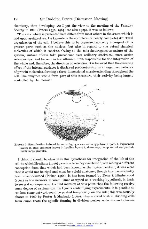
Ever since I used to watch under the microscope in the early 1920s the behaviour of ciliate protozoa, I have been fascinated by this problem of integration, taking place as it does in a jelly-like medium, which so far as I know is deficient in calcium; I do not think that others have reversed my findings in this respect. In 1906, Lyon (i906) centrifuged sea-urchin eggs, proving that the stratification so induced did not interfere with subsequent cell division (figure 3); hence the position of much of the cell contents did not matter. The implication that there was no reasonable chemical explanation of this seemed to me to be unsatisfactory, and accordingly I searched for one, finally finding a possible mechanism in the work on surface
L 11 ]
This content downloaded from 130.132.123.28 on Sun, 4 May 2014 22:19:03 PMAll use subject to JSTOR Terms and Conditions
chemistry, then developing. As I put the view to the meeting of the Faraday
Society in 1930 (Peters 1930, I963; see also 1929), it was as follows: 'The view which is presented here differs from most others in the stress which is
laid upon architecture. Its keynote is the complete (or nearly complete) structural
organization of the cell. I believe this to be organized not only in respect of its
grosser parts such as the nucleus, but also in regard to the actual chemical
molecules of which it consists. Owing to the microheterogeneous nature of the
system, surface effects take precedence over ordinary statistical, mass action
relationships, and become in the ultimate limit responsible for the integration of
the whole and, therefore, the direction of activities. It is believed that the directing
effect of the internal surfaces is displayed predominantly by an organized network
of protein molecules, forming a three-dimensional mosaic extending throughout the
cell. The enzymes would form part of this structure, their activity being largely
controlled by the mosaic.'
.4
FIGURE 3. Stratification induced by centrifuging a sea-urchin egg. Lyon (I906). 1, Pigmented
layer; 2, gray, granular layer; 3, hyaline layer; 4, dense cap, composed of compacted, fairly large granules.
I think it should be clear that this hypothesis for integration of the life of the
cell, to which Needham (I936) gave the term 'cytoskeleton', is in reality a different
conception from that which had been known as the 'cytosquelette'; it was clear
that it could not be rigid and must be a fluid anatomy, though this has evidently
been misunderstood (Picken I960). It has been termed by Dean & Hinshelwood
(I964) as the network theorem. Once accepted as a working hypothesis, it leads
to several consequences. I would mention at this point that the following receive
some degree- of explanation. In Lyon's centrifuging experiments, it is possible to
see how some network could be pushed temporarily on one side; this was actually
shown in 1960 by Porter & Machado (I960); they showed that in dividing cells
from onion roots the spindle forming in division pushes aside the endoplasmic
This content downloaded from 130.132.123.28 on Sun, 4 May 2014 22:19:03 PMAll use subject to JSTOR Terms and Conditions
reticulum. Where enzymes are attached to the cytoskeleton with a consequent control, it follows that Hofmeister's microkitchens will exist, with the possibility of compartmentation and of diverse chemical reactions taking place in the cell at the same time. The segregation of enzymes in mitochondria and microsomes is now a commonplace (Lehninger I 964). There was an extension of this which I never dared to develop at the time, namely that molecules could be handed spatially from one enzyme to another; interestingly Reed (I967) has inferred that this must happen in an oc-keto acid system. I emphasized then the need for exploring the interfacial action of individual groups of proteins, and especially those of the nucleoproteins. The presence of a cytoskeletal structure was supported by Wigglesworth (I953); in insect experiments, transplanted bristles grew in the original direction.
It is interesting to compare what Frey-Wyssling (I948) wrote after many years of study: 'The substratum in which life is inherent is not a dispersoid with indi- vidual particles or ultramicrons, it possesses a structure. The active groups which control metabolism and development, are juxtaposed in a given order. They are not intermingled by mere laws of chance and Brownian molecular movement; the fact is rather that the chain molecules arrange themselves into a delicate and very purposeful and flexible molecular frame activated as it were, by a purposeful coordinative impulse.' And again: 'For orderly biological processes are un- thinkable without presupposing structure-and it is, therefore, out of the question that any living constituent of protoplasm could consist of structureless, fluid, independently displaceable particles.'
I have never seen occasion to withdraw my views on the existence of a 'fluid' cytoskeleton, and have even developed them in the intervening years, envisaging that they would lead to the belief that the surface proteins of a cell, acting as receptors of stimuli, would be theoretically different from the cytoplasmic protein. In 1936 I drew attention to this in a Discussion to this Society on Pharmacological Action and developed this in an essay (I937a, b). In view of my own position over 40 years, I was somewhat surprised to see in a recent Abstract for a discussion (B.A. Record I968) the statement that 'Only recently the cell was, to the bio- chemist, a bag full of enzymes-and enzymes were simply biochemical catalysts. How such a system could behave in the purposive and controlled way charac- teristic of cells was, to say the least, obscure.'
Admittedly the statement had to be short for such an abstract, but it is not true that all biochemists thought like this. Many did, especially those who were actively engaged upon the isolation of various enzymes and the determination of their chemical properties. They did not find it necessary to go beyond this ele- mentary hypothesis, especially because it is true that when coupled with the astonishing specificity of these catalysts, much can be explained of the chemical reactions in cells. I am still surprised, however, that so many did not stop to think more deeply. There were several reasons for this-we might instance the lack of knowledge at that time of discoveries in surface chemistry; then Gowland Hopkins,
This content downloaded from 130.132.123.28 on Sun, 4 May 2014 22:19:03 PMAll use subject to JSTOR Terms and Conditions
very properly, had led a revolt against the nineteenth-century ideas of the giant molecule of protoplasm.
The statement in the abstract goes on to say: 'The electron microscope has shown that cells are highly structured systems.' If this is taken to mean that knowledge of internal structure had to wait for the advent of the electron microscope, it is not quite correct. Phase-contrast microscopy introduced by Zernicke (I935) had taught us much, and earlier. Under Dame Honor's direction it was used in the Strangeways Research Laboratory by Hughes & Swann (I948) on tissue cultures. In fact, phase contrast was most necessary at the time, because of the critical atmosphere created for histology, for many years, by that romantic genius, W. B. Hardy (later Sir William) (I899) who became a secretary of this Society. He made it clear that the different fixatives then used in histology produced different structures as seen under the light microscope, not only in tissue specimens, but even in gels of gelatin or in albumin solutions. For instance, figure 4, taken from Hardy's paper
7
6 Fig. 5. Solids per 100 ml. 4 g Fig. 6. Solids per 100 ml. 10 g Fig. 7. Solids per 100 ml. 25 g Fig. 8. Solids per 100 ml. 50 g
FIGURE 4. Structural effect seen by light microscope after fixation of a gel of gelatin by mercuric chloride. It differs much on changing from 4 to 10 % solids. Hardy (I899, figures 5 to 8.)
shows the effect of dilution of gelatin with water on structures obtained by fixing with mercuric chloride. It was even found that stress figures could be induced in
apparently homogeneous films by exposing them to minute globules of mercury. The conclusion was that ideas suggesting a foam structure for protoplasm, due mainly to Biitschli, could be merely a result of the fixative used.
The problem of the structure of a gel seems to bear intimately upon the internal structure of cells, and requires some further discussion. Even yet, 70 years after Hardy's publication it is not certainly solved. I had thought that it would be easy now to explain why Hardy obtained stress figures in an apparently homogenous gel, on the view that gels of gelatin would be shown by modern methods to contain a criss-cross network of fibrils, which, though invisible under the light microscope
This content downloaded from 130.132.123.28 on Sun, 4 May 2014 22:19:03 PMAll use subject to JSTOR Terms and Conditions
FIGURE 5. lFreeze-etched electron microscope picture of a gelatin gel. 20 % glycerol was present to prevent the formation of ice crystals. R. Reed (previously unpublished).
(Fsacing p. 14)
This content downloaded from 130.132.123.28 on Sun, 4 May 2014 22:19:03 PMAll use subject to JSTOR Terms and Conditions
FiG.uRE 6. Phase-contrast photographs of cells in a tissue culture from chick heart, showing filamentous mitochondria. (a) Normal. (b) After treatment with fluoroacetate.
This content downloaded from 130.132.123.28 on Sun, 4 May 2014 22:19:03 PMAll use subject to JSTOR Terms and Conditions
originally, would unite together to produce figures under the influence of a stress. Accordingly in preparing for this Discussion, I wrote to a number of those interested in gelatin to try to obtain a modern picture of a gelatin gel. Eventually, I am much indebted to Dr R. Reed (Leeds) for sending me an electron microscope picture of a gelatin gel in 20 % glycerol (figure 5, plate 1), prepared by the freeze-etch method; the glycerol was used to prevent the release of ice-crystals during preparation. The structure is not what I thought that it might be, and it is not easy to interpret. The literature (Bailey I968) indicates that gelatin contains peptides of varying length, some being coiled. One could believe either that one is looking at a cross-section of fibres or that the picture is one of discrete spherical particles, forming a 3-dimensional network. Dr R. Reed (personal communication) considers that one cannot assume 'that these particles are free and unconnected'. In his view, 'the problem of structure of these lyogels is still one of the most difficult in colloid science... People fail to take into account the great ordering propensity of water, and the large 3-dimensional or space network which it contains'. His interpretation reduces to the importance of considering water as a coordinating and structural agent. I think it is clear that in any final appraisal of the structure of a living cell, we shall be obliged to understand the part played by water; often in the past, this has been called 'bound' water.
Meanwhile, returning to present knowledge of organelles (figure 6, plate 2) taken from a film by Buffa, Godina & Barasa (see also p. 2) shows how much can be seen by phase-contrast photography. Of course with modern methods of fixation, the electron microscope shows much more, such as the details of the cristae in the mitochondria, the Golgi apparatus and above all the endoplasmic reticulum (ER); the electron microscope photographs published by Palade & Porter (I I954) also are most convincing. This is especially true of the photograph of two serial sections in the wall of a blood capillary, in which an isolated oval body appearing in one section is shown to be part of a tube in the second. The ER is known to exist in more than one form, smooth and rough, and to proliferate with some drugs. One can now begin to think seriously about the reality of macro- molecular structure and about transport in cells (see Northcote, this Discussion). With all their rigid appearance, mitochondrial structure are very labile. Seven years ago, Baker, Northcote & Peters (I962), and Peters (I963) found that a very short treatment of kidney mitochondria with laurate (the C12) fatty acid could disrupt mitochondria, and bring oxidative reactions to a standstill.
Clearly now we need no longer labour the point that the cell contains internal structure. We have come into the realm of macromolecular chemistry. But what is the position at the present of the conception of a cytoskeleton as an integrating entity ? This entails thinking of it as a kind of nervous system, with all the philo- sophical advantages and disadvantages of the conception of nervous reflexes. It is true that there is now possible a somewhat different interpretation of the Lyon centrifuging experiments on the sea-urchin egg. One can replace the idea of a continuous integrating structure by the view that the structure could be temporarily
This content downloaded from 130.132.123.28 on Sun, 4 May 2014 22:19:03 PMAll use subject to JSTOR Terms and Conditions
displaceable and subsequently able to rearrange to its original condition. This has been shown by Lester Reed and colleagues (Koike, Reed & Carroll I960; Willms et al. I967) to occur with the components of the ac keto oxidase systems. These contain three enormous molecules which can be dissociated with loss of activity. They can then, by appropriate biochemical means, be coaxed back into their original arrangements with a regain of activity. Also Weiss (I96I) has drawn attention to the great capacity of cell entities to arrange themselves in a regular order. But even these considerations imply fundamentally that there is an arranged structure-and we have often talked about the thixotropic gel in this connexion.
I still think that a cytoskeletal structure need not be much more than a few molecules or even one molecule thick, and base this view on surface chemistry. It would react with great speed. As long ago as 1911, Hardy & Harvey (I9 ii)
pointed out that when a microscope was focused on the most external, air-water layer of a glass cell containing animal and plant material exposed to an electric current, the most external layer was stationary, though the plant material, etc., moved with the current. This observation probably had much to do with his discovery that the interfacial tension between 08 substances with different end groups between benzene and water, depended upon the chemical constitution of the end group. This, together with the well-known work of Langmuir, Harkins & Adam (Adam I938), some of it based upon Devaux, gave a proof that chains of
molecules, one molecule thick, could have rigidity. I carried this further (Peters 193I) by showing that long chain fatty acids (CH2) carrying either -COOH or
-NH2 behaved in an interface as if the pK of this was altered towards the neutral point as compared with a bulk phase. The facts as to -0OOH groups were confirmed by Danielli (I937). These observations have not yet been much inte- grated into modern theory. An equation has been evolved by Hartley & Roe (I940)
for the difference in pH, and both MeLaren (I960) and L. Weiss (personal communi- cation) have recently shown its application to some enzyme studies.
Returning to the challenging question as to how a cell can integrate its chemistry with stimuli arriving at its surface, I have often referred to the fact given by Clark (I928) that in a heart cell stimulation can be induced by an amount of acetylcholine, which could only cover -6-L- of its surface. I think that the action
of this reduced to two possibilities: whether we are dealing only with a purely surface effect, or whether there are receptor groups of some component of the cytoskeleton on the surface, able speedily to relay a chemical action to the interior
parts of the cytoskeleton (possibly as a pulse of electrons). It is possible that a
stimulus applied at one point of a surface could be rapidly transmitted over the whole surface, provided that it is large enough to alter the interfacial tension; evidence exists of extension of effects beyond the actual area covered by protein molecules, as also of two-dimensional gas-like behaviour in surfaces. This would
be consistent with the Davson-Danielli model of a membrane; but in this case
I do not see how action would pass so selectively from an external protein through
This content downloaded from 130.132.123.28 on Sun, 4 May 2014 22:19:03 PMAll use subject to JSTOR Terms and Conditions
an essentially lipid layer even allowing for dynamic factors (Dingle & Lucy I965).
At some time the surface action must be transmitted to the interior of the cell. In more modern interpretations of the electron microscopic structure of membranes (Benson I966; Lenard & Singer I968; Wallach & Zahler I966), which include the specialized hydrophobic protein in membranes isolated by Green & Purdue (I966), it is thought that the central part of a membrane consists of a lipoprotein layer, with the polar groups lying externally and the hydrophobic chains of lipid and protein internally. With this model, the protein part could well act as the trans- mitter to an interior part of the cytoskeleton; the external polar groups would then be the receptors. The new conception seems to fit facts better, and is inci- dentally more consistent with Mitchell's (I966) ideas. When we remember that the surface of a bacillus, Escherichia coli, has space for about 50 million -COOH groups, there is plenty of room on a surface for many diverse receptors.
Coming back to the cytoskeletal hypothesis, I would make a plea for a serious consideration of this, because I think that a logical hypothesis of how a cell is coordinated as a whole is not merely of academic interest, important as this may be. Surely a useful hypothesis could accelerate the present progress of research on abnormal growth and can-cer. If it is a 'protein' cytoskeleton, it is easier to under- stand how changes in the proteins of this could alter the slant of cell activity. This has been suggested already for the action of insulin (Peters 1956; Krahl I967), as for that of developing tumours (Alexander & Lett I960; Peters I960). To my knowledge there has been no other hypothesis advanced as to how the cell is integrated in its reactions. The introduction of the high-speed centrifuge (Claude I946) made experimental tests for the first time really possible in this field.
It is true that the cell uses a battery of almost every kind of reaction known to physics and chemistry (Dean & Hinshelwood I964); but we can put the question quite simply, are we to believe that the cell with its endoplasmic reticulum and other organelles is merely an extremely complicated set of mass action equilibria, behaving in some sense like a rubber ball to surface stimuli; or must we look for some definite coordinating mechanism, as a chemical and physical means by which its purposeful activities are maintained? The vertebrate body is a chemical and physical system, but we should get a very little distance with physiological research, if we were not prepared to concede structure in the vascular system or the muscles and bones.
We now reach the question whether a controlling 'cytoskeleton', forms part of the endoplasmic reticulum. Hechter & Halkerston (I964) define it as a 'contractile solid-phase system of endoplasmic reticulum plus plasma membrane containing membrane-sited enzyme systems'. Perhaps then we have to search no further, except to show how this comes into contact with surface, probably protein receptors. But in any case, -recognition of an over-all microstructural physiology of the cell can be only a first step, giving the framework for the further and extensive molecular biochemical and biophysical research; the detail of this necessarily lies
2 Vol. I73. B.
This content downloaded from 130.132.123.28 on Sun, 4 May 2014 22:19:03 PMAll use subject to JSTOR Terms and Conditions
ahead, and includes an understanding of the vascular system, to be dealt with by Dr Northcote.
I am indebted to Dame Honor Fell, F.R.S., for helpful advice, and to Professor P. Buffa for the film.
FILM
At the conclusion of this paper, a shortened version was shown of a time lapse film prepared by Professor P. Buffa (Modena) in collaboration with Professor G. Godina and Dr A. Barasa (Turin). It was taken by phase-contrast photography, of a growing tissue culture from chick heart, and accelerated from 30 frames/min to 24 frames/s. The normal mitochondria, resembling eels are in rapid movement (figure 6, plate 2). Approximately 30 min after a treatment with fluoroacetate (F.CHR2. COONa) several of the mitochondria bulge and tend to break (figure 6b). This is interpreted as being due to an increased concentration of citrate arising in the mitochondria as a result of a block in the citric acid cycle at the stage of aconitase, which controls the tricarboxylic acids. The block is presumed to be due to the synthesis of fluorocitrate formed by a 'lethal synthesis' from fluoroacetate, a step which has been proved experimentally. Though the mitochondria were damaged, cell division took place normally.
POSTSCRIPT
During the course of our correspondence, Dr R. Reed introduced me to a review on water by Dr F. Franks (I968, Chem. and Ind. pp. 560-566). In this there was a mention of work on 'ortho' water by B. V. Deryagin. Dr A. D. Bangham (A.R.C., Institute of Animal Physiology, Babraham) has now told me that his father, Professor D. H. Bangham, published a number of papers in this field, see Nature, Lond. I968, 219, 1151. The fact that a different form of water can build up on surfaces is clearly of importance in relation to the conditions of water in cells.
REFERENCES (Peters)
Adam, N. K. 1938 Physics and chemistry of surfaces, 2nd ed. Oxford: Clarendon Press. Alexander, P. & Lett, J. T. I960 Biochem. Pharmacol. 4, 34-46. Bailey, A. J. I968 Comprehensive biochemistry, vol. 26B, ch. v, pp. 297-413. (Ed. M. Florkin
and E. H. Stotz.) Elsevier Publishing Co. Baker, P., Northcote, D. H. & Peters, R. A. I962 Nature, Lonid. 195, 661-662. Benson, A. A. I966 J. Am. Oil Chem. Soc. 43, 265-270. Claude, A. 1946 J. Exp. Med. 84, 51-89. Clark, A. J. 1928 J. Physiol. 64, 123-143. Danielli, J. I937 Proc. Roy. Soc. B 122, 155-174. Dean, A. C. R. & Hinshelwood, C. I964 Nature, Lond. 202, 1046-1052 and I965, 206,
546-553. Devaux, H. see Adam, N. K. Dingle, J. T. & Lucy, J. A. I965 Biol. Rev. 40, 422-461.
This content downloaded from 130.132.123.28 on Sun, 4 May 2014 22:19:03 PMAll use subject to JSTOR Terms and Conditions
Frey-Wyssling, A. 1948 Submicroscopic morphology of protoplasm and its derivatives, p. 230. Elsevier Publishing Co.
Green, D. E. & Purdue, J. F. I966 Proc. Natn. Acad. Sci. U.S.A. 55, 1295-1302. Hardy, W. B. I899 J. Physiol. 24, 158-210. Hardy, W. B. 1913 Proc. Roy. Soc. A 88, 303-333. Hardy, W. B. & Harvey, H. W. i9ii Proc. Roy. Soc. B 84, 217-226. Hartley, G. S. & Roe, J. N. 1940 Trans Faraday Soc. 36, 101-109. Hechter, 0. & Halkerston, I. D. K. I964 Hormones, vol. v, p. 781. (Ed. G. Pincus, K. V.
Thimann and E. B. Astwood.) New York: Academic Press. Hechter, O., Yoshinaga, K., Halkerston, I. D. K. & Birchall, K. I967 Archs Biochem.
Biophys. 122, 499-465. Hughes, A. P. & Swann, M. M. 1948 J. Exp. Biol. 25, 45-70. Koike, M., Reed, L. J. & Carroll, N. R. I960 J. Biol. Chem. 235, 1924-1930. Krahl, M. E. 1957 Perspectives in Biol. Med. 1, 69- 96. Lehninger, A. L. I964 The mitochondrion. New York, Amsterdam: W. A. Benjamin Inc. Lenard, J. & Singer, S. J. I968 Science, N.Y. 159, 738-739. Lyon, E. P. I906 Arch. Entuwick.Mech. 23, 151-173. McLaren, A. D. I960 Enzymologia 21, 356-364. Mitchell, P. I966 Biol. Rev. 41, 445-502. Needham, J. 1936 Terry Lectures. 'Order and life'. New Haven: Yale University Press. Palade, G. E. & Porter, K. R. 1954 J. Exp. Med. 100, 641-656. Peters, R. A. 1929 J. St. Med. 37, Harben Lecture 1. Peters, R. A. 1930 Trans. Faraday Soc. 26, 797-807. Peters, R. A. 1931 Proc. Roy. Soc. A 133, 140-145, and with R. WX. Wakelin (1938) Trans.
Faraday Soc. 34, 1537-1539. Peters, R. A. I937a Proc. Roy. Soc. B 121, 587-592. Peters, R. A. 1937 b Perspectives in biochemistry, pp. 30-44. (Ed. J. Needham and D. E.
Green.) Cambridge University Press. Peters, R. A. 1956 Nature, Lond. 177, 426-427. Peters, R. A. I960 Biological approaches to cancer chemotherapy, pp. 11-19. (Ed. R. J. C.
Harris.) New York: Academic Press. Peters, R. A. I963 Biochemical lesions and lethal synthesis, vol. 18. Oxford, London, New
York, Paris: Pergamon Press. Picken, L. I960 Organisation of cells, p. 162. Oxford University Press. Porter, K. R. & Machado, R. ID. I960 J. Biophys. Biochem. Cytol. 7, 167-184. Record, The B.A. I968 No. 4, p. 2. Reed, L. J. I967 Thiamine deficiency. Ciba Foundation Study Group, no. 28, pp. 67-76.
J. and A. Churchill Ltd. Wallach, D. F. H. & Zahler, P. H. I966 Proc. Natn. Acad. Sci. Wash. 56, 1552-1559. Weiss, P. I96I The molecular control of cellular activity, pp. 1-72. (Ed. J. M. Allen.) New York:
McGraw-Hill Book Co. Wigglesworth, V. B. 1953 J. Embryol. Exp. Morph. 1, 269-277. Willms, C. R., Cliver, R. H., Henney, H. R. J., Mukherjee, B. B. & Reed, L. J. I967 J. Biol.
Chem. 242, 889-897. Zernicke, F. 1935 Z. Tech. Phys. 16, 454-457.
2-2
This content downloaded from 130.132.123.28 on Sun, 4 May 2014 22:19:03 PMAll use subject to JSTOR Terms and Conditions