A DISSERTATION ON COMPARISON OF THE RESULTS OF OUTCOME OF CANAL WALL UP MASTOIDECTOMY AND CANAL WALL DOWN MASTOIDECTOMY FOR CHRONIC SUPPURATIVE OTITIS MEDIA DISSERTATION SUBMITTED FOR MASTER OF SURGERY BRANCH (IV) IN OTORHINOLARYNGOLOGY MARCH 2010 THE TAMIL NADU DR.M.G.R. MEDICAL UNIVERSITY CHENNAI – TAMILNADU
Transcript
A DISSERTATION ON COMPARISON OF THE
RESULTS OF OUTCOME OF CANAL WALL UP
MASTOIDECTOMY AND CANAL WALL DOWN
MASTOIDECTOMY FOR CHRONIC SUPPURATIVE
OTITIS MEDIA
DISSERTATION SUBMITTED FOR
MASTER OF SURGERY BRANCH (IV)
IN
OTORHINOLARYNGOLOGY
MARCH 2010
THE TAMIL NADU DR.M.G.R. MEDICAL UNIVERSITY
CHENNAI – TAMILNADU
DECLARATION
I hereby declare that this dissertation entitled
“COMPARISON OF THE RESULTS OF OUTCOME OF CANAL
WALL UP MASTOIDECTOMY AND CANAL WALL DOWN
MASTOIDECTOMY FOR CHRONIC SUPPURATIVE OTITIS
MEDIA” has been prepared by me under the guidance and supervision of
DR.K.R.KANNAPPAN M.S.D.L.O.M.ch, PROFESSOR AND HEAD
OF THE DEPARTMENT OF OTORHINOLARYNGOLOGY, Govt
Rajaji Hospital, Madurai.
This Dissertation is Submitted to The Tamil Nadu Dr.M.G.R
medical university in partial fulfillment of the university regulations for
the award of “THE MASTER OF SURGERY” in
OTORHINOLARYNGOLOGY. this work has not formed the basis of the
award of any degree/ diploma to me previously by any other university.
PLACE: MADURAI
DATE : SIGNATURE
CERTIFICATE
This is to certify that the thesis titled “A DISSERTATION ON
THE COMPARISON OF RESULTS OF OUTCOME OF CANAL
WALL UP MASTOIDECTOMY AND CANAL WALL DOWN
MASTOIDECTOMY FOR CHRONIC SUPPURATIVE OTITIS
MEDIA” submitted by DR. VIDHYA.M under my supervision &
guidance in partial fulfillment for the award of the degree of Master of
Surgery in Otorhinolaryngology by the Tamil Nadu Dr. M.G.R.
Medical University, Chennai, is a bonafide record of the work done by her
during the academic period 2007-2010. She has evinced keen interest in
collecting the cases from the ward and analysing them. I have great
pleasure in forwarding it.
Dr. KR. KANNAPPAN MS, DLO, M.Ch
The Professor & Head,
Dept of ENT Diseases,
Govt Rajaji Hospital,
Madurai.
ACKNOWLEDGEMENT
I would like to express my most sincere thanks to the following
persons who went the extra mile to help me complete this dissertation.
“The function of a teacher is not to tell only the meaning of words,
but to knock the doors of mind”.
I am highly indebted to my guide Dr.K.R. KANNAPPAN MS,
DLO, M.Ch, Professor and Head of Department of
OTORHINOLARYNGOLOGY, Govt Rajaji Hospital, Madurai., a true
teacher who taught me not only the art of surgery but also the etiquette of
a surgeon without whose guidance this work would have remained an
inconceivable dream.
I gratefully acknowledge and sincerely thank Prof.Dr.S.M.
SIVAKUMAR, Dean, Govt Rajaji Hospital, Madurai, for granting me
permission to utilise the resources of this institution for my study.
I express with a deep sense of gratitude, my sincere thanks to
Dr. S.SARAVANA MUTHU M.S., E.N.T, Associate professor, Govt
Rajaji Hospital for his encouraging directions and coaching throughout.
I am very much obliged to Dr. ARULSUNDARESHKUMAR M.S
and Dr.RAJAGANESH M S , Asst Profs of Dept of ENT , who helped
me in preparing and bringing a shape to this work..
I am also grateful to Asst Profs of Dept of ENT
Dr.RAJASEKARAN M.S DLO, Dr. ALAGUVADIVEL MS DLO,
Dr. SIVS SUBRAMANIAN MS DLO and Dr.RADHAKRISHNAN
MS DLO for their help and guidance.
I acknowledge with thanks, the unflaging support rendered by my
post graduate colleagues & friends.
I wish to record my sincere respect and thanks to all those patients
who despite their agony and suffering have helped me in moulding this
study.
Last but not the least I would like to thank my husband and parents
in providing me utmost cooperation needed for this study.
Place:
Date :
CONTENTS
S.NO CONTENTS PAGE NO
1. INTRODUCTION 1
2. AIMS AND OBJECTIVES 4
3. REVIEW OF LITERATURE 5
4. MATERIALS AND METHODS 54
5. SURGICAL PROTOCOL AND METHODOLOGY 56
6. ANALYSIS OF RESULTS 58
7. DISCUSSION 64
8. CONCLUSION 66
9. BIBLIOGRAPHY
10. ANNEXURES
*PROFORMA
*MASTERCHART
*KEY TO MASTER CHART
1
INTRODUCTION
The chronic discharging ear is still one of the common problems
that the Otorhinolaryngologist in India and other developing countries
are encountering. Although, thanks to the advent of newer antibiotics,
the incidence of acute suppurative otitis media and its complications
have reduced, chronic suppurative otitis media and their complications
are still prevalent.
The continuation of the infection and the bone eroding
properties of granulation tissue and cholesteatoma seen in CSOM are
known to be the major pathological process causing these
complications. As there is no simple means to eradicate this chronic
pathology, appropriate and timely intervention by an otologist goes a
long way in the prevention of these human maladies.
In cases of chronic suppurative otitis media with atticoantral /
postero superior marginal pathology, treatment modality is only
surgery. Surgical options available are the canal wall down
mastoidectomy and intact canal wall mastoidectomy.
Goals of surgical management of chronic otitis media include the
eradication of disease, restoration of hearing, and, to the extent
possible, maintenance or restoration of a normal anatomic
2
configuration. Prior to the mid-1950s, the first 2 of these goals were
usually accomplished by removal of the posterior external auditory
canal wall, resulting in a radical or modified radical mastoidectomy
cavity. The past 50 years have witnessed a trend away from mandatory
canal wall removal. Many otologic surgeons now prefer intact canal
wall mastoidectomy with tympanoplasty except when canal wall
removal is required because of extensive disease, inadequate access for
cholesteatoma excision, operation on an "only hearing ear," or
uncertainty of adequate follow-up. The popularity of intact canal wall
mastoidectomy stems from the benefits of maintaining a canal wall,
which include freedom from the need for frequent mastoid bowl
cleanings, freedom from water intolerance and calorically induced
vertigo, and less difficulty in fitting and use of hearing aids.
In canal wall down mastoidectomy, complete disease clearance
can be given. But this could be achieved only at the cost of post
operative cavity problem and considerable hearing loss.
Though these complications are not present in intact canal wall
mastoidectomy, disease clearance could not be achieved completely in
intact canal wall technique.
Though the complications of CSOM can be averted , still they
are on the rise due to poverty, ignorance of the patient and the non
3
availability of facilities on time. Therefore there is a need to make
public aware of the serious nature of this illness, the importance of
early diagnosis and managing so as not only to reduce the morbidity
and mortality but also to give them safe, dry and functioning ear.
The present study has been carried out to compare the post
operative results of canal wall up mastoidectomy and canal wall down
mastoidectomy in patients with atticoantral or postero superior
marginal pathology of chronic suppurative otitis media.
4
AIMS OF THE STUDY
1. To perform on CSOM patients, canal wall down mastoidectomy
and canal wall up mastoidectomy.
2. To follow up the patients post operatively by clinical
examination, otoendoscopy and pure tone audiogram.
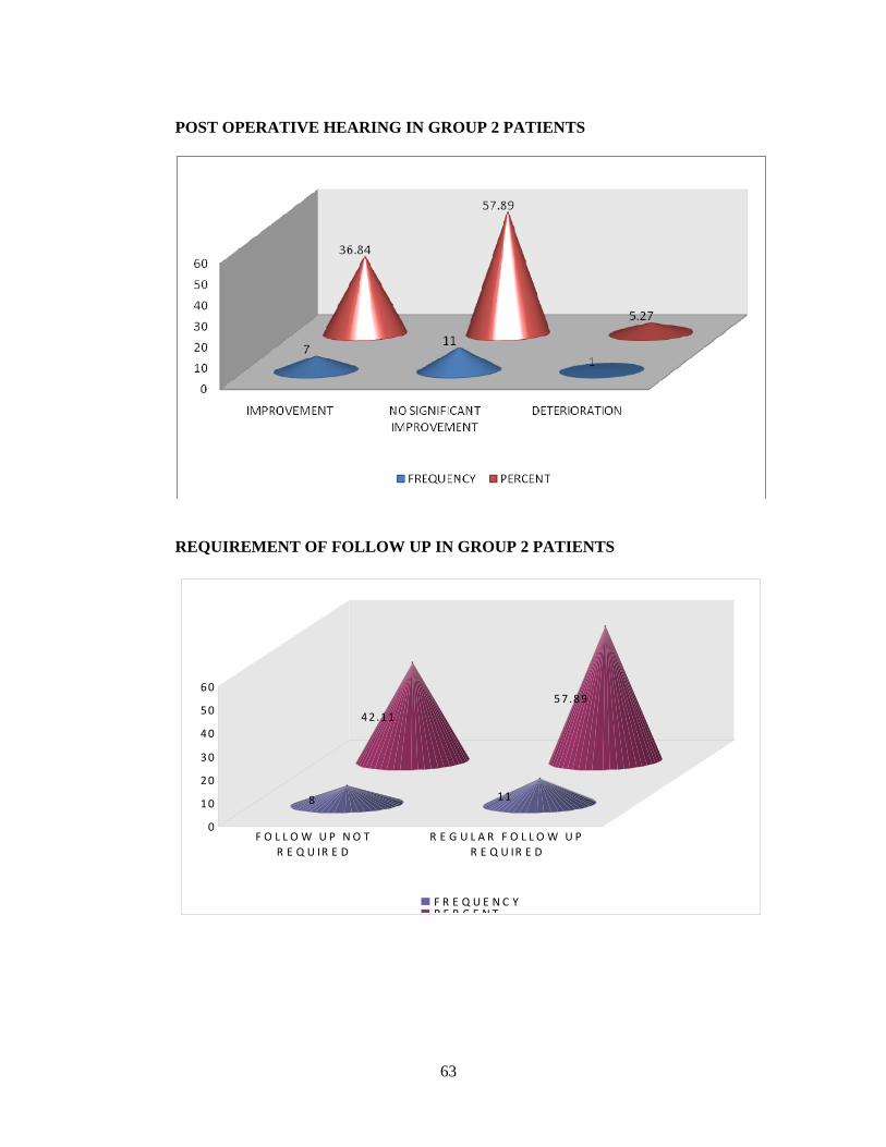
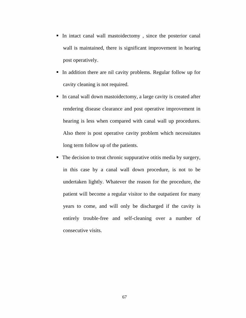
3. To study the post-operative results like recurrence of ear
discharge, improvement in hearing and requirement of post
operative follow up.
5
REVIEW OF LITERATURE
The history of Chronic Otorrhoea and its complications are as
old as the human race itself. Some of the Mummies found in the
pyramid show evidence of middle ear infection. The earliest written
history of ear discharge and its treatment are found in ‘PAPYRUS
EBEARS’ written more than 1500 years BC. It describes treatment
with olive oil sprinkled with spells.
Atharvana veda of 700 BC contains the earliest Indian medical
information including ear disease.
Sushrutha the Indian physician of 500 B C has written in detail
about CSOM describing it a ‘ Pooti Karna’ and is the first person to
describe its complications. He says that if ‘ Pooti Karna’ is not treated
properly, the patient may develop hallucinations and vomiting . He not
only used herbal tonics and ear drops but also devised many surgical
instruments to drain an abscess when it is pointing and to pack the
cavity daily with herbal medicines.
Hippocrates , the Father of Medicine (460BC) ,was probably the
first to inspect the tympanic membrane as dry thin spun web and to
recognize it as a part of organ of hearing. He mentioned that the patient
6
with acute ear pain and high continuous fever may become delirious
and die.
Medicines used till 1500 AD were honey, breast milk, one’s
own urine, bull’s urine , rain water, etc..
Gabriel Fallopious (1523-62) discovered the facial nerve canal
and also described the aural polyp and its treatment.
Joseph Dureuney (1648-1730) remembered as the Father of
Otology gave an account of scientific explanation with the tympanic
cavity.
Glovauni-Morgagni (1682-1771) was the first to clearly
recognise that the ear infection came first and the brain abscess was
secondary.
Morand S.F (1768) successfully operated upon a brain abscess
for the first time.
Hooper in 1826 first described ‘lateral sinus thrombosis’ from
middle ear infection.
William wilde in 1853 described the sub periosteal abscess and
popularized the concept of draining it with a post aural incision.
Herman an Schwartz in 1870 first published a report of having
opened the mastoid antrum.So well did Schwartz succeed, that by 19 th
7
century it attained a general acceptance and came to be known as
Simple or Cortical Mastoidectomy.
Emanuel Gaufal in 1890 described the technique of Radical
Mastoidectomy in detail and in 1891 Stacks advocated plastic meatal
skin flap for drainage. Then the Radical mastoid operation came to be
known as Gaufal or Stack operation.
Johannes Kessel in 1885 had done the first endaural Radical
Mastoidectomy.
Lane .W.A in 1890 established the surgical treatment for sinus
thrombosis.
Korner O.in 1902 demonstrated that infection can spread from
the lining mucosa of tympanum , through intact bone by means of
progressive thrombophlebitis.
It was Gluseppe Gradenigo who in 1967 described a symptom
complex consisting of Abducent Nerve in patents with suppurative
disease of the petrous apex.
Bezold F.S in 1908 described that infected air cells perforating
the inner surface of mastoid process can extend to the deep tissues of
neck calling it as “ Bezold’s abscess”.
8
Though the concept was very old , the first mastoid operation
trying to preserve the ossicular chain and hearing was described by
Bondi in 1910.
The credit for the first successful intra-temporal facial nerve
anastamosis and first successful facial nerve graft within temporal bone
in 1930 goes to Sterling Bunnel.
From 1930-1937 Kopetsky, Almor, Eagelton, Frenchkner,
Ramadier and Lempert described the various routes for draining the
petrous apex.
Alexander Fleming in 1928 discovered the antibiotic Penicillin
which revolutionized the treatment of CSOM.
Gerhard Domagic discovered Sulphonamides in 1932 and since
then many more useful broad spectrum antibiotics have been
introduced.
The reconstructive surgery to create a sound conductive system
and tympanoplasty operations started with Zollner in 1951 and
Wallstein in 1952.
Heerman in 1958 used temporalis fascia graft for tympanic
membrane perforations.
9
The invention of computerized axial tomography by Godfrey
Hounsfield in 1972 is described as the greatest step in the field of
radiology, after the invention of X- ray by Roentgen in 1895.
The basic principles of magnetic resonance imaging was
conceived by Bioch and Purcell in 1946 ; but it was Domadian and
Lauterber in 1973 who indicated its application in obtaining images in
the intact human body.
The development of Argon Laser microscope is considered as
the greatest advancement in the field of middle ear surgery, invented
by Rodney.C. Perkins in 1978.
In 1987 studies of Rubin R. showed that environmental factors
in addition to genetic factors exert a strong influence on the
development and ultimate size of mastoid air cells.
Aurim R.Eden in 1987 found preliminary evidence of neural
control of middle ear aeration controlled by respiratory centre.
Gates G.A in 1988 found out that Adenoidectomy in children
significantly reduced nasopharyngeal bacterial flora irrespective of the
size of adenoids and is beneficial for controlling middle ear infection.
In recent years many have studied the problems complications of
CSOM and further contributed to its better understanding .Browning
.G.G says that all active cases of CSOM with and without
10
Cholesteatoma, and whether or not they had previous surgery, should
be considered as liable to lead to major complications.
Even today the debate is still on, regarding an open or closed
technique to be adopted for eradicating cholesteatoma .The opinion
seems unanimous in a case of complication of CSOM, that the
eradication of the pathology from the middle ear is more important
than its auditory function.
11
SURGICAL ANATOMY OF MIDDLE EAR CLEFT
The middle ear cleft consists of the tympanic cavity, the
Eustachian tube, the mastoid air cell system and extension of the air
cell system into anterior and posterior petrous apex.
The tympanic cavity
It is an irregular laterally compressed air filled space in the
temporal bone, lined by mucous membrane. It is hour glass shaped
with a volume of about 2 cubic cm. For descriptive purpose it may be
thought of as a box with four walls, roof and floor.
The lateral wall of tympanic cavity
The tympanic membrane forms the central portion of the lateral
wall, while above and below there is bone, forming the outer lateral
walls of epitympanum and hypotympanum. Superiorly the scutum
separates the epitympanic recess from the roof of the external auditory
canal. Inferiorly a part of tympanic bone separates the tympanic cavity
from the medial part of temporomandibular joint. The pearly white
tympanic membrane is 0.1 mm thick and forms an angle of 55º with
the floor of meatus, is oval in shape, slightly broader above than below.
Its longest diameter from posterosuperior to anteroinferior is 9.1 mm,
perpendicular to this is the shorter diameter, 8.9mm. The
12
circumference of pars tensa is thickened to form a fibro-cartigenous
ring-the tympanic annulus, that sits in the tympanic sulcus. From the
superior limit of sulcus, the annulus becomes a fibrous band which
runs centrally as anterior and posterior malleolar fold to the lateral
process of malleus. The lax area above this fold,that does not have
annulus is pars flaccida. Below these malleolar folds is the pars tensa.
In pars tensa, the lateral most collagen fibres of the Lamina
propria, which are in direct contact with the basement membrane of the
epithelial layer, are arranged in regular radial orientation. These fibres
are believed to direct the migration of epithelium from the surface of
tympanic membrane to the outside. The upper limit of tympanic sulcus
is marked by anterior and posterior canaliculus for chorda tympanic
nerve which traverses the middle layer of tympanic membrane at the
level between malleus and incus. The anterior canaliculus is at the
medial end of the petrotympanic fissure which lodges the anterior
ligament of malleus and admits the anterior tympanic artery. The
posterior canaliculus is situated at the junction of lateral and posterior
wall at the level of upper end of handle of malleus. The opening leads
to a bony canal that descends through the posterior wall. It descends
obliquely and joins the nerve. A branch of stylomastoid artery
accompanies the nerve.
13
Arterial supply is through the deep auricular branch of maxillary
artery, anterior tympanic branch of maxillary artery, twigs from the
stylomastoid branch of posterior auricular artery and from the middle
meningeal artery.
The venous drainage occurs through the external jugular vein,
transverse sinus, dural veins and venous plexus around the Eustachian
tube.
Nerve supply is from the auriculotemporal nerve supplying
anterior portion,auricular branch of vagus supplying the posterior
portion and tympanic branch of glassopharyngeal nerve supplying the
medial surface.
Roof of the tympanic cavity
It is a thin plate of compact bone, the tegmen tympani, that
separates the middle cranial fossa from the tympanic cavity. It is
formed by petrous and squamous bone and the petrosquamous suture.
This suture is unossified in the young and does not close until adult
life. Veins from the tympanic cavity running to the superior petrosal
sinus pass through this line and this may allow infection to spread into
middle cranial fossa.
14
Floor of tympanic cavity
It is much narrower than the roof and consists of a convex plate
of bone separating the cavity from the superior bulb of internal jugular
vein. Occasionally the bone is deficient and the uncovered vein may
come upto the level of tympanic membrane covered only by mucous
membrane and fibrous tissue and lie in a dangerously exposed position
from the point of view of middle ear disease and surgery. The floor
sometimes may be thick and contain some accessory air cells.
Anteromedial to the vein, the tympanic branch of glassopharyngeal
nerve pierces the floor.
Medial wall of tympanic cavity
The medial wall is the lateral wall of the inner ear. The basal
turn of the cochlea forms a rounded elevation called promontory,
occupying much of the central portion of the medial wall. The
promontory surface is grooved by filaments of tympanic plexus.
Behind and below the promontory is the oval window leading to
vestibule, but in life is closed by the base of stapes and its surrounding
annular ligament. The round window lies below and behind the oval
window, separated by an extension of promontory called subiculum.
Occassionally a spicule of bone leaves the promontory above the
15
subiculum and runs to the pyramid on the posterior wall of tympanic
cavity. This spicule is called ponticulus. The round window is covered
by secondary tympanic membrane of 0.7mm thickness. The facial
nerve runs above the promontory and turns inferiorly behind the oval
window to descend in the posterior wall. Above the facial nerve, in the
epitympanum, is the dome of lateral semicircular canal. The triangular
area posterior to the round window is the sinus tympani which can
have cellular communication with the mastoid process and is thus of
significance in the pathology and surgery of the tympanic cavity, as it
can lodge cholesteatoma hidden during surgery.
Anterior wall of tympanic cavity
It is a narrow wall, as the medial and lateral walls of the
tympanic cavity converge. In the superior portion, there are two canals,
one for tensor tympanic above and the Eustachian tube below. The
septum between them extends backwards and forms the processus
cochleariformis on which the tendon of tensor tympanic glides. The
inferior part of the anterior wall consists of thin bony lamina forming
posterior wall of carotid canal and is perforated by superior and
inferior caroticotympanic nerves and the tympanic branch of internal
carotid artery.
16
Posterior wall of tympanic cavity
It is wider above than below and has in its upper part an opening
- the aditus-into the mastoid antrum. This is a large irregular hole that
leads backwards from the posterior epitympanum. Below this is the
fossa incudis, housing the short process of incus and the ligament
connecting the two. Below the fossa incudis and medial to chorda
tympani opening is the pyramid housing the stapedius muscle. The
bony portion of the posterior wall below the pyramid is the pyramidal
recess which may be perforated by air cells communicating with the
mastoid process. The facial nerve as it descends here is in danger from
air cells infection and from surgical attempts at air cells extirpation.
Between the promontory and the tympanic annulus is the facial recess.
Deep to both promontory and the facial nerve is the posterior,
extension of the mesotympanum, the sinus tympani. The importance
of facial recess is in posterior tympanotomy operation where an access
to middle ear from mastoid is made between facial nerve and
chordatympani nerve.
17
Contents of tympanic cavity
The tympanic cavity contains a chain of the small movable
bones - the malleus, incus and stapes, two muscles, the chordatympani
and tympanic plexus of nerves.
The malleus has a head, neck, two processes and a handle. The
head lies in the epitympanum and has a superior ligament which runs
to the tegmen tympani. Its posteromedial surface shows a facet for the
incudomalleolar joint. The anterior process receives a ligament from
the petrotypmpanic fissure, and the lateral process receives anterior
and posterior malleolar folds from tympanic annulus. The handle is
embedded between the mucosal and fibrous layers of tympanic
membrane and its rounded end forms the umbo. Tensor tympani
tendon is inserted on the deep surface of handle near its upper end.
The incus has a body and two processes. The body lies in the
epitympanum and has a cartilage covered facet for the incudo-
malleolar joint. The short process projects backwards to fossa incudis,
from which it receives a ligament. The ligament of incus is little more
than a fold of mucosa. The tip of long process – the lenticular process
is directed medially and forms a ball and socket joint with the head of
stapes.
18
The stapes consists of head, neck, two crura and a foot plate.
The stapedius tendon is inserted into the neck. The two crura arise
from the neck, and join the foot plate which covers the oval window
and is attached to its margins by the annular ligament of base of stapes.
The muscles of the tympanic cavity help in stabilizing the
ossicles, augmenting the sound signals and in protection of the inner
ear. The tensor tympani and stapedius are stimulated into activity by
sound and acting in combination, they exert a dampening effect on
amplitude of vibratory wave protecting the cochlea from excess
stimulation. The tensor tympani by pulling the tympanic membrane
medially may contribute to the functional role of the tensor palati in
cleaning the middle ear when the auditory tube is opened.
Compartments of middle ear
The middle ear is divided into three compartments: The
mesotympanum, epitympanum and hypotympanum.
The mesotympanum
It is that portion of the middle ear that lies between horizontal
plates drawn at the top and bottom edges of parstensa. It contains
stapes, long processes of malleus and incus, oval and round windows.
It contains stapes, long processes of malleus and incus, oval and round
19
windows. The eustachian tube exits from its anterior aspect. While
most of the mesotympanum is readily accessible to surgeon, two
crescent shaped recesses are extended posteriorly that are often
impossible to visualize directly. These spaces, the facial recess and
sinus tympani are the most common locations for cholesteatoma
persistence after chronic ear discharge surgery.
The epitympanum (attic)
It is that portion of the tympanic cavity that lies above the short
process of malleus. Superiorly it is bounded by tegmen tympani,
medially by the prominence of lateral semicircular canal and the
horizontal part of facial nerve, laterally by the scutum and posteriorly
by fossa incudis. It contains the head of malleus, body of incus and
their associated ligaments and mucosal folds. The epitympanum lies
within a fan shaped dehiscence on the tympanic bone – notch of
rivinus. It is bounded posteriorly by tympanomastoid line and
anteriorly by tympanosquamous line. The tympanic membrane here is
deficient of the dense fibre that forms the middle layer of pars tensa.
Because the pars flaccida lacks this structural support, it is more prone
to retraction in the face of negative middle ear pressure. This helps to
explain the propensity for cholesteatoma to form in the epitympanum.
20
The hypotympanum
It is that portion of middle ear that lies below the floor of bony
ear canal. It is an irregular bony groove that is seldom involved by
cholesteatoma. Occasionally the bone covering the jugular bulb may be
dehiscent in the hypotympanum.
Mucous membrane of middle ear cleft
The lining of the middle ear spaces is an extension and
modification of the respiratory mucous membrane that lines the nasal
cavity, nasopharynx and Eustachian tube. In all these regions the
mucous membrane consists of a layer of ciliated columnar cells with a
subepithelial layer of connective tissue. A film of mucous clothes the
membrane and is replenished by strategically located goblet cells and
mucous glands. The mucous film is kept in constant motion by the
continuous action of cilia, the direction of movement of the cilia being
from the tympanic cavity into the nasopharynx.A thin delicate mucous
membrane lines the whole of middle ear cavity and is reflected onto
the ossicles and tendons. It is continuous with the mucous membrane
of the mastoid antrum and Eustachian tube. It consists of non-ciliated
cuboidal epithelium, two or three cell deep, without a basement
membrane, but becomes ciliated columnar type especially near the
21
opening of Eustachian tube and hypotympanum, and changes to flat
pavement epithelium in the attic and air cells.
As one progresses from the cartilagenous to the bony portion of
Eustachian tube and from the tympanum to the antrum and air cells, the
sub epithelial connective tissue becomes thinner until the pavement
epithelium and the periosteum together form a thin delicate membrane.
The property to produce mucous is largely lost in the pavement
epithelium.These differences in the thickness of the mucous play an
important role in the genesis of ‘benign’ tubotympanic otitis media and
‘dangerous’ atticoantral type of disease.
Mucosal spaces of the middle ear
The mucous membrane is thrown into a series of folds by the
intratympanic structures dividing the middle ear into mucosal spaces of
surgical importance. The ossicular chain, ligaments, tendons of tensor
tympanic and stapedius muscles and the chorda tympani nerve are
called the ‘viscera’ of the middle ear and the mucosal folds are the
mesenteries.The attic is almost completely separated from the
mesotympanum by the ossicles and their folds except for two small but
constant openings called isthmus tympani anticus and isthmus tympani
posticus.
22
The transversely placed superior malleolar fold divides the attic
into a small anterior malleolar space which lies above the tensor
tympani fold that may prevent cholesteatoma from the attic reaching
the anterior mesotympanum and a larger posterior compartment. The
posterior compartment is further subdivided by the superior incudal
fold into a superior incudal space (lateral to the fold) and a medial
incudal space. The entrance into the Prussak’s space is usually located
between the lateral malleolar fold and lateral incudal fold. This latter
fold may arrest the passage of cholesteatoma, through a posterior
superior marginal perforation, into the attic.
The inferior incudal space
It is limited superiorly by the lateral incudal fold, medially by
the medial incudal fold, laterally by the posterior malleolar fold and
anteriorly by the interosseous fold, which lies between the long process
of incus and upper two thirds of the handle of malleus.
The anterior pouch of Von Troltsch
Lies between the anterior malleolar fold and that portion of the
tympanic membrane anterior to the handle of malleus.
23
The posterior pouch of Von Troltsch
Lies between the posterior malleolar fold and that portion of the
tympanic membrane posterior to the handle of malleus.
Prussack’s space
It is small space lying between the neck of malleus medially and
the pars flaccida laterally. It is bounded below by the short process of
malleus and above by the fibres of lateral malleolar fold, which fan
from the neck of malleus to be inserted along the entire rim of the
notch of Rivinus. A cholesteatoma may extend from Prussack’s space,
under lateral incudal fold, into the posterior mesotympanum.
The mucosal folds may limit the infection to one or several of
the compartments in the middle ear and if the disease is thus limited it
may be possible to control it in the affected compartment while
preserving the integrity and function of the adjacent structures.
From the Prussack’s space cholesteatoma may spread in three
directions.
Posterior route.
This is the commonest route. The extension would be into the
superior incudal space lateral to the body of incus which lies in the
24
posterolateral portion of the attic. From here it penetrates the aditus
and gains access to the mastoid.
Inferior route
This occurs frequently into the inferior incudal space or
posterior pouch of Von Troltsch into the posterior mesotympanum.
Cholesteatoma may then spread to the region of stapes, round window,
sinus tympani and facial recess.
Anterior route
It is less common. Penetration anterior to the malleus head leads
to involvement of the anterior epitympanum and supratubal recess.
Downward growth into the anterior mesotympanum may occur via the
anterior pouch of Von Troltsch.
Connection between middle ear and mastoid
A seris of mucosal folds and suspensory ligaments, known as the
tympanic diaphragm, nearly separates the mesotympanum from the
epitympanum and mastoid. The major components of this partition are
the malleus head and incus body, lateral and medial incudal folds,
anterior and lateral malleolar folds, and the tensor tympani fold. Only
two narrow passages anterior and posterior tympanic isthmus breach
25
this diaphragm. The anterior tympanic isthmus is larger, lies medial to
the body of the incus and passes between the stapes and the tensor
tympani tendon. The posterior isthmus is small and lies between the
medial incudal fold and posterior tympanic wall. The epitympanum is
connected to the mastoid antrum by a small triangular bony passage
known as aditus ad antrum.
The clinical importance of this tympanic diaphragm is that it
resists the spread of epitympanic cholesteatoma to the mesotympanum
and vice versa. Also, the patency of the aditus and antrum and
tympanic isthmus is important for aeration of the mastoid.
Mastoid antrum, aditus and antrum and air cells
Mastoid antrum
The mastoid antrum and its air cells lie within the petrous
portion of the temporal bone. The air filled spaces communicate with
the middle ear by way of the attic and some small spaces between the
suspensory ligaments of ossicles.
The roof of the mastoid antrum and mastoid air cell space forms
the floor of the middle cranial fossa, whereas the medial wall relates to
the posterior cranial fossa. Just deep to the aural plate of the posterior
cranial fossa is the saccus endolymphaticus, which derives from the
26
endolymphatic duct, which in turn has passed through the vestibular
aqueduct of the temporal bone. There are several straight blood vessels
running along the temporal bone. There are several straight blood
vessels running along the length of the sac on its mastoid surface.
Posterior to the endolymphatic system is the sigmoid sinus, which
curves downwards only to turn sharply upwards to pass medial to the
facial nerve and then become the dome of the jugular bulb in the
middle ear space. The posterior belly of the digastric muscle forms a
groove in the base of the mastoid bone. The corresponding ridge inside
the mastoid lies lateral not only to the sigmoid sins, but also to the
facial nerve and is a useful landmark for finding the nerve itself. The
periosteum of the digastric groove on the undersurface of the mastoid
bone continues anteriorly and part of it becomes the endosteum of the
stylomastoid foramen and subsequently of the facial nerve canal.
The outerwall of the mastoid lies just below the skin and is
easily palpable behind the pinna. Suprameatal triangle (Mac Ewan’s
triangle) is a direct lateral relation to the mastoid antrum and is formed
by a posterior prolongation of the line of the zygomatic arch and a
tangent to this that passes through the posterior border of external
auditory meatus. The mastoid antrum lies 15mm deep to this triangle.
27
Aditus ad antrum
It is a narrow communicating passage from the upper attic space
of the epitympanum into the mastoid antrum. The horizontal
semicircular canal lies between its medial wall and the floor and the
short process of incus lies on its floor. The facial nerve lies on a plane
below and deep to the opening of the aditus from the attic.
Mastoid process
It lies behind the tympanic portion of the temporal bone and on
the deeper aspect behind the styloid process. At birth it is flat and the
facial nerve which emerges from the stylomastoid foramen is
superficial. The development starts with development of
sternocleidomastoid muscle around two years of age, continuing till the
end of puberty.
During development the mastoid process excepting the antrum is
filled with bone marrow which later, in about 80% of cases, becomes
pneumatised resulting in a cellular mastoid. In some cases the bone
marrow persists, then it is known as diploic. In a third type the air cells
are totally absent, known as acellular or ivory of sclerosed mastoid.
The latter two types have relation to cholesteatoma.
28
The air cells also form in both the petrous and squamous parts of
the mastoid and when well developed they may be classified according
to the anatomical location into the following groups.
1. Zygomatic – Extending into the root of zygomatic process
2. Tegmen- Spread under tegmen tympani
3. Angle – Sinodural angle
4. Marginal – Behind the sigmoid sinus
5. Perisinus – Over the sigmoid sinus plate
6. Periantral – Close proximity to the antrum
7. Retrofacial – in gallories around the facial nerve or tunneling
medial to it to communicate with the middle ear.
8. Perilabyrinthine: Sometimes extending to the petrous apex,
perilabyrinthine cells may occur about the arch of the
superior semicircular canal (supralabyrinthine); beneath the
labyrinthe (infralabyrinthine) or behind it (retrolabyrinthine
cells).
9. Tip-Occasionally extending beyond the mastoid tip into the
styloid process.
10. Peritubal-Joining the cells in the hypotympanum.
29
Radiological evidence of pneumatisation is usually not present
till the age of three years. The two mastoids are similar except in
disease.
Facial nerve
The facial nerve is the nerve of the second branchial arch, which
explains its complex and intimate relationship with the middle ear cleft
and the ossicular chain. Facial nerve is a mixed nerve containing
motor, sensory and parasympathetic fibres.
The facial nerve enters the temporal bone through the porus
acousticus and internal auditory canal together with the cochlear nerve,
the nervus intermedius and the internal auditory artery and veins, all
these structures being ensheathed in a prolongation of the subarachnoid
space with its meninges.
At the fundus or lateral extremity of the internal auditory canal
the nerve continues with the nervus intermedius, into the bony
fallopian canal, which runs laterally above the vestibule (the
labyrinthine portion of the facial nerve which is the narrowest part of
facial canal, 0.7 mm in diameter at the site of entry) separated from the
middle cranial fossa by a thin layer of bone. Upon reaching the medial
wall of the epitympanic recess, it bends sharply backwards above the
30
promontory and arches downwards in the medial wall of the aditus to
the tympanic antrum. The point at which it bends sharply backwards is
the first genu, at which point it manifests a reddish ganglioform
swelling, the geniculate ganglion. In some cases the bony roof of the
canal is absent so that ganglion is directly related to the duramater.
From the geniculate ganglion the nerve runs posteriorly and
slightly inferiorly in the medial wall of tympanum. Here the bony
fallopian canal forms a cylindrical ridge- tympanic course of nerve,
lying slightly inferior to the horizontal semicircular canal and superior
to the oval window and promontory. The anterior limit of this section
of nerve is marked by the processus cochleariformis with its emerging
tensor tympani tendon, a valuable landmark.
In the bony floor of the aditus, the nerve makes a gradual bend,
the second genu, turning inferiorly 1 or 2 mm behind the pyramid to
the commencement of the vertical or mastoid segment. The descending
portion of the facial nerve runs directly inferior to the stylomastoid
foramen and is surrounded by the mastoid air cells.
Branches of facial nerve
1. Greater superficial petrosal nerve which comes off at the
geniculate ganglion.
31
2. Nerve to stapedius which arises from the facial nerve
opposite the pyramidal eminence on the posterior wall of the
mesotympanum. It passes forwards through a small canal to
reach the stapedius muscles.
3. The chorda tympani nerve- which arises from the facial nerve
usually 6mm from the stylomastoid foramen, but the distance
is variable and it may be anywhere from 1-2 mm below the
nerve to stapedius to the stylomastoid foramen.
4. The posterior auricular nerve supplying the occipitofrontails
and external auricular muscles.
5. The digastric branch to the posterior belly of digastric
muscle, and the stylohyoid branch to the stylohyoid muscle
all arise close to the stylomastoid foramen.
Finally, the fanwise branching of the facial nerve has five main
branches:
Temporal branch
Zygomatic branch
Buccal branch
Mandibular branch
32
Cervical branch
Venous sinuses of skull
There are four systems of veins that drain blood from the brain
and skull. The superficial venous system drains blood from the skin
and soft tissue away from the skull and consists of facial, temporal,
internal maxillary, posterior auricular and occipital veins. This system
leaves via the external jugular vein and to some extent the internal
jugular vein also.
A second system of veins drains the blood from the cranial
bones and consists of dipoloic veins.
A third system which is concerned to our subject, complications
of CSOM, drains blood from membranes and consists of meningeal
veins and venous sinuses of dura matter.
A fourth system drains the blood from brain tissue and consists
of cerebral veins.
The last three systems leave the head via the internal jugular
vein to a lesser extent via vertebral and external jugular and also
through various anastamoses. The second and fourth system go into the
venous sinuses before leaving the skull.
33
APPLIED PHYSIOLOGY
Sound conduction
Sound can be transmitted to the inner ear in one of three ways:
1. By way of the ossicular chain, from the vibrating tympanic
membrane to the oval window. This is the most important
route.
2. Directly across the middle ear, when waves fall on the round
window membrane. This may occur when there is a large
perforation of the drumhead.
3. By bone conduction, sound energy is taken up and
transmitted to the inner ear through the bones of the skull.
Effective functioning of tympanic membrane and ossicular chain
is necessary to conduct sound energy selectively to oval window. They
also help to overcome the impedance mismatch between the air and
cochlear fluid. The most important factor for efficient impedance
transfer mechanism is the difference in the area of tympanic membrane
to that of oval window. The effective ratio of these areas is about 14:1.
The ossicles themselves constitute a lever mechanism (acting through
the rotational axis of malleus and incus) which has a mechanical
34
advantage of 1.3:1. The product of these areal and lever ratios (14 and
1.3) is about 18:1, which represents the transformer ratio of the whole
mechanism. By this effect the amplitude of vibration at the stapes is
reduced as compared with that of the membrane, while the force
exerted by the stapes upon the labyrinthine fluids is increased in same
proportion.
The directly measured values of tympanic membrane impedance
suggest that some 68% of the incident energy would be absorbed by
the cochlea, as against 1% expected in the absence of middle ear
transformer. Tensor tympani and stapedius muscle helps in regulation
of sound pressure applied at oval window, thus protecting the cochlea
from excessively loud sound.
A perforation in the tympanic membrane, describes the
resistance offered to the traveling sound wave. Even a small
perforation in the posterior part of tympanic membrane can hamper the
perception of sound, by reducing the ‘baffle’ effect of round window,
when acoustic separation of this window is lost. Impairment of
conductive function of middle ear can occur due to underlying middle
ear pathology such as mucosal edema, fluids, granulation,
cholesteatoma, osteitis and ossicular necrosis.
35
A cholesteatoma mass or granulations can bridge the necrosed
ossicles and increase the sound conduction too. Sensory neural hearing
loss can occur in CSOM probably because of diffusion of toxins from
inflammation into scala tympani via round window membrane or
serous labyrinthitis. Recent studies indicate that the pathology is
confined to the basal turn of cochlea.
Pressure equalization
The middle ear in health should contain air at atmospheric
pressure, which is achieved by a normally functioning Eustachian tube.
The Eustachian tube allows the passage of air inside the middle ear by
contracting the levator palati muscle, thus dilating the pharyngeal
opening and the tensor palati muscle opening the cartilagenous tube
during swallowing. The Eustachian tube also allows the secretions of
the middle ear to pass onto nasopharynx and also prevents pharyngeal
secretions from entering the middle ear by the action of cilia.
Interference of this basic function of the tube in equalization of air
pressure may arise from intrinsic or extrinsic causes which affects both
the normal physiology of sound conduction and resistance to infection.
The Eustachian tube function is deficient in 1/3 of chronically infected
ears with cholesteatoma.
36
When the middle ear is cut off from the atmosphere by blockage
of tube, absorption of air initially leads to retraction of tympanic
membrane, and further absorption of air promotes exudation from the
blood and lymphatic vessels of the mucoperiosteum. This secretion is a
ready culture medium for bacteria if the tube obstruction is of long
standing, fibrotic changes in the tympanic membrane and the ossicular
joint may cause fixation of the membrane. Though the middle ear
mucosa is not less effective in controlling middle ear infection than the
respiratory epithelium, a malfunctioning Eustachian tube makes it
impossible to function normally. Even when the infection is cured, the
hypertrophied glands in the middle ear can cause continued otorrhoea,
making the ear wet and prone to recurrent infection. A long standing
negative middle ear pressure can cause thinning of the tympanic
membrane making it more prone for perforation.
Vestibular system
The vestibular sense organ contains three semi-circular canals,
the utricle and saccule. Hair cells in the ampulla of canals sense the
rotational acceleration of the head in three mutually perpendicular
planes. The macular receptors in utricle and saccule sense linear
37
acceleration and static head position in gravitational field. This sensory
information is analysed by the brain for balancing the body.
When there is bony dehiscence and the perilymph is only
separated by mucosa, spontaneous vertigo occurs. When the labyrinth
is invaded by bacteria, a total and permanent loss of vestibular and
auditory function of that ear will result, with distressing episodes of
vertigo lasting several days, unless treatment is initiated very early in
the course of invasion of bacteria. When complete labyrinthine loss in
one ear occurs, compensation occurs due to relearning by the other
vestibular apparatus and within a few weeks the patient no longer
experience vertigo.
Immunology
The role of immune system in middle ear disease has received a
considerable amount of attention in the recent years. In essence, the
reaction of middle ear to disease process is highly involved with
immune response including the immunoglobulins, complement system
and the cellular reaction which includes migration factors.
Normally Langerhan’s cells are present only in the suprabasal
portion of the epithelium, however in the cholesteatoma matrix the
Langerhan’s cells are situated at various levels within the epithelium.
38
It is believed that Langerhan’s cells are able to bind antigens and
present them to lymphocytes either in the epithelium or regional lymph
nodes. It appears that the Langerhan’s cells are able to initiate an
immunological response in the presence of antigen, the end product
being an inflammatory reaction to its subsequent bone destruction.
As in any part of the body, in the middle ear too, specific
antibodies are produced against the chronically infecting organisms.
The persistence of organisms in middle ear, even in the presence of
specific antibodies is noteworthy.
39
GENERAL CONSIDERATIONS OF CSOM
Chronic suppurative otitis media is defined as “an inflammatory
process within the middle ear cleft associated with irreversible tissue
pathology. It may be active with continuous suppuration or inactive
with sequelae of a burnt out infection”.
The disease nearly always is associated with a tympanic
membrane perforation and may be active when infection and otorrhea
are present, or quiescent when they are absent. The length of active and
quiescent periods varies from patient to patient. Individuals prone to
upper respiratory infections and allergies tend to experience more
frequent and lenghthier episodes of active disease.
Complications of otitis media
Temporal bone Extra temporal
Middle ear Intracranial
i) Facial nerve paralysis i) Extradural abscess
ii) Ossicular lesions ii) Subdural abscess
iii) Perforation of the tympanic iii) Brain abscess


![Microsoftinteroperability.blob.core.windows.net/files/MS-CSOM/[M… · Web view[MS-CSOM]: SharePoint Client Query Protocol. Intellectual Property Rights Notice for Open Specifications](https://static.documents.pub/doc/80x56/605fb7a8af5ae9745d5c00d2/micro-m-web-view-ms-csom-sharepoint-client-query-protocol-intellectual-property.jpg)


