A Fiducial-Based Tangible User Interface for White Matter Tractography Steven R. Gomez, Radu Jianu, and David H. Laidlaw Department of Computer Science Brown University {steveg,jr,dhl}@cs.brown.edu Abstract. We present a method for interacting with 3D brain tract visualizations using a webcam and a fiducial marker that can be con- structed cheaply in any home or office. Our contributions are a fiducial- based tracking architecture in the context of white matter tractography, and a preliminary evaluation with domain scientists providing usability and design insights. Expert feedback indicates that model positioning in our system is easier than in previous methods using traditional input devices or two-dimensional input interfaces, and that tract selection may be faster to execute using our tool, given training and practice. 1 Introduction Scientists can now explore the shape and connectivity of fibrous tissues, such as muscle and brain white matter, through visualizations of Diffusion Tensor Magnetic Resonance Imaging (DTI) data that commonly render variations of streamlines, such as streamtubes and hyperstreamlines, in 3D. In the case of the brain, these streamline models are visually dense as a consequence of the brain’s complex circuitry. As a result, typical interactions with white matter tracts, such as bundle selection or inspection of the model, may be difficult for a user to perform with conventional interface tools. In this paper, we present a new method for interacting with neural fiber tracts using a computer vision-based interface that allows for intuitive manipulation of the DTI model. We use fiducial tracking to position the brain and perform 3D selection of fiber tracts. In lieu of a typical interface, e.g. keyboard and mouse, or specialized input devices, the user holds and moves a homemade marker object in front of a webcam to manipulate the model. This marker can be constructed inexpensively from a pattern and common household materials. In our experiments, we con- structed a cardboard cube and decahedron whose faces are covered with paper Augmented Reality (AR) patterns, which can be printed on any printer. Figure 1 shows a user interacting with a DTI brain model using our system. We have obtained feedback from experts in an anecdotal study for an initial prototype. Results suggest that this type of lightweight 3D interaction has the potential to enable faster interaction with dense fiber tract collections. Expert feedback indicates that our new representation is more intuitive – and may be easier to use and learn – than conventional DTI model interaction methods.
Transcript
A Fiducial-Based Tangible User Interface forWhite Matter Tractography
Steven R. Gomez, Radu Jianu, and David H. Laidlaw
Department of Computer ScienceBrown University
{steveg,jr,dhl}@cs.brown.edu
Abstract. We present a method for interacting with 3D brain tractvisualizations using a webcam and a fiducial marker that can be con-structed cheaply in any home or office. Our contributions are a fiducial-based tracking architecture in the context of white matter tractography,and a preliminary evaluation with domain scientists providing usabilityand design insights. Expert feedback indicates that model positioningin our system is easier than in previous methods using traditional inputdevices or two-dimensional input interfaces, and that tract selection maybe faster to execute using our tool, given training and practice.
1 Introduction
Scientists can now explore the shape and connectivity of fibrous tissues, suchas muscle and brain white matter, through visualizations of Diffusion TensorMagnetic Resonance Imaging (DTI) data that commonly render variations ofstreamlines, such as streamtubes and hyperstreamlines, in 3D. In the case of thebrain, these streamline models are visually dense as a consequence of the brain’scomplex circuitry. As a result, typical interactions with white matter tracts,such as bundle selection or inspection of the model, may be difficult for a userto perform with conventional interface tools. In this paper, we present a newmethod for interacting with neural fiber tracts using a computer vision-basedinterface that allows for intuitive manipulation of the DTI model.
We use fiducial tracking to position the brain and perform 3D selection offiber tracts. In lieu of a typical interface, e.g. keyboard and mouse, or specializedinput devices, the user holds and moves a homemade marker object in front of awebcam to manipulate the model. This marker can be constructed inexpensivelyfrom a pattern and common household materials. In our experiments, we con-structed a cardboard cube and decahedron whose faces are covered with paperAugmented Reality (AR) patterns, which can be printed on any printer.
Figure 1 shows a user interacting with a DTI brain model using our system.We have obtained feedback from experts in an anecdotal study for an initialprototype. Results suggest that this type of lightweight 3D interaction has thepotential to enable faster interaction with dense fiber tract collections. Expertfeedback indicates that our new representation is more intuitive – and may beeasier to use and learn – than conventional DTI model interaction methods.
2 Steven R. Gomez, Radu Jianu, and David H. Laidlaw
Fig. 1. The user holds both the brain marker and selection marker to identify tractsof interest in the visualization.
2 Related Work
2.1 Interacting with DTI Models
White matter tracts in the brain are frequently visualized as tubes renderedfrom streamline traces in DTI data [1–4]. Common interaction tasks for thesevisualizations include exploration of the 3D model and tract of interest (TOI)selections in the tract bundles that make up the model. Tract selections aretraditionally performed directly in 3D: regions of interest (ROIs) are placed in thevolume along the presumed path of the desired tract bundle; the application thenselects only fibers that intersect those ROIs [5–7]. At the same time, masteringthe interactions required for 3D ROI manipulation using conventional inputdevices, e.g. mouse and keyboard, often slows inexperienced scientists in theirexploration.
Due to these limitations, recent work has explored new input techniques thatallow brain scientists to perform tract bundle selection with minimal overhead.In CINCH [8], Akers presents a sketch and gesture interface for identifying tractbundles by curve similarity and other heuristics. Jianu et al. [4] and Zhanget al. [3] present methods that use tract similarity to create 2D abstractionsof the DTI space along the original 3D model, and use brushing and linkingto map interactions performed in 2D onto the 3D model. While simplifyinginteraction, these methods require scientists to operate on abstract, unfamiliarrepresentations and maintain a mapping between the different views of the samedata. Closer to our work is a technique introduced by Zhou et al. [9] for lassoing
Lecture Notes in Computer Science 3
fiber tracts into bundles using a finger-mounted haptics device that provides forcefeedback and 3D tracking. Recently, Klein et al. [10] created a tract selectioninterface that allows a user to create 3D ROIs using a Nintendo Wii remotecontroller. Our system, which uses fiducial marker tracking with the ARToolKit[11] and a simple webcam, differs from these by offering an interface for thebrain model that allows the user to interact in 3D space, as opposed to planarsketching, but requires no specialized hardware.
2.2 Fiducial Tracking
Fiducial-based tracking has grown in popularity with the release of systems likeARToolKit and ARTag [12, 13], which provide pattern creation and recognitionas library functions. Fiducials have been broadly applied in applications rangingfrom robot navigation and communication [14], games and learning [15], and thedesign of tangible user interfaces (TUIs) [16] where real objects are used as inputdevices. We find a novel application area for TUI design principles in interactivescientific visualizations.
Fig. 2. Brain model markers constructed as polyhedra from cardboard and augmentedreality (AR) patterns on the flat faces. Top, left to right: cube model; side view ofdecahedron; top view of decahedron. Bottom, faces A and B of the two-sided selectionmarker.
3 Methods
3.1 Brain Model Manipulation
We use the ARToolKit [11] for tracking fiducial patterns in the video feed fromthe webcam. Each pattern is a monochrome character sequence against a whitebackground and inside a black square frame. We choose each sequence to be
4 Steven R. Gomez, Radu Jianu, and David H. Laidlaw
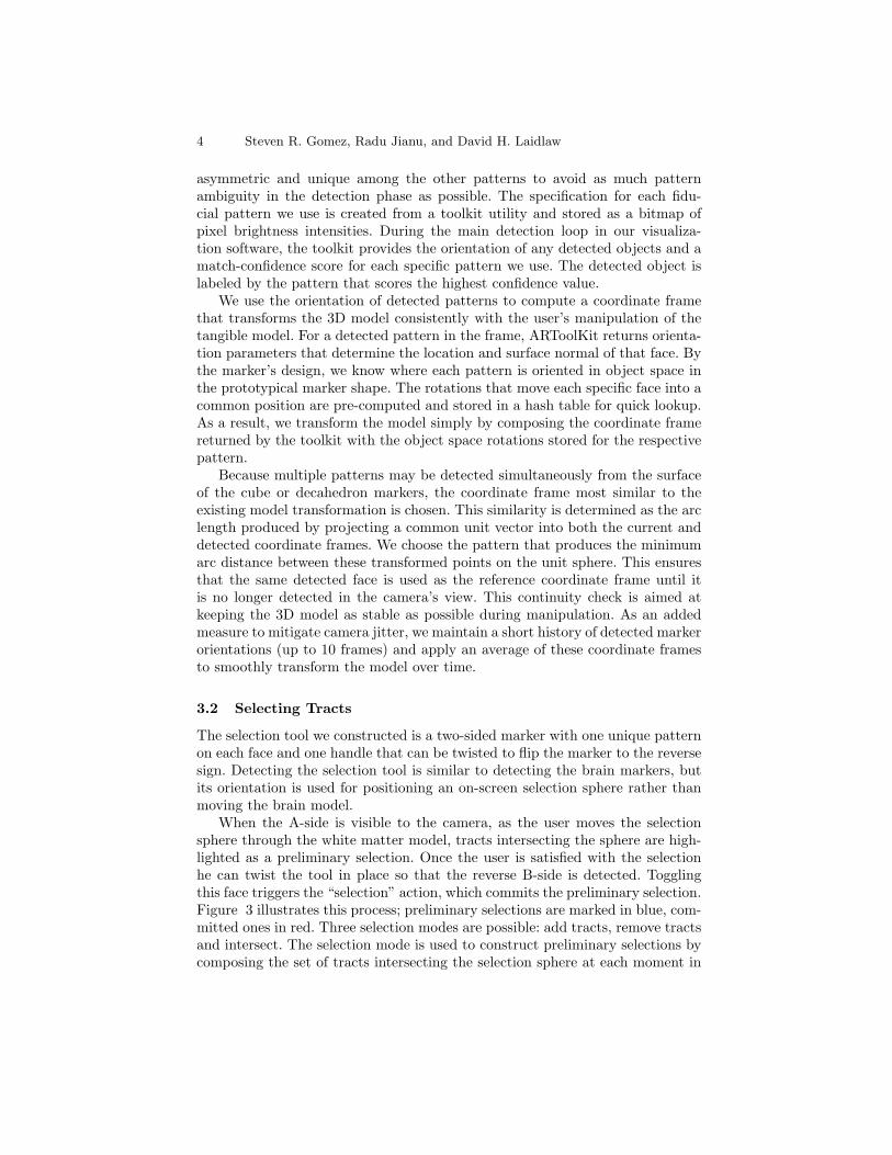
asymmetric and unique among the other patterns to avoid as much patternambiguity in the detection phase as possible. The specification for each fidu-cial pattern we use is created from a toolkit utility and stored as a bitmap ofpixel brightness intensities. During the main detection loop in our visualiza-tion software, the toolkit provides the orientation of any detected objects and amatch-confidence score for each specific pattern we use. The detected object islabeled by the pattern that scores the highest confidence value.
We use the orientation of detected patterns to compute a coordinate framethat transforms the 3D model consistently with the user’s manipulation of thetangible model. For a detected pattern in the frame, ARToolKit returns orienta-tion parameters that determine the location and surface normal of that face. Bythe marker’s design, we know where each pattern is oriented in object space inthe prototypical marker shape. The rotations that move each specific face into acommon position are pre-computed and stored in a hash table for quick lookup.As a result, we transform the model simply by composing the coordinate framereturned by the toolkit with the object space rotations stored for the respectivepattern.
Because multiple patterns may be detected simultaneously from the surfaceof the cube or decahedron markers, the coordinate frame most similar to theexisting model transformation is chosen. This similarity is determined as the arclength produced by projecting a common unit vector into both the current anddetected coordinate frames. We choose the pattern that produces the minimumarc distance between these transformed points on the unit sphere. This ensuresthat the same detected face is used as the reference coordinate frame until itis no longer detected in the camera’s view. This continuity check is aimed atkeeping the 3D model as stable as possible during manipulation. As an addedmeasure to mitigate camera jitter, we maintain a short history of detected markerorientations (up to 10 frames) and apply an average of these coordinate framesto smoothly transform the model over time.
3.2 Selecting Tracts
The selection tool we constructed is a two-sided marker with one unique patternon each face and one handle that can be twisted to flip the marker to the reversesign. Detecting the selection tool is similar to detecting the brain markers, butits orientation is used for positioning an on-screen selection sphere rather thanmoving the brain model.
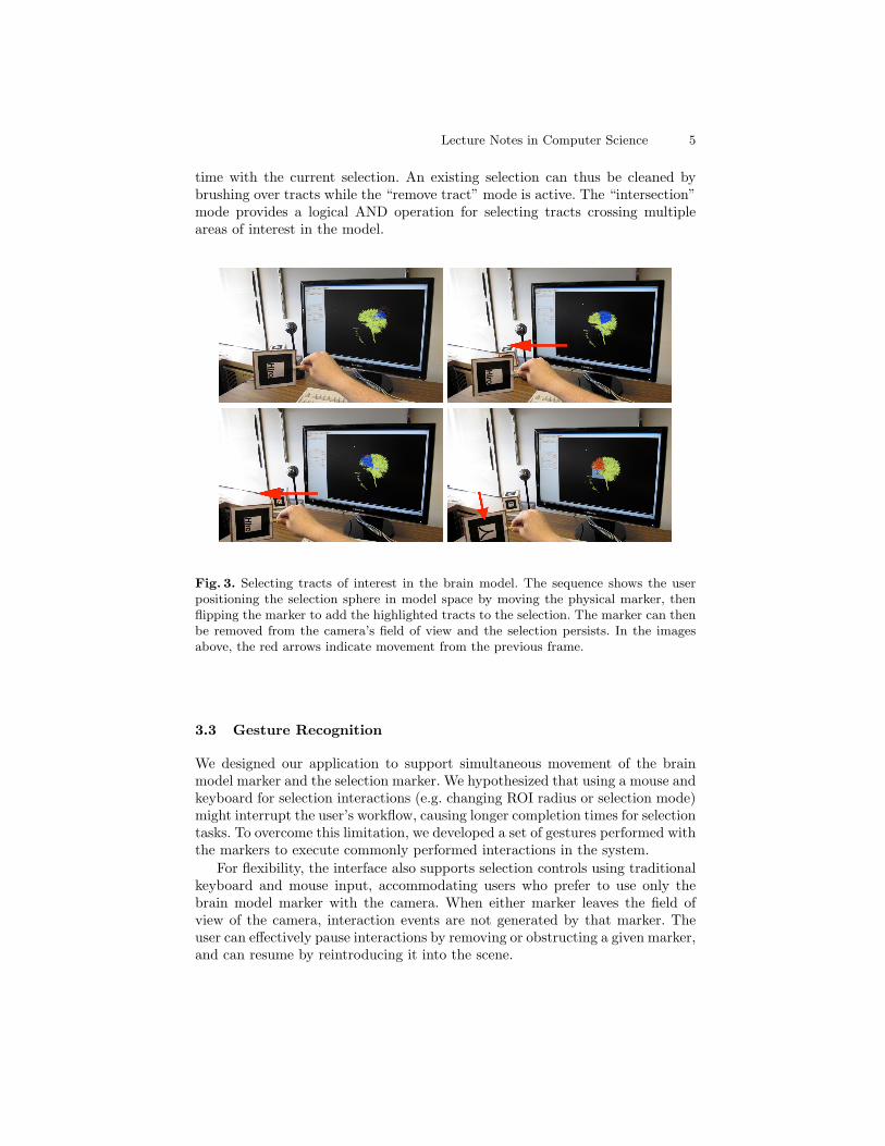
When the A-side is visible to the camera, as the user moves the selectionsphere through the white matter model, tracts intersecting the sphere are high-lighted as a preliminary selection. Once the user is satisfied with the selectionhe can twist the tool in place so that the reverse B-side is detected. Togglingthis face triggers the “selection” action, which commits the preliminary selection.Figure 3 illustrates this process; preliminary selections are marked in blue, com-mitted ones in red. Three selection modes are possible: add tracts, remove tractsand intersect. The selection mode is used to construct preliminary selections bycomposing the set of tracts intersecting the selection sphere at each moment in
Lecture Notes in Computer Science 5
time with the current selection. An existing selection can thus be cleaned bybrushing over tracts while the “remove tract” mode is active. The “intersection”mode provides a logical AND operation for selecting tracts crossing multipleareas of interest in the model.
Fig. 3. Selecting tracts of interest in the brain model. The sequence shows the userpositioning the selection sphere in model space by moving the physical marker, thenflipping the marker to add the highlighted tracts to the selection. The marker can thenbe removed from the camera’s field of view and the selection persists. In the imagesabove, the red arrows indicate movement from the previous frame.
3.3 Gesture Recognition
We designed our application to support simultaneous movement of the brainmodel marker and the selection marker. We hypothesized that using a mouse andkeyboard for selection interactions (e.g. changing ROI radius or selection mode)might interrupt the user’s workflow, causing longer completion times for selectiontasks. To overcome this limitation, we developed a set of gestures performed withthe markers to execute commonly performed interactions in the system.
For flexibility, the interface also supports selection controls using traditionalkeyboard and mouse input, accommodating users who prefer to use only thebrain model marker with the camera. When either marker leaves the field ofview of the camera, interaction events are not generated by that marker. Theuser can effectively pause interactions by removing or obstructing a given marker,and can resume by reintroducing it into the scene.
6 Steven R. Gomez, Radu Jianu, and David H. Laidlaw
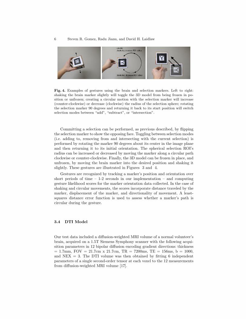
Fig. 4. Examples of gestures using the brain and selection markers. Left to right:shaking the brain marker slightly will toggle the 3D model from being frozen in po-sition or unfrozen; creating a circular motion with the selection marker will increase(counter-clockwise) or decrease (clockwise) the radius of the selection sphere; rotatingthe selection marker 90 degrees and returning it back to its start position will switchselection modes between “add”, “subtract”, or “intersection”.
Committing a selection can be performed, as previous described, by flippingthe selection marker to show the opposing face. Toggling between selection modes(i.e. adding to, removing from and intersecting with the current selection) isperformed by rotating the marker 90 degrees about its center in the image planeand then returning it to its initial orientation. The spherical selection ROI’sradius can be increased or decreased by moving the marker along a circular pathclockwise or counter-clockwise. Finally, the 3D model can be frozen in place, andunfrozen, by moving the brain marker into the desired position and shaking itslightly. These gestures are illustrated in Figures 3 and 4.
Gestures are recognized by tracking a marker’s position and orientation overshort periods of time – 1-2 seconds in our implementation – and computinggesture likelihood scores for the marker orientation data collected. In the case ofshaking and circular movements, the scores incorporate distance traveled by themarker, displacement of the marker, and directionality of movement. A least-squares distance error function is used to assess whether a marker’s path iscircular during the gesture.
3.4 DTI Model
Our test data included a diffusion-weighted MRI volume of a normal volunteer’sbrain, acquired on a 1.5T Siemens Symphony scanner with the following acqui-sition parameters in 12 bipolar diffusion encoding gradient directions: thickness= 1.7mm, FOV = 21.7cm x 21.7cm, TR = 7200ms, TE = 156ms, b = 1000,and NEX = 3. The DTI volume was then obtained by fitting 6 independentparameters of a single second-order tensor at each voxel to the 12 measurementsfrom diffusion-weighted MRI volume [17].
Lecture Notes in Computer Science 7
4 Results
We evaluated our prototype anecdotally with two neuropsychologists who hadexperience using white matter streamtube visualizations in clinical research.Each received a demonstration and brief training on the interface and gesturesbefore exploring the tool and selecting TOIs. We gathered feedback using athink-aloud protocol during our demonstration and the users’ exploration of thetool.
The workflow that both our subjects suggested, and said they were likelyto use with such an application, was to position the model in a specific pose,freeze it, and use the selection marker to perform a selection. They found that forcertain types of interactions, such as model positioning using the fiducial marker,our method would be helpful in supplementing the mouse and keyboard. Bothagreed that different users may have varying preferences for using the fiducialsor a typical keyboard/mouse interface, for either coarse model manipulation orfine-scale TOI selection. For instance, one expert said he preferred the fiducial forcoarse-scale rotation of the model, then favored switching to the mouse for precisetract selection; the other felt he could set the model position more accuratelywith a keyboard and mouse, but preferred the fiducial for tract selection becausehe could quickly select tract bundles in 3D and preview the selection.
Our subjects stated that while the selection gesture is useful, they preferredusing the keyboard and mouse to alter other selection parameters (e.g. adding,removing, or intersecting modes; freezing and unfreezing the model). They notedthat the selection marker was at times difficult to use with precision, given thatexecuting the twist “select” gesture can change the marker’s position, causingdifferent tracts to be selected once the gesture is recognized. One noted that thisbecame easier after a few minutes of practice, and that more extensive initialtraining may give more confidence to the user. Both agreed that adding a newmode to hide non-selected tracts would allow the user to refine the selectionmore easily by reducing visual clutter. Additionally, one expert suggested thatother medical applications requiring quick and easy inspection of a 3D model,such as a heart or tumor visualization, could benefit from an interaction methodlike ours.
5 Discussion
5.1 System Robustness
One finding in our work is that a vision-based approach to model manipulationintroduces challenges from external factors. Fiducial recognition relies on somelevel of light and camera quality beyond our control. Occlusion of the fiducialpatterns can also cause recognition problems, especially when concurrently ma-nipulating both the selection marker and brain maker. One marker may blockthe camera’s view of the other as they try to occupy an overlapping position inmodel space. Even a user’s grip on a marker can introduce occlusion problems.In fact, we noticed that some improved usability of the decahedron over the cube
8 Steven R. Gomez, Radu Jianu, and David H. Laidlaw
was not due to the visibility of multiple patterns at the same time, as initiallyintended, but instead by the ease of holding and moving it while having at leastone marker visible at all times.
5.2 Accommodating User Workflow
In the selection task, the largest challenge we identified was designing interac-tions that were appropriate for the user’s intended level of precision for selection.For instance, a large ROI volume that moves relatively quickly in model spacewhen manipulated may be ideal for a coarse tract bundle selection; however, forrefinements of the selection, a smaller, slower moving selection volume may beeasier for the user to navigate and use without error. Ideally, we would like oursystem to determine the user’s tract selection goals in order to set these parame-ters automatically, to reduce manual specification that may slow the user down.We hypothesize that a coarseness estimate can be made from the level of modelzoom in the 3D visualization; a user who is closely examining the model likelywants to make finer scale selections.
As revealed by our evaluation, the workflow suggested by the users was toposition the model, freeze it, and then make a selection. We believe this is due toour subjects’ familiarity with streamtube visualizations where model positioningand selection cannot happen concurrently. We hypothesize that new users, orthose with training, will prefer to perform selections while manipulating boththe selection marker and the model marker concurrently. We expect this woulddecrease the time required to execute a selection with minimal error becausepositioning the brain and selection markers can be done simultaneously. Thismay require further extension and refinement of the gesture set to obviate allkeyboard and mouse interactions.
5.3 Reaching a Broad Audience
The power of this approach lies in its affordability. Many laptops are now soldwith integrated cameras, and for other personal computers, external webcamsare relatively inexpensive to purchase. In the released version of our application,we will make available marker templates that can be printed, cut, and foldedinto cubes. Users should be able to reproduce our setup in less than half an hourfrom downloading our tool.
Furthermore, we plan to design meaningful pictures, such as projections oftypical brain views (i.e. coronal, sagittal, axial), to distribute as fiducial patternson the marker. Each of these images offers a preview of the model’s orientationbefore the marker face is presented to camera. We believe these fiducials will helpnew users more intuitively understand how manipulation of the marker objectchanges the brain visualization.
Lecture Notes in Computer Science 9
6 Conclusion
We present a lightweight, affordable interaction method for DTI brain visualiza-tions using paper fiducial markers that are easy to create. This method allowsscientists to manipulate 3D white matter models directly by manipulating aphysical analogue in real space in front of a camera. Our contributions includea fiducial-based tracking architecture for interacting with white matter tractog-raphy models, and an evaluation revealing the advantages of this method andsuggesting design guidelines. Furthermore, by using a physical marker that canbe printed, constructed easily, and used without specialized hardware, we pro-vide a simple method for distributing the visualization tool to a wide base ofusers.
References
1. Basser, P., Pajevic, S., Pierpaoli, C., Duda, J., Aldroubi, A.: In vivo fiber tractog-raphy using DT-MRI data. Magnetic Resonance in Medicine 44 (2000) 625–632
2. Mori, S., Van Zijl, P.: Fiber tracking: principles and strategies-a technical review.NMR in Biomedicine 15 (2002) 468–480
4. Jianu, R., Demiralp, C., Laidlaw, D.: Exploring 3d dti fiber tracts with linked 2drepresentations. IEEE Transactions on Visualization and Computer Graphics 15(2009) 1449–1456
5. Catani, M., Howard, R.J., Pajevic, S., Jones, D.K.: Virtual in vivo interactivedissection of white matter fasciculi in the human brain. NeuroImage 17 (2002) 77– 94
6. Wakana, S., Jiang, H., Poetscher, N.L.M., van Zijl, P.C., Mori, S.: Fiber tract-basedatlas of human white matter anatomy. Radiology 230 (2004) 77–87
7. Maddah, M., Mewes, A.U.J., Haker, S., Eric, W., Grimson, L., Warfield, S.K.:Automated atlas-based clustering of white. In: In MICCAI. (2005) 188–195
8. Akers, D.: Cinch: a cooperatively designed marking interface for 3d pathway se-lection. In: UIST ’06: Proceedings of the 19th annual ACM symposium on Userinterface software and technology, New York, NY, USA, ACM (2006) 33–42
9. Zhou, W., Correia, S., Laidlaw, D.H.: Haptics-assisted 3D lasso drawing for tracts-of-interest selection in DTI visualization. IEEE Visualization 2008 Poster Com-pendium (Best Poster Nominee) (2008)
10. Klein, J., Scholl, M., Kohn, A., Hahn, H.K.: Real-time fiber selection using the wiiremote. Proceedings of the SPIE 7625 (2010)
11. (ARToolKit) http://www.hitl.washington.edu/artoolkit/.12. Fiala, M.: Artag, a fiducial marker system using digital techniques. Computer
Vision and Pattern Recognition, IEEE Computer Society Conference on 2 (2005)590–596
13. Fiala, M.: Designing highly reliable fiducial markers. Pattern Analysis and MachineIntelligence, IEEE Transactions on 32 (2010) 1317 –1324
14. Dudek, G., Sattar, J., Xu, A.: A visual language for robot control and program-ming: A human-interface study. In: ICRA. (2007) 2507–2513
10 Steven R. Gomez, Radu Jianu, and David H. Laidlaw
15. Kostandov, M., Schwertfeger, J., Jenkins, O.C., Jianu, R., Buller, M., Hartmann,D., Loper, M., Tsoli, A., Vondrak, M., Zhou, W., Fiala, M.: Robot gaming andlearning using augmented reality. In: SIGGRAPH ’07: ACM SIGGRAPH 2007posters, New York, NY, USA, ACM (2007) 5
16. Kato, H., Billinghurst, M., Poupyrev, I., Imamoto, K., Tachibana, K.: Virtual ob-ject manipulation on a table-top ar environment. Augmented Reality, InternationalSymposium on 0 (2000) 111
17. Basser, P.J., Mattiello, J., LeBihan, D.: Estimation of the effective self-diffusiontensor from the nmr spin echo. Journal of magnetic resonance. Series B 103 (1994)247–254