Pearls A Foot in the Door for Dermatophyte Research Rebecca Rashid Achterman 1 , Theodore C. White 2 * 1 Department of Biology, Western Washington University, Bellingham, Washington, United States of America, 2 Division of Cell Biology and Biochemistry, University of Missouri at Kansas City, Kansas City, Missouri, United States of America What Diseases Do Dermatophytes Cause? Dermatophytes are a group of filamentous fungi that cause infections of the skin (see Figure 1 for diseases, Figure 2 for typical dermatophyte species). Diseases caused by dermatophytes include athlete’s foot, ringworm, jock itch, and nail infections (onycho- mycosis). The medical terminology for dermatophyte infections is to use the word tinea (to denote a fungal infection of the skin) followed by a word that describes the site of infection. For example, tinea pedis refers to athlete’s foot and tinea capitis is scalp ringworm. In general, dermatophytes remain localized to keratinized surfaces such as skin, hair, and nails and do not invade deeper tissues. That said, dermatophyte infections in immuno- compromised patients can be quite severe. Dermatophytes are grouped into three general categories based on their natural environment: anthropophilic (live exclusively on humans), zoophilic (live on an animal host), and geophilic (live in the soil) [1]. The majority of human infections are caused by anthropophilic species; however, species from all three groups have been associated with human disease. For example, pets with ringworm can transmit the infection to their owners, and stray cats carrying dermatophytes are considered to be a vector for infection in several eastern and southern European countries [2]. Is the Most Prevalent Disease the Same from Country to Country? Although dermatophytes are found throughout the world, the most prevalent strains and the most common sites of infection vary by region [2,3]. Hot, humid climates and overcrowding predispose populations to skin diseases, including tinea infections [4]. Developing countries have high rates of tinea capitis, while developed countries have high rates of tinea pedis and onycho- mycosis [2]. Low socioeconomic conditions are strongly linked to higher prevalence rates for skin infections, including tinea infections. A review of 18 studies representing large geographical areas determined that tinea capitis is present in up to 19.7% of the general population in developing countries [4]. A recent study found tinea capitis present in more than 30% of children at certain grade levels in some urban areas of the United States [5]. High prevalence rates of tinea pedis and onychomycosis have been linked to increased urbanization, community showers, sports, and the use of occlusive footwear [2,3,6]. These factors are thought to contribute to the high prevalence of tinea pedis in certain occupational groups, including marathon runners (22%– 31% prevalence), miners (21%–72.9% prevalence), and soldiers (16.4%–58% prevalence) [2]. Several of these studies also found high rates of onychomycosis presenting with tinea pedis. Although tropical and subtropical climates have a higher overall prevalence of skin mycoses, tinea pedis and onychomycosis are rare in India and rural Africa [6]. Why Can People Get Athlete’s Foot (and Other Dermatophyte Diseases) More Than Once? Dermatophyte diseases recur at a high rate following treatment with an antifungal [7]. It is currently unknown whether this is due to insufficient clearing of the fungus during treatment and reemergence of disease, and thus an example of relapse, or if these represent new infections (Figure 3). The high false-negative culture rate from clinical samples contributes to this problem. The advent of molecular biology tools may provide a means by which clinicians can more accurately determine the presence (or absence) of dermatophytes [7]. Certainly, such tools will help determine whether a new infection is indeed caused by the same strain as a previous infection in the same patient. Treatment of dermatophyte infections represents a significant cost burden. It has been estimated that over US$500 million per year is spent worldwide on drugs to treat dermatophytoses [8]. Treatment for skin infections is generally by a topical antifungal. If hair roots or nail beds are infected, an oral antifungal agent is generally prescribed. Nail infections are often recalcitrant to treatment. Immunocompromised patients can experience dissem- inated dermatophyte disease, which has a particularly high treatment failure rate (30.8%) [9]. Why Aren’t There More Drug-Resistant Dermatophytes? Over-the-counter antifungals are commonly used to self-treat athlete’s foot and jock itch. This might be predicted to lead to drug resistance. Surprisingly, drug resistance among dermato- phytes is rare. Two large clinical studies looking at drug susceptibility in dermatophytes did not find significant increases in the minimum inhibitory concentration of several antifungal drugs used to treat dermatophytes [10,11]. That said, occasional drug resistance has been documented. For example, a single amino acid substitution in the target enzyme was found to confer resistance to terbinafine in a clinical isolate from a patient with onychomycosis [12,13]. The question remains, why aren’t mutations conferring drug resistance a more widespread occurrence? One possibility is that dermatophytes are able to tolerate drugs without acquiring point mutations in the target enzyme. In the yeast Candida albicans, Citation: Achterman RR, White TC (2012) A Foot in the Door for Dermatophyte Research. PLoS Pathog 8(3): e1002564. doi:10.1371/journal.ppat.1002564 Editor: Joseph Heitman, Duke University Medical Center, United States of America Published March 29, 2012 Copyright: ß 2012 Achterman, White. This is an open-access article distributed under the terms of the Creative Commons Attribution License, which permits unrestricted use, distribution, and reproduction in any medium, provided the original author and source are credited. Funding: The authors’ research on dermatophytes is funded by NIH R21 AI081235. The funders had no role in study design, data collection and analysis, decision to publish, or preparation of the manuscript. Competing Interests: The authors have declared that no competing interests exist. * E-mail: [email protected]PLoS Pathogens | www.plospathogens.org 1 March 2012 | Volume 8 | Issue 3 | e1002564
Transcript
Pearls
A Foot in the Door for Dermatophyte ResearchRebecca Rashid Achterman1, Theodore C. White2*
1 Department of Biology, Western Washington University, Bellingham, Washington, United States of America, 2 Division of Cell Biology and Biochemistry, University of
Missouri at Kansas City, Kansas City, Missouri, United States of America
What Diseases Do Dermatophytes Cause?
Dermatophytes are a group of filamentous fungi that cause
infections of the skin (see Figure 1 for diseases, Figure 2 for typical
dermatophyte species). Diseases caused by dermatophytes include
athlete’s foot, ringworm, jock itch, and nail infections (onycho-
mycosis). The medical terminology for dermatophyte infections is
to use the word tinea (to denote a fungal infection of the skin)
followed by a word that describes the site of infection. For
example, tinea pedis refers to athlete’s foot and tinea capitis is
scalp ringworm. In general, dermatophytes remain localized to
keratinized surfaces such as skin, hair, and nails and do not invade
deeper tissues. That said, dermatophyte infections in immuno-
compromised patients can be quite severe.
Dermatophytes are grouped into three general categories based
on their natural environment: anthropophilic (live exclusively on
humans), zoophilic (live on an animal host), and geophilic (live in
the soil) [1]. The majority of human infections are caused by
anthropophilic species; however, species from all three groups
have been associated with human disease. For example, pets with
ringworm can transmit the infection to their owners, and stray cats
carrying dermatophytes are considered to be a vector for infection
in several eastern and southern European countries [2].
Is the Most Prevalent Disease the Same fromCountry to Country?
Although dermatophytes are found throughout the world, the
most prevalent strains and the most common sites of infection vary
by region [2,3]. Hot, humid climates and overcrowding predispose
populations to skin diseases, including tinea infections [4].
Developing countries have high rates of tinea capitis, while
developed countries have high rates of tinea pedis and onycho-
mycosis [2].
Low socioeconomic conditions are strongly linked to higher
prevalence rates for skin infections, including tinea infections. A
review of 18 studies representing large geographical areas
determined that tinea capitis is present in up to 19.7% of the
general population in developing countries [4]. A recent study
found tinea capitis present in more than 30% of children at certain
grade levels in some urban areas of the United States [5].
High prevalence rates of tinea pedis and onychomycosis have
been linked to increased urbanization, community showers, sports,
and the use of occlusive footwear [2,3,6]. These factors are
thought to contribute to the high prevalence of tinea pedis in
certain occupational groups, including marathon runners (22%–
31% prevalence), miners (21%–72.9% prevalence), and soldiers
(16.4%–58% prevalence) [2]. Several of these studies also found
high rates of onychomycosis presenting with tinea pedis. Although
tropical and subtropical climates have a higher overall prevalence
of skin mycoses, tinea pedis and onychomycosis are rare in India
and rural Africa [6].
Why Can People Get Athlete’s Foot (and OtherDermatophyte Diseases) More Than Once?
Dermatophyte diseases recur at a high rate following treatment
with an antifungal [7]. It is currently unknown whether this is due
to insufficient clearing of the fungus during treatment and
reemergence of disease, and thus an example of relapse, or if
these represent new infections (Figure 3). The high false-negative
culture rate from clinical samples contributes to this problem. The
advent of molecular biology tools may provide a means by which
clinicians can more accurately determine the presence (or absence)
of dermatophytes [7]. Certainly, such tools will help determine
whether a new infection is indeed caused by the same strain as a
previous infection in the same patient.
Treatment of dermatophyte infections represents a significant
cost burden. It has been estimated that over US$500 million per
year is spent worldwide on drugs to treat dermatophytoses [8].
Treatment for skin infections is generally by a topical antifungal. If
hair roots or nail beds are infected, an oral antifungal agent is
generally prescribed. Nail infections are often recalcitrant to
treatment. Immunocompromised patients can experience dissem-
inated dermatophyte disease, which has a particularly high
treatment failure rate (30.8%) [9].
Why Aren’t There More Drug-ResistantDermatophytes?
Over-the-counter antifungals are commonly used to self-treat
athlete’s foot and jock itch. This might be predicted to lead to
drug resistance. Surprisingly, drug resistance among dermato-
phytes is rare. Two large clinical studies looking at drug
susceptibility in dermatophytes did not find significant increases
in the minimum inhibitory concentration of several antifungal
drugs used to treat dermatophytes [10,11]. That said, occasional
drug resistance has been documented. For example, a single
amino acid substitution in the target enzyme was found to confer
resistance to terbinafine in a clinical isolate from a patient with
onychomycosis [12,13].
The question remains, why aren’t mutations conferring drug
resistance a more widespread occurrence? One possibility is that
dermatophytes are able to tolerate drugs without acquiring point
mutations in the target enzyme. In the yeast Candida albicans,
Citation: Achterman RR, White TC (2012) A Foot in the Door for DermatophyteResearch. PLoS Pathog 8(3): e1002564. doi:10.1371/journal.ppat.1002564
Editor: Joseph Heitman, Duke University Medical Center, United States ofAmerica
Published March 29, 2012
Copyright: � 2012 Achterman, White. This is an open-access article distributedunder the terms of the Creative Commons Attribution License, which permitsunrestricted use, distribution, and reproduction in any medium, provided theoriginal author and source are credited.
Funding: The authors’ research on dermatophytes is funded by NIH R21AI081235. The funders had no role in study design, data collection and analysis,decision to publish, or preparation of the manuscript.
Competing Interests: The authors have declared that no competing interestsexist.
several mechanisms of resistance have been documented that do
not require a point mutation in the target enzyme. More research
is needed to determine if something similar is happening with
dermatophytes.
Why Don’t We Know More about HowDermatophytes Cause Disease?
Despite the prevalence of dermatophyte infections worldwide, a
sophisticated understanding of how they cause disease is lacking
[14]. The historic difficulties in working with dermatophytes have
been two-fold: technical difficulties due to poor virulence models
and a lack of genetic tools, and an under-appreciation of the need
to study these organisms.
There have been several recent advances that minimize the
technical difficulties of working with these organisms. Performing
genetics in dermatophytes has historically posed a challenge;
however, recent advances in the field have provided a foundation
for genetic manipulation of several species of dermatophytes
[15,16].
Currently, the most common animal model for studying
dermatophyte virulence is the guinea pig. Although this has been
useful for zoophiles, guinea pigs and other dermatophyte animal
models do not provide accurate infection models for most
anthropophilic species [15]. Other virulence models include
determining the ability of the organism to grow on keratinized
surfaces such as sterilized nail fragments, which is a non-
quantitative model. Recently, skin explants have been used to
study dermatophyte adherence and invasion. Human epidermal
tissues are commercially available and represent a possible
virulence model to study the initial stages of dermatophyte
infection [15].
One other reason that we don’t know more about dermatophyte
disease is that most scientists, including many mycologists, do not
consider dermatophytes as important as other infectious diseases.
Therefore, there are a limited number of researchers working on
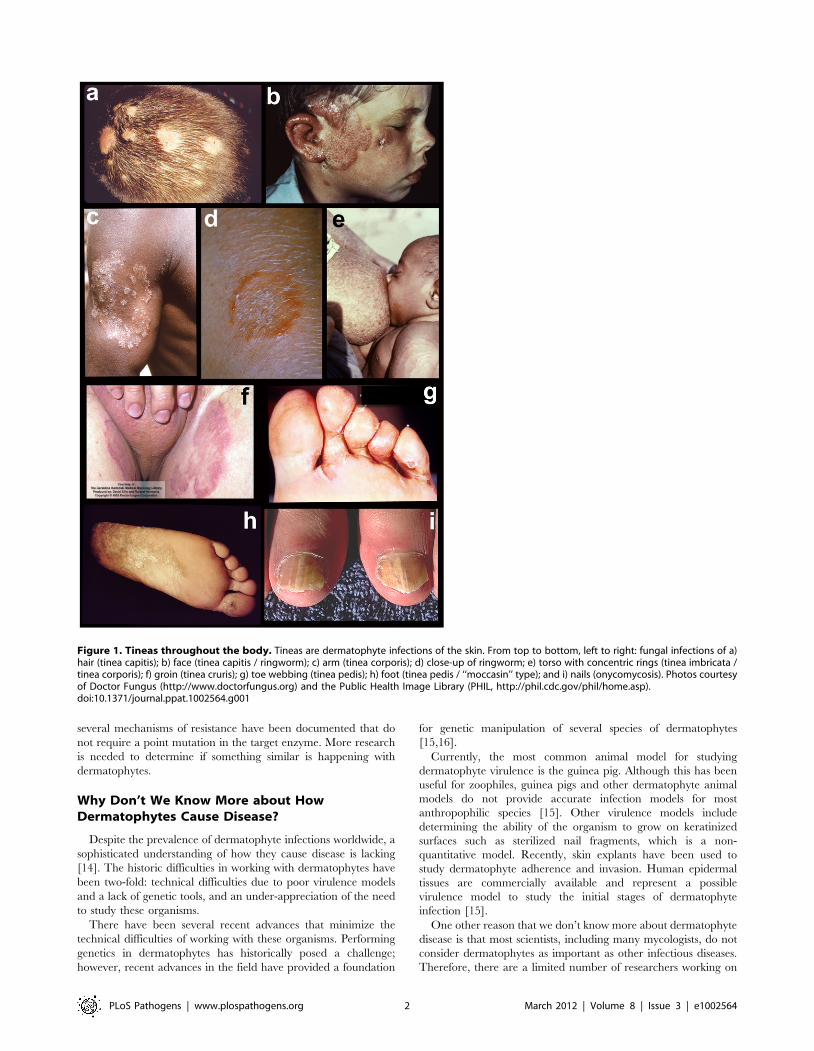
Figure 1. Tineas throughout the body. Tineas are dermatophyte infections of the skin. From top to bottom, left to right: fungal infections of a)hair (tinea capitis); b) face (tinea capitis / ringworm); c) arm (tinea corporis); d) close-up of ringworm; e) torso with concentric rings (tinea imbricata /tinea corporis); f) groin (tinea cruris); g) toe webbing (tinea pedis); h) foot (tinea pedis / ‘‘moccasin’’ type); and i) nails (onycomycosis). Photos courtesyof Doctor Fungus (http://www.doctorfungus.org) and the Public Health Image Library (PHIL, http://phil.cdc.gov/phil/home.asp).doi:10.1371/journal.ppat.1002564.g001
Disseminated dermatophytosis in a patient with hereditary hemochromatosis andhepatic cirrhosis: case report and review of the literature. Med Mycol 48: 518–527.
10. Ghannoum M, Isham N, Sheehan D (2006) Voriconazole susceptibilities of
dermatophyte isolates obtained from a worldwide tinea capitis clinical trial. J Clin
Microbiol 44: 2579–2580.
11. Ghannoum MA, Wraith LA, Cai B, Nyirady J, Isham N (2008) Susceptibility ofdermatophyte isolates obtained from a large worldwide terbinafine tinea capitis
clinical trial. Br J Dermatol 159: 711–713.
12. Mukherjee PK, Leidich SD, Isham N, Leitner I, Ryder NS, et al. (2003) Clinical
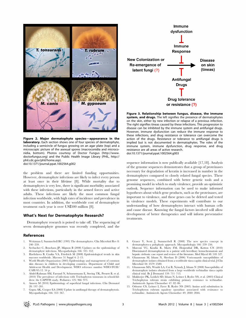
Figure 2. Major dermatophyte species—appearance in thelaboratory. Each section shows one of four species of dermatophyte,including a semicircle of fungus growing on an agar plate (top) and amicroscopic picture of the asexual spores (macroconidia and microco-nidia, bottom). Photos courtesy of Doctor Fungus (http://www.doctorfungus.org) and the Public Health Image Library (PHIL, http://phil.cdc.gov/phil/home.asp).doi:10.1371/journal.ppat.1002564.g002
Figure 3. Relationship between fungus, disease, the immunesystem, and drugs. The left signifies the presence of dermatophyteson the skin, either by new infection or relapse of a previous infection.The right signifies tineas caused by these infections. This progression todisease can be inhibited by the immune system and antifungal drugs.However, immune dysfunction can reduce the immune response tothese infections, and drug resistance or tolerance can overcome theaction of the drugs. Resistance or tolerance to antifungal drugs isimplied but is not documented in dermatophytes. The roles of theimmune system, immune dysfunction, drug response, and drugresistance are all areas of active research.doi:10.1371/journal.ppat.1002564.g003


![Non-dermatophyte onychomycosis · 2016. 1. 14. · gens of already diseased nails. The prevalence of non-dermatophyte molds as nail invaders ranges between 1.45% and 17.60% [3], The](https://static.documents.pub/doc/80x56/60d6204a3fc9a30f0a11bfaa/non-dermatophyte-onychomycosis-2016-1-14-gens-of-already-diseased-nails-the.jpg)


