A fungal pathogen secretes plant alkalinizing peptides to increase infection Sara Masachis, David Segorbe, David Turrà, Mercedes Leon-Ruiz, Ursula Fürst, Mennat El Ghalid, Guy Leonard, Thomas A. Richards, Georg Felix & Antonio Di Pietro SUPPLEMENTARY INFORMATION ARTICLE NUMBER: 16043 | DOI: 10.1038/NMICROBIOL.2016.43 NATURE MICROBIOLOGY | www.nature.com/naturemicrobiology 1
Transcript
Supplementary Information
A fungal pathogen secretes plant alkalinizing peptides to increase infection
Sara Masachis, David Segorbe, David Turrà, Mercedes Leon-Ruiz, Ursula Fürst, Mennat El Ghalid, Guy Leonard, Thomas A. Richards,
Supplementary Figure 1. F. oxysporum induces alkalinization during infection of tomato roots. Roots of tomato plants were immersed in water in the absence (empty circles) or presence (full circles) of F. oxysporum microconidia. Extracellular pH was measured at the indicated times (*, P < 0.01, versus tomato root according to unpaired Student´s t-test) Error bars, s.d., n = 3 biological replicates. Experiments performed twice.
Supplementary Figure 2. F-RALF inhibits root elongation and root hair growth in Arabidopsis. a. Primary sequence of F. oxysporum F-RALF. Arrow indicates the cleavage site of the secretion signal peptide predicted by SignalP. b, c. Effect of F-RALF peptide on root growth and morphology. Seedlings of Arabidopsis (Col-0) were incubated for two days in ½ MS medium supplemented with the indicated concentration of F-RALF peptide or with an equivalent volume of 50% (v/v) methanol (control). In (c) the medium was supplemented with 20 mM MES buffered to pH 5.5 or 6.8. Upper panels: Representative plants from each treatment were imaged. Scale bar 2 mm. Middle panels: Mean length of roots was measured (*, P < 0.0001, versus control according to unpaired Student´s t-test). Error bars, s.d.; n = 20 per treatment. Lower panel: close up photographs of roots. Scale bar 0.5 mm. Experiments performed twice.
Supplementary Figure 3. Generation of f-ralf null mutants, complemented and overexpressing strains. a. Identification of f-ralfΔ deletion mutants by Southern blot analysis. Genomic DNA of the wild type and four independent transformants was treated with the restriction enzymes SacI (left panel) or HindIII (right panel), separated on a 0.7% agarose gel, transferred to a nylon membrane and hybridised with a DNA probe corresponding to the 5' flanking region of the f-ralf gene. Transformants showing banding patterns consistent with ectopic (ectopic) or homologous integration (f-ralfΔ) are indicated. Molecular sizes of the hybridizing fragments are on the left. b. Physical map of the Ptef::f-ralf overexpression construct. The promoter of the F. oxysporum tef-1 gene (Translation Elongation Factor alpha-1) was fused either to the wild type f-ralf allele with its terminator (Ptef::f-ralf), or to a point-mutated f-ralf allele (Ptef::f-ralf(I10A)). c, d. Analysis of f-ralfΔ+Ptef::f-ralf (left panel) and f-ralfΔ+Ptef::f-ralf(I10A) overexpressing strains. c. Genomic DNA of three independent transformants for each construct was used as template for PCR with the primer pair Ptef-ralf-for + Ralf-nest-rev(2) (indicated in b). Presence of an amplification product indicates the presence of the construct in the genome of the f-ralfΔ recipient strain. d. Transcript levels of the f-ralf gene were measured by rt RT-qPCR of cDNA obtained from mycelium of the indicated strains grown for 2 h in liquid minimal medium. Transcript levels were calculated by the ΔΔCt method and normalized to the F. oxysporum actin gene (*, P < 0.0001, versus wild type according to unpaired t-test). Error bars, s.d., n = 3 biological replicates from one representative experiment. Experiments performed twice.
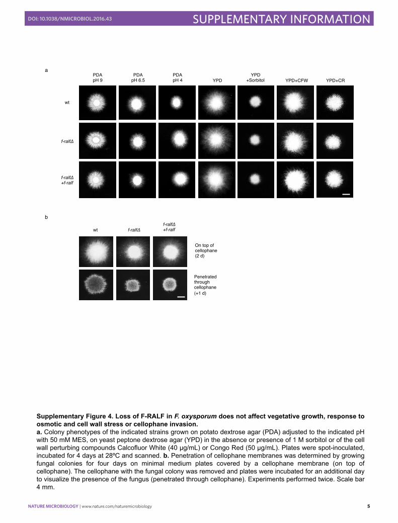
Supplementary Figure 4. Loss of F-RALF in F. oxysporum does not affect vegetative growth, response to osmotic and cell wall stress or cellophane invasion. a. Colony phenotypes of the indicated strains grown on potato dextrose agar (PDA) adjusted to the indicated pH with 50 mM MES, on yeast peptone dextrose agar (YPD) in the absence or presence of 1 M sorbitol or of the cell wall perturbing compounds Calcofluor White (40 µg/mL) or Congo Red (50 µg/mL). Plates were spot-inoculated, incubated for 4 days at 28ºC and scanned. b. Penetration of cellophane membranes was determined by growing fungal colonies for four days on minimal medium plates covered by a cellophane membrane (on top of cellophane). The cellophane with the fungal colony was removed and plates were incubated for an additional day to visualize the presence of the fungus (penetrated through cellophane). Experiments performed twice. Scale bar 4 mm.
Supplementary Figure 5. Virulence of the f-ralfΔ mutant is partially restored by exogenous alkalinization. a. Kaplan-Meier plot showing survival of tomato plants infected with F. oxysporum f. sp. lycopersici. Groups of 20 plants (cultivar Monika) were inoculated by dipping roots in a suspension of 5 x 10⁶ freshly obtained microconidia/ml of the indicated strains, planted in minipots and watered either with unbuffered water or with 1 mM MES adjusted to pH 7. Mortality caused by the f-ralfΔ mutant and the wild type strain was significantly higher at pH 7 (P < 0.001) according to log-rank test. Data shown are from one representative experiment. Experiments performed twice. b. Representative plants from each treatment were imaged 20 days after inoculation.
Supplementary Figure 6. Loss of F-RALF in F. oxysporum results in increased activation of plant defense responses. a. Transcript levels of tomato defense-related genes PR-1 (Pathogenesis-related protein 1), GLUB (basic β-1,3-glucanase), CHI3 (acidic chitinase) and CEVI-1 (pathogen-induced anionic peroxidase) were measured by rt RT-qPCR of cDNA obtained from roots and stems of tomato plants at 5 days after inoculation with the indicated fungal strains or from the non-inoculated control (H2O). b. Transcript levels of Arabidopsis immunity marker genes WRKY53 (salicylic acid-responsive) and PDF1.2 (jasmonic acid-responsive) were measured by rt RT-qPCR of cDNA obtained from plants at 2 days after inoculation with the indicated fungal strains or from the non-inoculated control (H2O). Transcript levels for each sample were calculated by the ΔΔCt method, normalized to the tomato GADPH or the Arabidopsis ACTIN2 gene, respectively, and expressed relative to the non-inoculated control (H2O) (*, P < 0.01, versus wt according to unpaired t-test). Error bars, s.d., n = 3 biological replicates from one representative experiment. Experiments performed twice.
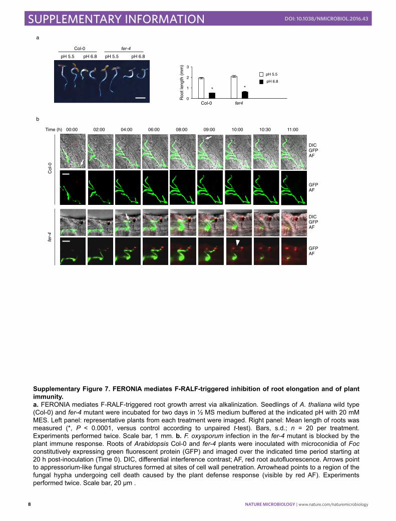
Supplementary Figure 7. FERONIA mediates F-RALF-triggered inhibition of root elongation and of plant immunity. a. FERONIA mediates F-RALF-triggered root growth arrest via alkalinization. Seedlings of A. thaliana wild type (Col-0) and fer-4 mutant were incubated for two days in ½ MS medium buffered at the indicated pH with 20 mM MES. Left panel: representative plants from each treatment were imaged. Right panel: Mean length of roots was measured (*, P < 0.0001, versus control according to unpaired t-test). Bars, s.d.; n = 20 per treatment. Experiments performed twice. Scale bar, 1 mm. b. F. oxysporum infection in the fer-4 mutant is blocked by the plant immune response. Roots of Arabidopsis Col-0 and fer-4 plants were inoculated with microconidia of Foc constitutively expressing green fluorescent protein (GFP) and imaged over the indicated time period starting at 20 h post-inoculation (Time 0). DIC, differential interference contrast; AF, red root autofluorescence. Arrows point to appressorium-like fungal structures formed at sites of cell wall penetration. Arrowhead points to a region of the fungal hypha undergoing cell death caused by the plant defense response (visible by red AF). Experiments performed twice. Scale bar, 20 µm .
Supplementary Figure 8. Full images of western and Southern blots included in figures. a. Full images of western blots included in Figure 1c. Antibodies used are indicated on the right. Arrows point to hybridising bands corresponding to the indicated proteins. Relative positions of molecular size markers (kDa) are on the left. Note that the anti-phospho-p44/42 MAPK a n t i b o d y a l s o r e c o g n i z e s t h e phosphorylated form of the cell wall integrity MAPK Mpk1 which has a higher molecular weight than Fmk1; that the hybridization solution with the anti-Fus3 antibody also contained anti-Mpk1 antibody for simultaneous detection of both MAPKs; and that the anti-mouse-α-tubulin antibody used as loading control detects a number of unspecific bands in the F. oxysporum protein extract, besides the 50 kDa α-tubulin band. b. Full images of the Southern blots included in Supplementary Data Figure 3a. Molecular size markers (kb) are on the left .
ATTACATTATC Ralf-rev AATGTCGTGGTGGTGTTGGTG Ptef1t-for (2) AACACACAGGCGCAAGACCAA Ralf-nest-rev CCGCCACTTTAGTCTTTCCGA Mut-ralf-for CGGTGAGGCCTCATATGGTGC Mut-ralf-rev CCATATGAGCGCTCACCGCTC ralf-RT-rev GTCCTCTCTTAGTTGCCACCA Real time qPCR primer (f-ralf) act-q7 ATGTCACCACCTTCAACTCCA Real time qPCR primer (actin1) act-q8 CTCTCGTCGTACTCCTGCTT Real time qPCR primer (actin1) pr1-7 GCATCCCGAGCACAAAACTA Real time qPCR primer (PR1) pr1-8 TGGTAGCGTAGTTATATCTG Real time qPCR primer (PR1) gluB-7 ATTCTGTTTATGCTGCGATGG Real time qPCR primer (GLUB) gluB-8 CTTTCTCGGACTACCTTCTTT Real time qPCR primer (GLUB) chi3-5 TCTTGTCTCTTTTTCTTGTTCC Real time qPCR primer (CHI3) chi3-6 GCAGTATCATCACCAGCAGT Real time qPCR primer (CHI3) gadph-1 TGATTTGAACTCGTCGCAG Real time qPCR primer (GADPH) gadph-2 CCAAAAACAGTAACAGTAACAGCCTTC Real time qPCR primer (GADPH) six-1-1 ATAGCATGGTACTCCTTGGCG Real time qPCR primer (six1) six-1-2 CCTGATGGTGACGGTTACGAA Real time qPCR primer (six1) cevi1-forRTPCR TCCATTTGAAAGCCT Real time qPCR primer (CEVI1) cevi1-revRTPCR AAGTCTTTGTTGAAA Real time qPCR primer (CEVI1) Actin2-for TCCCTCAGCACATTCCAGCAGAT Real time qPCR primer (ACTIN2) Actin2-rev AACGATTCCTGGACCTGCCTCATC Real time qPCR primer (ACTIN2) PDF1.2-for TGTTCTCTTTGCTGCTTTCGACGC Real time qPCR primer (PDF1.2) PDF1.2-rev TGTGTGCTGGGAAGACATAGTTGC Real time qPCR primer (PDT1.2) WRKY53-for GCGACAAGACACCAGAGTCA Real time qPCR primer (WRKY53) WRKY53-rev ACCGTTGGATTGAACCAGTC Real time qPCR primer (WRKY53) FRK1-for GGAAGCGGTCAGATTTCAAC Real time qPCR primer (FRK1) FRK1-rev AGCTTGCAATAGCAGGTTGG Real time qPCR primer (FRK1)