A F U R T H E R S T U D Y O N Q U I C K E R R E - E N T R Y O F C H R O M O S O M A L L Y A B N O R M A L C E L L S T O A C T I V E P R O L I F E R A T I O N A N D P R E F E R E N T I A L D E A T H O F
C H R O M O S O M A L L Y N O R M A L C E L L S D U R I N G A G E I N G
O F S P E C I M E N S IN A C U T E M Y E L O I D L E U K A E M I A
( A M L )
You-SHENG Ll, JOHN G. MATTHEWS and FRANK G. J. HAYHOE
Department of Haematological Medicine, University Clinical School, Cambridge, U.K.
(Received 15 December 1983. Accepted 22 December 1983)
Abstract--Quicker re-entry of chromosomally abnormal cells to active proliferation under culture conditions was observed in cultured peripheral blood samples, old or fresh, but not in cultured bone marrow samples. In 10 patients with AML studied using peripheral blood, the mitotic index (MI) rose after 24 h in culture and reached a peak after 48 h in culture in AA patients (with all abnormal karyotypes); the MI rose after 24 h in culture and reached a peak after 48, 72 or 96 h in culture in AN patients (mixed normal and abnormal karyotypes); the MI remained at much lower level until after 72, 96 or 120 h of culture in NN patients (all normal karyotypes). The MI at 24 and 48 h of culture was significantly higher in AN and AA patients than in NN patients. A significantly increased frequency of chromosomally normal cells after prolonged culture was observed in one patient with t(8;21 ). The results of this study also suggest that chromosomally normal cells are more likely to die off than are abnormal ones during ageing of specimens.
IN THE cytogenetic study of haematological disorders a short-term of culture, up to a few days, has some advantage over the so-called direct method. It not only improves the quality of metaphases but also increases the yield of mitoses, both of which are prime re- quirements in chromosome analysis of leukaemic cells. However, whether a short term of culture stimulates preferentially the growth of chromosomally abnormal (aneuploid and pseudodiploid) cells or the growth of chromosomally normal (diploid) cells has been hitherto a matter for debate [10, 14, 15, 17, 20, 21]. In the study of cytogenetic prepara- tions from samples sent by post we observed that chromosomally abnormal cells appeared to re-enter active proliferation more quickly than chromosomally normal cells after they were put in culture [12]. The number of patients studied was relatively small, but the fre- quency of chromosome abnormalities was significantly higher in harvests of 20-30 h culture (5/5) than in harvests of 30-120 h culture (14/30). More direct experimental evidence for the quicker re-entry of chromosomally abnormal leukaemic cells into active proliferation was obtained by analysis of the cytogenetic results of serially harvesting leukaemic cultures. The results of this study support the previous conclusion that chromosomally normal cells are more likely to die off during ageing of specimens than chromosomally abnormal cells [12].
Abbreviations: AML, acute myeloid leukaemia; AMML, acute myelomonocytic leukaemia; AMyL, acute myeloblastic leukaemia; AA, all abnormal karyotypes; AN, mixed normal and abnormal karyotypes; NN, all normal karyotypes; MI, mitotic index.
Correspondence to: Professor F. G. J. Hayhoe, Department of Haematological Medicine, University Clinical School, Hills Road, Cambridge CB2 2QL. U.K.
659
660 You-SHENG LI, JOHN O. MAFTHEWS and FRANK G. J. HAYHOE
M A T E R I A L S A N D M E T H O D S
Patients A total of 13 patients with AML, either acute myel0blastic'(AMyL) or acute myelomonocytic leukaemia
(AMML), were entered in this study. Nine patients were non-selected and four (cases $7, S136, $294 and $363) were added after preliminary chromosome analysis revealed abnormal clones in Leishman stained preparations. Two patients (cases $7 and SI36), in whom the cell cultures were harvested only twice, but at 5 day intervals. were also included.
Specimens The material used was bone marrow in two patients (cases S136 and $280), both peripheral blood and bone
marrow in one patient (case $285), and peripheral blood only in the remaining 10 patients. No patient was leucopenic. Four patients (cases $276, $278, $279 and $368) were from outside Cambridge, the blood samples having been sent by post. For the remaining nine patients admitted to Addenbrooke's Hospital all fresh samples were put in culture within a few hours of being taken except for case $280 where a bone marrow sample was kept at 5°C for 16 h before culture. An aliquot was set aside from all peripheral blood samples (except in one patient. case $7) kept on the bench for 2 or 3 days, and then cultured again for chromosome study in order to observe the effects of specimen ageing. For all blood samples which were not fresh the percentage of dead cells was roughly determined on Leishman stained smears counting cells with disrupted or homogeneously stained nuclei as dead cells. Some cells counted as "l ive" might of course have reduced viability and might have died off soon after culture.
Cytogenetics, mitotic index and cytochemistry Cells were cultured at a cell concentration of 1-2 × 10⁹/1 in 10 ml of medium in a tightly capped Universal
bottle. McCoy's medium was used for marrow cultures and TC199 medium for peripheral blood cultures. Several cultures at the same cell concentration were set up in each case for serial harvests. Leishman staining and the G-banding technique were both applied to each case [12]. Karyotypes were determined by banding for every patient but not for each occasion of harvesting. The ratio of chromosomally abnormal cells to chromosomally normal cells for each harvest was determined mainly on Leishman stained preparations, which we found more reliable. In cases $279 and $283 it was difficult to identify the minor abnormalities on Leishman stained prepara- tions while the banded preparations contained many ambiguous metaphases. Since too tentative an interpreta- tion of chromosome abnormalities might be misleading the ratio of chromosomally abnormal to chromosomally normal cells was not determined in these cases.
The mitotic index (MI) was determined by counting the percentage of cells in mitosis on Leishman stained preparations. Although a 10- to 100-fold rise in M! in the patients could by no means have been due to an uneven distribution of metaphases on slides, caution was taken to avoid only counting cells on a certain area. Twenty or more mitoses were usually surveyed for each harvest if possible. If no metaphases were found and the MI was recorded as 0°70, usually more than 50,000 cells had been searched and counted. Dual esterase staining [71 was attempted in cases $278 and $279 after 120 h in culture.
In order to rule out the possibility of the transformation of normal T lymphocytes a control study was per- formed in a normal individual and I l patients with chronic lymphocytic leukaemia or hairy cell leukaemia.
R E S U L T S
T h e r e l e v a n t d a t a a n d the resu l t s o f the c y t o g e n e t i c s t u d y f r o m the 13 pa t i en t s w i th
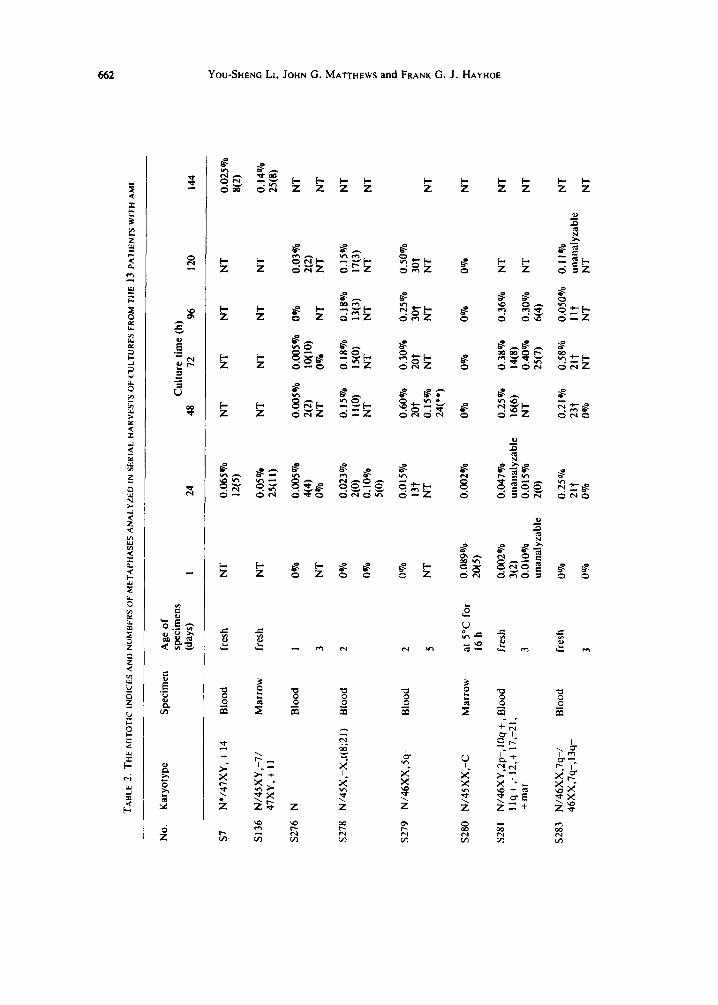
A M L a re s h o w n in T a b l e s l a n d 2. T h e q u i c k e r r e - en t ry o f c h r o m o s o m a i l y a b n o r m a l cells
to ac t i ve p r o l i f e r a t i o n u n d e r c u l t u r e c o n d i t i o n s was o n l y o b s e r v e d in p e r i p h e r a l b l o o d
cu l t u r e s bu t n o t in b o n e m a r r o w cu l tu res . T h e f o l l o w i n g d i s cus s ion on this m a t t e r re fe rs
on ly to p e r i p h e r a l b l o o d cu l tu res .
T h e pa t i en t s w e r e c lass i f i ed as A A (wi th o n l y a b n o r m a l cells) , A N (mixed n o r m a l a n d
a b n o r m a l cells) , o r N N (all n o r m a l cells). In the t w o A A pa t i en t s , the MI rose a f t e r 24 h o f c u l t u r e , r e a c h e d a p e a k a f t e r 48 h o f cu l t u r e , a n d then fell s l ight ly . In the f ive A N pa t i en t s ,
the M l a l so rose a f t e r 24 h o f c u l t u r e a n d r e a c h e d a p e a k a f t e r 48 h in o n e pa t i en t a n d a f t e r
72 o r 96 h in the r e m a i n i n g f o u r pa t i en t s . In the 3 N N pa t i en t s the M I r e m a i n e d at a l o w e r
level unt i l 72 h o r l a te r in cu l t u r e , the peak o f MI o c c u r r i n g at 72 o r 96 h in two o f the
three N N pa t i en t s a n d at 120 h in c u l t u r e in the th i rd . T h e m a j o r i t y o f the cells were
pres t~mably d e a d o r u n d i v i d i n g i f t he re were no mi to se s f o u n d in cu l t u r e f r o m an o ld
s p e c i m e n . W h e n u n y i e l d i n g ha rves t s f r o m such cu l tu re s were e x c l u d e d , the MI at 24 h in
cu l tu re was h i g h e r in all the l 0 ha rves t s f r o m A N a n d A A pa t i en t s t h a n in all th ree
ha rves t s f r o m N N pa t i en t s . T h e d i f f e r e n c e is a l so s i gn i f i c an t (p = 0 .0045) . T h e
c h r o m o s o m a l l y a b n o r m a l cells thus a p p e a r e d to r e s p o n d m o r e q u i c k l y to t he s t imu lus o f
c u l t u r e c o n d i t i o n s a n d en t e r i n t o ac t i ve p r o l i f e r a t i o n s o o n e r t h a n n o r m a l ones .
TABI.E I. S[.x, AGE AND OIHER DETAILS OF I
HE
13 P
ArlEN FS WI IH AML
No.
A
ge
Sta
ge o
f Se
x (y
ears
) D
iagn
osis
di
seas
e
Leu
cocy
te
Age
of
coun
ts
spec
imen
s (%
) ( ×
10
'/I)
S
peci
men
s (d
ays)
(%
) o
f bl
asts
o
f de
ad c
ells
$7
SI3
6
$276
$278
$279
$280
$281
m m f f f f
$283
f
$285
f
$294
m
$363
f
$367
f
$368
m
69
AM
yL
diag
nosi
s 61
A
MyL
di
agno
sis
42
AM
yL
diag
nosi
s
29
AM
yL
diag
nosi
s
52
AM
yL
diag
nosi
s
34
AM
yL
rela
pse
68
AM
ML
di
agno
sis
18
AM
ML
di
agno
sis
52
AM
ML
di
agno
sis
10
AM
yL
diag
nosi
s
78
AM
ML
di
agno
sis
53
AM
yL
rela
pse
23
AM
ML
di
agno
sis
150
bloo
d marrow
180
bloo
d
10
bloo
d
16
bloo
d
32
mar
row
47
bloo
d
350
bloo
d
120
mar
row
bl
ood
21
bloo
d
33
bloo
d
30
bloo
d
110
bloo
d
fres
h 82
N
T
fres
h 36
N
T
I 20
7
3 70
99
2 68
8
5 50
30
2 55
21
5
12
55
at 5
°C f
or
65
NT
1
6h
fres
h 80
N
T
3 68
21
fres
h 83
N
T
3 74
94
fres
h 60
N
T
fres
h 28
N
T
3 68
85
fres
h 60
N
T
2 50
12
fres
h 50
N
T
3 73
50
fres
h 90
N
T
3 N
T
60
1 34
46
3
50
50
O
O
,-i o O 8 W
~r
O 5"
>
r-
NT
, no
t te
sted
.
TA
BL
E 2
. T
HE
MIT
OT
IC I
ND
ICE
S A
ND
NU
MB
ER
S O
F M
ET
AP
HA
SE
S A
NA
LY
ZE
D I
N S
ER
IAL
HA
RV
ES
TS
OF
CU
LT
UR
ES
FR
OM
TH
E 1
3 P
AT
IEN
TS
WIT
H A
ML
No.
K
aryo
type
S
peci
men
A
ge o
f sp
ecim
ens
Cul
ture
tim
e (h
) (d
ays)
1
24
48
72
96
120
144
$7
N*/
47X
Y, +
14
Blo
od
fres
h
SI3
6 N
/45
XY
,-7
/ M
arro
w
fres
h 47
XY
, +
I 1
$276
N
B
lood
$278
N
/45X
,-X
,t(8
;21)
B
lood
NT
NT
I 0%
3 N
T
2 0
%
0%
$279
N
/46X
X, 5
q-
Blo
od
2 5
$280
N
/45
XX
,-C
M
arro
w
at 5
°C f
or
16
h
$281
N
/46X
Y,2
p-,I
Oq
+,B
loo
d
fres
h I i
q+
,-1
2,+
17,
-21,
+
mar
3
$283
N
/46
XX
,7q
-/
Blo
od
fres
h 46
XX
,7q-
, 13q
-
3
0%
NT
0.08
9o/0
20(5)
0.00
2%
3(2)
0,01
0%
unan
alyz
able
0%
O%
0.06
5%
NT
N
T
NT
N
T
12(5
)
0.05
%
NT
N
T
NT
N
T
2500
0.00
5%
0.00
5%
0.00
5%
0%
0.
03%
4(
4)
2(2)
10
(10)
2(
2)
0%
N
T
0%
N
T
NT
0.02
3%
0.15
%
0.18
%
0.18
%
0.15
%
2(0)
Ii
~)
15(0
) 13
(3)
170)
0.
10%
N
T
NT
N
T
NT
5(
0)
0.02
5%
8(2)
0.14
070
25(8)
NT
NT
NT
NT
0.01
5%o
0.60
0?0
0.30
%
0.25
0"/0
0.
50%
e ! 3
t 2o
t 2o
t 3o
t 3o
t N
T
0.15
%o
NT
N
T
NT
N
T
24(*
*)
0.00
2o/0
0%
0 00
70
0%0
0%0
NT
0.04
7%
0.25
%
0.3
80
/0
0.36
0/0
NT
N
T
unan
alyz
able
16
(6)
14(8
) 0.
015%
0 N
T
0.40
%0
0.30
%
NT
N
T
2(0)
25
(7)
6(4)
0.25
%
0.21
%
0.58
%
0.05
0%
0.11
%
NT
21
? 23
t 2
it
I1?
unan
alyz
able
0
%
0%
N
T
NT
N
T
NT
o b~
O t-
b"
Z O
K
>
e~
"rl
> Z O
>
TAB
LE 2
CO
NT
INU
ED
.
No.
K
aryo
type
S
peci
men
A
ge o
f sp
ecim
ens
(day
s)
i C
ultu
re t
ime
(h)
24
48
72
96
120
144
$285
N
M
arro
w
fres
h 0.
050o
/0
1808
) B
lood
fr
esh
0.00
3%
3 N
T
0.00
8%
0.01
9%
NT
0.
005%
0.
020%
un
anal
yzab
le
2(2)
2(
2)
0.005%
0.005%
NT
0.07
9%
0.015%
5(5)
0%
0o
70
NT
N
T
NT
O%
0.03
4%
2(2)
N
T
NT
NT
NT
NT
NT
$294
45
X,-
Y,t
(8;2
1)
Blo
od
fres
h 0.002%
unan
alyz
able
2
NT
0.04
8%
0.80
0/0
0.20
%
NT
0.
19%
4(0)
I(0)
10(0)
12(0)
NT
N
T
NT
0
%
NT
$363
47
XX
, + 2
0, +
DM
C B
lood
fr
esh
0.01
0%
2(0)
3
NT
0.03
0%
0.04
0%
0.03
0%
0.01
5%
0.00
8%
20(0
) 10
(0)
15(0
) 7(
0)
NT
0.
i 0
%
NT
N
T
NT
30
(0)
$367
N
/4S
XX
,-C
B
lood
fr
esh
0%
3 N
T
0.01
2%
0.30
%
0.42
%
NT
0.
069%
23
(15)
8(
6)
NT
N
T
0.82
%
NT
N
T
12(1
0)
$368
N
B
lood
1
0%
3 N
T
0.00
56o7
o 0.
015%
0.
31°7
o N
T
0.12
o7o
8(8)
10
(10)
10
(10)
N
T
NT
0
.04
8%
o
NT
N
T
lO(l
O)
NT
NT
O
o O B m
t~
o B [.-
*N 4
6XX
or
46X
Y.
)'Fig
ures
not
sco
rabl
e w
ith
cert
aint
y; s
ee t
ext.
N
umbe
r o
f ch
rom
osom
ally
nor
mal
cel
ls i
n br
acke
ts.
NT
,Not
tes
ted.
664 YOU-SHENG L,, JOHN G. MATTHEW$ and'FRANK G. J. HAYHOE
Significantly increased frequency of chromosomally normal cells after prolonged culture was only observed in one patient (case $278).
The quicker re-entry of chromosomally abnormal cells to active proliferation was observed in cultures both from old and fresh peripheral blood specimens.
Attempts were made to study the effects of specimens ageing by leaving the specimen on the bench for a few days. In cultures from peripheral blood specimens of three or more days old metaphases were found in five out of six (83%) AN and AA patients but only one out of three (33°70) NN patients and the MI was much lower in the NN patient than in the AA and AN patients.
The percentage of blast cells decreased in five patients and increased in four patients (cases $276, $285, $363, $368) after peripheral blood samples had become older (Table 1). Three out of the four patients whose percentage of blast cells increased had a normal karyotype and the fourth, though karyotypically abnormal, showed a low level of MI which was similar to that in NN patients but different from that in the remaining AN and AA patients. This might reflect the low frequency of blast cells in the original specimens. Mature monocytes died quickly during ageing of specimens. As shown in Table l, the frequency of dead cells in old specimens was parallel with their age and the height of the leucocyte counts. It might also be influenced by the karyotype and other characteristics of the cells, but the number of patients studied was too small for these possibilities to be explored.
In case $279, more than half the metaphases harvested after 48 and 96 h of culture showed numerous fine granules in the cytoplasm. Dual esterase staining in cases $278 and $279 revealed 40 and 50% of chloroacetate esterase positive cells but no butyrate esterase positivity. This suggests that the cells studied were granulocyte precursors, which supports the view that spontaneously transformed lymphocytes are too few to cause any confusion.
In case $279 the harvest after 24 h of culture from a two-day-old specimen and the harvest after 48 h of culture from a five-day-old specimen both revealed nearly half the metaphases to be chromosomally abnormal with non-clonal abnormalities including four metaphases with 47 chromosomes.
A challenge to the prolonged culture up to 5 or 6 days may be that prolonged cultures might stimulate normal lymphocytes to divide. Peripheral blood or spleen cells from a normal individual and I I patients with chronic lymphoproliferative disorders were cultured for 120-144 h and the highest MI was 0.006%. In 8 of those 11 patients cultures were also set up with PHA and the MI was from 0.1 to 1.4% in six. Apparently spon- taneously transformed lymphocytes did not cause any confusion in this study.
DISCUSSION
Results of several recent investigations indicated that a period of in vitro culture substantially increased the likehood of detecting an abnormal clone of leukaemic cells [2, 5, 9, 14, 10, 21]. This seems incompatible with the generally held view that culture condi- tions lead to the proliferation of normal marrow elements [14, 20]. However, all these in- vestigations were based on bone marrow study and a comparison between the direct method and the culture method. Different periods of culture have not been subjected to comparison in detail. It was mentioned in a previous paper [12] that the frequency of chromosome abnormalities decreased when the culture time was extended from 30 to 120 h. The explanation was that the chromosomally abnormal cells respond to the improve- ment of living conditions more quickly than chromosomally normal ceils. This explana- tion has been confirmed by the present study.
In this study the quicker re-entry of chromosomally abnormal cells to active prolifera- tion was observed in cultures both from fresh and old peripheral blood specimens but not in bone marrow culture. In the patients studied using peripheral blood, the peak of MI
Proliferation of ¢hromosomally abnormal cells in AML 665
was found at 48 h culture in AA patients, at 48, 72 or 96 h culture in AN patients, and at 72, 96 or 120 h culture in NN patients. The MI was significantly lower in NN patients than in AN and AA patients at 24 or 48 h culture.
The majority of peripheral blood cells in AML are non-cycling ceils and therefore in Go phase. The delayed re-entry of chromosomally normal cells to active proliferation com- pared to chromosomally abnormal cells might imply that they are less readily induced to cell cycle. The low percentage of blast cells might have contributed to the generally low MI in the three NN patients. However, this was not necessarily related to the delayed appearance of the MI peak as the peak was found at 48 h culture in case $363 where the percentage of blasts in the specimen and the M1 in subsequent culture were both very low.
The quicker re-entry of chromosomally abnormal cells to active proliferation was not observed in bone marrow cultures, perhaps because a higher proportion of bone marrow ceils were actively in cycle before culture, thus masking the re-entry of Go cells into cycle under culture conditions. As to the decreased frequency of chromosomally normal cells in bone marrow culture noticed by other investigators [2, 5, 10, 19, 21], it might reflect the loss of the capacity for division in erythrocyte precursors soon after culture (?matura- tion), as pointed out by Berger et al. [3]. Non-leukaemic erythrocyte precursors were not suppressed by the leukaemic myeloid cells in bone marrow [6]. Nucleated erythrocytes are much less common in peripheral blood.
By the study of premature chromosome condensation Hittelman et al. concluded that under growth-limiting conditions transformed cells accumulated in late G, phase while normal cells accumulated in early G~ phase [8]. Moreover, some malignant cells might be able to enter into S phase and die before metaphase. It is understandable that chromosomally abnormal leukaemic cells reached the metaphase stage before normal-cells after they had been induced to cycle if we ignore the difference of cell cycle time between leukaemic and normal cells. However, this cannot explain the delayed MI peak of chromosomally normal cells as long as a few days in cases $276 and $285 compared to that in the chromosomally abnormal cases.
A delayed response of T lymphocytes to PHA [11, 16] was commonly found in B cell disorders and in AML. This was supposed to be due to the diminished frequency of T lym- phocytes among leukaemic populations [4, 11, 19]. The quicker response of chromosomally abnormal cells to the stimulus of culture conditions might reflect the predominance of chromosomally abnormal leukaemic cells and relatively low concentra- tion of chromosomally normal non-leukaemic cells. This may explain the delayed response of chromosomally normal cells in case $278, but not that in the NN patients, from whom the specimens contained many blast cells.
A tentative explanation is that the blast cells in such patients might be leukaemic but suppressed by more mature non-leukaemic monocytoid cells. In our experience with the cytogenetic study of AMML, when the leucocyte count was high and the predominant cells in peripheral blood were promonocytes or monocytes, the cells from peripheral blood were difficult to grow in vitro and died off readily during ageing of specimens. Moreover, the karyotype tended to be normal when the cells grew. Barak et al. [1] sug- gested that the marrows presenting diminished or no growth patterns in soft agar culture were presumably those from AMyL patients with a monocytic reaction, and Richman and Rowley found marrows presenting such growth patterns were more likely to have a nor- mal karyotype [13]. The presence of reactive monocytoid components in AMML was also supported by the observation that the cells from patients with monocytic leukaemia were in a state of activation, ready to phagocytose [18].
All the explanations given above are based on the presumption that the chromosomally normal cells were non-leukaemic cells. This might not be true in all cases. A fourth pos- sible, more likely, explanation is that the presence of detectable chromosome abnor- malities is associated with great activity of malignant cells, featured by the quick re-entry
666 You-SHENG LI, JOHN G. MATTHEWS and FRANK G. J. HAYHOE
tO active pro l i fe ra t ion and high MI in this s tudy.
The preferent ia l death of ch romosoma l ly n o r m a l cells dur ing ageing of specimens was further conf i rmed . We have received 34 peripheral b lood samples of A M L sent by post
since the s tudy repor ted earlier was completed [12]. A m o n g the 23 samples which were
two days or less old enough metaphases were ob ta ined in 12 (50o70) and four (33o70) were
found to have a b n o r m a l clones. A m o n g the 11 samples more than two days old enough
metaphases were ob ta ined in five (45o70) a nd three (60°7o) showed a b n o r m a l clones.
However , the a t t empt to ob ta in direct evidence for the preferent ia l death of ch romosomal ly n o r m a l cells by leaving specimens on the bench for a certain period ended
in failure. The pa t t e rn of cell death dur ing ageing of specimens may be more compl ica ted
than expected and the under ly ing reason remains unclear .
Acknowledgements--YSL is the holder of a grant from the Chinese Academy of Medical Sciences. FGJH is the Leukaemia Research Fund Professor of Haematoiogical Medicine.
R E F E R E N C E S
1. BARAK Y., MA A. & SHORE N. A. (1979) Acute myelomonocytic leukaemia in children. Acta Haemat. 62, 137.
2. BEROER R., BERNHEIM A. & FLANDRIN G. (1980) Absence d 'anomali¢ chromosomique et leucemie aigue; relations avec les cdlules medulaires normales. C.r. Acad. Sci. D, Paris 290, 1557.
3. BERGER R., BERNHEIM A., DANIEL M-T., VALENSI T., SIGAUX F. & FLANDRIN G. (1982) Cytologic characterization and significance of normal karyotypes in t(8;21) acute myeloblastic leukaemia. Blood 59, 171.
4. BLOmGREN H. (1976) Modification of the response of human T lymphocytes to phytomitogens by cocultiva- tion with unresponsive non-T leukocytes. Scand. J. lmmunol. 5,467.
5. CARBONELL F., GRILU G. & FLIEDNER T. M. (1981) Cytogenetic evidence for a clonal selection of leukemic cells in culture. Leukemia Res. $, 395.
6. FIALKOW P. J., SINGER J. W., ADAMSON J. W., VAIDVA K., DOW L. D., OCHS J. & MOOHR J. W. (1981) Heterogeneity of stem cell origin. Blood 57, 1068.
7. HAYHOE F. G. J. & FLEMANS R. J. (1982) A CoiourAtlas o f Haematological Cytology, 2nd Edn. Wolfe Medical Publications, London.
8. HffTELMAN W. & RAO P. N. (1978) Mapping GI phase by the structural morphology of the prematurely condensed chromosomes. J. Cell Physiol. 9$, 333-342.
9. KNUUTILA S., VUOPIO P., BORGSTROM G. H, & DE LA CHAPELLE A. (1980) Higher frequency of 5q- clone in bone marrow mitoses after culture than by a direct method. Scand. J. Haemat. 2.5, 358.
10. KNUUTILA S., VUOPIO P., ELONEN i~., SOMES M., KOVANEN R., BORGSTROM G. H. & DE LA CHAPELLE A. (1981) Culture of hone marrow reveals more cells with chromosomal abnormalities than direct method in patients with haematologic disorders. Blood 58, 369.
11. LELE K. P., FILLIPPA D. A. & CHAGANTI R. S. K. (1981) Cytogenetic studies of hairy cell leukemia. Cancer Genet. Cytogenet. 4, 325.
12. LI Y. S. & HAYHOE F. G. J. (1982) Cytogenetic study in acute myeloid leukaemia using peripheral blood samples sent by post. J. clin. Path. 35, 861.
13. RICHMAN C. M. & ROWLEY J. D. (1983) Correlation of in vitro culture pattern and Q-banded karyotype in acute non-lymphocytic leukemia. Am. J. Haemat. 14, 37.
14. ROWLEY J. D. (1981) Do all leukemic cells have an abnormal karyotype? N. Engl. J. Med. 305, 164. 15. SANDaERG A. A. (1980) The Chromosomes in Human Cancer and Leukaemia. Elsevier, New York. 16. SHIRAISHI Y., TAGUCHI a. , NUYA K., SHIOMI F., KIKUKAWA K., KYBONISHI S., OHMURA T., HAMAWAK! M.
& UEOA N. (1982) Diagnostic and prognostic significance of chromosome abnormalities in marrow and mitogen response of lymphocytes of acute noniymphocytic leukemia. Cancer C, enet. Cytogenet. 5, 1.
17. TESTA J. R., OGUMA N., POLLAK A. & WIERNIK P. H. (1983) Neartetraploid clones in acute leukemia. Blood 61, 71.
18. VAN FURTH R., LEIJH P. C. J., VAN ZWET T. L. & VAN DEN BARSELAAR T. (1982) Phagocytosis and in- tracellular killing by peripheral blood monocytes of patients with monocytic leukemia. Blood 59, 1234.
19. WYBRAN J., CHANTLER S. & FUDENnERG H. H. (1973) Isolation of normal T cells in chronic lymphocytic leukaemia. Lancet i, 126.
20. YUN~S J. J. (1981) Specific fine chromosomal defects in cancer: an overview. Hum. Path. 12, 503. 21. YUNIS J. J., BLOOMFIELD C. D. & ENSRUD K. (1981) All patients with acute non-lymphocytic leukaemia may
have a chromosome defect. N. Engl. J. Med. 305, 135.