Published: September 28, 2011 r2011 American Chemical Society 8484 dx.doi.org/10.1021/ac2017037 | Anal. Chem. 2011, 83, 8484–8491 ARTICLE pubs.acs.org/ac A General Protease Digestion Procedure for Optimal Protein Sequence Coverage and Post-Translational Modifications Analysis of Recombinant Glycoproteins: Application to the Characterization of Human Lysyl Oxidase-like 2 Glycosylation Kathryn R. Rebecchi, Eden P. Go, Li Xu, Carrie L. Woodin, Minae Mure, and Heather Desaire* Department of Chemistry, University of Kansas, 2030 Becker Drive, Lawrence, Kansas 66047, United States b S Supporting Information ABSTRACT: Using recombinant DNA technology for expression of protein therapeutics is a maturing field of pharmaceutical research and development. As recombinant proteins are increasingly utilized as biotherapeutics, improved methodologies ensuring the characterization of post-translational modifications (PTMs) are needed. Typically, proteins prepared for PTM analysis are proteolytically digested and analyzed by mass spectrometry. To ensure full coverage of the PTMs on a given protein, one must obtain complete sequence coverage of the protein, which is often quite challenging. The objective of the research described here is to design a protocol that maximizes protein sequence coverage and enables detection of post-translational modifications, specifically N-linked glycosylation. To achieve this objective, a highly efficient proteolytic digest protocol using trypsin was designed by comparing the relative merits of denaturing agents (urea and Rapigest SF), reducing agents [dithiothreitol (DTT) and tris(2- carboxyethyl)phophine (TCEP)], and various concentrations of alkylating agent [iodoacetamide (IAM)]. After analysis of human apo-transferrin using various protease digestion protocols, ideal conditions were determined to contain 6 M urea for denaturation, 5 mM TCEP for reduction, 10 mM IAM for alkylation, and 10 mM DTT, to quench excess IAM before the addition of trypsin. This method was successfully applied to a novel recombinant protein, human lysyl oxidase-like 2. Furthermore, the glycosylation PTMs were readily detected at two glycosylation sites in the protein. These digestion conditions were specifically designed for PTM analysis of recombinant proteins and biotherapeutics, and the work described herein fills an unmet need in the growing field of biopharmaceutical analysis. R ecombinant proteins are designed and produced for a variety of reasons, most notably for use as therapeutic agents 1 3 and vaccine candidates. 4 8 Utilizing recombinant DNA technology and genetic engineering to produce a wide-range of proteins has been shown to be beneficial in the development of various pharmaceuticals, as demonstrated by pharmacological studies involving interferons, 2 reproductive hormones, 9 and monoclonal antibodies. 1,10 More recently, the use of recombinant proteins has focused on the development of potential biopharmaceutical protein drugs that contain novel post-translational modifications (PTMs), in an effort to alter the solubility, efficacy, half-life, and in vivo clearance rate in comparison to the characteristics of the corresponding native protein sequences. 1,10,11 For potential protein pharmaceuticals, full characterization including PTMs is critical in the drug development process. Mass spectrometry (MS) is an important tool for protein identification 10,12 and quantification 11,13 15 and is especially powerful in the analysis of post-translationally modified proteins. MS is perhaps the most commonly utilized technique for primary sequence characterization of recombinant proteins, as well as for Received: July 8, 2011 Accepted: September 28, 2011
Transcript
Published: September 28, 2011
r 2011 American Chemical Society 8484 dx.doi.org/10.1021/ac2017037 |Anal. Chem. 2011, 83, 8484–8491
ARTICLE
pubs.acs.org/ac
A General Protease Digestion Procedure for Optimal ProteinSequence Coverage and Post-Translational Modifications Analysisof Recombinant Glycoproteins: Application to the Characterizationof Human Lysyl Oxidase-like 2 GlycosylationKathryn R. Rebecchi, Eden P. Go, Li Xu, Carrie L. Woodin, Minae Mure, and Heather Desaire*
Department of Chemistry, University of Kansas, 2030 Becker Drive, Lawrence, Kansas 66047, United States
bS Supporting Information
ABSTRACT:
Using recombinant DNA technology for expression of protein therapeutics is a maturing field of pharmaceutical research anddevelopment. As recombinant proteins are increasingly utilized as biotherapeutics, improved methodologies ensuring thecharacterization of post-translational modifications (PTMs) are needed. Typically, proteins prepared for PTM analysis areproteolytically digested and analyzed by mass spectrometry. To ensure full coverage of the PTMs on a given protein, one mustobtain complete sequence coverage of the protein, which is often quite challenging. The objective of the research described here is todesign a protocol that maximizes protein sequence coverage and enables detection of post-translational modifications, specificallyN-linked glycosylation. To achieve this objective, a highly efficient proteolytic digest protocol using trypsin was designed bycomparing the relative merits of denaturing agents (urea and Rapigest SF), reducing agents [dithiothreitol (DTT) and tris(2-carboxyethyl)phophine (TCEP)], and various concentrations of alkylating agent [iodoacetamide (IAM)]. After analysis of humanapo-transferrin using various protease digestion protocols, ideal conditions were determined to contain 6 M urea for denaturation,5 mM TCEP for reduction, 10 mM IAM for alkylation, and 10 mM DTT, to quench excess IAM before the addition of trypsin.This method was successfully applied to a novel recombinant protein, human lysyl oxidase-like 2. Furthermore, the glycosylationPTMs were readily detected at two glycosylation sites in the protein. These digestion conditions were specifically designed forPTM analysis of recombinant proteins and biotherapeutics, and the work described herein fills an unmet need in the growing field ofbiopharmaceutical analysis.
Recombinant proteins are designed and produced for a varietyof reasons, most notably for use as therapeutic agents1�3 and
vaccine candidates.4�8 Utilizing recombinant DNA technologyand genetic engineering to produce a wide-range of proteins hasbeen shown to be beneficial in the development of variouspharmaceuticals, as demonstrated by pharmacological studiesinvolving interferons,2 reproductive hormones,9 and monoclonalantibodies.1,10 More recently, the use of recombinant proteinshas focused on the development of potential biopharmaceuticalprotein drugs that contain novel post-translational modifications(PTMs), in an effort to alter the solubility, efficacy, half-life, andin vivo clearance rate in comparison to the characteristics of the
corresponding native protein sequences.1,10,11 For potentialprotein pharmaceuticals, full characterization including PTMsis critical in the drug development process.
Mass spectrometry (MS) is an important tool for proteinidentification10,12 and quantification11,13�15 and is especiallypowerful in the analysis of post-translationally modified proteins.MS is perhaps the most commonly utilized technique for primarysequence characterization of recombinant proteins, as well as for
Received: July 8, 2011Accepted: September 28, 2011
the detection of PTMs.2,16 A common preparatory step prior toMS of proteins (recombinant or native) includes a proteasedigestion procedure, where the primary protein sequence iscleaved into peptides. These proteolized peptides typically retaintheir PTMs, thereby allowingMS and tandemMS experiments tobe used to detect the modifications, while maintaining informa-tion about the site that was modified.17,18 For detection of all thedifferent PTMs present in proteins, it is advantageous to achievefull protein sequence coverage.19 Therefore, efficient proteasedigestions are crucial in order to achieve accurate characteriza-tion and full detection for peptides containing PTMs.3,20
In order to develop optimized methods for MS analysis ofpeptides and PTMs on proteins, previous work has focused onseveral different stages of the protein preparation process,ranging from evaluation of different types of mass spectro-meters21,22 or separation methods20 to comparing specific as-pects of a protease digestion procedure.23�26 Inefficient proteasedigestion procedures inevitably result in poor mass spectrometrydata, no matter how efficient the separation method or massspectrometer parameters.23,25�28 In many instances, if a proteinis not properly unfolded prior to addition of protease, the pro-tease will not efficiently cleave the protein; therefore, MS datainterpretation suffers because several peptides, consisting ofdifferent degrees of enzymatic mis-cleavage, would be presentand diluted over multiple m/z values. Those peptides that aredifficult to ionize will not be detected, leading to lower proteinsequence coverage.10,19,23,27
Proteolytic digestion methods consist of several proceduralsteps prior to the addition of an enzyme to cleave a protein intopeptides, including denaturation, reduction of disulfide bonds,and subsequent alkylation or “capping,” of reduced cysteineresidues. Previous studies have focused on each of the individualsteps in the protease digestion process, and much of the optimiza-tion research has concentrated on denaturation.14,24,25,27,29 Ad-ditionally, most of the recent emphasis has centered on the MSanalysis of membrane proteins27,29 and cellular proteome elucida-tion.24,25 Analysis of cellular proteomes incorporates numerousmembrane-bound proteins; thus, it is not surprising that re-searchers found ideal denaturants for these types of proteins toinclude MS friendly detergents, such as Rapigest SF, sincedetergents are known to aid in the solubilization of membrane-bound proteins.29 It is unknown whether these conditions wouldbe optimal for recombinantly expressed proteins, which aretypically secreted, post-translationally modified, and not mem-brane-bound.
Due to the common presence of disulfide bonds present inmany proteins, researchers also have focused on optimizingconditions for reduction30,31 and alkylation.23,32,33 When reduc-tion and alkylation are incomplete, a lower signal-to-noise ratio is
often observed, as peaks may be present in the MS data thatcorrespond to both derivatized and underivatized peptides.15
Thus, peptides with already low ionization efficiencies, such asglycosylated peptides, may not be detected because splittingpeptide ions over multiple m/z values can result in ion abun-dance too low for MS detection.15 Moreover, overalkylation ofpeptides, or alkylation of theN-terminus or other amino acid sidechains besides cysteine, can also occur when the alkylating agentis allowed to incubate with the sample for long periods of time,such as when the alkylating agent is not removed during theprotease digestion step.34 Therefore, optimizing reduction andalkylation conditions is also essential for maximizing proteinsequence coverage by MS.
The work described herein focuses on designing an idealprotease digestion protocol for readily soluble proteins contain-ing both disulfide bonds andN-linked glycosylation, with a morespecific goal of identifying reaction conditions yielding highprotein sequence coverage, as well as effective detection ofN-linked glycosylation. Multiple parameters in the proteasedigestion process were assessed by developing several differentreaction conditions on a model protein for determinationof the optimal digestion strategy. These optimal digestionconditions were applied for the MS analysis of a recombinantform of human lysyl oxidase-like 2 (hLOXL2). hLOXL2 hasbeen shown to be a very important protein in the progressionof breast and ovarian cancers, as well as having the potential tobe a glycoprotein therapeutic drug, as the primary sequence ofhLOXL2 contains potential N-linked glycosylation sites.35�39
hLOXL2 has not been isolated, and its extent of PTMs, includingglycosylation, has not been examined. Therefore, analysis ofhLOXL2 by mass spectrometry is necessary prior to drugdevelopment, especially for the detection of its PTMs, mostnotably glycosylation.
’EXPERIMENTAL SECTION
Materials and Reagents. All reagents, except for hLOXL2,were purchased from common commercial sources. More detailsare in the Supporting Information.Glycoprotein Protease Digestion Denatured with Rapi-
Gest SF. Human apo-transferrin (10 mg/mL) was dissolved in0.1% RapiGest SF containing 50 mMNH4HCO3, pH 7.8 buffer.For reduction of disulfide bonds, either dithiothreitol (DTT) ortris(2-carboxyethyl)phosphine (TCEP) was added. Table 1shows the concentrations and type of reducing agent added foreach of the seven different reaction conditions. Samples wereincubated for 45 min at 60 �C. Iodoacetamide (IAM) was addedas the alkylating agent for 60 min at room temperature in thedark. As shown in Table 1, reaction condition 3 contained a step
Table 1. Protease Digestion Preparation Conditions for Human Apo-Transferrin
7b 6 M urea 5 mM TCEP 10 mM IAA 10 mM DTTa Initial reaction conditions tested. bReaction condition predicted as optimal after analysis of conditions 1�6.
where DTT was added to quench the alkylation reaction afterIAM had incubated with the protein samples for 1 h in the dark.Trypsin was added at a 1:30 (w/w) enzyme:protein ratio, and allsamples were incubated at 37 �C for 18 h. HCl was added to afinal concentration of 50 mM to stop the tryptic digestion, aswell as provide an acidic solution for RapiGest SF precipitation.Samples were reincubated at 37 �C for an additional 45 min andthen centrifuged to pellet out RapiGest SF. The supernatantwas removed and stored at �20 �C until analysis with massspectrometry.Glycoprotein Protease Digestion Denatured with Urea.
Urea (6 M) was added to glycoproteins (>2 mg/mL), which hadbeen dissolved in 50mMNH4HCO3, pH 7.8. DTT or TCEPwasadded to reduce the disulfide bonds (see Table 1), and sampleswere incubated at room temperature for 1 h before IAM wasadded to alkylate Cys residues by incubation in the dark for 1 h.Reaction conditions 5 and 7 contained an additional step whereDTT was added to quench the alkylation reaction after IAM hadbeen allowed to incubate in the dark for 1 h. The NH4HCO3
buffer was added to dilute the urea concentration to 1 M beforethe addition of trypsin, at a 1:30 (w/w) enzyme:protein ratio.Samples were then incubated at 37 �C for 18 h. To stop thetrypsin reaction, 1 μL acetic acid was added per 100 μL ofsolution before storing samples at �20 �C until analysis withmass spectrometry.Liquid Chromatography/Mass Spectrometry. All samples
were analyzed using the same LC�MS parameters, where anESI-LTQ-FTICR-MS instrument was used for detection. Moredetails are available in the Supporting Information.Data Analysis. MS and MS/MS data acquired on the hybrid
LTQ FTICR mass spectrometer were searched using Mascot(Matrix Science, London,UK, version 2.2.04) against the SwissProtdatabase (v2011x). The peak list from Xcalibur raw files wereextracted using DTA SuperCharge (version 1.19, http://msquant.
sourceforge.net). The mgf files were searched using the followingparameters: (a) enzyme, trypsin; (b) missed cleavage, 2; (c)fixed modification, carbamidomethyl; (d) variable modification,deamidation (NQ), Glnfpyro-Glu (N-term Q), Glufpyro-Glu(N-term E), methionine oxidation, carbamidomethyl (N-term),carbamyl; (e) peptide tolerance of 1.0 Da; and (f) MS/MStolerance of 0.8 Da. Search results with Mascot ion score thresholdof 44 for transferrin and 33 for hLOXL2 indicate peptide identifica-tion at 95% confidence level. To evaluate underalkylation of Cysresidues, a second passMascot error tolerant searchwas performed.A list of all possible combinations of Cys modifications for peptideswithmore than oneCyswas generated. Significant peptidematcheswere confirmed manually to eliminate false positives. Glycopep-tides could not be detected using Mascot. More details regardingdetection and analysis of glycopeptides can be found in theSupporting Information.
’RESULTS AND DISCUSSION
The goal of the study described herein is to determine ideallysuited reaction conditions for proteolytic digestion of recombi-nant proteins and glycoproteins. To achieve the objectives,various reaction conditions were tested on a model protein, sothat the optimal protein digest condition could be identified.Human apo-transferrin (transferrin) was chosen as the modelglycoprotein, because transferrin possesses a high number of co-and post-translational modifications, including 19 disulfidebonds and two N-linked glycosylation sites. As validation thatthe conditions are highly effective, they are used to characterizean important pharmaceutical target, hLOXL2. Figure 1 shows theamino acid sequence for transferrin and hLOXL2.Survey of Various Protease Digestion Conditions. Trans-
ferrin was subjected to six different reaction conditions (Table 1),and the resulting MS and MS/MS data were assessed for the
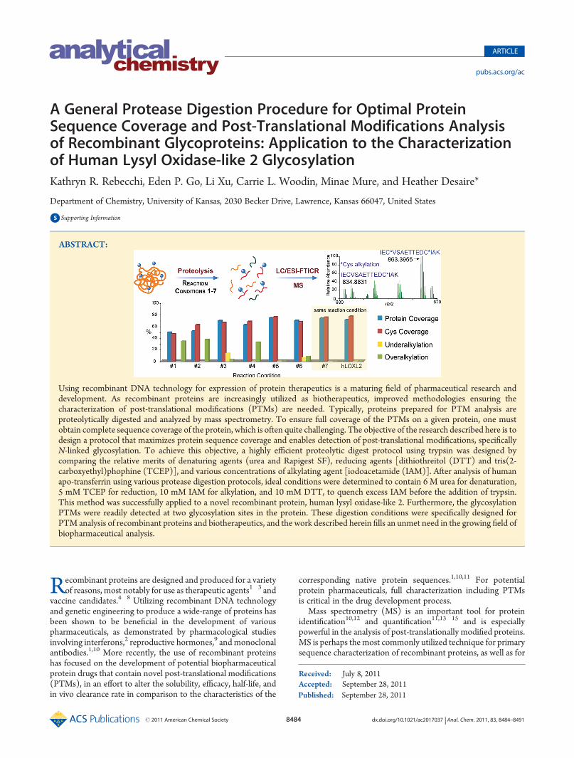
Figure 1. (A) Amino acid sequence of human apo-transferrin. (B) Amino acid sequence of a recombinant hLOXL2. Black text indicates the signalpeptide. Blue text signifies amino acids that were detected using digestion conditions 7 (see Table 1). Red text illustrates amino acids that were notdetected using these conditions.
detection of peptides. The different reaction conditions chosenallowed for the comparison of two different denaturants, Rapi-Gest SF and urea, as well as the reducing agents DTT and TCEPand various concentrations of IAM. Additionally, the necessity ofadding an extra procedural step where DTTwas added to quenchthe excess IAM, preventing unwanted side reactions and com-plications in MS data analysis, was investigated.In addition to simply detecting transferrin peptides in the MS
and MS/MS data, other factors governing digestion efficiencywere also assessed, including (1) complete alkylation (or under-alkylation) of the Cys residues, (2) alkylation on the N-terminusof peptides (overalkylation), and (3) incomplete detection of
glycans. For an efficacious digestion, one would expect to obtaincomplete alkylation of Cys residues, no overalkylation on theN-terminus, and complete detection of the glycans present.MS andMS/MS analysis was performed to evaluate these factorsfor each of the different reaction conditions in an effort todetermine optimal protease digestion conditions for transferrin.All LC�MS/MS data files were submitted to a Mascot search.Using the parameters described in the Experimental Section, thenonglycosylated peptides were identified. From the list of thebest peptide matches, we determined the overall coverage alongwith the species that were either under- or overalkylated. Thedata were also manually inspected to identify the glycopeptides
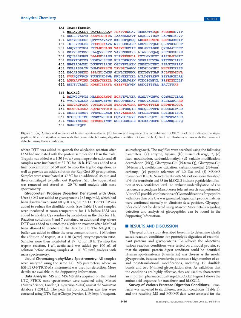
Figure 2. Representative human apo-transferrin data from condition 3 (see Table 1). (A) Total ion chromatogram. Highlighted in red is the region(41�42 min) where the MS data in part B are shown. (B) The two labeled peptides illustrate both the fully and nonalkylated peptideSAGWNIPIGLLYCDLPEPR. The peak that is circled in red is the m/z where MS/MS was acquired, as shown in part C. Additional labeled peaksindicate other detected human apo-transferrin peptides. The * indicates the alkylated Cys residue. (C) MS/MS data for the nonalkylatedSAGWNIPIGLLYCDLPEPR with a retention time of 41.55 min.
and to provide additional assurance that the automated assign-ments were correct.Figure 2 shows an example of some of the data. In Figure 2A,
an HPLC chromatogram from condition 3 in Table 1 is shown.The highlighted region from 41 to 42 min corresponds to theretention time averaged for the high-resolution mass spectrumshown in Figure 2B. TheMS/MS data in Figure 2C resulted fromthe circled peak labeled in the mass spectrum (Figure 2B). Theseparticular MS andMS/MS data correspond to a peptide that wasnot alkylated, indicating that the alkylation reaction was incom-plete in this case. Thus, as shown in Figure 2B, the peptide isdiluted between the fully alkylated peak (at m/z 1086) and thenonalkylated form (at m/z 1057). Fortunately, this particularpeptide happened to ionize efficiently, so both the alkylated andnonalkylated forms were detected. However, if the ionizationefficiency of this species had been lower, it would be likely thatneither of these peaks would be detected in the MS/MS data,leading to lower protein sequence coverage.Comparison of Denaturing and Reducing Agents. The
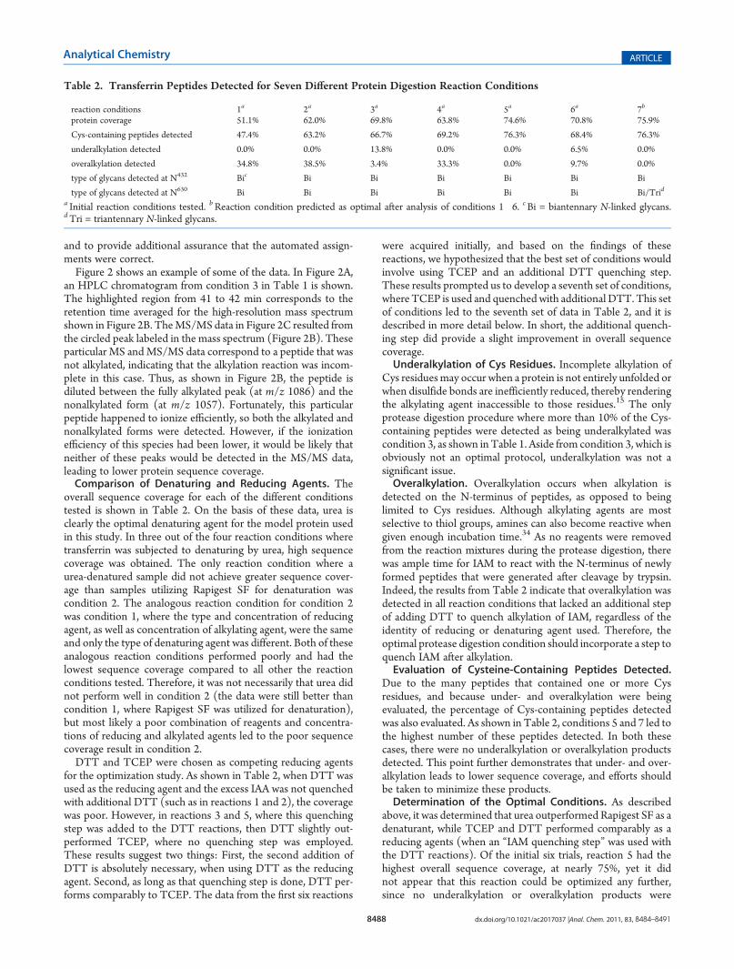
overall sequence coverage for each of the different conditionstested is shown in Table 2. On the basis of these data, urea isclearly the optimal denaturing agent for the model protein usedin this study. In three out of the four reaction conditions wheretransferrin was subjected to denaturing by urea, high sequencecoverage was obtained. The only reaction condition where aurea-denatured sample did not achieve greater sequence cover-age than samples utilizing Rapigest SF for denaturation wascondition 2. The analogous reaction condition for condition 2was condition 1, where the type and concentration of reducingagent, as well as concentration of alkylating agent, were the sameand only the type of denaturing agent was different. Both of theseanalogous reaction conditions performed poorly and had thelowest sequence coverage compared to all other the reactionconditions tested. Therefore, it was not necessarily that urea didnot perform well in condition 2 (the data were still better thancondition 1, where Rapigest SF was utilized for denaturation),but most likely a poor combination of reagents and concentra-tions of reducing and alkylated agents led to the poor sequencecoverage result in condition 2.DTT and TCEP were chosen as competing reducing agents
for the optimization study. As shown in Table 2, when DTT wasused as the reducing agent and the excess IAA was not quenchedwith additional DTT (such as in reactions 1 and 2), the coveragewas poor. However, in reactions 3 and 5, where this quenchingstep was added to the DTT reactions, then DTT slightly out-performed TCEP, where no quenching step was employed.These results suggest two things: First, the second addition ofDTT is absolutely necessary, when using DTT as the reducingagent. Second, as long as that quenching step is done, DTT per-forms comparably to TCEP. The data from the first six reactions
were acquired initially, and based on the findings of thesereactions, we hypothesized that the best set of conditions wouldinvolve using TCEP and an additional DTT quenching step.These results prompted us to develop a seventh set of conditions,where TCEP is used and quenched with additional DTT. This setof conditions led to the seventh set of data in Table 2, and it isdescribed in more detail below. In short, the additional quench-ing step did provide a slight improvement in overall sequencecoverage.Underalkylation of Cys Residues. Incomplete alkylation of
Cys residuesmay occur when a protein is not entirely unfolded orwhen disulfide bonds are inefficiently reduced, thereby renderingthe alkylating agent inaccessible to those residues.15 The onlyprotease digestion procedure where more than 10% of the Cys-containing peptides were detected as being underalkylated wascondition 3, as shown in Table 1. Aside from condition 3, which isobviously not an optimal protocol, underalkylation was not asignificant issue.Overalkylation. Overalkylation occurs when alkylation is
detected on the N-terminus of peptides, as opposed to beinglimited to Cys residues. Although alkylating agents are mostselective to thiol groups, amines can also become reactive whengiven enough incubation time.34 As no reagents were removedfrom the reaction mixtures during the protease digestion, therewas ample time for IAM to react with the N-terminus of newlyformed peptides that were generated after cleavage by trypsin.Indeed, the results from Table 2 indicate that overalkylation wasdetected in all reaction conditions that lacked an additional stepof adding DTT to quench alkylation of IAM, regardless of theidentity of reducing or denaturing agent used. Therefore, theoptimal protease digestion condition should incorporate a step toquench IAM after alkylation.Evaluation of Cysteine-Containing Peptides Detected.
Due to the many peptides that contained one or more Cysresidues, and because under- and overalkylation were beingevaluated, the percentage of Cys-containing peptides detectedwas also evaluated. As shown in Table 2, conditions 5 and 7 led tothe highest number of these peptides detected. In both thesecases, there were no underalkylation or overalkylation productsdetected. This point further demonstrates that under- and over-alkylation leads to lower sequence coverage, and efforts shouldbe taken to minimize these products.Determination of the Optimal Conditions. As described
above, it was determined that urea outperformed Rapigest SF as adenaturant, while TCEP and DTT performed comparably as areducing agents (when an “IAM quenching step” was used withthe DTT reactions). Of the initial six trials, reaction 5 had thehighest overall sequence coverage, at nearly 75%, yet it didnot appear that this reaction could be optimized any further,since no underalkylation or overalkylation products were
Table 2. Transferrin Peptides Detected for Seven Different Protein Digestion Reaction Conditions
reaction conditions 1a 2a 3a 4a 5a 6a 7b
protein coverage 51.1% 62.0% 69.8% 63.8% 74.6% 70.8% 75.9%
detected. Condition 6, which contained both urea and TCEP fordenaturation and reduction, respectively, was nearly as good ascondition 5, in terms of its sequence coverage, yet the resultsfrom condition 6 included several overalkylated peptides (and afew underalkylated ones). On the basis of these findings, theseventh reaction condition was developed with the same pro-tease digestion procedure from condition 6, but with an addi-tional step after alkylation, where DTT was added to quenchunreacted IAM (see Table 1 for full details). The transferrinpeptides identified in the MS data from the condition 7 digestionprotocol are highlighted in Figure 1A. As described in Table 2,condition 7 had the greatest sequence coverage, and no under- oroveralkylation was detected. Thus, condition 7 was determinedto be optimal for protease digestion.Detection andAnalysis of Glycopeptides. In addition to the
criteria above, one key feature of an ideal digestion protocol isthat it produces high coverage of the PTMs on the protein beinganalyzed. Therefore, in addition to checking for sequence cover-age and alkylation state, the seven data sets were also searched forthe known PTMs on transferrin, which contain two N-linkedglycosylation sites. For a set of reaction conditions to beconsidered optimal, the glycopeptides detected in the transferrinMS data needed to encompass all the glycoforms described in theliterature for this protein sample. The major glycoform known tobe present on transferrin is an N-linked biantennary sialylatedcomplex type glycan.40 However, transferrin also has anN-linkedtriantennary sialylated complex type glycan present in lowerabundance.40 Therefore, the MS data were searched for bothbiantennary and triantennary sialylated complex type glycopep-tides in the data sets from all seven reaction conditions. As shownin Table 2, biantennary N-linked sialylated glycopeptides weredetected at both glycosylation sites in transferrin in data sets fromall seven reaction conditions. This was expected, since the bian-tennary glycans are the most abundant glycoforms in transferrin.40
The only data set where the lesser abundant triantennary glycanswere detected was from condition 7. The triantennary N-linkedglycan details are listed in Table 1 of the Supporting Information.The presence of the less common glycoform detected only in theMS data for condition 7 was further confirmation that condition7 was indeed the most optimal reaction protocol.Application of an Optimized Proteolytic Digest to
hLOXL2. After identifying and validating an ideal set of reactionconditions for the model protein, transferrin, the final objectiveof this work was to demonstrate that these conditions werealso effective in the analysis of a biologically important protein,highlighting the general utility of the method. Lysyl oxidase(LOX) is a secreted copper-containing amine oxidase thatforms reactive aldehydes by oxidizing the ε-amino group oflysine side chains in collagen and elastin. LOX contains a cross-linked quinone cofactor arising from the PTM of its lysyl andtyrosol residues, which are conserved across all LOX and lysyloxidase-like (LOXL) proteins.35,41,42 Research involving lysyloxidase and lysyl oxidase-like proteins (LOXL, LOXL2, LOXL3,and LOXL4) has implicated that these enzymes participate in avariety of biological processes, including extracellular matrixstabilization, cellular growth, and homeostasis.41,43,44 Moreover,LOX family participants are attractive pharmacological targets,since dysregulation of LOX has been found to correlate withnumerous diseases and adverse physiological states, includingcancer formation and metastasis, connective tissue disorders, neuro-degenerative pathologies, and cardiovascular abnormalities.36,38,39,44
Specifically, LOXL2 has been shown to be involved in abnormal
collagen deposition, tumor invasion, lymph node metastasis, andcancer progression in breast and ovarian cancers.35�39 As such,characterization of LOXL2 is as essential step in assessing itsviability as a pharmaceutical candidate of interest.To confirm the sequence of the recombinant hLOXL2, as well
as to detect its PTMs, specifically N-linked glycosylation, pro-teolytic digestion followed by mass spectrometry was performed.As described above, the optimal set of digestion conditions onthe model protein transferrin was protocol 7 from Table 1. Theprotein, hLOXL2, was subjected to these conditions, followed byLC�MS and MS/MS analysis of the rendered peptides.A summary of the hLOXL2 amino acid residues detected isillustrated in Figure 1B. The overall results from the MS dataanalysis of hLOXL2 are described in Table 3, and high-resolutionMS data for each detected peptide are shown in Table 1 of theSupporting Information. The percent protein sequence coveragewas 71% and the percent Cys-containing peptides was 78%. Infact, only two peptides containing Cys residues were not detectedin the analysis of hLOXL2. The nondetected Cys-containingpeptides are highlighted in red in Figure 1B. The first suchpeptide is very large, 72 amino acids in length. Additionally, thepeptide has 11 acidic residues (D or E) and only one basicresidue. With that in mind, it is unlikely that this peptide wouldionize well in the mass range of the experiment (up tom/z 2000),as the lowest detectable charge state for this ion would be +4,producing an m/z of 1980. Even upon manual inspection of thedata, an ion at this mass and charge state was not detected.Likewise, a +5 charged ion for this peptide, atm/z 1584, was alsosearched for manually, but could not be conclusively identified.Therefore, it seems most probable that the inability to detect thispeptide was not due to the reaction conditions, but rather due tothe fact that the peptide does not ionize in the mass range of theexperiment. The second Cys-containing peptide, which was notdetected, also was not expected to be observable by this MSexperiment, because its tryptic peptide is only CR, which wouldhave an m/z of 335. This value is outside the scan range used forthis experiment.A few additional peptides (that do not contain Cys) were also
not detected. These peptides include LNGGR, NPYEGR, LLR,SR, VAEGHK, and YDGHR. All these undetected peptides areshort, with six amino acid residues or fewer, as highlighted in redin Figure 1B. Of these peptides LLR and SR were not expected tobe detected because their masses fall below the scan range (i.e., <500 Da). The other four peptides are five or six amino acidresidues in length and could potentially be detected by MS withthe scan range utilized. However, we expect that these peptideshave low ionization efficiencies because of the presence of acidicamino acid residues (D and E) in three of the four undetectedpeptides. In summary, this work illustrates that the digestionconditions were clearly optimized in that most peptides were
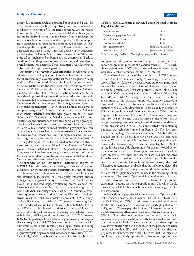
Table 3. hLOXL2 Peptides Detected Using Optimal ProteaseDigest Conditions
protein coverage 71.3%
Cys-containing peptides detected 77.8%
underalkylation detected 0.0%
overalkylation detected 0.0%
major glycorform at N31 a[Hex]3[HexNAc]2[Fuc]1
major glycorform at N220 a[Hex]3[HexNAc]2[Fuc]1aHex = hexose, HexNAc = N-acetylhexosamine, Fuc = fucose.
detected, and those that were not detected could reasonably beexpected to suffer from poor ionization efficiency. Additionally,no underalkylated or overalkylated peptides were detected in thehLOXL2 sample, further supporting the conclusion that thedigestion conditions were optimal for this sample.Glycopeptide Analysis of hLOXL2.The CIDMS/MS data of
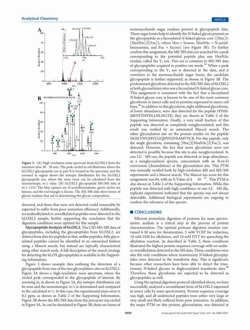
glycopeptides, including the glycopeptides from hLOXL2, aredistinct from data for peptides in that, unlike peptides, fully glyco-sylated peptides cannot be identified in an automated fashionusing a Mascot search, but instead are typically characterizedusing other search tools. A full description of the search strategyfor detecting the hLOX glycopeptides is available in the Support-ing Information.Figure 3 shows example data outlining the detection of a
glycopeptide from one of the two glycosylation sites in hLOXL2.Figure 3A shows a high-resolution mass spectrum, where thecircled peak corresponded to the glycopeptide of interest. Byzooming in, as shown in Figure 3A, the isotopic distribution canbe seen and the monoisotopic m/z is determined and comparedto the calculated m/z. In this case, the experimental mass error is8.2 ppm, as shown in Table 2 of the Supporting Information.Figure 3B shows the MS/MS data from the precursor ion circledin Figure 3A. As can be elucidated in Figure 3B, there are losses of
monosaccharide sugar residues present in glycopeptide data.These sugar losses help to identify theN-linked glycan present onthe glycopeptide as a fucosylated N-linked glycan core ([Hex]3-[HexNAc]2[Fuc]1, where Hex = hexose, HexNAc = N-acetyl-hexosamine, and Fuc = fucose) (see Figure 3B). To furtherconfirm this assignment, theMS/MS data are searched for a peakcorresponding to the potential peptide plus one HexNAcresidue, called the Y1 ion. This ion is common to MS/MS dataof glycopeptides acquired in positive ion mode.45 When a peakcorresponding to the Y1 ion is detected in the data, and itcorrelates to the monosaccharide sugar losses, the candidateglycopeptide is further supported, as shown in Figure 3B. Thepredominant glycoform detected in theMS/MS data of hLOXL2at both glycosylation sites was a fucosylatedN-linked glycan core.This assignment is consistent with the fact that a fucosylatedN-linked glycan core is known to be one of the most commonglycoforms in insect cells and in proteins expressed in insect celllines.46 In addition to this glycoform, eight additional glycoforms,of lower abundance, were also detected for the peptide HYHS-MEVFTHYDLLNLNGTK; they are shown in Table 2 of theSupporting Information. Finally, a very small fraction of thispeptide was detected as completely nonglycosylated, and thisresult was verified by an automated Mascot search. Theother glycosylation site on the protein resides on the peptideNGSLVWGMVCGQNWGIVEAMVVCR. For this peptide, onlythe single glycoform, containing [Hex]3[HexNAc]2[Fuc]1, wasdetected. However, the fact that more glycoforms were notdetected is possibly because this site is also nonglycosylated. Inone LC�MS run, the peptide was detected, in large abundance,as a nonglycosylated species, concomitant with an N-to-Dconversion (deamidation) at the glycosylation site. This PTMwas manually verified both by high-resolution MS and MS/MSexperiments and a Mascot search. The Mascot ion score for thisassignment was 84, with an E-Value of 4 � 10�10 . This PTM isalso shown in Table 2 of the Supporting Information. While thispeptide was detected with high confidence in one LC�MS file,replicate experiments indicated that the species was not alwaysdetectable. Additional biological experiments are ongoing toconfirm the relevance of this species.
’CONCLUSIONS
Efficient proteolytic digestion of proteins for mass spectro-metric analysis is a critical step in the process of proteincharacterization. The optimal protease digestion reaction con-tained 6 M urea for denaturation, 5 mM TCEP for reduction,10 mM IAM for alkylation, and 10 mM DTT for quenching thealkylation reaction. As described in Table 2, these conditionsillustrated the highest protein sequence coverage with no under-or overalkylation detected in theMS data. These conditions werealso the only conditions where triantennary N-linked glycopep-tides were detected in the transferrin data. This is significant,because other researchers have been able to detect the trian-tennary N-linked glycans in deglycosylated transferrin data.40
Therefore, these glycoforms are expected to be detected asglycopeptides as well.
Using the optimal digestion protocol identified above, we havesuccessfully analyzed a recombinant form of hLOXL2 expressedin insect cells by mass spectrometry. Protein sequence coveragewas high, and all undetected peptides were either very large orvery small and likely suffered from poor ionization. In addition,the major PTM on this protein, glycosylation, was detected at
Figure 3. (A) High-resolution mass spectrum from hLOXL2 from theretention time 38�39 min. The peak circled in red illustrates where thehLOXL2 glycopeptide ion in part B is located in the spectrum, and thezoomed in region shows the isotopic distribution for the hLOXL2glycopeptide ion, where the mass error can be calculated from themonoisotopic m/z value. (B) hLOXL2 glycopeptide MS/MS data atm/z 1153. The blue squares are N-acetylhexosamine, green circles arehexose, and the red triangle is fucose. The MS/MS data show losses ofglycan residues that aid in determining the glycan composition.
both putative glycosylation sites. The glycoforms identified wereconsistent with the glycoforms typically present from the cellline used.
’ASSOCIATED CONTENT
bS Supporting Information. Additional information as notedin the text. Thismaterial is available free of charge via the Internet athttp://pubs.acs.org.
The authors acknowledge financial support from an NSFCAREER award (project number 0645120) to H.D., an NSFFellowship to K.R. andC.W. (DGE-0742523), andNSFCAREERaward (MCB-0747377) and NIH funding (5R01GM079446-02)to M.M.
S.; Yang, X.; Nagabhushan, T. L.; Pramanik, B. N. Anal. Biochem. 2011, 408,105–117.(3) Walsh, G.; Jefferis, R. Nat. Biotechnol. 2006, 24, 1241–1252.(4) Koff, R. J. Parasitol. 2003, 33, 517–523.(5) Madrid-Marina, V.; Torres-Poveda, K.; Lopez-Toledo, G.;
Garcia-Carranca, G. Arch. Med. Res. 2009, 40, 471–477.(6) Go, E. P.; Irungu, J.; Zhang, Y.; Dalpathado, D. S.; Liao, H.-X.;
Sutherland, L. L.; Alam, S. M.; Haynes, B. F.; Desaire, H. J. Proteome Res.2008, 7, 1660–1674.(7) Go, E. P.; Chang, Q.; Liao, H.-L.; Sutherland, L. L.; Alam, S. M.;
Baynes, B. F.; Desaire, H. J. Proteome Res. 2009, 8, 4231–4242.(8) Barouch, D. H. Nature 2008, 455, 613–619.(9) Thakur, D.; Rejtar, T.; Karger, B.; Washburn, N. J.; Bosques,
C. J.; Gunay, N. S.; Shriver, Z.; Venkataraman, G. Anal. Chem. 2009,81, 8900–8907.(10) Barnes, C. A. S.; Lim, A.Mass Spectrom. Rev. 2007, 26, 370–388.(11) Ezan, E.; Dubois, M.; Becher, F. Analyst 2009, 134, 825–834.(12) Chen, G.; Mirza, U. A.; Pramanik, B. N. Adv. Chromatogr. 2009,
47, 1–29.(13) Rebecchi, K. R.; Wenke, J. L.; Go, E. P.; Desaire, H. J. Am. Soc.
Mass Spectrom. 2009, 20, 1048–1059.(14) Norrgran, J.; Williams, T. L.; Woolfitt, A. R.; Solano, M. I.;
Pirkle, J. L.; Barr, J. R. Anal. Biochem. 2009, 393, 48–55.(15) Hamdan,M.; Righetti, P. G.Mass Spectrom. Rev. 2002, 21, 287–302.(16) Itoh, S.; Kawasaki, N.; Ohta, M.; Hayakawa, T. J. Chromatogr. A
2002, 978, 141–152.(17) Dalpathado, D.; Desaire, H. Analyst 2008, 133, 731–738.(18) Wuhrer, M.; Catalina, M. I.; Deelder, A. M.; Hokke, C. H.
J. Chromatogr. B 2007, 849, 115–128.(19) Meyer, B.; Papasotiriou, D. G.; Karas, M. Amino Acids 2010,
41, 291–310.(20) Zhang, Y.; Go, E. P.; Desaire, H. Anal. Chem. 2008, 80,
3144–3158.(21) Second, T. P.; Blethrow, J. D.; Schwartz, J. C.; Merrihew, G. E.;
MacCoss, M. J.; Swaney, D. L.; Russell, J. D.; Coon, J. J.; Zabrouskov, V.Anal. Chem. 2009, 81, 7757–7765.(22) Garza, S.; Moini, M. Anal. Chem. 2006, 78, 7309–7316.
(23) Ren, D.; Julka, S.; Inerowicz, H. D.; Regnier, F. E. Anal. Chem.2004, 76, 4522–4530.
(24) Chen, E. I.; Cociorva, D.; Norris, J. L.; Yates, J. R., III J. ProteomeRes. 2007, 6, 2529–2538.
(25) Arnold, R. J.; Hrncirova, P.; Annaiah, K.; Novotny, M. V.J. Proteome Res. 2004, 3, 653–657.
(26) Strader, M. B.; Tabb, D. L.; Hervey, W. J.; Pan, C.; Hurst, G. B.Anal. Chem. 2006, 78, 125–134.
(27) Yu, Y.-Q.; Gilar, M.; Lee, P. J.; Bouvier, E. S. P.; Gebler, J. C.Anal. Chem. 2003, 75, 6023–6028.
(28) Hervey, W. J., IV; Strader, M. B.; Hurst, G. B. J. Proteome Res.2007, 6, 3054–3061.
(29) Masuda, T.; Tomita, M.; Ishihama, Y. J. Proteome Res. 2008,7, 731–740.
(30) Getz, E. B.; Xiao, M.; Chakrabarty, T.; Cooke, R.; Selvin, P. R.Anal. Biochem. 1999, 273, 73–80.
(31) Cline, D. J.; Redding, S. E.; Brohawn, S. G.; Psathas, J. N.;Schneider, J. P.; Thorpe, C. Biochemistry 2004, 43, 15195–15203.
(32) Jacobs, J. M.; Mottaz, H. M.; Yu, L.-R.; Anderson, D. J.; et al.J. Proteome Res. 2004, 3, 68–75.
(33) Sechi, S.; Chait, B. T. Anal. Chem. 1998, 70, 5150–5158.(34) Boja, E. S.; Fales, H. M. Anal. Chem. 2001, 73, 3576–3582.(35) Vadasz, Z.; Kessler, O.; Akiri, G.; Gengrinovitch, S.; Kagan,H.M.;
Baruch, Y.; Izhak, O. B.; Neufeld, G. J. Hepatol. 2005, 43, 499–507.(36) Kirschmann, D. A.; Seftor, E. A.; Fong, S. F. T.; Nieva, D. R. C.;
Sullivan, C. M.; Edwards, E. M.; Sommer, P.; Csiszar, K.; Hendrix,M. J. C. Cancer Res. 2002, 62, 4478–4483.
(38) Barker, H. E.; Chang, J.; Cox, T. R.; Lang, G.; Bird, D.; Nicolau,M.; Evans, H. R.; Gartland, A.; Erler, J. T.Cancer Res. 2011, 71, 1561–1572.
(39) Fong, S. F. T.; Dietzsch, E.; Fong, K. S. K.; Hollosi, P.;Asuncion, L.; He, Q. P.; Parker, M. I.; Csiszar, K. Genes, ChromosomesCancer 2007, 46, 644–655.
(40) Wada, Y.; Azadi, P.; Costello, C. E.; Dell, A.; et al. Glycobiology2007, 17, 411–422.
(41) Molnar, J.; Fong, K. S. K.; He, Q. P.; Hayashi, K.; Kim, Y.; Fong,S. F. T.; Fogelgren, B.; Szauter, K. M.; Mink, M.; Csiszar, K. Biochim.Biophys. Acta 2003, 1647, 220–224.
(42) Seve, S.; Decitre, M.; Gleyzal, C.; Farjanel, J.; Sergeant, A.;Ricard-Blum, S.; Sommer, P. Connect. Tissue Res. 2002, 43, 613–619.
(43) Lucero,H.A.; Kagan,H.M.Cell.Mol. Life Sci.2006, 63, 2304–2316.(44) Rodriguez, C.; Rodriguez-Sinovas, A.; Martinez-Gonzalez, J.
Drug News Perspect. 2008, 21, 218–224.(45) Segu, Z. M.; Mechref, Y. Rapid Commun. Mass Spectrom. 2010,
24, 1217–1225.(46) Fabini, G.; Freilinger, A.; Altmann, F.; Wilson, I. B. H. J. Biol.