48

A Joint Project Produced on Behalf of:
International Network of Agencies for Health Technology Assessment
Positron Emission Tomography:Experience with PET and Synthesis of theEvidence
Elizabeth Adams1, José Asua2, José Conde Olasagasti3Martin Erlichman4, Karen Flynn1, Iraida Hurtado-Saracho2
1Department of Veterans Affairs Health Services Research and Development, MDRCTechnology Assessment Program, Boston, Massachusetts, USA.
2Basque Office for Health Technology Assessment (OSTEBA), Vitoria-Gasteiz, SPAIN.3Agencia de Evaluación de Tecnologías Sanitarias (AETS), Madrid, SPAIN.4Agency for Health Care Policy and Research, Center for Practice and Technology
Assessment Washington, DC, USA.
November 1999
Established in 1993 the International Network of Agencies for HealthTechnology Assessment (INAHTA) provides a forum for the identification andpursuit of interests common to health technology assessment agencies.INAHTA consists of non-profit agencies throughout the world that assess healthcare technologies, relate to a regional or national government and are funded atleast 50 percent by public sources.
INAHTA joint projects are collaborations among member agencies to evaluatemedical technologies of mutual interest. In 1996 INAHTA completed its firstjoint project on bone density measurement. In 1999 INAHTA conducted jointprojects in telemedicine and prostate cancer screening, in addition to PETscanning.
INAHTA Newsletter provides updates on recently published reports, currentinitiatives and activities among member agencies, new projects within theNetwork, recent developments and trends in health policy research, publicationsin the field and upcoming events. INAHTA Newsletter is published quarterly inEnglish, Spanish and French.
Information about INAHTA Newsletter, INAHTA and its members, includinglinks to member agencies’ websites and the database of published reports andcurrent projects, is available on the web at www.inahta.org or from theINAHTA Secretariat directly.
INAHTA Secretariat c/o SBU, Box 5650, S-114 86 Stockholm, Swedentel +46 8 412 3200 or fax +46 8 411 3260
This report may be freely copied. If citing this report please use the following information:
Adams EJ, Asua J, Conde Olasagasti JG, Erlichman M, Flynn K, Hurtado-Saracho I. On behalfof INAHTA. Positron Emission Tomography: Experience with PET and Synthesis of theEvidence. Stockholm: International Network of Agencies for Health Technology Assessment,1999.

INAHTA PET Collaboration 1999 Page i
International Network of Agencies for Health Technology Assessment(as of September 30, 1999)
AETS Agencia de Evaluación de Tecnologías Sanitarias SPAIN
AETSA Agencia de Evaluación de Tecnologías Sanitarias de Andalucía SPAIN
AHCPR Agency for Health Care Policy and Research, Center for Practice and Technology Assessment USA
AHFMR Alberta Heritage Foundation for Medical Research CANADA
ANAES L'Agence Nationale d'Accréditation et d'Evaluation en Santé FRANCE
CAHTA Catalan Agency for Health Technology Assessment SPAIN
CCOHTA Canadian Coordinating Office for Health Technology Assessment CANADA
CEDIT Comité d’Evaluation et de Diffusion des Innovations Technologiques FRANCE
CETS Conseil d’Evaluation des Technologies de la Santé CANADA
CVZ College voor Zorgverzekeringen/Health Care Insurance Board THE NETHERLANDS
DIHTA Danish Institute for Health Technology Assessment DENMARK
DIMDI German Institute for Medical Documentation and Information GERMANY
DSI Danish Institute for Health Services Research and Development DENMARK
ETESA Unidad De Tecnologías De Salud CHILE
FINOHTA Finnish Office for Health Care Technology Assessment (Stakes) FINLAND
GR Health Council of the Netherlands (Gezondheidsraad) THE NETHERLANDS
HSC NHS Horizon Scanning Center UNITED KINGDOM
ICTAHC Israel Center for Technology Assessment in Health Care ISRAEL
INHEM Instituto Nacional de Higiene Epidemiologia y Microbiologia Infanta CUBA
ITA HTA Unit of the Institute of Technology Assessment, Austrian Academy of Science AUSTRIA
MSAC Medicare Services Advisory Committee AUSTRALIA
NCCHTA National Coordinating Centre for Health Technology Assessment UNITED KINDGOM
NHSCRD NHS Centre for Reviews and Dissemination UNITED KINGDOM
NZHTA New Zealand Health Technology Assessment NEW ZEALAND
OSTEBA Basque Office for Health Technology Assessment (OSTEBA) SPAIN
SBU Swedish Council on Technology Assessment in Health Care SWEDEN
SFOSS Medical Technology Section of the Swiss Federal Office of Social Security SWITZERLAND
SMM The Norwegian Centre for Health Technology Assessment NORWAY
SWISS/TA Swiss Science Council/Technology Assessment SWITZERLAND
TNO TNO Prevention and Health THE NETHERLANDS
VATAP US Department of Veterans Affairs Technology Assessment Program USA
Source: www.INAHTA.org

INAHTA PET Collaboration 1999 Page ii
Special Acknowledgments
INAHTA extends its full appreciation to the following organizations for contributing theirassessment findings to the report. Their assessments are proprietary and may be obtained directlyfrom each organization.
Blue Cross and Blue Shield Association676 North St. Clair StreetChicago, IL 60611USATel. (312) 440-5514Fax (312) 440-6320
ECRI5200 Butler PikePlymouth Meeting, PA 19462-1298USATel. (610) 825-6000Fax (610) 834-1275E-mail [email protected] site http://www.ecri.org
HAYES, Inc.Suite 200157 South Broad StreetLansdale, PA 19446USATel. (215) 855-0615Fax (215) 855-5218
The authors also wish to thank Jennifer Jacoppo, Maria Fonseca, Frank Roth, Jennifer Cheslog,Elaine Alligood and Scott Brigante for their assistance in bringing the report to completion.

INAHTA PET Collaboration 1999 Page iii
INAHTA JOINT PROJECTPositron Emission Tomography:
Experience with PET and Synthesis of the EvidenceExecutive Summary
• INAHTA conducted this joint collaboration in response to an increasing global interest in the clinicalpotential of positron emission tomography (PET). The project documents PET use and related publichealth coverage in countries represented by INAHTA members and synthesizes technologyassessments of PET conducted by INAHTA members and three private US organizations. Itconsiders all PET systems, that is, conventional full ring models, newer partial ring models andSPECT cameras modified for imaging positron emitters.
• PET is a functional imaging technology that uses a radioactive tracer to assess perfusion andmetabolic activity in the human body. Introduced first as a research tool, PET has undergonetechnological advances that make it feasible for clinical use.
• PET’s availability is still quite limited, as evidenced by the low numbers and relative under use ofscanners in each country or region. Most health systems used their PET scanners for both researchand diagnostic purposes, but there were wide variations in use across systems. Local regulatorypolicies and the availability of private funding sources likely contributed to these differences.
• The vast majority of reimbursed clinical PET activity is concentrated in relatively few health systemsand is confined to comparatively few indications.
• Public health systems in Australia, Switzerland, Denmark and the US (VHA) conducted 85%of the activity.
• The most frequently covered PET indications, presented in descending order, were fordiagnosing head and neck cancer, lung cancer and lymphoma followed closely bydifferentiating brain tumor from radiation necrosis and diagnosing colorectal cancer, breastcancer and melanoma.
• 70% of the oncology activity comprised melanoma, lung cancer staging, and an undefinedcategory of “other”. The vast majority of neurology activity was for distinguishing braintumor from radiation necrosis and for localizing epileptic foci in potential surgical candidateswith intractable epilepsy.
• Many health systems refer to US experiences, particularly Medicare policy, to establish localreimbursement policy. In the US in 1997, supporters of clinical PET were instrumental in changingFDA regulation of PET drugs. As a result, Medicare has expanded coverage of PET scans beyondcardiac perfusion imaging to include diagnosing indeterminate solitary pulmonary nodules, recurrentmetastatic melanoma and recurrent colorectal cancer and staging non-small cell lung cancer,Hodgkin’s and non-Hodgkin’s lymphoma.
• Regarding PET’s utility, evidence of diagnostic accuracy is largely based on traditional full ring PET,is limited by bias and often relates only to small patient numbers. In all of the advocated clinicalindications there was uniform agreement that critical research is needed to define the clinical andeconomic consequences of using PET on treatment decisions and health outcome relative to othermethods now in clinical use.
• Many INAHTA agencies identified clinical PET as a major research priority and are initiatingrigorous evaluation efforts. Most recommended that, if used at all, PET should be used under researchprotocols designed to evaluate PET’s relative cost-effectiveness.

INAHTA PET Collaboration 1999 Page iv
Table of Contents
Introduction .............................................................................................................................. 1
Background .............................................................................................................................. 1Part I. PET Use and Reimbursement Among Project Participants.............................................. 3
A. Methods ........................................................................................................................ 3B. Results .......................................................................................................................... 4
1. 1997 Survey ............................................................................................................ 42. 1999 Survey ............................................................................................................ 7
3. Trends in the US.....................................................................................................10C. Summary/Discussion....................................................................................................15
Part II. Synthesis of Technology Assessments of PET..............................................................16
A. Methods .......................................................................................................................16B. Results .........................................................................................................................16
1. Neuropsychiatry .....................................................................................................172. Cardiology..............................................................................................................19
3. Oncology (non-central nervous system tumors) ......................................................22C. Summary......................................................................................................................23
Appendix A. 1997 INAHTA PET Survey ................................................................................26Appendix B. 1999 INAHTA PET Survey.................................................................................27Appendix C. INAHTA PET Collaboration Assessments of Clinical PET .................................28
Bibliography............................................................................................................................38

INAHTA PET Collaboration 1999 Page 1
INAHTA JOINT PROJECTPositron Emission Tomography:
Experience with PET and Synthesis of the Evidence
INTRODUCTION
At the fifth annual meeting of the International Network of Agencies for Health TechnologyAssessment (INAHTA) in 1997, members initiated a joint project on positron emissiontomography (PET) scanning in clinical medicine in response to a growing interest in thetechnology worldwide. INAHTA members have experienced different motivations or hindrancesto the diffusion of PET, and their assessments reflect the different health care systemenvironments. One goal of this collaborative effort is to bring together the range of experiencesinto a broadly applicable document. A collaborative INAHTA project drawing on an array ofapproaches and research questions will expand the scope of individual agencies’ assessments.
Four agencies agreed to coordinate the project: The Agency for Health Care Policy and Research(AHCPR), United States; Agencia de Evaluación de Tecnologías Sanitarias (AETS), Spain;Basque Office for Health Technology Assessment Health Department (OSTEBA), Spain; and theDepartment of Veterans Affairs Technology Assessment Program (VA TAP), United States.
This report will document:
I. the use of PET and public health care coverage policies for PET in countries representedby INAHTA members,
II. a synthesis of technology assessments of PET conducted by INAHTA members and threeprivate US organizations.
Interest in PET now extends beyond the traditional full ring PET scanners to partial ring modelsand to single photon emission computed tomography (SPECT) modified for imaging positronemitters. Thus, the scope of this project will include positron emitting imaging modalities thatuse principles of coincidence detection or high energy 511 keV collimation to form the rawimage.
BACKGROUND
PET is a minimally invasive imaging procedure that uses a radioactive tracer to assess perfusionand metabolic activity in various organ systems of the human body. The tracer decays byemitting a positively charged electron, called a positron, from the nucleus. The positron collideswith a negatively charged electron resulting in two high energy (511 keV) photons traveling inopposite directions. The high energy photon is subject to less absorption or scatter by tissue.
A positron camera (tomograph) arranged in a ring around the patient detects the two photonssimultaneously (coincidence detection) to produce cross-sectional tomographic images.Traditional PET scanners come in full ring and, more recently, partial ring models. Dual-headedSPECT cameras with coincidence detection capability and multi-headed SPECT camerasadapted for high energy 511 keV collimation are now available for imaging positron emitters.

INAHTA PET Collaboration 1999 Page 2
Relative to other nuclear medicine technologies, traditional PET systems provide superior imagequality and quantitative information (Lewellen 1999).
Charged particle accelerators (e.g. generators and cyclotrons) produce the radiopharmaceuticalsused in PET scanning. Both generators and cyclotrons are commercially available. Most PETfacilities use cyclotron-produced, short-lived positron emitting radionuclides. These areprincipally oxygen (O-15), nitrogen (N-13), carbon (C-11) and fluorine (F-18). Radionuclidegenerators typically use longer-lived parent radionuclides to produce a singleradiopharmaceutical for routine use, the most common being rubidium (Rb-82) chloride.
Ter-Pogossian (1992) summarized the development of PET from a research instrument to adiagnostic test. Since the invention of the first cyclotron and discovery of the positron in the1930s, PET has been used in the study of basic physiology. Over the years, advances in detectorinstrumentation, knowledge of the properties of many short-lived positron emitters, and rapidchemical labeling procedures allowing for in vivo nuclear medicine imaging contributed to thedevelopment and understanding of PET as a research tool.
Further advances have made PET more feasible for clinical use:
• greater ease of operation, reliability and competitive pricing of cyclotrons;• less expensive and easier-to-install accelerators for generating PET radionuclides;• lower cost and more accessible generator-produced radionuclides; and• improved sensitivity and resolution of PET instrumentation.
Despite technical improvements and an increasing interest in its clinical uses, PET has evolvedslowly as a clinical tool relative to other imaging modalities such as CT or MRI. Ter-Pogossian(1992) noted two important barriers limiting PET’s acceptance and use in clinical medicine.PET, unlike CT and MRI, was initially developed for research purposes, and PET requires acharged particle accelerator, usually a cyclotron, to produce short-lived positron emitters astracers for PET studies. Thus, the clinical use of PET using these tracers had been largelyrestricted to affiliated research departments equipped with cyclotrons and expertise inradiochemistry.
Early clinical PET studies were first conducted in the brain for localizing seizure foci anddifferentiating causes of dementia. Cardiology studies followed using PET with Rb-82 to tracetissue perfusion and FDG to trace tissue metabolism. More recently, whole body scanningcapability has generated considerable attention in using PET to manage oncology patients.
Like many large diagnostic imaging systems, PET is an expensive technology to purchase andmaintain. Technical improvements now permit a wider of range of options for buyers of PET.Several PET manufacturers provided the following costs approximated in USD, which may notnecessarily reflect the actual negotiated costs (personal communications 1999). Full ring modelsrange from $1.25 to $1.5 million USD, partial ring models cost $900,000 USD, and cyclotronssell for $1.5 million USD. There may be additional costs associated with installation,construction and operation.
The costs of traditional PET systems are prohibitive for many providers. Accordingly, many areupgrading more readily available dual-headed gamma cameras for coincidence imaging. The cost

INAHTA PET Collaboration 1999 Page 3
of the upgrade is $250,000; dual-headed gamma cameras without the upgrade sell for $400,000to $600,000 (personal communications 1999). A strontium generator for producing Rb-82 costs$300,000 USD (ECRI 1996).
Annual operating costs for a PET facility can vary considerably and are often related to thecomplexity of operations (Flynn 1996). Typically, PET facilities that conduct only clinical scansoffer the least costly alternative. Mobile PET scanner models and regional suppliers ofradiopharmaceuticals such as F-18 fluorodeoxyglucose (FDG) may eliminate the need for an on-site cyclotron. Adding research capability can increase the complexity of operations, asradiolabeling complex tracer substances may require an on-site cyclotron and expertise inradiochemistry. Other research personnel may be needed for patient care, computer function anddata analysis. A cyclotron and radiochemistry lab may require a larger facility.
PART I. PET USE AND REIMBURSEMENT AMONG PROJECT PARTICIPANTS
Interest in clinical PET has changed over the last two decades. Facing an uncooperative USmarket in the early 1990s, manufacturers looked overseas to increase market share. Lately,clinical PET has gained in popularity in the US and in many other parts of the world. Thissection explores the changing global interest in clinical PET through the experiences ofINAHTA members.
A. Methods
The authors used several strategies to gather data:
1. OSTEBA surveyed 12 INAHTA members, excluding those from the US, and eight non-INAHTA participants in the HTA Europe Project1 to obtain annual estimates of research andclinical utilization in private and public PET facilities and to gather information on publicreimbursement for clinical PET scans performed in 1997 (see Appendix A).
2. Increases in INAHTA membership since 1997 and a continuing interest in PET’s clinicalpotential warranted a new survey. In 1999 OSTEBA surveyed all 31 INAHTA memberslisted on page i on the availability of public reimbursement for clinical PET scans for thetime period July 1, 1998 through June 31, 1999 (see Appendix B).
3. In formulating local policy on PET many public health systems have looked to the USexperience. The heterogeneity of the US health care system made it difficult to obtaincomplete utilisation data for the US and required that a different approach be used. VA TAPand AHCPR reported on the primary factors affecting the diffusion of clinical PET in the USand on trends in coverage for clinical PET, as represented by the major health care payersand providers. For this section the authors searched MEDLINE®, HealthSTAR® and severalPET-related web sites to obtain descriptive information, contacted major payers andproviders for coverage policies, and obtained reports from technology assessmentorganizations in the US that conducted evaluations of clinical PET.
1 HTA Europe is a Project whose aim is to develop a coordinated approach to health care technology assessment in Europe.

INAHTA PET Collaboration 1999 Page 4
B. Results
1. 1997 Survey
Surveyed organizations represented 19 countries and one province. Fifteen HTA agencies(ten INAHTA members and five HTA Europe Project participants) responded indicating a75% response rate (See Table 1). However, participants from Belgium and Germany did notprovide complete information. Since three countries have no PET scanners, survey findingsrepresent 13 countries with 33 PET scanners. Thirty scanners (90.9%) are situated in publichealth care systems, while only three scanners are situated privately. We observed a highpercentage of whole body PET (84.8%) relative to smaller partial body scanners.
Table 1. 1997 INAHTA PET Survey Respondents by Country or Province
Country or provinceNumber of
PET scanners Country or provinceNumber of
PET scannersAustralia 3 Germany 2*Austria 1 Greece 0Belgium 2* Ireland 0Canada (except Québec) 5 The Netherlands 2Québec (Canada) 1 New Zealand 0Denmark 3 Spain 2Finland 2 Sweden 5France 3 Switzerland 2
* Incomplete questionnaires
Figures 1 and 2 depict PET utilization across countries represented in the survey. Themajority used their PET scanners for both clinical and research purposes, but there wereexceptions. Austria and Switzerland dedicated their scanners to clinical diagnosis only, whileCanada, France and Sweden set aside some of their scanners solely for research. Holland andDenmark performed the most investigative tests, whereas Australia, followed by Spain andBelgium, conducted the most diagnostic studies, bearing in mind the incomplete informationreceived from Belgium.
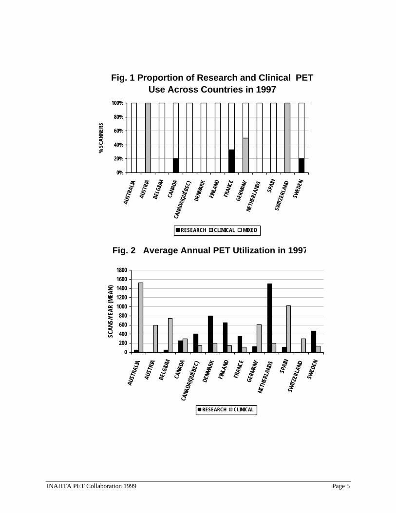
INAHTA PET Collaboration 1999 Page 5
Fig. 1 Proportion of Research and Clinical PET Use Across Countries in 1997
0%
20%
40%
60%
80%
100%
AUST
RALI
A
AUST
RIA
BELG
IUM
CANA
DACA
NADA
(QUÉ
BEC)
DENM
ARK
FINL
AND
FRAN
CEGE
RMAN
YNE
THER
LAND
S
SPAI
NSW
ITZE
RLAN
D
SWED
EN
% S
CANN
ERS
RESEARCH CLINICAL MIXED
Fig. 2 Average Annual PET Utilization in 1997
0
200
400
600
800
1000
1200
1400
1600
1800
AUST
RALI
A
AUST
RIA
BELG
IUM
CANA
DACA
NADA
(QUÉ
BEC)
DENM
ARK
FINL
AND
FRAN
CEGE
RMAN
YNE
THER
LAND
S
SPAI
NSW
ITZE
RLAN
D
SWED
EN
SCAN
S/YE
AR (M
EAN)
RESEARCH CLINICAL

INAHTA PET Collaboration 1999 Page 6
Analysis of individual scanner utilization showed that 51.5% of the scanners conductedfewer than 250 clinical scans per year, and 48.5% performed less than 250 scans per year forresearch purposes (See Table 2). It was very infrequent for an individual scanner to conductmore than 500 scans per year. Accounting for the two incomplete survey results, six (18.2%)scanners were used for clinical use only, three (9.1%) scanners were used exclusively forresearch. The remainder were used for both research and clinical studies.
Table 2. Average Annual Utilization of 33 PET Scanners in 1997
Number (%) of scanners Number (%) of scannersAverage #Scans/year Research Clinical
Average #Scans/year Research Clinical
None 4 (12.1) 3 (9.1) 1001-1250 1 (3.0) 2 (6.1)1-250 12 (36.4) 14 (42.4) 1251-1500 2 (6.1) 1 (3.0)251-500 7 (21.2) 5 (15.2) > 1500 0 1 (3.0)501-750 5 (15.2) 5 (15.2) unknown 1 (3.0) 1 (3.0)751-1000 1 (3.0) 1 (3.0) TOTAL 33 (100) 33 (100)
Table 3. Diagnostic Uses and Public Reimbursement Among SurveyParticipants in 1997
DiagnosticUse
PublicReimbursement
PublicReimbursement
Diagnostic applicationsNumber of Scanners
(% of total)Number of Scanners
(% of total)Number of Countries
(% of total)Neurology Epilepsy 27 (81.8) 18 (54.4) 9 (69.2)
Tumor vs necrosis 27 (81.8) 21 (63.6) 9 (69.2)
Neurodegenerative disorders 26 (78.8) 14 (42.4) 7 (53.8)
ACV 5 (15.2) 5 (15.2) 1 (7.7)
Encephalopathy 2 (6.1) 2 (6.1) 2 (15.4)
Psychiatry 1 (3.0) 1 (3.0) 1 (7.7)
Cardiology Myocardial viability 24 (72.7) 16 (48.5) 8 (61.5)
Myocardial perfusion 19 (57.6) 8 (24.2) 4 (30.8)
Oncology Lung 21 (63.6) 11 (33.3) 5 (38.5)
(non-central Soft tissue 21 (63.6) 17 (51.5) 7 (53.8)
nervous system) Head and neck 18 (54.5) 13 (39.4) 7 (53.8)
Solitary pulmonary nodules 18 (54.5) 9 (27.3) 4 (30.8)
Colorectal 16 (48.5) 7 (21.2) 3 (23.1)
Breast 16 (48.5) 9 (27.3) 4 (30.8)
Gynecological 14 (42.4) 11 (33.3) 4 (30.8)
Hematological 12 (36.4) 7 (21.2) 3 (23.1)
Genitourinary 10 (30.3) 7 (21.2) 3 (23.1)
Hepatobiliary 7 (21.2) 7 (21.2) 4 (30.8)
Melanoma 7 (21.2) 5 (15.2) 3 (23.1)
Adrenal 4 (12.1) 4 (12.1) 1 (7.7)
Thyroid 2 (6.1) 1 (3.0) 1 (7.7)

INAHTA PET Collaboration 1999 Page 7
Data in Table 3 indicated that there was considerable variability in diagnostic use across the33 scanners represented in the survey. In general, the most commonly performed diagnosticapplications were also the ones most likely to be reimbursed. More than 50% of the PETscanners were used for neurologic diagnoses of epilepsy, neurodegenerative disorders, anddistinguishing brain tumor from necrosis, followed by evaluations of myocardial viability,ischemic heart disease, and cancers of the lung, head and neck, and soft tissue.
The majority (>50%) of diagnostic PET scans for epilepsy, distinguishing brain tumor fromnecrosis, and soft tissue cancers were reimbursed, but coverage for the remaining diagnosticapplications was far less common. A comparison of reimbursement among the 13 countriesin the survey yielded similar results. It is interesting to note that staging lung cancer, acondition that constitutes a considerable burden to most health systems, was among the mostcommon diagnostic uses but was less frequently reimbursed. Conversely, the slightly lowerPET use in head and neck cancer was among the most often reimbursed indications.
2. 1999 Survey
Thirty agencies, representing 18 countries or regions, responded to the survey yielding aresponse rate of 96%. Survey findings show that Chile, New Zealand, Andalucía (Spain) andAlberta (Canada) had no PET scanners. Providers in Israel, Germany, Norway and Austriaused their PET scanners solely for research. Members in Great Britain and France wereunable to provide data for their countries.
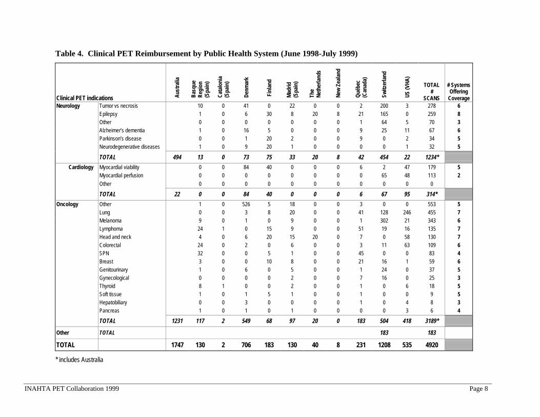
The remaining respondents provided data for analysis. New Zealand has arranged with ahospital in Melbourne, Australia to provide PET scans for patients with epilepsy who arecandidates for surgical treatment, and these data were included under New Zealand in theanalysis. Australia provided only aggregate data for each clinical category. Table 4 listsclinical PET applications reimbursed for each of the 11 public health systems within 13countries or regions, and Table 5 further summarizes these findings. Table 6 provides arelative comparison of reimbursed clinical PET activity in 1999 across health systems.
Data collection time periods varied among some respondents. VA TAP provided datacovering June 1997 through July 1998 and only for veterans scanned at VA PET facilities,labeled herein as US (VHA). Data from Switzerland covered January 1998 throughDecember 1998. Denmark and New Zealand provided six months of data from which theyestimated annual activity. All other respondents provided data for the time period July 1,1998 to June 31, 1999.
INAHTA PET Collaboration 1999 Page 8
Table 4. Clinical PET Reimbursement by Public Health System (June 1998-July 1999)
Clinical PET indications Aust
ralia
Basq
ueRe
gion
(Spa
in)
Cata
loni
a(S
pain
)
Denm
ark
Fin
land
Mad
rid(S
pain
)
The
Neth
erla
nds
New
Zea
land
Qué
bec
(Can
ada)
Switz
erla
nd
US (V
HA)
TOTAL#
SCANS
# SystemsOffering
CoverageNeurology Tumor vs necrosis 10 0 41 0 22 0 0 2 200 3 278 6
Epilepsy 1 0 6 30 8 20 8 21 165 0 259 8Other 0 0 0 0 0 0 0 1 64 5 70 3Alzheimer’s dementia 1 0 16 5 0 0 0 9 25 11 67 6Parkinson’s disease 0 0 1 20 2 0 0 9 0 2 34 5Neurodegenerative diseases 1 0 9 20 1 0 0 0 0 1 32 5
TOTAL 494 13 0 73 75 33 20 8 42 454 22 1234*
Cardiology Myocardial viability 0 0 84 40 0 0 0 6 2 47 179 5Myocardial perfusion 0 0 0 0 0 0 0 0 65 48 113 2Other 0 0 0 0 0 0 0 0 0 0 0
TOTAL 22 0 0 84 40 0 0 0 6 67 95 314*
Oncology Other 1 0 526 5 18 0 0 3 0 0 553 5Lung 0 0 3 8 20 0 0 41 128 246 455 7Melanoma 9 0 1 0 9 0 0 1 302 21 343 6Lymphoma 24 1 0 15 9 0 0 51 19 16 135 7Head and neck 4 0 6 20 15 20 0 7 0 58 130 7Colorectal 24 0 2 0 6 0 0 3 11 63 109 6SPN 32 0 0 5 1 0 0 45 0 0 83 4Breast 3 0 0 10 8 0 0 21 16 1 59 6Genitourinary 1 0 6 0 5 0 0 1 24 0 37 5Gynecological 0 0 0 0 2 0 0 7 16 0 25 3Thyroid 8 1 0 0 2 0 0 1 0 6 18 5Soft tissue 1 0 1 5 1 0 0 1 0 0 9 5Hepatobiliary 0 0 3 0 0 0 0 1 0 4 8 3Pancreas 1 0 1 0 1 0 0 0 0 3 6 4
TOTAL 1231 117 2 549 68 97 20 0 183 504 418 3189*
Other TOTAL 183 183
TOTAL TAL 1747 130 2 706 183 130 40 8 231 1208 535 4920
* includes Australia

INAHTA PET Collaboration 1999 Page 9
Table 5. Summary of the Number of Reimbursed PET Scans by Public Health System(June 1998-July 1999)
Public Health System byCountry or Region Neurology Cardiology Oncology Other TotalAustralia 494 22 1231 0 1747
Switzerland* 454 67 504 183 1208
Denmark* 73 84 549 0 706
US (VHA)* 22 95 418 0 535
Québec (Canada) 42 6 183 0 231
Finland 75 40 68 0 183
Madrid (Spain) 33 0 97 0 130
Basque Region (Spain) 13 0 117 0 130
The Netherlands 20 0 20 0 40
New Zealand* 8 0 0 0 8
Catalonia (Spain) 0 0 2 0 2
TOTAL (% total) 1234 (25) 314 (6) 3189 (65) 183 (4) 4920 (100)
* data collection periods vary. See page 7.
Table 6. Number of Reimbursed PET Scans per 100,000 inhabitants(June 1998-July 1999)
Public Health System byCountry or Region Neurology Cardiology Oncology Other TotalUS (VHA)* 0.73 3.17 13.93 0 17.8
Switzerland* 6.48 0.95 7.2 2.61 17.2
Denmark * 1.46 1.68 10.98 0 14.2
Australia 2.74 0.12 6.83 0 9.7
Basque Region (Spain) 0.65 0 5.85 0 6.5
Finland 1.5 0.8 1.36 0 3.6
Québec (Canada) 0.6 0.09 2.61 0 3.3
Madrid (Spain) 0.1 0 0.3 0 0.4
The Netherlands 0.13 0 0.13 0 0.3
New Zealand* 0.22 0 0 0 0.2
Catalonia (Spain) 0 0 0.03 0 0.03
* data collection periods vary. See page 7.
Neurology. Neurology indications constituted 25% of all reimbursed clinical PET activityamong survey respondents. Excluding Australia, the majority of public health systemsoffered coverage for clinical PET evaluations in epilepsy, Alzheimer’s dementia, braintumors and neurodegenerative disorders. The survey also revealed considerable variability inthe volume of scans for each indication. Of note, PET scans in evaluations of brain tumorand epilepsy comprised 75% of the total volume in neurology.

INAHTA PET Collaboration 1999 Page 10
Switzerland, Australia and Finland reimbursed the most neurologic PET scans. InSwitzerland epilepsy and brain tumors constituted 36% and 44%, respectively, of the totalvolume of neurological diagnostic studies. Finland conducted 60% of neurologic activity inassessments of Alzheimer’s disease, Parkinson’s disease and other neurodegenerativedisorders.
Cardiology. Cardiac indications represented the smallest portion (6%) of reimbursed clinicalvolume in the survey, of which approximately 60% of the scans were performed for viabilitydetermination. Fewer than half of the public health systems surveyed offered funding formyocardial viability determination (five systems) or cardiac perfusion studies (two systems).
US (VHA), Switzerland and Denmark had the highest volume of cardiac PET scans. Thevolume was roughly equivalent for viability and perfusion in US (VHA), whereas themajority of studies in Switzerland were for myocardial perfusion. Denmark, Quebec andFinland only reimbursed viability studies, while US (VHA) and Switzerland covered bothindications.
Oncology. Oncology indications constituted the vast majority (65%) of publicallyreimbursed diagnostic PET scans. The majority of health systems reimbursed for head andneck cancer, lung cancer and lymphoma, with the remainder in breast cancer, colorectalcancer and melanoma. The highest volume of diagnostic PET scans was in evaluationscategorized as “other” primarily in Denmark, but no specific information was madeavailable. Other high volume uses were in lung cancer, lymphoma, head and neck cancerand colorectal cancer.
Further comparison in Table 5 indicates that 85% of reimbursed clinical PET scans wereconcentrated in four public health systems in Australia, Switzerland, Denmark and US(VHA). Except for Finland, The Netherlands and New Zealand, oncology indicationsconstituted the majority of clinical PET reimbursement.
Table 6 shows the relative comparison of clinical PET activity taking into consideration thevolume of patients served by each health system. US (VHA), Switzerland and Denmarkreimburse the most PET scans per 100,000 patients. US (VHA) and Denmark concentratedactivity in oncology, whereas Switzerland split most of its volume between neurology andoncology indications.
3. Trends in the US
Trends in acceptance and use of clinical PET noted by Ter-Pogossian (1992) persistthroughout the 1990s, as most clinical PET activity continues to be concentrated in largermedical centers with affiliated research departments capable of supporting the cyclotrons.Other factors have been identified that influence clinical PET use in the US:
• the complexity and high cost of the technology (Coleman 1992; Flynn 1996);• Food and Drug Administration regulations (Coleman 1992; Flynn 1996);• slow development of reimbursement policies by third-party payers (Coleman 1992; Flynn
1996);• health care reform (Flynn 1996);

INAHTA PET Collaboration 1999 Page 11
• lack of demonstrated clinical utility (Flynn 1996).
Early efforts by PET advocates were ineffective in cultivating acceptance of its clinicalpotential within health care policy and medical communities. In 1991 individuals, institutionsand industry officials in the PET community organized the Institute for Clinical PET (ICP)whose mission is to advance the science of PET and its development in the clinical setting.The ICP actively promotes clinical PET to providers, payers, patients and regulators onbehalf of the clinical PET community (ICP 1999).
Complexity/Costs. The initial purchase and maintenance of a PET facility is a majorimpediment to capital investment in and, as a consequence, access to PET. The primarymarkets for traditional PET scanners are large regional hospitals and academic institutions.
PET manufacturers are focusing research and development on instrumentation and softwarethat not only improve image quality and scanning performance, but also are lower in cost anduser friendly. Lower cost traditional PET systems, mobile units, modified SPECT systemsand networks of Radiopharmaceutical suppliers are expanding the market to include smallerhospitals and clinics. The ICP reports that there are approximately 150 PET centers witheither traditional PET or modified SPECT systems in the US (ICP 1999).
Regulation--Food and Drug Administration (FDA). FDA has either approved or clearedfor marketing both traditional PET cameras and gamma PET cameras to image radionuclidesin the body. Considerable controversy has surrounded the jurisdiction of the FDA inregulating PET radiopharmaceuticals. Prior to 1997, FDA had approved only two PETradiopharmaceuticals for clinical PET use:
• Rb-82 limited to rest alone or rest with pharmacologic stress PET scans and used fornoninvasive imaging of the perfusion of the heart for the diagnosis and management ofpatients with known or suspected coronary artery disease.
• FDG indicated for identifying regions of abnormal glucose metabolism associated with fociof epileptic seizure. Approval for use is restricted to The Methodist Medical Center inPeoria, Illinois.
Supporters in the United States Senate were instrumental in codifying changes to the FDAModernization Act of 1997 pertaining to FDA regulation of PET (Connell 1999). As a resultof this law passing, FDA withdrew its previous regulation of PET drug products andmanufacturing guidelines and is drafting new procedures to replace them (FDA 1997). Anofficial ruling on the safety and effectiveness of three PET drugs, F-18 FDG, N-13 ammoniaand O-15 water and on the proposed procedures for obtaining marketing approval for themwill be forthcoming (FDA 1999).
Reimbursement. To a great extent, reimbursement for PET studies can influence itsdiffusion into clinical medicine. Because of the controversy surrounding FDA jurisdictionover PET radiopharmaceuticals, many payers have been reluctant to include PET as acovered benefit, until the dispute was resolved. Not surprisingly, the recent changes in theFDA regulation of PET radiopharmaceuticals have had a positive influence onreimbursement for clinical PET in the US.

INAHTA PET Collaboration 1999 Page 12
Medicare—Medicare coverage policies frequently reflect FDA decisions and shape coveragedecisions of other payers. Until recently, FDA approval of each site as a manufacturer ofPET radiotracers had delayed Medicare reimbursement for clinical PET scans. The HealthCare Financing Administration2 (HCFA) has been continuously reviewing the scientificliterature on clinical PET scans and, until the end of 1997, offered limited coverage for PETscans (See Table 7).
Table 7. HCFA-Approved Medicare Benefits for PET Scanning
Date Indication(s) Conditions for coverageMarch 1995 Cardiac Perfusion
Imaging Using Rb-82
• Medically necessary• Does not unnecessarily duplicate other covered diagnostic tests• Does not involve investigational drugs or procedures using investigational
drugs• Used in place of SPECT or in addition to an inconclusive SPECT• Not for screening asymptomatic patients
January 1998 SPNs and Non-Small Cell LungCancer (NSCLC)Using FDG
• Uses traditional PET scanners or coincidence imaging gamma cameras• To characterize SPNs initially detected, usually by CT• To stage mediastinum in patients with confirmed primary NSCLC• Payment for the use of routine biopsy following a negative PET scan will be
denied for these conditions, unless the claim is supported by evidenceexplaining the medical necessity of the biopsy
March 1999 Additional OncologyIndications UsingFDG
• Detecting and localizing recurrent colorectal cancer with risingcarcinoembryonic antigen (CEA)
• Staging and characterizing both Hodgkin’s and non-Hodgkin’s lymphoma inplace of a gallium scan or lymphangiogram
• Identifying metastases in melanoma recurrence in place of gallium studies
Source: HCFA 1998
Intense pressure on the part of clinical PET advocates has influenced expansion of PETcoverage in oncology and inclusion of SPECT with coincidence detection capability(Anonymous 1998). Interim coverage for PET scans is conditioned upon its ability toprovide useful information for the management and treatment of patients with theseconditions. HCFA will use the claims process to evaluate the impact of PET on clinical careand determine the extent to which they should modify future policy.
2 Health Care Financing Administration of the Department of Health and Human Services administers the Medicare, Medicaid, and ChildHealth Insurance Programs. In addition, HCFA performs a number of quality-focused activities including development of coveragepolicies. http://www.hcfa.gov/

INAHTA PET Collaboration 1999 Page 13
Veterans Health Administration (VHA)—Currently, VHA has ten PET scanners, and nearlyall share operations with academic affiliates. VHA does not have a national coverage policyfor PET scans; coverage decisions are made by each institution. Access to VHA PET centersis limited, and they are not evenly distributed throughout the system. Consequently, manyVA medical centers without traditional PET facilities are upgrading dual-headed gammacameras for coincidence imaging, or are affiliating with local PET centers in the privatesector.
VHA is capitalizing on its investment in PET in several ways:• by establishing a registry to collect important utilization data from VHA PET centers;• by funding prospective outcomes research; and• by conducting regular systematic review updates to track new data in the literature.
Private sector carriers—The ICP has successfully lobbied national and local carriers and isactively involving patients in its lobbying efforts (ICP 1999; Connell 1999). The ICP reportsthat, since 1996, there has been an increase in the number of insurance carriers now coveringPET scans (See Table 8). Table 8 is not a complete listing of all carriers with approvedcoverage policies for PET, but it does illuminate the trend in PET coverage among privatesector carriers in the US in recent years. Coverage decisions emphasize PET’s use inoncology, particularly of the lung. However, most still reimburse on a case-by-case basis,and many require pre-approval.
Table 8. Approved PET Indications of Selected Private Carriers in the US
Source=ICP (personal communication: Ruth Tesar, ICP, May 1998).
Carrier Approved indicationsCalifornia Blue Cross Lung, colorectal, head and neck, melanoma, SPN
California Blue Shield Lung, colorectal, head and neck, melanoma, SPN
Empire BC/BS New York SPN, lung cancer staging
Cigna/Health Source Provident Colorectal, head and neck, melanoma, lung
Duke University Brain tumors, colorectal
Kaiser-Permanente* Epileptic foci when not identified by other meansSPN in selected low risk patients
Aetna/USHealthCare** Cardiac-Rb82
Brain tumor vs. necrosis, epileptic foci, and lung cancer on a case-by case basisOthers, not specified
United Health Care Several, not specified
Several, not specified Brain tumors
Many, not specified Epilepsy
Few, not specified Cardiac-Rb82
*personal communication: M. Sugarman, Kaiser-Permanente (May 1999)**personal communication: R. McDonough, M.D., Aetna/USHealthCare (May 1999)

INAHTA PET Collaboration 1999 Page 14
Health care reform. Unless otherwise noted, background information on health care reformis taken from Zelman (1996). Over the last two decades health care reform initiatives relatedprincipally to payment system restructuring are driving more explicit decisions about the useof health care resources. This section presents a condensed account of the current and mostrelevant reforms measures and the impact of these measures on the diffusion of PET intoclinical care.
The introduction of a prospective, pre-determined payment system (called the ProspectivePayment System) in 1983, departed from the traditional retrospective fee-for-service system.This has had a dramatic effect on health industry behavior (Holmes 1992). The new systemof payment sharply curtailed public sector revenue to providers, who responded byincreasing premiums to private payers, including employers. Private insurers quickly adoptedthe new payment system, which further decreased revenue to providers.
As premiums continued to rise at an alarming rate, payers, providers and consumers becamemore value-driven. Maximizing outcomes and productivity while containing costs haveresulted in a growth in underwriting practices, managed care alternatives, outcomes researchand patient consumerism. Increasingly, government intervention is sought: 1) to protectconsumer value by lowering or limiting the rise in costs and maintaining or elevating quality,and 2) to foster or maintain competition, for example, by establishing basic minimumstandards of care and requiring evidence of cost-effectiveness for benefit coverage.
In the above scenario, regulation and reimbursement primarily defined the market for PET.Early proponents for clinical PET sought to capitalize on a cutting-edge technology thatshowed potential both as a clinical tool and as a source of revenue (Flynn 1996). However,PET was introduced into clinical medicine when trends in health care decision making weretransitioning from a rationale based primarily on resources and opinions to a rationalederived from research findings.
Today in response to reform pressures decision makers, who seek to optimize patient care byinvesting in diagnostic technologies, are insisting on scientific evidence that supportsimproved patient outcomes and cost-savings in addition to safety and improved diagnosticperformance. The clinical research community is beginning to collaborate on evaluations ofPET’s utility in selected clinical areas (Adams 1998).
Clinical utility. Flynn (1996) reviewed assessments, guidelines and policy statements onclinical PET use produced by US organizations through 1994. Early findings or commentsabout the use of PET in clinical applications were generally based on expert opinion and non-systematic qualitative reviews. Comments emphasized PET’s clinical “potential” in severalareas, mainly neurology and cardiology, and its ability to provide unique clinicalinformation. The added value of this information was not proven.
Since 1994 emphasis has shifted toward more rigorous methods of technology assessment.Results of such assessments of PET from US organizations published after 1994, which arein the public domain or otherwise made available to the authors for this report, are includedand discussed in Part II.

INAHTA PET Collaboration 1999 Page 15
C. Summary/Discussion
INAHTA members’ experiences with PET since 1997 show wide variations in use and publicreimbursement for clinical scans. PET’s availability and use is still quite limited, as evidencedby the small numbers and relative under use of scanners in each country or region. Most PETscanners are confined to academically affiliated facilities, and most health systems (excludingVHA) have fewer than five PET scanners. FDG is the most common radiotracer used in clinicalPET studies, and its availability is critical to PET facilities wishing to conduct diagnostic tests.FDG produced on-site requires a cyclotron, and commercial vendors of FDG can varyregionally.
The majority of PET scanners are used for both research and diagnostic studies, but there areexceptions. Local regulatory policies and funding sources may explain the variations in PET useamong survey participants. For example, regulatory policies that stress clinical research toaddress gaps in knowledge of PET’s clinical utility may explain the high frequency of researchstudies in some countries. In countries with a high concentration of clinical studies, privatefunding sources may be available to enhance clinical activity that would otherwise not becovered with public funding. The size and geographic distribution of the population served byeach health system may further contribute to the variations in use.
The latest utilization data show that most clinical PET activity is concentrated in relatively fewhealth systems and primarily in a few select indications in oncology and neurology. Whereaspublic financing is available for several oncology indications, nearly 70% of the volume isconfined to evaluations of melanoma, lung cancer staging, and an undefined category of “other.”Similarly, reimbursement is available for a number of neurological indications, but the vastmajority of neurological PET activity is confined to distinguishing brain tumor versus radiationnecrosis and to localizing epileptic foci in potential surgical candidates with intractable epilepsy.
High cost and technical complexity, regulatory policies, value-driven health care decisionmaking and lack of demonstrated clinical utility had hampered the diffusion of clinical PET inthe US. However, PET use and acceptance have been on the rise, particularly since 1997. Lowercost PET systems, mobile units, modified SPECT systems and commercialization ofradiopharmaceuticals allow smaller hospitals and clinics to offer clinical PET capability. Well-organized promotions by the clinical PET community both in the US and abroad have beeninstrumental in supporting initiatives that increase demand for the technology and in changingpolicies that now permit limited reimbursement for PET despite the continued lack of evidencesupporting its clinical or economic benefit.

INAHTA PET Collaboration 1999 Page 16
PART II. SYNTHESIS OF TECHNOLOGY ASSESSMENTS OF PET
Several INAHTA members have conducted technology assessments of PET in response to theneeds of their health care systems. Likewise, several private TA organizations in the US haveevaluated PET for a range of clinical indications to support clinical policy makers. Each assessmentaddressed various research questions using an array of approaches. Synthesizing the assessmentswill expand the scope of each assessment into a single, comprehensive document.
A. Methods
The authors surveyed each INAHTA member for completed technology assessments on the clinicalutility of PET. Three private US organizations also agreed to collaborate on the project: Blue CrossBlue Shield Association Technology Evaluation Center (BCBSA TEC), Emergency Care andResearch Institute (ECRI), and HAYES, Inc. (HAYES).
B. Results
Appendix C contains information abstracted from each PET assessment. Full reports fromINAHTA members are kept on file at the VA TAP; reports from BCBSA TEC, ECRI, andHAYES are proprietary. Thirty-one assessments from 13 organizations were synthesized for thisreview. The assessments reflect a variety of motivations for conducting the assessment, researchquestions, inclusion criteria and methodologies. As is the nature of health care technologyassessment, organizations produced assessments for decision-makers in response to pressures touse or evaluate PET for one or more clinical indications.
Most assessments were qualitative systematic reviews and focused on evaluations of PET’s clinicalutility. The remainder comprised quantitative analyses, non-systematic reviews, report synthesesand expert panel consensus. ECRI, BCBSA TEC and MSAC used quantitative methods to analyzePET’s utility and/or its economic impact in health care. Reports from CEDIT (Baffert 1999) inFrance and SFOSS in Switzerland outlined proposals for systematically evaluating PET in severalclinical indications within their respective health systems; VA TAP (Adams 1998) reported resultsof an ongoing system-wide PET registry data collection effort.
To the extent that the inclusion criteria were either specified in the report or made available to theauthors of this synthesis, most reports appraised literature published or otherwise available since1990. BCBSA TEC (1997) and AHCPR (1998) extended their literature searches to 1985 and1977, respectively. Organizations used electronic data sources extensively. Several reportsincluded literature published in a range of languages, thus helping to minimize potential languagebias. Unless otherwise stated, assessments appraised studies using PET with FDG, reflecting theradiopharmaceutical most often used in clinical PET studies.
All organizations assessed traditional full ring PET systems. Partial ring PET scanners, gammacameras (SPECT) modified for coincidence detection and 511 keV collimated PET imaging arenewly available, lower cost alternatives to traditional full ring PET systems but are not yetoptimized for clinical use. However, they attract considerable interest, and their rapid diffusion intoclinical care could have a substantial impact on national health systems. Accordingly, AHFMR,

INAHTA PET Collaboration 1999 Page 17
CEDIT, NCCHTA and VA TAP addressed one or more of the alternatives to traditional PET intheir reviews.
Report syntheses and updates are ongoing efforts of participating agencies to track the PETliterature and are included in this report. OSTEBA and AETS (1995) synthesized several reports,including those from INAHTA members. AETS and NCCHTA used methodologies similar to VATAP to expand on the VA TAP (Flynn 1996) report, and VA TAP updated its review in 1998.BCBSA TEC is in the process of updating its assessments, but the updates are not yet available tothe authors of this synthesis. HAYES continues to update its assessments and has made themavailable for this report.
The major uses for clinical PET are grouped into three main categories: neuropsychiatry,cardiology and oncology. Technology assessments of PET in each category are discussed below.Indications for which assessment findings disagree or make for compelling discussion are describedin more detail within each category.
1. Neuropsychiatry
Early PET activity focused on clinical and research indications in neurology and psychiatry.PET allows the qualitative and quantitative evaluation of cerebral physiology and the study ofthe biochemical bases for clinical diseases. The majority of indications in Table 9 employ FDGto study glucose metabolism using traditional PET systems.
Table 9. Assessments of Clinical PET in Neuropsychiatry byOrganization and Indication
Brain tumor
OrganizationReportDate Al
zhei
mer
’s d
isea
se
Park
inso
nism
Oth
er n
euro
dege
nera
tive
diso
rder
s
Epile
psy
Diffe
rent
ial
diag
nosi
s
Gra
ding
mal
ig-
nant
glio
mas
Recu
rren
ce v
s.ne
cros
is
Gui
ding
ste
reo-
tact
ic b
iops
y
Mon
itorin
gtre
at-m
ent
Cere
brov
ascu
lar
diso
rder
s
Oth
er
AETS May 1999 4 4 4 4 4
AHCPR Jul 1998 4
AHFMR Aug 1998 4
BCBSA TEC Mar 1997 4 4 4 4 4 4 4 4
CAHTA 1993 4 4
CAHTA 1996 4
CAHTA 1997 4
HAYES Jul 1997 4 4 4 4 4 4
MSAC Nov 1990 4
NCCHTA Feb 1999 4 4 4 4
OSTEBA Sep 1998 4 4 4
VA TAP Sep 1996 4
VA TAP Dec 1998 4
INAHTA PET Collaboration 1999 Page 18
Intractable epilepsy. The majority of patients with epilepsy use anti-epileptic medication tocontrol seizure activity. Among patients whose seizures remain uncontrolled with medication(medically refractory or intractable epilepsy), most suffer from temporal lobe epilepsy.Resective temporal lobe surgery has been shown to be an effective treatment for appropriatelyselected patients with medically refractory complex partial seizures to avoid progressive braininjury due to uncontrolled seizures and the adverse effect of anti-epileptic medication.
Patients undergo pre-surgical evaluation to identify and delineate the epileptogenic foci and todetermine resectability. In many cases the decision to proceed with surgery can be made basedon information from a detailed history, observation and review of non-invasiveelectrophysiologic, neurophysiologic and structural imaging and can result in good postsurgicaloutcomes. Other tests such as invasive electroencephalography (EEG), MRI and functionalimaging modalities may be used to help identify additional surgical candidates. Interictal(between seizures) PET using FDG records glucose hypometabolism that appears to beassociated with the epileptogenic zone. PET has been suggested in the work up of thesepatients to complement MRI data and potentially to supplant or reduce the use of invasiveEEG.
Eight organizations appraised the literature on PET for the pre-surgical localization ofepileptogenic foci in patients with medically refractory complex partial seizures. Reportsfocused primarily on the use of interictal FDG PET to measure hypometabolic regions of thetemporal lobe. Findings conflict regarding the quality of the available evidence on which toestablish PET’s efficacy for this indication. BCBSA TEC found that FDG PET imaging forthese patients met their methodologic quality criteria; other assessments remarked on thelimited quantity and quality of evidence available to establish PET’s clinical utility.
Assessments suggested that the diagnostic accuracy of interictal FDG PET was comparable orsuperior to other functional imaging modalities used to confirm epileptogenic foci indicated byEEG or structural lesions on MRI. However, available evidence was insufficient to supportreplacing either invasive EEG or structural imaging with PET and had not supported using PETfor many patients with non-temporal lobe epilepsy.
PET could potentially benefit a small minority of patients whose epilepsy is difficult to manage,but its impact on patient management decisions, eventual outcome and costs are as yetunknown. Several reports suggested that the use of PET in managing patients with intractableepilepsy be done in the context of well-designed prospective research protocols.
Alzheimer’s disease3. In Alzheimer’s disease (AD) the primary role of diagnostic testing hasbeen the differential diagnosis of AD from reversible or treatable diseases. Functional imagingtechnologies have been used to improve diagnostic certainty and provide information on thepathophysiologic basis of AD and can aid in early diagnosis. While there is no cure for AD,
3 A definitive diagnosis of AD is based on a typical clinical picture and histopathologic findings in samples of braintissue at autopsy. In the absence of histologic confirmation of AD, patients are referred to as having a diagnosis ofDementia of the Alzheimer’s Type (DAT). For simplicity, in this report AD is used to mean patients with DAT.

INAHTA PET Collaboration 1999 Page 19
psychosocial techniques and drug therapies aimed at slowing disease progression are nowavailable to help improve quality of life.
For differentiating Alzheimer’s disease from other dementias, assessments generally agreed thatthe diagnostic accuracy of PET appeared comparable or superior to competing technologieslike CT, MRI, SPECT or EEG, but was little and of low quality. Assessments concurred on theneed for additional rigorous study. Reports acknowledged the importance of improveddiagnostic information to AD patients and their families for coping and for future planning, butthe value of improved diagnostic information to management of AD patients or to theimprovement in clinical results was unknown. Further, the potential utility and accuracy of PETin AD should be viewed in the context of no available effective cure and of the accessibility andaccuracy measurements of other diagnostic modalities, many of which have been morerigorously studied.
Brain tumors. For managing patients with brain tumors, primarily gliomas, BCBSA TEC foundthat in none of the indications was there sufficient scientific evidence to permit conclusionsabout the effect of PET on health outcomes. In the differential diagnosis of radionecrosisversus residual or relapsing tumor, CAHTA (1993) concluded that PET’s diagnosticperformance was superior to conventional diagnostic techniques (CT, MRI). AETS (1999)similarly pointed out that while PET appeared to be superior to MRI for this indication, PETwas not superior to SPECT. Further, PET’s impact on clinical management wasundocumented, the overall quality of available evidence was limited, and further controlledstudy of PET was warranted.
Cerebrovascular and other neurodegenerative and neuropsychiatric disorders. Studiesemploy a variety of radiotracers with PET to evaluate cerebral metabolism and perfusion, tomap cerebral regions and to locate receptor sites within the brain. Potential indications includedifferential diagnosis, assessing response to therapy and improved knowledge of diseasemechanisms.
Reviews of the evidence recognized PET’s contribution to the knowledge of the biochemicaland physiological mechanisms of many cerebrovascular and neuropsychiatric conditions, but itwas unclear whether the added information improved patient management or outcomes.Evidence comprising small and methodologically inconsistent studies and lack of normative dataprevented definitively establishing PET’s efficacy or cost-effectiveness for these indications.AETS, NCCHTA and OSTEBA recommended further prospective study to define thecontribution of PET in these areas.
2. Cardiology
There is an extensive array of noninvasive strategies for diagnosing coronary artery disease(CAD). For patients with chronic left ventricular dysfunction who are being considered forrevascularization by coronary artery bypass surgery or angioplasty, there is a need to accuratelydetermine whether the myocardium is viable and likely to respond to improved blood flow.Traditionally, clinicians use noninvasive coronary perfusion imaging in these patients todiagnose and evaluate CAD and to determine viable and hibernating myocardium for potential

INAHTA PET Collaboration 1999 Page 20
response to revascularization. Conventional techniques identify viable tissue by measuringperfusion, contractile reserve and cell membrane integrity (Cowley 1999). Standard coronaryperfusion imaging consists of single photon emission computed tomography (SPECT) andplanar scintigraphy using intravenous administration of thallium-201 or technetium-99msestamibi during exercise or pharmacologic stress. These techniques often employ delayedimaging methods to identify viable tissue. Technetium-99m is also used for gated blood poolscanning to measure left ventricular ejection fraction (the capacity of the heart muscle tocontract). Stress echocardiography further assesses global and regional wall motionabnormalities that may not be present at rest, and the use of magnetic resonance technologies inthis area is evolving.
Traditional PET systems reportedly offer higher quality images over conventional testing. PETcan image and quantify myocardial perfusion with a variety of tracers such as nitrogen-13ammonia, oxygen-15 water, or rubidium-82. Most PET studies of myocardial viability utilizethe radiotracers FDG and, to a lesser extent, C-11 acetate to detect active metabolism.
In this setting the metabolic information from PET may improve patient selection forrevascularization and consequently the likelihood of successful surgery. PET may offer costsavings by eliminating unnecessary angiography and revascularization in inappropriate patients.Coincidence detection SPECT, high energy SPECT and 511 keV collimated positron imaginghave been advocated as less costly, technically simpler and potentially more accessiblealternatives to traditional PET systems.
Table 10. Assessments of Clinical PET in Cardiologyby Organization and Indication
OrganizationReportDate
Myocardialperfusion
Myocardialviability
Monitoringtreatmentresponse
AETS Dec 1995 4 4
AHCPR Jan 1995 4
AHFMR1999(pending)
4
BCBSA TEC Oct 1995 4
BCBSA TEC 1996 4
CAHTA 1993 4 4
CEDIT Apr 1998 4 4
HAYES May 1997 4 4 4
HAYES Jul 1999 4 4
MSAC Nov 1990 4 4
NCCHTA Feb 1999 4 4
OSTEBA Sep 1998 4 4
Myocardial perfusion. AHCPR and BCBSA TEC confined their reviews to studies using thetracer rubidium-82, and BCBSA TEC further restricted its review to studies of patients at

INAHTA PET Collaboration 1999 Page 21
intermediate risk of having CAD.4 BCBSA TEC produced a cost-effectiveness analysis (Garber1996) using the societal perspective and analytic assumptions from their 1995 clinicalassessment. CEDIT appraised studies on the use of coincidence detection SPECT forconventional scintigraphy.
Reports generally agreed on the comparable or superior performance of PET to othermyocardial perfusion imaging alternatives, particularly to thallium-201 SPECT, but the extentof the improvement in performance and its contribution to managing patients with CAD wasunclear. PET was more costly than all other individual noninvasive strategies, and it had notbeen able to replace coronary angiography as the definitive standard for assessment of CAD inmost symptomatic patients. PET for patients at intermediate risk as determined by BCBSATEC was an unlikely cost-effective alternative to immediate angiography or to othernoninvasive tests such as stress echocardiography or SPECT. Consideration should be given tothe most overall cost-effective approach, but at present, evidence is needed to establish therelative cost-effectiveness of PET in diagnosing CAD.
Coincidence detection SPECT has the theoretical advantage of being able to image bothpositron-emitting and gamma-emitting radiopharmaceuticals. CEDIT found that there was noscientific evidence to support the use of coincidence detection SPECT as a replacement forconventional scintigraphy. Experts agreed that for either myocardial perfusion or viabilitystudies no practical problem arose when coincidence detection SPECT was carried out usingtechnetium and higher energy radiotracers, but that the quality of coincidence detection SPECTusing low energy thallium could not be guaranteed.
Myocardial viability. For determining myocardial viability and/or predicting risk for cardiacevents, most assessments found that PET appeared to have comparable sensitivity and superiorspecificity to other modalities, but the studies comparing PET’s diagnostic performance toother functional imaging modalities were few and methodologically flawed. With regard toimproving the likelihood of successful revascularization and cost savings, the data wereinsufficient to confirm the relative cost-effectiveness of PET. AHFMR determined that the datawere similarly limited for all other functional imaging modalities (SPECT, dobutamineechocardiography and MRI). However, CAHTA suggested a limited role for FDG-PET forpatients with inconclusive results on delayed thallium-201 reinjection imaging. HAYES (1999)concluded from the evidence that PET information is clinically useful only for patients who aresuitable candidates for revascularization.
NCCHTA identified as a major research priority the relative cost-effectiveness of coincidencedetection SPECT and 511 keV collimated PET for selecting patients for myocardialrevascularization. AHFMR stated that any use of PET for this indication in Alberta, Canadashould be associated with prospective studies involving long-term follow up.
4 defined as 25% – 75% probability of having either a 50% or greater left main coronary artery occlusion or a 70% or greater occlusion of any othercoronary artery.
INAHTA PET Collaboration 1999 Page 22
Monitoring response to treatment. HAYES (1997) concluded that PET’s efficacy had notbeen firmly established for assessing the effects of pharmacologic therapy or risk factormodification techniques in subjects with CAD, hypertension or cardiomyopathy.
3. Oncology (non-central nervous system tumors)
Reliance on tumor histology and anatomy limits the oncologist’s tools for selecting the mostfavorable treatment. Since tumor metabolism and blood flow often differ from adjacent tissue,adding functional information may expand the oncologists’ ability to optimize treatment. PEThas been used to detect, stage and grade tumors, discern recurrence from treatment changes,predict tumor response to therapy and monitor response to therapy. FDG is the mostcommonly employed radiopharmaceutical in PET cancer studies.
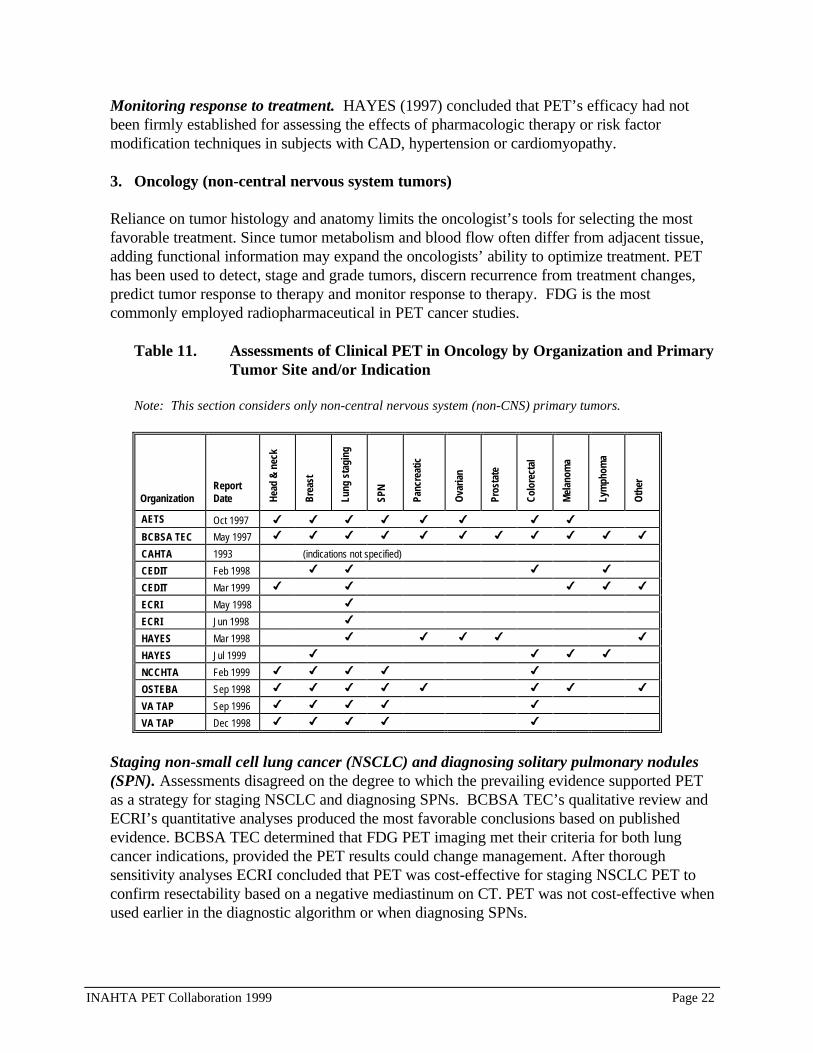
Table 11. Assessments of Clinical PET in Oncology by Organization and PrimaryTumor Site and/or Indication
Note: This section considers only non-central nervous system (non-CNS) primary tumors.
OrganizationReportDate He
ad &
nec
k
Brea
st
Lung
sta
ging
SPN
Panc
reat
ic
Ova
rian
Pros
tate
Colo
rect
al
Mel
anom
a
Lym
phom
a
Oth
er
AETS Oct 1997 4 4 4 4 4 4 4 4
BCBSA TEC May 1997 4 4 4 4 4 4 4 4 4 4 4
CAHTA 1993 (indications not specified)
CEDIT Feb 1998 4 4 4 4
CEDIT Mar 1999 4 4 4 4 4
ECRI May 1998 4
ECRI Jun 1998 4
HAYES Mar 1998 4 4 4 4 4
HAYES Jul 1999 4 4 4 4
NCCHTA Feb 1999 4 4 4 4 4
OSTEBA Sep 1998 4 4 4 4 4 4 4 4
VA TAP Sep 1996 4 4 4 4 4
VA TAP Dec 1998 4 4 4 4 4
Staging non-small cell lung cancer (NSCLC) and diagnosing solitary pulmonary nodules(SPN). Assessments disagreed on the degree to which the prevailing evidence supported PETas a strategy for staging NSCLC and diagnosing SPNs. BCBSA TEC’s qualitative review andECRI’s quantitative analyses produced the most favorable conclusions based on publishedevidence. BCBSA TEC determined that FDG PET imaging met their criteria for both lungcancer indications, provided the PET results could change management. After thoroughsensitivity analyses ECRI concluded that PET was cost-effective for staging NSCLC PET toconfirm resectability based on a negative mediastinum on CT. PET was not cost-effective whenused earlier in the diagnostic algorithm or when diagnosing SPNs.

INAHTA PET Collaboration 1999 Page 23
Other agencies found the existing body of evidence on PET’s efficacy insufficient to establish arole for PET in staging NSCLC or diagnosing SPNs. Lung cancer constitutes a considerableburden to the health systems represented by INAHTA agencies. Consequently, severaladvocated rigorous comparative study of PET, including coincidence detection SPECT andpartial ring PET versus traditional PET, to alternative strategies to clarify PET’s role in stagingNSCLC.
All other indications. Based on evidence from 1977 through 1998, reviews were unable tofirmly established PET’s role in all other oncology indications. Recent 1999 reviews by HAYESconfirmed these findings in breast cancer, melanoma and lymphoma but suggested a plausiblecomplementary role for PET with conventional imaging to confirm suspicious post-treatmentcolorectal cancer recurrence, if results will significantly alter patient management or improveoutcome. However, within certain health systems there is focused interest in PET fordiagnosing and staging patients with breast cancer, lymphoma and melanoma.
Several agencies stressed the need for further research to define or support the relativecontribution of PET in the management of patients with these cancers. Recent assessments byNCCHTA and VA TAP (Adams 1998) advocated rigorous study of positron coincidenceimaging alternatives in the oncologic work up. CEDIT (Baffert 1999) and SFOSS areinstituting clinical protocols to systematically collect data on the use of PET in selectedoncology indications.
C. Summary
Assessments, founded collectively on evidence available since 1977, provide valuable insight intoclinical trends and the body of knowledge used to suggest PET’s clinical utility thus far. Availableresearch assessed the feasibility of using primarily traditional full ring PET in certain clinicalsituations and on defining its accuracy as a diagnostic test.
There was uniform agreement that critical research into defining the clinical consequences of usingPET on treatment decisions and health outcome has not been studied. While deficiencies in theevidence prevented most organizations from firmly establishing a clinical role for PET, someidentified plausible roles in view of the clinical context and PET’s availability and diagnosticperformance relative to alternative modalities (Table 12).
Differences among report conclusions generally indicated the degree to which the evidence met theassessments’ quality assessment criteria, where reported. Other possible reasons may have includedthe rationale for the assessment, focus of the report, inclusion criteria and analytical methods.Therefore, methodologic transparency is critical to health care organizations wishing to make validcomparisons of technology assessments and establish policies based on the best available evidence.

INAHTA PET Collaboration 1999 Page 24
Table 12. Potential Clinical PET Indications Identified by INAHTA PETCollaboration Participants
Clinical indication Evidence suggests…Diagnosing seizure foci in intractable epilepsy • PET’s diagnostic accuracy was comparable or superior to other functional
imaging modalities used to confirm foci identified by EEG or MRI, but PETis not yet able to replace invasive EEG or structural imaging.
• Diagnostic contribution of all functional imaging for this indication is stillquestioned.
Diagnosing Alzheimer’s dementia PET’s diagnostic accuracy was comparable or superior to competingtechnologies (CT, MRI, SPECT, EEG), but the value of improved diagnosticinformation to management of AD patients or to improved clinical results wasunknown.
Diagnosing brain tumor recurrence vs. radiationnecrosis
PET’s diagnostic accuracy was superior to conventional diagnostic techniques(CT, MRI) but not to SPECT.
Assessing myocardial perfusion in patients withcoronary artery disease (CAD)
• PET’s diagnostic accuracy is improved over other imaging alternatives,particularly thallium-201 SPECT, but the extent of improvement is unclear.
• PET is more costly than all other individual noninvasive strategies.• PET is unable to replace coronary angiography as the definitive standard for
CAD assessment in most patients. Assessing myocardial viability • PET has comparable sensitivity and superior specificity to other modalities.
• Quality of data for evaluating the performance of SPECT, dobutamineECHO and MRI are similarly limited.
Diagnosing and staging non-small cell lung cancer PET may be cost-effective for staging lung cancer to confirm resectability inpatients with a negative mediastinum on CT.
Characterizing solitary pulmonary nodules PET may have utility when other diagnostic tests are inconclusive.
It should be noted that the evidence supporting many technologies used in routine practice and forwhich coverage policies exist are similarly deficient and often show diagnostic performance inferiorto PET (e.g. myocardial viability diagnostic testing). Further, the value of improved diagnosticaccuracy with PET or with other modalities is questioned in certain indications for which there isno cure or effective treatment to improve prognosis (e.g. Alzheimer’s dementia).
Review of assessments shows both encouraging and disturbing trends in the advancement ofknowledge regarding PET’s clinical utility. Neuropsychiatric and, to a lesser extent, cardiac clinicalindications have been studied since the early 1980s. Yet after almost two decades questions ofPET’s clinical utility persist and hamper its diffusion into clinical practice.
In recent years there have been positive trends in the regulation and use of PET in oncology in spiteof the lack of supportive evidence. As pressure on our health care resources increases, similartrends are seen in the use of lower-cost nuclear medicine systems modified for positron emissioncoincidence detection. Questions of PET’s utility in oncology could follow a path similar to itsother indications, unless rigorous, prospective clinical research is conducted.
In light of limited health care resources, some organizations recommended approving use on a case-by-case basis in select indications for which there are limited diagnostic options. Some conditionedPET use on its ability to affect patient management decisions or restricted its use to subgroups ofpatients who met explicit selection criteria. Most identified clinical PET as a major research

INAHTA PET Collaboration 1999 Page 25
priority for their respective organizations and recommended well-designed prospective clinicalstudies to assess the relative contribution of traditional and/or modified PET. Several INAHTAagencies reported on rigorous research efforts either underway or proposed within their healthsystems to help clarify the contribution of PET to clinical medicine, which for now remains elusive.
INAHTA PET Collaboration 1999 Page 26
APPENDIX A. 1997 INAHTA PET SURVEY
1. Number, type and use of PET scanners (human subjects only).
PUBLIC scanners
Type Scans/ yearOrganisation and city Body PET Small Research Clinical
PRIVATE scanners
Type Studies/ yearOrganisation and city Body PET Small Research Clinical
2. Diagnostic application and public reimbursement
Diagnosticuse
PublicReimbursement
Diagnostic applications Yes No Yes No
EpilepsyNeurology Tumor vs necrosis
Neurodegenerative disordersOther.....................................Viability
Cardiology Ischemic heart diseaseOther....................................Head and neckColorectalBreastLungSolitary pulmonary nodules
Oncology HaematologicalHepatobiliarySoft tissueGenitourinaryGynaecologicalOther....................................
Other

INAHTA PET Collaboration 1999 Page 27
APPENDIX B. 1999 INAHTA PET SURVEY
Activity for the time period (1 July 1998 – 30 June 1999)
PublicReimbursement
Diagnostic applications Yes(Number)
No
Neurology Epilepsy
Alzheimer disease
Parkinsonisms
Neurodegenerative disorders
Tumour vs necrosis
Other.....................................
Cardiology Myocardial Viability
Coronary artery disease
Other....................................
Oncology Head and neck
Colorectal
Breast
Lung
Solitary pulmonary nodules
Lymphomas
Hepatobiliary
Pancreas
Thyroid
Soft tissue
Melanoma
Genitourinary
Gynaecological
Other....................................
Other............................................
............................................
Type of Reimbursement: __ Case by Case __ Regular Basis __Other………
Number of PET Scans in the Country or Region: ………….
Health Care Financing Organisation …………………………..
Level (National, Regional or Local): …………………………..

INAHTA PET Collaboration 1999 Page 28
APPENDIX C. INAHTA PET COLLABORATION ASSESSMENTS OF CLINICAL PET
Organiza-tion
ReportDate Indications studied
Sources and DatesCovered in Search
AssessmentType Quality Assessment Conclusions/Recommendations
AETS Dec 1995 Myocardial perfusionMyocardial viability
Not specified Synthesis ofreports fromECRI andAHCPR
Criteria not specified • PET and SPECT appear to perform similarly fordiagnosing coronary perfusion in coronary diseaseand for making subsequent management decisions(AHCPR).
• Substituting PET for SPECT or vice versa fordetermining myocardial viability depends on whichis more cost-effective to use (ECRI).
AETS Oct 1997 Head and neckRecurrent colorectalBreast cancerLung cancerSPNPancreaticMetastatic melanomaOvarian cancer
VA TAP PET reportthrough 1995,MEDLINE andHEALTHPLANthrough 1996
Systematicreview
EBM criteria used forgrading quality of evidence
Fryback and Thornburyscale used for classifyingselected articles
• No definitive conclusions can be made relative tothe contribution of PET in the management of theoncologic patient.
• PET seems to offer a good alternative for lungcancer staging and SPN diagnosis.
• As there exists no controlled clinical trials, PET isdeemed an investigative technology.
• Rigorous, clinical trials are needed to assess theclinical benefit of PET in all clinical indications.
AETS May 1999 Alzheimer diseaseParkinsonismsEpilepsyBrain TumorsOther less frequent
MEDLINE 1995-Dec 1997Extended to the endof 1998 forradionecrosis vs.Residual-relapsinglesions.
Systematicreview
EBM criteria used forgrading quality of evidence
Fryback and Thornburyscale used for classifyingthe 48 selected articles
• FDG-PET has proven clinical utility in themanagement of:• Refractory complex partial seizures and
temporal epilepsy candidates for surgery, as acomplementary diagnostic and prognostic tool.FDG-PET does not preclude invasive methodsin most cases.
• Differential diagnosis between radionecrosis andresidual or relapsing tumoral lesions.
• FDG-PET aids in the early diagnosis of AD. Thisfact doesn’t modify the current clinicalmanagement of this disorder.
• There is a remarkable lack of studies and ofmethodological quality guided to establish thePET's utility in handling of specific clinicalsituations, and its contribution in improvingtherapeutic results.
• Recommend developing appropriately designedprospective studies mainly to answer questions ofgreat interest for the National Health System inorder to use PET most effectively.
• PET use should be controlled according to aresearch protocol.
AHCPR Jan 1995 myocardial perfusion Not specified HealthTechnologyReview
Criteria not specified • Rb-82 PET and Thallium 201 SPECT appear to beuseful for evaluating myocardial perfusion andmaking further management and therapeuticdecision in cardiac patients.
• It was not apparent from the available data, whichvaried over the same wide range, whether improvedimages with RB-82 PET led to better sensitivitiesand specificities than those of Thallium 201SPECT.
• PET with Rb-82 is more costly than planarscintigraphy, 201 Thallium SPECT,echocardiography, but costs less than angiography.Whether using a more expensive technology isnecessary in particular situations might beconsidered when making mgmt, and therapeuticdecisions in cardiac patients.

INAHTA PET Collaboration 1999 Page 29
Organiza-tion
ReportDate Indications studied
Sources and DatesCovered in Search
AssessmentType Quality Assessment Conclusions/Recommendations
AHCPR
Jul 1998 Localization ofepileptogenic foci
MEDLINE andHealthline from 1977through 1996
Systematicreview
Criteria not specified • 70-80% of patients with interictal PET scansdemonstrated hypometabolism in areas concordantwith the epileptogenic foci indicated by otherdiagnostic tests such as EEG and MRI.
• Many PET scans appear to miss a substantialnumber of EEG-identified foci and appear toindicate abnormalities that were discordant withEEG findings.
• Available data were insufficient to determinewhether PET scans might serve as a reliablesubstitute for EEG or what PET contributes to themanagement of patients with intractable, complexpartial seizures.
• Further studies are needed before a role for FDGPET in complex partial seizure can be defined.
AHFMR Aug 1998 medically refractoryepilepsy (MRE)
Embase, MEDLINE,HealthStar, ECRIfrom 1993-Nov 1997
Systematicreview
Scope of studies based onFineberg et al. 1977classificationMethodolgic quality basedon:Study designDescription of studypopulationDiagnostic methodDetermination ofdiagnostic accuracy andvalidityInfluence on managementInfluence on outcomes
• PET would be used as a complement to anatomicalimaging methods such as MRI and would increasethe cost of management.
• PET has advantages over existing functionalimaging methods in terms of accuracy oflocalization of lesions in patients with MRE.
• PET is not helpful for many patients with non-tem-poral lobe epilepsy.
• Quality of the available evidence on PET'sperformance and impact is limited.
• Further work is needed to define PET’s role andeconomic costs and benefits.
• Any use of PET in managing Albertan patients withMRE should be in the context of well designedstudies to evaluate PET's clinical & economicimpact.

INAHTA PET Collaboration 1999 Page 30
Organiza-tion
ReportDate Indications studied
Sources and DatesCovered in Search
AssessmentType Quality Assessment Conclusions/Recommendations
AHFMR 1999(releasepending)
Functional diagnosticimaging in theassessment ofmyocardial viability inpatients consideredfor revascularizationincludes use of FDGPET and somereference also to N-13, C-11 and Rb-82studies(Two volumes)
MEDLINE, EMBASE,ECRI
1993-November 1998
Systematicreview
Interest inaccuracy,effects onpatientmanagementand outcome.
Quality of evidence onaccuracy: Poor – Fair
Quality of evidence onOutcomes: Poor Detailed criteria for thefollowing attributes:Determination of diag.accuracy & validityStudy designDescription of study pop.Characteristics of theassessed FDI techniqueFollow-up & outcomeanalysis:
• For accuracy, in terms of identifying viable regionsof the myocardium, PET and echo seem to offersimilar levels of performance. However, given thequality of the studies, there is limited evidence ofaccuracy of these methods in this application.
• There is little information on the contribution ofthese methods to patient outcomes. There is someevidence that PET is able to predict outcomes, butthis is not conclusive.
• The promise of PET in assessment of MV is not yetmatched by convincing evidence of benefit to healthcare, data on comparative performance are limitedand technical development continues to be rapid.Any use in Alberta should be associated withprospective studies involving long term follow up ofpatients.
BCBSATEC
May 1997 Lung CancerBreast CancerPancreatic CancerColorectal CancerHead and NeckCancerLymphomaMelanomaMusculo-skeletalTumorsMiscellaneousThyroid, Parathyroid,Ovarian,Hepatocellular,Thymoma, Prostate,Germ Cell, andesophageal
MEDLINEJanuary 1985 – April1997
Systematicreview(dedicatedPET systemsonly)
Study design: prospective,retrospective, uncertainRepresentative patientsample: yes, no, uncertainPET interpretation:quantitative, qualitative,uncertainMasked observers: yes,no uncertainWithin-subjectscomparison of alternativeimaging technique: yes,no, uncertainConsistent andappropriate referencestandard: yes, no,uncertainClear and completepresentation of data topermit 2x2 tablecalculation
• FDG PET imaging meets the BCBSA TEC criteriafor 2 indications in lung cancer:• Staging mediastinal lymph nodes.• Diagnosing solitary pulmonary nodule in patients
in whom chest x-ray and computed tomographyhave failed to distinguish benign from malignantdisease, when the results of the test couldchange management.
• FDG PET imaging does not meet BCBSA TECcriteria for all other uses in imaging non-CNStumors because the scientific evidence did notpermit conclusions concerning the effect of thetechnology on health outcomes.
• NOTE: This TEC Assessment is currentlybeing updated and these conclusions maychange based on additional evidence review.
BCBSATEC
Mar 1997 Neurologicindications:Differential dx ofsymptomaticintracranial massesDifferentiation of low-grade and high-gradebrain tumorsGuidance ofstereotactic biopsy orbiopsies ofdocumentedintracranial massesrecurrent brain tumorfrom radionecrosisMonitoring treatmentresponse in patientswith brain tumors
MEDLINE1985 – February 1997
Systematicreview
Criteria as above • FDG PET imaging does not meet BCBSA TECcriteria for any of the CNS tumor indicationsreviewed because the scientific evidence did notpermit conclusions concerning the effect of thetechnology on health outcomes.
• NOTE: This TEC Assessment is currentlybeing updated and these conclusions maychange based on additional evidence review.
INAHTA PET Collaboration 1999 Page 31
Organiza-tion
ReportDate Indications studied
Sources and DatesCovered in Search
AssessmentType Quality Assessment Conclusions/Recommendations
BCBSATEC Mar 1997
medically refractorycomplex partialseizures in potentialsurgical candidates
MEDLINE 1985 – Feb1997
Systematicreview
Criteria as above • FDG PET imaging meets the BCBSA TEC criteriafor the evaluation of patients who have medicallyrefractory complex partial seizures and are potentialcandidates for surgery. All other uses of PET forthe management of seizure disorders do not meetBCBSA TEC criteria.
BCBSATEC
Mar 1997 Detecting AcuteIschemiaAssessing Aphasia
MEDLINE 1985 – Feb1997
Systematicreview
Criteria as above • FDG PET imaging does not meet the BCBSA TECcriteria for the evaluation of cerebrovasculardisease because the evidence was not sufficient topermit conclusions about the diagnosticperformance characteristics of PET.
BCBSATEC
1996 PET MyocardialPerfusion Imagingfor the Detection ofCoronary ArteryDiseasecompanion toclinical assessmentbelow
Cost-Effectiveness Analysisfromsocietalperspective
• The CEA compared immediate angiography versususing PET, SPECT, Stress Echo, Planar Thallium,or Exercise Treadmill Testing (ETT) as diagnostictests to select patients for angiography in apopulation with intermediate risk* of CAD. The basecase was a 55 yr old man with 50% risk of CAD.
• CE ratio of PET was quite high and not within therange of other technologies generally accepted tobe cost-effective. The incremental cost-effectiveness of PET compared with SPECT was$900,000 per life year or $490,000 per QALY.
* defined as 25% – 75% probability of having either a50% or greater left main coronary artery occlusion or a70% or greater occlusion of any other coronary artery.
BCBSATEC
Oct 1995 Myocardial perfusionin patients at“intermediate” risk ofhaving CAD
MEDLINE throughAug 1995
Systematicreviewand pooledanalysis ofPETperformance
Criteria as above • PET imaging using 82Rb for the detection ofcoronary artery disease in patients at intermediaterisk* of having coronary artery disease meets theBCBSA TEC criteria.
* defined as 25% – 75% probability of having either a50% or greater left main coronary artery occlusion or a70% or greater occlusion of any other coronary artery.
CAHTA 1993(internaluse)
Myocardial perfusionMyocardial viabilityBrain tumorrecurrence vs.necrosisAlzheimer's diagnosisOncology
Not Specified LiteratureReview
Criteria not specified(however methodologicallimitations are addressed)
• For myocardial perfusion, differences in sensitivityand specificity between PET and SPECT (usingnew isotopes) are negligible.
• FDG18-PET can be a support technology to identifymyocardial viability and to assess the feasibility fora revascularization procedure for those patientswith an inconclusive diagnosis using conventionaltechnologies (Thallium-201 reinjection after 4 hours)
• PET has shown to be superior to diagnosticconventional techniques (CT, MRI) in thedifferential diagnosis between post-radiation tissuenecrosis and tumor recurrence.
• PET is useful in the differential diagnosis betweenAlzheimer's and other dementias. However, thetherapeutic approach of the Alzheimer patient doesnot change with the information. This indication isstill considered experimental.
• PET seems to have a great potential in the earlydetection of cancer. However, its use is still in theexperimental stage.
CAHTA 1996(briefing)
Autism Medline 1986-96Search strategyspecified
Synthesis ofthe scientificevidence
Criteria not specified(however quality of thescientific evidence wasaddressed and discussed)
• The scientific evidence shows lack of a consistentanatomical or metabolic image which can beassociated with the presence of autism. Theavailable studies have a low methodological quality.PET is still an experimental technology for thisclinical indication.

INAHTA PET Collaboration 1999 Page 32
Organiza-tion
ReportDate Indications studied
Sources and DatesCovered in Search
AssessmentType Quality Assessment Conclusions/Recommendations
CAHTA 1997(briefing)
PET & StereotacticSurgery for patientswith neuropsychiatricdisorders
Mediline 1986-1996Search strategyspecified
Synthesis ofthe scientificevidence
Criteria not specified(however quality of thescientific evidence wasaddressed and discussed)
• No conclusive scientific evidence has shown aconsistent PET brain image pattern associated todifferent neuropsychiatric disorders. Study resultsare questioned due to their methodologicallimitations. PET is still an experimental technology.
CEDIT Feb 1998 FDG-PET andcoincidence detectionPET (CDPET)imaging in AssistancePublique-Hôpitaux deParis (AP-HP)
Not specified Expert panel Criteria not specified • Assessment addressed technical aspects, clinicaluses, economics, regulatory issues, andrecommendations from the perspective of the AP-HP system
• Literature is inconclusive but appears to supportpositron imaging in prostatic cancer and haspotential value in at least 4 areas:bronchopulmonary cancer, colorectal cancer,lymphoma, and breast cancer.
• CEDIT recommends establishing a PET center forAP-HP cancer patients for routine oncologic useand funding comparative studies of PET versusCDPET in pre-operative staging patients with lungcancer for diagnostic contribution andeffectiveness.
CEDIT
Feb 1998 CDPET forconventionalscintigraphy
Not specified Expert panel Criteria not specified • Evidence is non-existent• Using CDPET to conduct scans using Technitium
and higher-energy tracers does not seem to poseany problem
• The quality of Thallium scans using CDPET is notguaranteed.
• CEDIT does not recommend that a comparativestudy of conventional gamma cameras vs. CDPETusing Thallium be carried out.
CEDIT March1999
Patient-care protocolsin AP-HP to evaluateFDG-PET in :lung cancerdigestive cancerlymphomaENT cancerFuture clinicalresearch programs in:biliary tract cancermelanomachildhood cancer
not applicable not applicable not applicable • CEDIT approved recommendations issued inOctober 1997.
• For conditions in which literature is deficient,protocols will include medical/economic studiestaking into account feasibility, effectiveness inimproving patient care and estimating thepopulation impact. Expected annual patientenrollment=1,600.
• Scientific committee will be assembled to overseepatient accessibility and scientific quality,comprised of experts in nuclear medicine, PET,radiopharmacology, disease treatment, scientificmethodology and external scientific authority, andrepresentatives from AP-HP and CEDIT.
• Organizing committee will supervise PET Centeroperations and assess accessibility to otherhospitals.
• CEDIT recommends that AP-HP central pharmacyestablish procedures to ensure quality andpermanence of FDG supplies.
ECRI May &June1998
Non-small cell lungcancer
Multiple sourcesincluding CochraneCD, CurrentContents, EMBASE,HealthSTAR,HSRPROJ, IHTA,MEDLINE, variousweb sites, grayliterature from1990-97
Systematicreview, meta-analysis,cost-effectivenessanalysis
No specific criteria used • PET is not cost-effective for diagnosing an SPN asmalignant or benign. Instead, CT should be used,with positive results confirmed with needle biopsy.
• PET is cost-effective for staging proven NSCLCwhen it is used only for confirming negative CTfindings of suspected metastases (mets) tounresectable lymph nodes of the mediastinum.
• Mediastinal biopsy is preferred to PET forconfirming positive CT findings of mediastinal nodemets.

INAHTA PET Collaboration 1999 Page 33
Organiza-tion
ReportDate Indications studied
Sources and DatesCovered in Search
AssessmentType Quality Assessment Conclusions/Recommendations
HAYES May 1997 myocardial perfusionmyocardial viabilitymonitoring responseto treatment forcardiac disorders
MedlineHEALTHSTAREMBASECurrent ContentsAdditional Info: FDA,the ACC, and theAHA
evidence-basedevaluation orsystematicreview
HAYES Rating system:[description]
B rating for use as anoninvasive method fordetecting the presenceand severity of CAD,determining myocardialviability, and assessingresponse to therapy insymptomatic patientsD for diagnosingmyocardial viability insubjects with LBBB
• The evidence of PET's efficacy is fraught withmethodologic flaws and inconsistent methodology.
• PET's efficacy for assessing response torevascularization, pharmacologic intervention, orrisk factor modification in subjects with CAD, HTN,or cardiomyopathy is not firmly established.
• Because of limited availability, multicenter trials ormeta-analyses are required to confirm efficacy.Additional study is needed to determine the cost-effectiveness of PET and to clarify controversialissues.
HAYES Jul 1999 myocardial perfusionmyocardial viability
MedlineHEALTHSTAREMBASECurrent ContentsAdditional Info: FDA,the ACC, and theAHA1966-7/99
evidence-basedevaluation orsystematicreview
A rating for determiningmyocardial viability inindividuals with CAD andleft ventricular dysfunction(LVD) who are suitablecandidates forrevascularizationB rating for use as anoninvasive method fordetecting the presenceand severity of CAD insymptomatic patientsD for diagnosing CAD inasymptomatic individuals
• Evidence suggests that PET is the most accurateand reliable noninvasive strategy for detecting thepresence and severity of CAD. PET will not replacecor. angiography in most symptomatic patients withsuspected CAD.
• The primary cardiac indication is assessingmyocardial viability and identifying those with CADand LVD who are at high risk for cardiac events andwho would most benefit from revascularization.PET information is clinically useful only in those inwhom successful revascularization is likely.
HAYES July 1997 Alzheimer's d. (AD)Huntington's d. (HD)Wilson's d. (WD)Parkinson's d. (PD)Epilepsyschizophreniaaddiction, chronicsubstance abuseADHDhead traumacerebrovasculardisease
MEDLINEEMBASECurrent ContentsHealthSTAR1ascuto May, 1997
evidence-basedevaluation orsystematicreview
C for all indicationsexcept:B for localizing seizurefoci in subjects withintractable epilepsyD for assessing ADHD,head trauma, andschizophrenia
• For the applications reviewed, the efficacy of PEThas not been firmly established dueuto the paucityof evidence or quality of evidence available foreach. Data suggest that:• FDG PET, particularly in combination with
surface EEG, is highly effective in localizingseizure foci
• FDG PET is valuable in understandingmechanisms of disease and drug intervention,which could lead to improved management ofmany conditions
• In identifying foci, PET may be superior uto MRI,CT and SPECT.
• FDG PET may be comparable or better thanother current diagnostic modalities indifferentiating AD from other neurologicdiagnoses.
• PET may have limited value in assessing certainpsychiatric or psychologic disorders, as well ascerebrovascular disorders and ischemia.
HAYES March1998
lung cancer Medline-MESH1992-1997 completed11/24/97
evidence-basedevaluation orsystematicreview
B for differentiating benignand malignant lesions andfor staging with FDG PETC for differentiatingrecurrence and treatment-induced changes withFDG PETD for monitoring responseto treatment with C-11-MET PET
• The efficacy of PET for each application reviewedhas not been firmly established due to the paucityand/or quality of available evidence.
• No cost-effectiveness studies could be foundregarding the use of PET for lung cancer. It hasnot been proven in most cases whether theadditional information provided by PET translatesinto improved patient management or outcomes.
• Further study is required to define the role of PETfor each application.
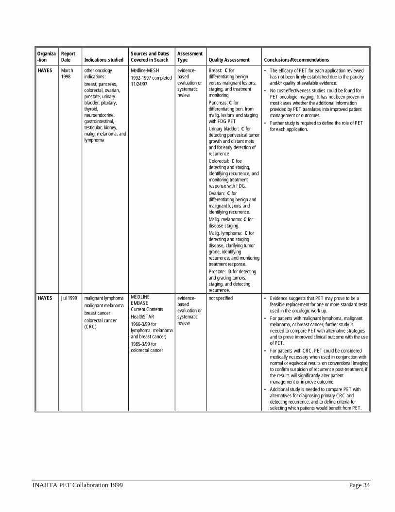
INAHTA PET Collaboration 1999 Page 34
Organiza-tion
ReportDate Indications studied
Sources and DatesCovered in Search
AssessmentType Quality Assessment Conclusions/Recommendations
HAYES March1998
other oncologyindications:breast, pancreas,colorectal, ovarian,prostate, urinarybladder, pituitary,thyroid,neuroendocrine,gastrointestinal,testicular, kidney,malig. melanoma, andlymphoma
Medline-MESH1992-1997 completed11/24/97
evidence-basedevaluation orsystematicreview
Breast: C fordifferentiating benignversus malignant lesions,staging, and treatmentmonitoring Pancreas: C fordifferentiating ben. frommalig. lesions and stagingwith FDG PET Urinary bladder: C fordetecting perivesical tumorgrowth and distant metsand for early detection ofrecurrence Colorectal: C foedetecting and staging,identifying recurrence, andmonitoring treatmentresponse with FDG. Ovarian: C fordifferentiating benign andmalignant lesions andidentifying recurrence. Malig. melanoma: C fordisease staging. Malig. lymphoma: C fordetecting and stagingdisease, clarifying tumorgrade, identifyingrecurrence, and monitoringtreatment response. Prostate: D for detectingand grading tumors,staging, and detectingrecurrence.
• The efficacy of PET for each application reviewedhas not been firmly established due to the paucityand/or quality of available evidence.
• No cost-effectiveness studies could be found forPET oncologic imaging. It has not been proven inmost cases whether the additional informationprovided by PET translates into improved patientmanagement or outcomes.
• Further study is required to define the role of PETfor each application.
HAYES Jul 1999 malignant lymphomamalignant melanomabreast cancercolorectal cancer(CRC)
MEDLINEEMBASECurrent ContentsHealthSTAR1966-3/99 forlymphoma, melanomaand breast cancer;1985-3/99 forcolorectal cancer
evidence-basedevaluation orsystematicreview
not specified • Evidence suggests that PET may prove to be afeasible replacement for one or more standard testsused in the oncologic work up.
• For patients with malignant lymphoma, malignantmelanoma, or breast cancer, further study isneeded to compare PET with alternative strategiesand to prove improved clinical outcome with the useof PET.
• For patients with CRC, PET could be consideredmedically necessary when used in conjunction withnormal or equivocal results on conventional imagingto confirm suspicion of recurrence post-treatment, ifthe results will significantly alter patientmanagement or improve outcome.
• Additional study is needed to compare PET withalternatives for diagnosing primary CRC anddetecting recurrence, and to define criteria forselecting which patients would benefit from PET.

INAHTA PET Collaboration 1999 Page 35
Organiza-tion
ReportDate Indications studied
Sources and DatesCovered in Search
AssessmentType Quality Assessment Conclusions/Recommendations
MSAC(formerlyNHTAP)
Nov 1990 Myocardial perfusionMyocardial viabilityLocalization ofepileptic foci insurgical candidateswith medicalrefractory epilepsygrading malignantcerebral gliomasrecurrent glioma vs.radiation necrosis
Not specified Narrativereview withcost analysis
Criteria not specified • Sufficient case has not yet been established forroutine use of PET as a clinical service in Australia.
• If proposed PET units are introduced into Australia,they should be subject to a coordinated evaluationof clinical and cost benefits. No further units shouldbe considered until evaluations are completed.
NCCHTA Feb 1999 Head and neckcancerBreast cancerLung cancerSPNColorectal cancerAlzheimer’s DiseaseCerebrovasculardisordersEpilepsyParkinson’s DiseaseDementiaMyocardial perfusionMyocardial viability
Ovid MEDLINE andCochrane Libraryfrom 1996-98
Systematicreview(updates andexpands 1996VA TAPreport)three-roundDelphi surveyto identifyresearchpriorities forthe NHS
• Under Council review• Report includes myocardial and neuropsychiatric
applications and all positron imaging modalities• Evidence related to diagnostic accuracy is limited
by bias and often relates only to small patientnumbers.
• Evidence is needed on the cost-effectiveness ofpositron imaging modalities in all of the advocatedclinical indications
• Research priorities identified in descending order:• relative cost-effectiveness of full ring PET and
gamma camera PET to pre-operatively stagepatients with lung cancer
• compare partial ring to full ring PET in oncology• relative cost-effectiveness of full ring PET and
gamma camera PET to stage and monitortreatment response in patients with breastcancer
• relative cost-effectiveness of gamma PET tocollimated 511 keV positron imaging forselecting patients for myocardialrevascularization surgery
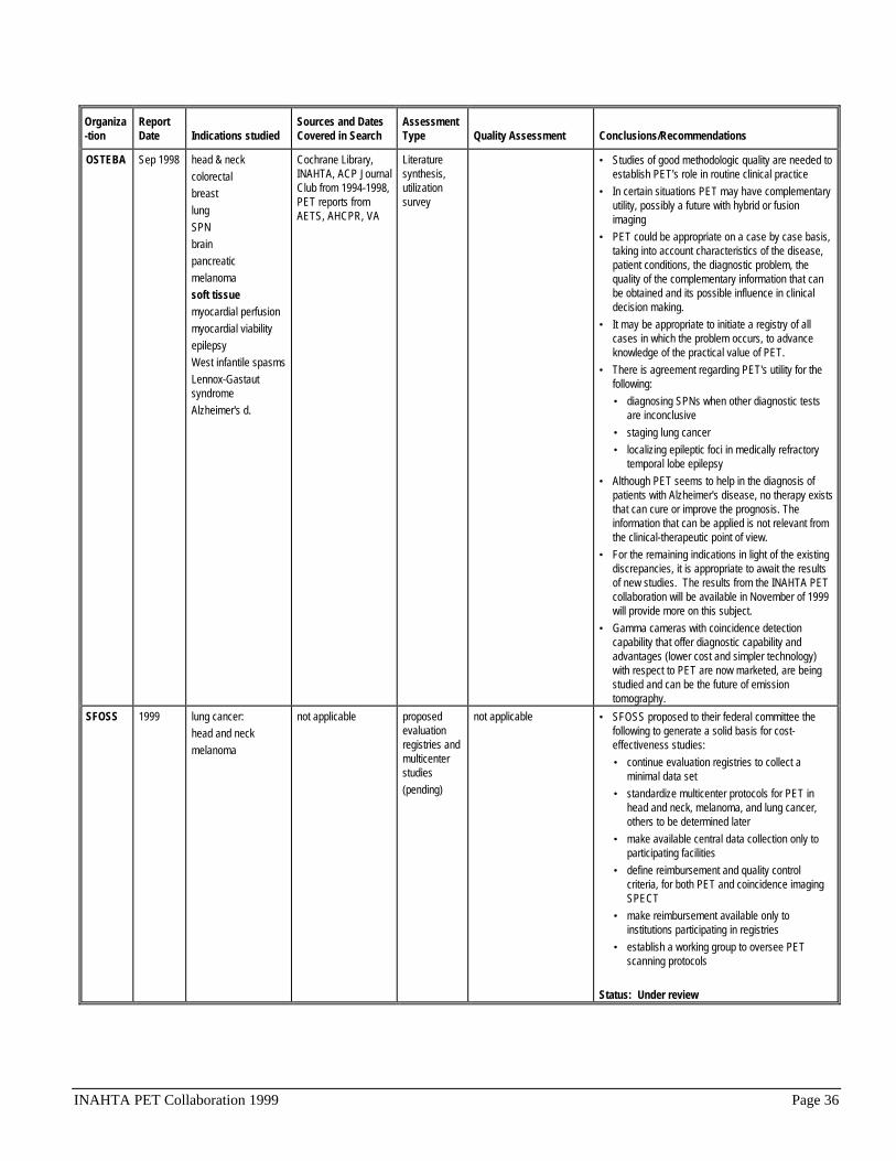
INAHTA PET Collaboration 1999 Page 36
Organiza-tion
ReportDate Indications studied
Sources and DatesCovered in Search
AssessmentType Quality Assessment Conclusions/Recommendations
OSTEBA Sep 1998 head & neckcolorectalbreastlungSPNbrainpancreaticmelanomasoft tissuemyocardial perfusionmyocardial viabilityepilepsyWest infantile spasmsLennox-GastautsyndromeAlzheimer's d.
Cochrane Library,INAHTA, ACP JournalClub from 1994-1998,PET reports fromAETS, AHCPR, VA
Literaturesynthesis,utilizationsurvey
• Studies of good methodologic quality are needed toestablish PET's role in routine clinical practice
• In certain situations PET may have complementaryutility, possibly a future with hybrid or fusionimaging
• PET could be appropriate on a case by case basis,taking into account characteristics of the disease,patient conditions, the diagnostic problem, thequality of the complementary information that canbe obtained and its possible influence in clinicaldecision making.
• It may be appropriate to initiate a registry of allcases in which the problem occurs, to advanceknowledge of the practical value of PET.
• There is agreement regarding PET's utility for thefollowing:• diagnosing SPNs when other diagnostic tests
are inconclusive• staging lung cancer• localizing epileptic foci in medically refractory
temporal lobe epilepsy• Although PET seems to help in the diagnosis of
patients with Alzheimer's disease, no therapy existsthat can cure or improve the prognosis. Theinformation that can be applied is not relevant fromthe clinical-therapeutic point of view.
• For the remaining indications in light of the existingdiscrepancies, it is appropriate to await the resultsof new studies. The results from the INAHTA PETcollaboration will be available in November of 1999will provide more on this subject.
• Gamma cameras with coincidence detectioncapability that offer diagnostic capability andadvantages (lower cost and simpler technology)with respect to PET are now marketed, are beingstudied and can be the future of emissiontomography.
SFOSS 1999 lung cancer:head and neckmelanoma
not applicable proposedevaluationregistries andmulticenterstudies(pending)
not applicable • SFOSS proposed to their federal committee thefollowing to generate a solid basis for cost-effectiveness studies:• continue evaluation registries to collect a
minimal data set• standardize multicenter protocols for PET in
head and neck, melanoma, and lung cancer,others to be determined later
• make available central data collection only toparticipating facilities
• define reimbursement and quality controlcriteria, for both PET and coincidence imagingSPECT
• make reimbursement available only toinstitutions participating in registries
• establish a working group to oversee PETscanning protocols
Status: Under review

INAHTA PET Collaboration 1999 Page 37
Organiza-tion
ReportDate Indications studied
Sources and DatesCovered in Search
AssessmentType Quality Assessment Conclusions/Recommendations
VA TAP Sept1996
Head and neckcancerBreast cancerLung cancerSPNColorectal cancerAlzheimer’s disease
MEDLINE,HealthSTAR,EMBASE, CurrentContents, and BIOSISfrom 1991 throughSep 1996
Systematicreview
EBM criteria used forgrading quality of evidence
Fryback and Thornburyscale used for classifyingincluded articles
• Research into the clinical utility of PET for selectedoncology conditions is preliminary. The evidence ofFDG-PET's diagnostic accuracy is methodologicallyweak, and PET's contribution to improvingoutcomes has not been systematically assessed.
• PET is an accurate test for dementia of theAlzheimer's type. However, evidence arguesagainst routine clinical use of PET for diagnosingAD until more effective treatments and riskmodification interventions are developed, and untilmeaningful and robust predictive values areobtained from an ongoing European multicenterPET study.
• VA should maximize the value of its existingcommitment, rather than establish additional PETcenters.
VA TAP Dec 1998 Head and neckcancerBreast cancerLung cancerSPNColorectal cancerAlzheimer’s disease
MEDLINE, Health,Current Contents,from Sep 1996-Dec1998
Systematicreview
EBM criteria used forgrading quality of evidence
Fryback and Thornburyscale used for classifyingincluded articles
• The prevailing evidence does not support usingeither dedicated or camera-based PET using FDGas a diagnostic test for the applications in thisreview.
• Several cooperative studies of PET are ongoing orplanned in the US. Clinicians should await theresults of these efforts before incorporating PETinto routine diagnostic strategies.

INAHTA PET Collaboration 1999 Page 38
BIBLIOGRAPHY
Adams E, Flynn K. Positron Emission Tomography: descriptive analysis of experience withPET in VA. A systematic review update of FDG-PET as a diagnostic test in cancer andAlzheimer's Disease. Report Number 10. 1998, VA TAP: Boston.
AETS. Tomografia por emision de positrones en cardiologia. 1995, Agencia de Evaluacion deTechnologias Sanitarias: Madrid.
AETS. Tomografia por emision de positrones (PET) en oncologia clinica no neurologica.1997, Agencia de Evaluation de Technologias Sanitarias: Madrid.
AETS. Tomografia por emision de positrones con fluordeoxiglucosa (FDG-PET) enNeurologia. Report Number 10. 1999, Agencia de Evaluacion de Technologias Sanitarias,Instituto de Salud "Carlos III": Madrid.
AHCPR. 18F-Labeled 2-Deoxy-2-Fluoro-D-Glucose Positron-Emission Tomography scans forthe localization of the epileptogenic foci. Report Number 12. 1998, Agency for Health CarePolicy Research: Rockville.
Anonymous. Spect coincidence detection equipment added to FDG PET medicare coveragedecision following ADAC presentation to HCFA; interim coverage effective Jan. 1. The GraySheet M-D-D-I Reports 1998; (January 12): 8.
Asua J, Hurtado de Saracho I. Tomografia por emision de possitrones: su utilidad clinica. 1998,Osteba: Vitoria-Gasteiz.
Baffert S, Lajonchere JP, Fery-Lemonnier E. Tomographie par emission de positons. 1999,Committee for Evaluation and Diffusion of Innovative Technologies (CEDIT): Paris.
BCBSA TEC. Evaluations of clinical PET - various indications. 1997, Technology EvaluationCenter. Blue Cross Blue Shield Association: Chicago.
CAHTA. Assessments of clinical PET - various indications (internal document). 1993, CatalanAgency for Health Technology Assessment: Barcelona.
CAHTA. Clinical PET in autism (briefing). 1996, Catalan Agency for Health TechnologyAssessment: Barcelona.
CAHTA. PET and stereotactic surgery for patients with neuropsychiatric disorders (briefing).1997, Catalan Agency for Health Technology Assessment: Barcelona.
CEDIT. La tomographie par emission de positons. 1998, Committee for Evaluation andDiffusion of Innovative Technologies: Paris.
INAHTA PET Collaboration 1999 Page 39
CEDIT. Utilisation des cameras TEDC pour la scintigraphie conventionnelle. 1998, Committeefor Evaluation and Diffusion of Innovative Technologies: Paris.
Coleman RE, Briner WH, Siegel BA. Clinical PET scanning: a "short-lived" orphan.International Journal of Technology Assessment in Health Care 1992; 8(4): 610-622.
Connell EJ. U.S. Senator champions PET [news]. Journal of Nuclear Medicine 1999; 40(3):20N, 23N, 31N.
Corabian P, Hailey D. Functional diagnostic imaging in epilepsy. 1998, Alberta HeritageFoundation for Medical Research (AHFMR): Edmonton.
Cowley DE. Positron Emission Tomography. 1990, Australian Health Technology AdvisoryCommittee: Canberra.
Cowley D, Corabian P, Hailey D. Functional diagnostic imaging in the assessment ofmyocardial viability, Part 1 - Overview. 1999, Alberta Heritage Foundation for MedicalResearch (AHMFR): Edmonton.
Cowley D, Corabian P, Hailey D. Functional diagnostic imaging in the assessment ofmyocardial viability, Part 2. Details of studies. 1999, Alberta Heritage Foundation for MedicalResearch (AHFMR): Edmonton.
ECRI. Diagnostic Imaging. In: Wallace C, Ed. Health Technology Forecast. PlymouthMeeting: ECRI; 1996.
ECRI. Positron Emission Tomography (PET) for the diagnosis and staging of non-small-celllung cancer, Part 1: Results of ECRI's meta-analysis. Report Number 72. 1998, ECRI:Plymouth Meeting.
ECRI. Positron Emission Tomography (PET) for the diagnosis and staging of non-small-celllung cancer, Part 2: Cost-effectiveness analysis. Report Number 73. 1998, ECRI: PlymouthMeeting.
FDA. Revocation of certain guidance documents on Positron Emission Tomography drugproducts. Federal Register 1997; 62(244): 66636.
FDA. Medical imaging drugs advisory committee; notice of meeting. Federal Register 1999;64(109): 30526.
Flynn K, Adams E. Positron Emission Tomography: descriptive analysis of experience withPET in VA systematic reviews: FDG-PET as a diagnostic test for cancer and Alzheimer'sDisease. Report Number 3. 1996, VA TAP: Boston.

INAHTA PET Collaboration 1999 Page 40
Garber AM, Solomon NA. PET myocardial perfusion imaging for the detection of coronaryartery disease - cost-effectiveness analysis. Special Assessment. 1996, Technology EvaluationCenter. Blue Cross and Blue Shield Association: Chicago.
Hayes Inc. Positron Emission Tomography (PET) for neurologic applications--ExecutiveSummary. Report Number POSI203.8. 1997, Winifred S. Hayes, Inc: Lansdale, PA.
Hayes Inc. Positron Emission Tomography (PET) for cardiac applications--Executive Summary.Report Number POSI1303.07. 1997, Winifred S. Hayes, Inc: Lansdale, PA.
Hayes Inc. Positron Emission Tomography (PET) for lung cancer--Executive Summary. ReportNumber POSI1103.18. 1998, Winifred S. Hayes, Inc: Lansdale, PA.
Hayes Inc. Positron Emission Tomography (PET) for other malignancies--Executive Summary.Report Number POSI1103.19. 1998, Winifred S. Hayes, Inc: Lansdale, PA.
Hayes Inc. Positron Emission Tomography (PET) for cardiac applications--Draft Update--Executive Summary--Internal Document. Report Number None. 1999, Winifred S. Hayes, Inc:Lansdale, PA.
Hayes Inc. Positron Emission Tomography (PET) for other malignancies: Colorectal cancer,malignant lymphoma, malignant melanoma, breast cancer--Draft Update--Executive Summary--Internal Document. Report Number None. 1999, Winifred S. Hayes, Inc: Lansdale, PA.
HCFA. U.S. Health Care Financing Administration. (1998). Claims Review and AdjudicationProcedures. [Web Site]. Available: http://www.hcfa.gov/, [July 7, 1999].
Holmes BL. Current strategies for the development of medical devices. In: Gelijns AC, Ed.Technology and Health Care in An Era of Limits. Washington, DC: National Academy Press;1992.
Hotta SS. Myocardial Perfusion Imaging with Rubidium 82 Positron Emission Tomography.Report Number 10. 1995, AHCPR: Rockville.
INAHTA. (1999). Agency listing. Available: http://www.inahta.org, [September 30, 1999].
Institute for Clinical PET. (1999). Available: http://www.icppet.org, [August 1999].
Lewellen TK, Miyaoka RS, Swan WJ. PET imaging using dual-headed gamma cameras: Anupdate. Nuclear Medicine Communications 1999; 20(1): 5-12.
Robert G, Milne R. Positron Emission Tomography: Establishing priorities for healthtechnology assessment. 1999, National Co-ordinating Centre for Health TechnologyAssessment: Southhampton.

INAHTA PET Collaboration 1999 Page 41
Stevens T. Stevens announces long-awaited agreement regarding PET scan Medicarereimbursement. Press Release, November 6, 1997. 1997, U.S. Senate: Washington, D.C.
Ter-Pogossian MM. The Origins of Positron Emission Tomography. Seminars in NuclearMedicine 1992; 22(3): 140-149.
Zelman WA. The changing health care marketplace: Private ventures, public interests. SanFrancisco: Jossey-Bass Publishers; 1996.


![4. 5. 6. JOINT] JOINT T P JOINT T P JOINT C 18 H JOINT C T ... ken-syoumei.pdf4. 5. 6. JOINT] JOINT T P JOINT T P JOINT C 18 H JOINT C T. P JOINT JOINT a C (2) JOINT x (3) JOINT x](https://static.documents.pub/doc/80x56/611edb438155026709151f58/4-5-6-joint-joint-t-p-joint-t-p-joint-c-18-h-joint-c-t-ken-4-5-6-joint.jpg)


