Page 1
A-level: Gaseous Exchange
Page 1 of 23: Gayaza High School 2017. Biology Department
GASEOUS EXCHANGE
This is the diffusion of gases from an area of higher concentration to an area of lower
concentration, especially the exchange of oxygen and carbon dioxide between an organism and
its environment.
VENTILATION is an active mechanism which draws over and expels respiratory gases away
from the gaseous exchange surfaces.
Pulmonary ventilation
It is simply taking in of air from the atmosphere and giving out of air from the lungs. It is carried
out by breathing which constantly renews the air present in the lungs. It involves two processes -
inspiration and expiration
Difference between inspiration and expiration
Note: One breathe includes one inspiration and one expiration, the respiratory rate is the number
of breathes taken per minute. For a person breathing normally at rest, it is equal to 12-14 breaths
per minute.
CONDITIONS NEEDED FOR GAS EXCHANGE
a) The supply of oxygen
(1) Air - About 21% of air is oxygen.
(2) Water - Amount of oxygen in water varies (about 1.03% in fresh water and 0.85% in sea
water) but is always much less than in air, being even lower in warmer water than colder water.
b) Diffusion
Diffusion is faster when the
surface area to volume ratio is large
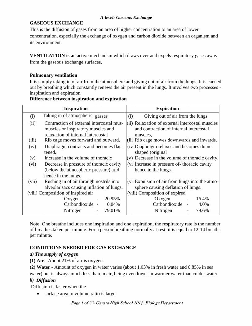
Inspiration Expiration
(i) Taking in of atmospheric
air.
gasses (i) Giving out of air from the lungs.
(ii) Contraction of external intercostal mus- (ii) Relaxation of external intercostal muscles
muscles or inspiratory muscles and
relax-
and contraction of internal intercostal
mus- relaxation of internal intercostal
muscles.
muscles, (iii) Rib cage moves forward and outward. (iii
)
Rib cage moves downwards and inwards.
(iv) Diaphragm contracts and becomes flat- (iv
)
Diaphragm relaxes and becomes dome
tened. shaped (original
position):
(v) Increase in the volume of thoracic
cavity.
(v) Decrease in the volume of thoracic cavity.
(vi) Decrease in pressure of thoracic cavity (vi
)
Increase in pressure of- thoracic cavity
and (below the atmospheric pressure) arid hence in the lungs.
hence in the lungs,
(vii) Rushing in of air through nostrils into
the
(vi
)))
)i)
Expulsion of air from lungs into the atmo-
alveolar sacs causing inflation of lungs. sphere causing deflation of lungs.
(viii) Composition of inspired air (viii) Composition of expired
air
Oxygen - 20.95% Oxygen - 16.4% Carbondioxide - 0.04% Carbondioxide - 4.0%
Nitrogen - 79.01% Nitrogen - 79.6%
Page 2
A-level: Gaseous Exchange
Page 2 of 23: Gayaza High School 2017. Biology Department
distance travelled is small
concentration gradient of the diffusing substance is high.
c) A moist surface is required because oxygen and carbondioxide must be dissolved in water to
diffuse across a membrane.
d) Permeable membranes
(e) large surface area to volume ratio
Therefore, an efficient gas exchange surface must
(1) Have a large surface area relative to the volume of the organism to ensure a faster diffusion
rate of respiratory gases
(2) provide a short distance (be thin) for gases to diffuse across
(3) be moist to enable dissolving of respiratory gases
(4) permeable to the respiratory gases to enable their diffusion
(5) be organized or operate in a way that maintains a favourable concentration gradient for the
diffusion of respiratory gases
Hence a circulatory system may operate in tandem with the gas exchange system to maintain
the concentration gradient.
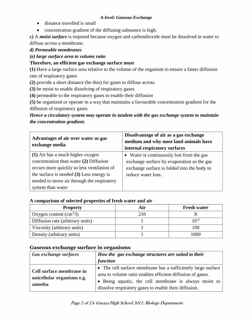
Advantages of air over water as gas
exchange media
Disadvantage of air as a gas exchange
medium and why most land animals have
internal respiratory surfaces
(1) Air has a much higher oxygen
concentration than water (2) Diffusion
occurs more quickly so less ventilation of
the surface is needed (3) Less energy is
needed to move air through the respiratory
system than water
Water is continuously lost from the gas
exchange surface by evaporation so the gas
exchange surface is folded into the body to
reduce water loss.
A comparison of selected properties of fresh water and air
Property Air Fresh water
Oxygen content (cm3/l) 210 8
Diffusion rate (arbitrary units) 1 10-5
Viscosity (arbitrary units) 1 100
Density (arbitrary units) 1 1000
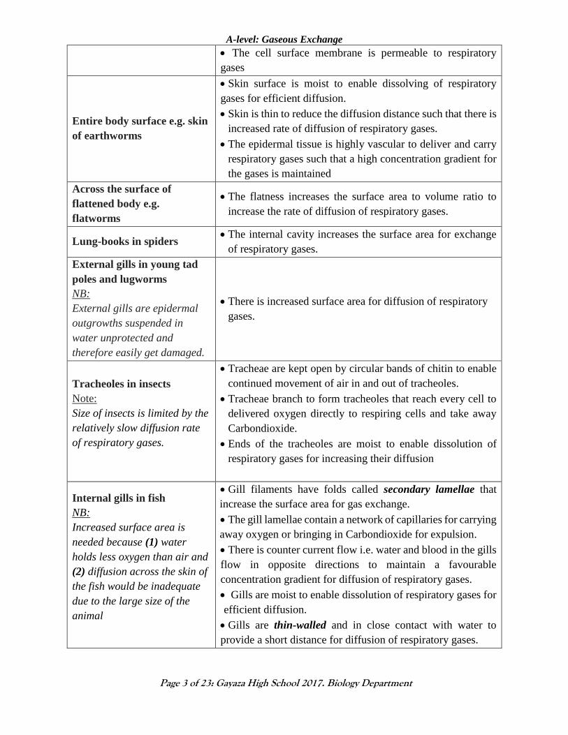
Gaseous exchange surface in organisms
Gas exchange surfaces How the gas exchange structures are suited to their
function
Cell surface membrane in
unicellular organisms e.g.
amoeba
The cell surface membrane has a sufficiently large surface
area to volume ratio enables efficient diffusion of gases.
Being aquatic, the cell membrane is always moist to
dissolve respiratory gases to enable their diffusion.
Page 3
A-level: Gaseous Exchange
Page 3 of 23: Gayaza High School 2017. Biology Department
The cell surface membrane is permeable to respiratory
gases
Entire body surface e.g. skin
of earthworms
Skin surface is moist to enable dissolving of respiratory
gases for efficient diffusion.
Skin is thin to reduce the diffusion distance such that there is
increased rate of diffusion of respiratory gases.
The epidermal tissue is highly vascular to deliver and carry
respiratory gases such that a high concentration gradient for
the gases is maintained
Across the surface of
flattened body e.g.
flatworms
The flatness increases the surface area to volume ratio to
increase the rate of diffusion of respiratory gases.
Lung-books in spiders The internal cavity increases the surface area for exchange
of respiratory gases.
External gills in young tad
poles and lugworms
NB:
External gills are epidermal
outgrowths suspended in
water unprotected and
therefore easily get damaged.
There is increased surface area for diffusion of respiratory
gases.
Tracheoles in insects
Note:
Size of insects is limited by the
relatively slow diffusion rate
of respiratory gases.
Tracheae are kept open by circular bands of chitin to enable
continued movement of air in and out of tracheoles.
Tracheae branch to form tracheoles that reach every cell to
delivered oxygen directly to respiring cells and take away
Carbondioxide.
Ends of the tracheoles are moist to enable dissolution of
respiratory gases for increasing their diffusion
Internal gills in fish
NB:
Increased surface area is
needed because (1) water
holds less oxygen than air and
(2) diffusion across the skin of
the fish would be inadequate
due to the large size of the
animal
Gill filaments have folds called secondary lamellae that
increase the surface area for gas exchange.
The gill lamellae contain a network of capillaries for carrying
away oxygen or bringing in Carbondioxide for expulsion.
There is counter current flow i.e. water and blood in the gills
flow in opposite directions to maintain a favourable
concentration gradient for diffusion of respiratory gases.
Gills are moist to enable dissolution of respiratory gases for
efficient diffusion.
Gills are thin-walled and in close contact with water to
provide a short distance for diffusion of respiratory gases.
Page 4
A-level: Gaseous Exchange
Page 4 of 23: Gayaza High School 2017. Biology Department
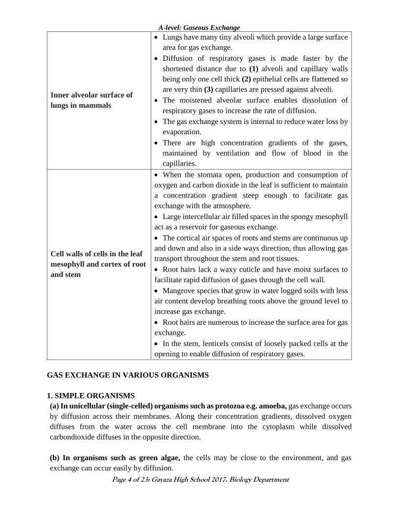
Inner alveolar surface of
lungs in mammals
Lungs have many tiny alveoli which provide a large surface
area for gas exchange.
Diffusion of respiratory gases is made faster by the
shortened distance due to (1) alveoli and capillary walls
being only one cell thick (2) epithelial cells are flattened so
are very thin (3) capillaries are pressed against alveoli.
The moistened alveolar surface enables dissolution of
respiratory gases to increase the rate of diffusion.
The gas exchange system is internal to reduce water loss by
evaporation.
There are high concentration gradients of the gases,
maintained by ventilation and flow of blood in the
capillaries.
Cell walls of cells in the leaf
mesophyll and cortex of root
and stem
When the stomata open, production and consumption of
oxygen and carbon dioxide in the leaf is sufficient to maintain
a concentration gradient steep enough to facilitate gas
exchange with the atmosphere.
Large intercellular air filled spaces in the spongy mesophyll
act as a reservoir for gaseous exchange.
The cortical air spaces of roots and stems are continuous up
and down and also in a side ways direction, thus allowing gas
transport throughout the stem and root tissues.
Root hairs lack a waxy cuticle and have moist surfaces to
facilitate rapid diffusion of gases through the cell wall.
Mangrove species that grow in water logged soils with less
air content develop breathing roots above the ground level to
increase gas exchange.
Root hairs are numerous to increase the surface area for gas
exchange.
In the stem, lenticels consist of loosely packed cells at the
opening to enable diffusion of respiratory gases.
GAS EXCHANGE IN VARIOUS ORGANISMS
1. SIMPLE ORGANISMS
(a) In unicellular (single-celled) organisms such as protozoa e.g. amoeba, gas exchange occurs
by diffusion across their membranes. Along their concentration gradients, dissolved oxygen
diffuses from the water across the cell membrane into the cytoplasm while dissolved
carbondioxide diffuses in the opposite direction.
(b) In organisms such as green algae, the cells may be close to the environment, and gas
exchange can occur easily by diffusion.
Page 5
A-level: Gaseous Exchange
Page 5 of 23: Gayaza High School 2017. Biology Department
(i) In the dark, no photosynthesis occurs in the chloroplast, no oxygen is made. Dissolved oxygen
diffuses from the water across the cell membrane into the mitochondria while dissolved
carbondioxide diffuses in the opposite direction, along their concentration gradients.
(ii) In the light, photosynthesis in chloroplasts releases oxygen, some of which diffuses into the
mitochondria, the excess diffuses out.
2. GAS EXCHANGE IN EARTHWORMS
Earthworms exchange oxygen and carbon dioxide directly through their skin. The oxygen diffuses
into tiny blood vessels in the skin surface, where it combines with the red pigment haemoglobin.
Hemoglobin binds loosely to oxygen and carries it through the animal's bloodstream. Carbon
dioxide is transported back to the skin by the hemoglobin from which it detaches and diffuses out.
3. GAS EXCHANGE IN INSECTS
Terrestrial insect e.g. grasshopper
In grasshopper, the tracheal system consists of 10 pairs of spiracles, located laterally on the body
surface. Of these, 2 pairs are thoracic and 8 pairs are abdominal. The spiracles are guarded by fine
hairs to keep the foreign particles out and by valves that function to open or close the spiracles as
required. The spiracles open into small spaces called the atria that continue as air tubes called the
tracheae. The tracheae are fine tubes that have a wall of single layered epithelial cells.
The cells secrete spiral cuticular thickenings called taenidia around the tube that gives support to
the tubes.
The tracheal tubes branch further into finer tracheoles that enter all the tissues and sometimes,
even the cells of the insect. The ends of the tracheoles that are in the tissue are filled with fluid and
lack the cuticular thickenings.
The main tracheal tubes join together to form three main tracheal trunks- dorsal, ventral and lateral.
At some places, the tracheae enlarge to form air sacs which are devoid of cuticle and serve to store
air.
ventilation and gaseous exchange in insects
Increased CO2 is detected by chemoreceptors, causing relaxation of the abdominal muscles and
lowering of pressure. The spiracles valves open and air rich in oxygen is drawn into the tracheal
system.
Spiracles valves then close and oxygen is forced along the tracheal system into the fluid-filled
tracheoles, which are in direct contact with the tissue fluid. Gaseous exchange takes place due to
difference in concentration gradients of oxygen and carbon dioxide.
Air is expelled out when muscles contract and flatten the insect body, decreasing the volume of
the tracheal system.
During increased metabolic activity, the water potential of tissue lowers causing osmotic efflux
of water from the tracheoles; and hence air replaces the fluid of the tracheoles.
Page 6
A-level: Gaseous Exchange
Page 6 of 23: Gayaza High School 2017. Biology Department
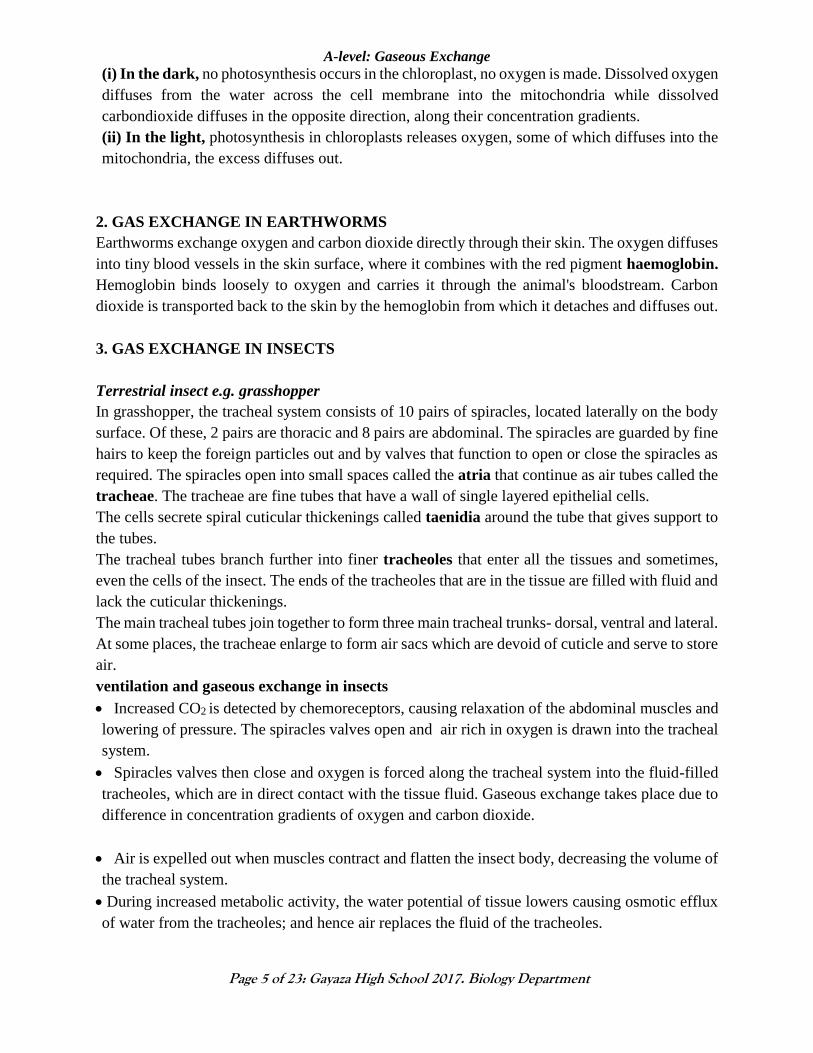
In resting tissues, the water potential of tissue fluid increases resulting in the diffusion of much
water into the tracheoles.
Appearance of tracheoles at rest Appearance of tracheoles during activity
4. GAS EXCHANGE IN FROGS
Gaseous exchange in the frog takes place in three main parts of the body:
(1) The skin - especially during low activity when hibernating (2) the mouth [buccal cavity] (3)
the lungs.
The Skin:
Air from the atmosphere diffuses through the moist thin skin; it into the dense capillary below the
skin.
Due to its low concentration in the blood than in the skin surface, oxygen is then taken to the
tissues via the red blood cells. Carbon dioxide moves from the blood into the skin surface then to
the atmosphere. This happens due to its high concentration in the blood tissues than in the surface
of the skin.
The mouth (Buccal cavity):
The muscles of the mouth contract and then lower the surface of the mouth hence reducing its
pressure than that of the atmosphere.
Air rich in oxygen is inhaled through the nostrils into the mouth cavity
There exists dense capillary network in the mouth cavity and as such, gaseous exchange takes
place. Oxygen due to its high concentration diffuses into the blood and is transported by the red
blood cells. Carbon dioxide diffuses from the blood tissues to the buccal cavity; then exhaled
through the nostrils when the mouth floor is raised.
The lungs:
The mouth muscles contract then lower the floor of the mouth hence increasing its volume.
Pressure reduces in the mouth cavity than the atmosphere’s, causing air to move into the mouth
through the nostrils.
The nostril then closes and the mouth’s floor is raised. This forces the air into the lungs.
Page 7
A-level: Gaseous Exchange
Page 7 of 23: Gayaza High School 2017. Biology Department
Gaseous exchange takes place between the alveoli of the lungs and the blood; oxygen due to its
high concentration in the alveoli than the blood diffuse into the blood while Carbon dioxide
diffuses out of the blood tissue to the alveoli where it is exhaled out through the nostrils by the
muscles of the lungs which contract and relax rhythmically.
5. GAS EXCHANGE IN BONY FISH
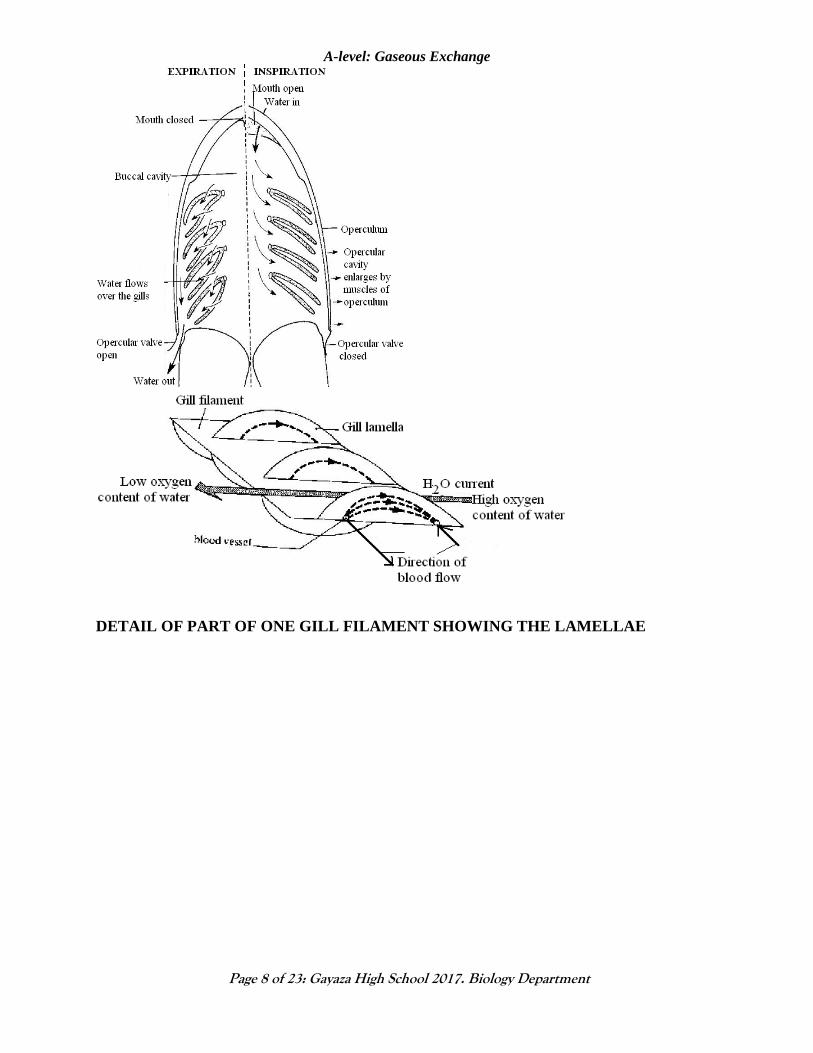
Mechanism of ventilation in bony fish
Contraction of the mouth muscles lowers the floor of the mouth, reducing its pressure as the
mouth opens. Water (with dissolved oxygen) moves into the mouth and at the same time the
operculum remain closed.
The operculum muscles relax; causing operculum to bulge open; this increases the volume but
lowers the pressure in the gill region as the mouth closes.
Water from the cavity mouth moves into the gill region due to the reduced pressure; and bathes
the gill filaments in opposite direction to the flow of the blood. This is termed countercurrent
flow. Oxygen diffuses into the blood capillaries due to its high concentration in the gill region
than the blood capillaries; it combines with haemoglobin and is transported as oxyhaemoglobin
to the respiring tissues.
Carbon dioxide and toxic metabolic wastes, like ammonia which are at higher concentration in
the blood than the gill filaments are excreted into the gills and exhaled through the water that
moves out when the operculum opens.
The higher internal water pressure in gill chamber forces operculum to open to exit the water
Page 8
A-level: Gaseous Exchange
Page 8 of 23: Gayaza High School 2017. Biology Department
DETAIL OF PART OF ONE GILL FILAMENT SHOWING THE LAMELLAE
Page 9
A-level: Gaseous Exchange
Page 9 of 23: Gayaza High School 2017. Biology Department
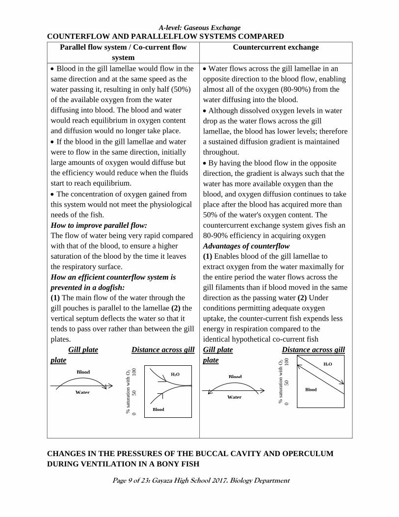
COUNTERFLOW AND PARALLELFLOW SYSTEMS COMPARED
Parallel flow system / Co-current flow
system
Countercurrent exchange
Blood in the gill lamellae would flow in the
same direction and at the same speed as the
water passing it, resulting in only half (50%)
of the available oxygen from the water
diffusing into blood. The blood and water
would reach equilibrium in oxygen content
and diffusion would no longer take place.
If the blood in the gill lamellae and water
were to flow in the same direction, initially
large amounts of oxygen would diffuse but
the efficiency would reduce when the fluids
start to reach equilibrium.
The concentration of oxygen gained from
this system would not meet the physiological
needs of the fish.
How to improve parallel flow:
The flow of water being very rapid compared
with that of the blood, to ensure a higher
saturation of the blood by the time it leaves
the respiratory surface.
How an efficient counterflow system is
prevented in a dogfish:
(1) The main flow of the water through the
gill pouches is parallel to the lamellae (2) the
vertical septum deflects the water so that it
tends to pass over rather than between the gill
plates.
Gill plate Distance across gill
plate
Water flows across the gill lamellae in an
opposite direction to the blood flow, enabling
almost all of the oxygen (80-90%) from the
water diffusing into the blood.
Although dissolved oxygen levels in water
drop as the water flows across the gill
lamellae, the blood has lower levels; therefore
a sustained diffusion gradient is maintained
throughout.
By having the blood flow in the opposite
direction, the gradient is always such that the
water has more available oxygen than the
blood, and oxygen diffusion continues to take
place after the blood has acquired more than
50% of the water's oxygen content. The
countercurrent exchange system gives fish an
80-90% efficiency in acquiring oxygen
Advantages of counterflow
(1) Enables blood of the gill lamellae to
extract oxygen from the water maximally for
the entire period the water flows across the
gill filaments than if blood moved in the same
direction as the passing water (2) Under
conditions permitting adequate oxygen
uptake, the counter-current fish expends less
energy in respiration compared to the
identical hypothetical co-current fish
Gill plate Distance across gill
plate
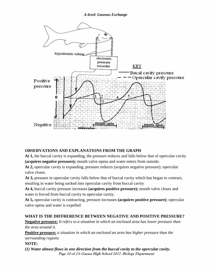
CHANGES IN THE PRESSURES OF THE BUCCAL CAVITY AND OPERCULUM
DURING VENTILATION IN A BONY FISH
% s
atu
rati
on
wit
h O
2
0
5
0
1
00
H2O
Blood
Water
Blood
Water
Blood
% s
atu
rati
on
wit
h O
2
0
5
0
1
00
H2O
Blood
Page 10
A-level: Gaseous Exchange
Page 10 of 23: Gayaza High School 2017. Biology Department
OBSERVATIONS AND EXPLANATIONS FROM THE GRAPH
At 1, the buccal cavity is expanding, the pressure reduces and falls below that of opercular cavity
(acquires negative pressure); mouth valve opens and water enters from outside.
At 2, opercular cavity is expanding, pressure reduces (acquires negative pressure); opercular
valve closes.
At 3, pressure in opercular cavity falls below that of buccal cavity which has began to contract,
resulting in water being sucked into opercular cavity from buccal cavity
At 4, buccal cavity pressure increases (acquires positive pressure); mouth valve closes and
water is forced from buccal cavity to opercular cavity.
At 5, opercular cavity is contracting, pressure increases (acquires positive pressure); opercular
valve opens and water is expelled
WHAT IS THE DIFFRERENCE BETWEEN NEGATIVE AND POSITIVE PRESSURE?
Negative pressure: It refers to a situation in which an enclosed area has lower pressure than
the area around it.
Positive pressure: a situation in which an enclosed an area has higher pressure than the
surrounding regions
NOTE:
(1) Water almost flows in one direction from the buccal cavity to the opercular cavity.
Page 11
A-level: Gaseous Exchange
Page 11 of 23: Gayaza High School 2017. Biology Department
EVIDENCE: Throughout the ventilation cycle, except for one short period when the buccal
cavity expands (see 1above), the pressure in the buccal cavity is higher than that in the
opercular cavity forcing water to flow from the buccal cavity to the opercular cavity along the
pressure gradient. Expansion of buccal cavity lowers the pressure below that of opercular
cavity, causing the water to enter the buccal cavity but at the same time the opercular valves
close to prevent entry of water.
(2) The buccal cavity acts as a force pump while the opercular cavity as a suction pump.
Sample questions
Qn. 1. (a) Explain why when fish are taken out of the water, they suffocate.
b) Under what circumstances do fish suffocate in the water?
Qn. 2. The graph below shows the changes in pressure in the buccal cavity and in the
opercular cavity during a ventilation cycle.
a) Calculate the rate of ventilation in cycles per minute
b) (i) With evidence from the graph, explain why water almost flows in one direction over
the gills.
ii) How does the fish increase buccal cavity pressure?
6. GAS EXCHANGE IN MAN
Humans have a high metabolic rate which necessitates a fast rate of gas exchange.
This is enabled by two key features the human system has evolved:
(1) A blood transport system with red blood cells containing haemoglobin
(2) A mechanism of ventilation to get the gases to and from the gas exchange surface.
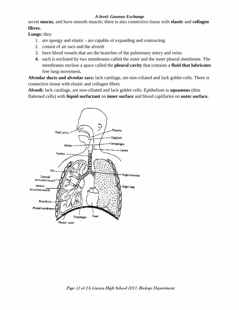
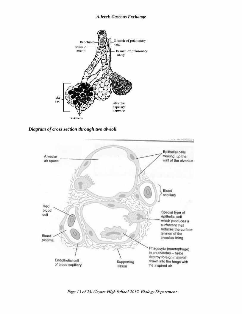
Main features of the respiratory system
Trachea (wind pipe), the two bronchi and the bronchioles: They are held open (without
collapsing) by the C-shaped cartilaginous rings. Epithelium is ciliated, have goblet cells that
Page 12
A-level: Gaseous Exchange
Page 12 of 23: Gayaza High School 2017. Biology Department
secret mucus, and have smooth muscle; there is also connective tissue with elastic and collagen
fibres.
Lungs: they
1. are spongy and elastic - are capable of expanding and contracting
2. consist of air sacs and the alveoli
3. have blood vessels that are the branches of the pulmonary artery and veins
4. each is enclosed by two membranes called the outer and the inner pleural membrane. The
membranes enclose a space called the pleural cavity that contains a fluid that lubricates
free lung movement.
Alveolar ducts and alveolar sacs: lack cartilage, are non-ciliated and lack goblet cells. There is
connective tissue with elastic and collagen fibres
Alveoli: lack cartilage, are non-ciliated and lack goblet cells. Epithelium is squamous (thin
flattened cells) with liquid surfactant on inner surface and blood capillaries on outer surface.
Page 13
A-level: Gaseous Exchange
Page 13 of 23: Gayaza High School 2017. Biology Department
Diagram of cross section through two alveoli
Page 14
A-level: Gaseous Exchange
Page 14 of 23: Gayaza High School 2017. Biology Department
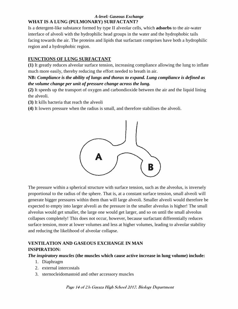
WHAT IS A LUNG (PULMONARY) SURFACTANT?
Is a detergent-like substance formed by type II alveolar cells, which adsorbs to the air-water
interface of alveoli with the hydrophilic head groups in the water and the hydrophobic tails
facing towards the air. The proteins and lipids that surfactant comprises have both a hydrophilic
region and a hydrophobic region.
FUNCTIONS OF LUNG SURFACTANT
(1) It greatly reduces alveolar surface tension, increasing compliance allowing the lung to inflate
much more easily, thereby reducing the effort needed to breath in air.
NB: Compliance is the ability of lungs and thorax to expand. Lung compliance is defined as
the volume change per unit of pressure change across the lung.
(2) It speeds up the transport of oxygen and carbondioxide between the air and the liquid lining
the alveoli.
(3) It kills bacteria that reach the alveoli
(4) It lowers pressure when the radius is small, and therefore stabilises the alveoli.
The pressure within a spherical structure with surface tension, such as the alveolus, is inversely
proportional to the radius of the sphere. That is, at a constant surface tension, small alveoli will
generate bigger pressures within them than will large alveoli. Smaller alveoli would therefore be
expected to empty into larger alveoli as the pressure in the smaller alveolus is higher! The small
alveolus would get smaller, the large one would get larger, and so on until the small alveolus
collapses completely! This does not occur, however, because surfactant differentially reduces
surface tension, more at lower volumes and less at higher volumes, leading to alveolar stability
and reducing the likelihood of alveolar collapse.
VENTILATION AND GASEOUS EXCHANGE IN MAN
INSPIRATION:
The inspiratory muscles (the muscles which cause active increase in lung volume) include:
1. Diaphragm
2. external intercostals
3. sternocleidomastoid and other accessory muscles
Page 15
A-level: Gaseous Exchange
Page 15 of 23: Gayaza High School 2017. Biology Department
The external intercostal muscles contract and the inner ones relax.
The rib cage moves upwards and outwards.
The muscles of the diaphragm contract and it flattens (loses its dome shape).
These movements increase the volume of the thoracic cavity; and that of the lungs.
Pressure in the lungs is then decreased; lower than the atmospheric pressure. Because air
always flows from a region of high pressure to a region of lower pressure, it rushes in through
the nostrils, through the nasal passages, into the pharynx, through the larynx, down the trachea,
into the main bronchi, then into smaller bronchioles, through even smaller alveolar ducts, and
into alveoli.
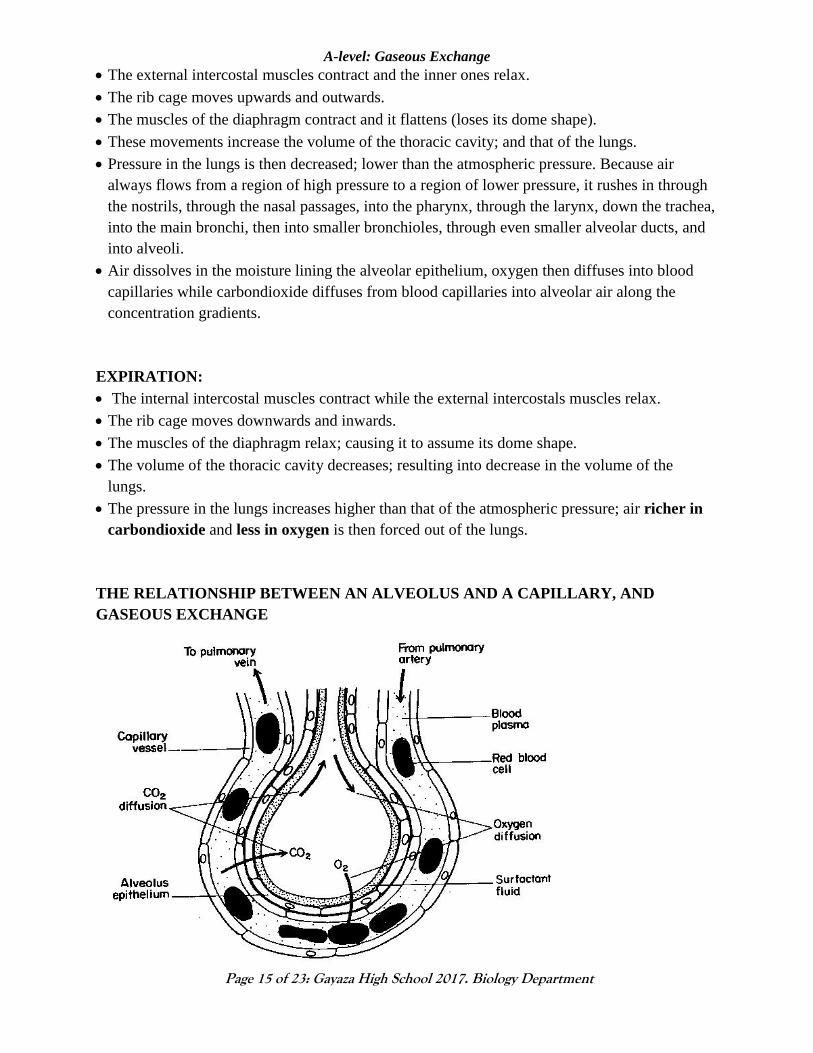
Air dissolves in the moisture lining the alveolar epithelium, oxygen then diffuses into blood
capillaries while carbondioxide diffuses from blood capillaries into alveolar air along the
concentration gradients.
EXPIRATION:
The internal intercostal muscles contract while the external intercostals muscles relax.
The rib cage moves downwards and inwards.
The muscles of the diaphragm relax; causing it to assume its dome shape.
The volume of the thoracic cavity decreases; resulting into decrease in the volume of the
lungs.
The pressure in the lungs increases higher than that of the atmospheric pressure; air richer in
carbondioxide and less in oxygen is then forced out of the lungs.
THE RELATIONSHIP BETWEEN AN ALVEOLUS AND A CAPILLARY, AND
GASEOUS EXCHANGE
Page 16
A-level: Gaseous Exchange
Page 16 of 23: Gayaza High School 2017. Biology Department
PHYSICAL CHANGES THAT OCCUR TO AIR DURING GAS EXCHANGE
It is
1. warmed by the capillary blood in the nostrils
2. moistened by mucus lining the trachea, bronchi and bronchioles
3. filtered and cleaned of particles and dust by hair (whiskers) in the nostrils, cilia
and mucus in the trachea, bronchi and bronchioles
4. the composition of air changes as indicated in the table below:
GAS PERCENTAGE BY VOLUME
Inspired air Alveolar air Expired air
Oxygen 20.90 13.90 15.30
Nitrogen 78.60 No available data 74.90
Carbondioxide 0.03 4.90 3.60
Water vapour 0.47 (usually varies) No available data 6.20 (saturated)
OBSERVATIONS AND EXPLANATIONS FROM THE TABLE ABOVE
Observations Explanations
1. Inspired air contains more oxygen
and nitrogen than exhaled air, yet
exhaled air contains more
carbondioxide (120 times) and water
vapour than those in inhaled air
Some of the oxygen in inhaled air diffuses into blood
capillaries while carbondioxide and water vapour
from blood diffuse into the air to be expired. The
percentage of nitrogen decreases in expired air
because of the increased partial pressure of
carbondioxide and water vapour.
2. The volume of oxygen and
carbondioxide in expired air is
intermediate between the inspired and
alveolar values.
Some oxygen in alveolar air diffuses into blood
capillaries while carbondioxide from blood diffuses
into alveolar air. The air that remains in the alveoli
mixes with the incoming fresh air hence lowering the
percentage of oxygen in alveolar air
TYPICAL QUESTION
The table below shows the rate and depth of breathing in a group of students during rest and
during strenuous exercise.
Student
Breathing during rest Breathing during exercise
Volume of
inspired air (cm3)
Number of
inspirations per
minute
Volume of
inspired air (cm3)
Number of
inspirations per
minute
1 480 13 2300 19
2 508 12 2250 20
3 496 12 2290 21
4 515 11 2340 20
5 490 12 2280 20
Page 17
A-level: Gaseous Exchange
Page 17 of 23: Gayaza High School 2017. Biology Department
(i) Calculate the average number of inspirations per minute during rest and during exercise.
During rest: 12; during exercise: 20
(ii) Calculate the average tidal volume during rest and during exercise
During rest: 497.8cm3; during exercise: 2292cm3
(b) Explain how the oxygen requirements of a mammal are met under different conditions of
physical activity.
NORMAL LUNG VOLUMES AND LUNG CAPACITIES IN RESTING ADULTS
Lung volumes and lung capacities refer to the volume of air associated with different phases of
the respiratory cycle. Lung volumes are directly measured. Lung capacities are determined from
lung volumes. Lung capacities are subdivisions of total volume that include two or more of the 4
basic lung volumes.
Lung volumes and capacities as shown by a spirometer
Pulmonary air volumes:
1. Tidal volume (TV): It is the volume of air breathed in and out during normal breathing
or in in each respiratory cycles. TV= 500 ml (0.5 litre)
2. Inspiratory reserve volume (IRV). It is an extra volume of air over and above the tidal
volume that can be taken in during deep breath. IRV = 1500-2500 ml (l.5 - 2.5 litres)
3. Expiratory reserve volume (ERV). After a normal expiration (tidal expiration), one can
still expel a large volume of air. This is known as expiratory reserve volume. ERV =
1500 ml (l.5 litres)
4. Vital capacity. It is the total volume of air expired after a maximum inspiration-
followed by a maximum expiration.
Vital capacity of a normal adult = TV + IRV + ERV = 3500 – 4500 ml (3.5 – 4.5 litres)
The higher the vital capacity, the higher the amount of air exchanged in each breath
Page 18
A-level: Gaseous Exchange
Page 18 of 23: Gayaza High School 2017. Biology Department
Note:
5. Total lung capacity. It refers to the amount of air present in the lungs after the maximum
inhalation. It is equivalent to 5000-5500 ml
6. Residual volume (RV). It is the amount of air left in the lungs even after the maximum
expiratory effort. It can never be forced out of lungs.
RV = 1500 ml. (1.5litres)
Residual volume is the air that is always present in the lungs meaning that the exchange
of gases continues even during expiration, or even when you hold the breath
7. Dead space air. It is the amount of air that is present in the respiratory tubes where
gaseous exchange does not occur. With each expiration, it is expelled out without
undergoing any change in oxygen or carbon dioxide concentration.
It is equivalent to about 150 ml (0.15 litres). Out of the tidal volume of 500 ml, 150 ml
remains in respiratory tubes as dead space air and only the rest 350 ml is present in
alveolar sacs in the lungs for exchange of gases.
8. Alveolar ventilation: Total volume of fresh air entering the alveoli per minute.
VENTILATION RATE
Ventilation = (Tidal volume - anatomic dead space) x respiratory rate. An average human
breathes some 12-20 times per minute.
Minute ventilation = Tidal volume x Respiratory rate. Units: (ml/min) = (ml/breath)
x (breaths/minute)
Since a fixed volume of each tidal volume goes to dead space, increased depth of breathing is
more effective in elevating alveolar ventilation than increased breathing rate.
Higher vital capacity Lower vital capacity
Athletes Non-athletes
Mountain dwellers People living in plains
Men Women Youth Old individuals
Non cigarette smokers Cigarette smokers
Page 19
A-level: Gaseous Exchange
Page 19 of 23: Gayaza High School 2017. Biology Department
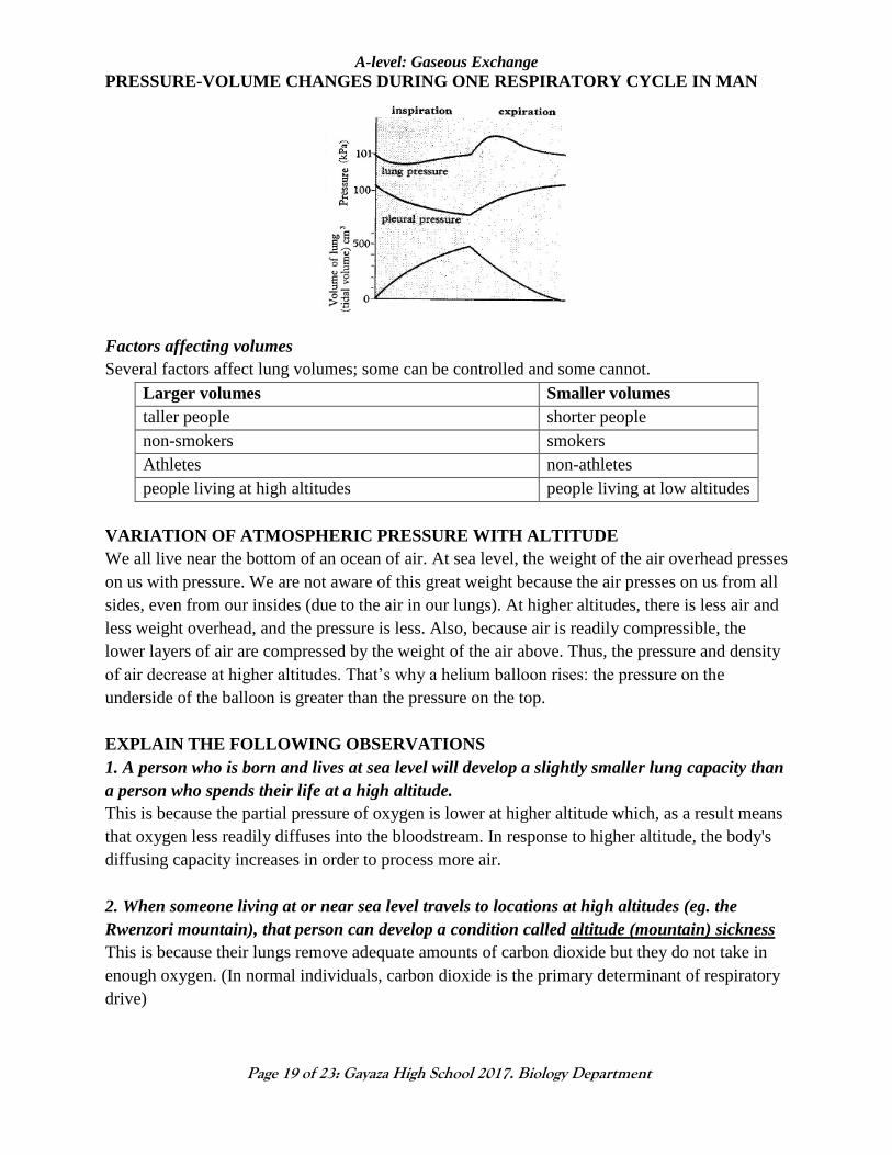
PRESSURE-VOLUME CHANGES DURING ONE RESPIRATORY CYCLE IN MAN
Factors affecting volumes
Several factors affect lung volumes; some can be controlled and some cannot.
Larger volumes Smaller volumes
taller people shorter people
non-smokers smokers
Athletes non-athletes
people living at high altitudes people living at low altitudes
VARIATION OF ATMOSPHERIC PRESSURE WITH ALTITUDE
We all live near the bottom of an ocean of air. At sea level, the weight of the air overhead presses
on us with pressure. We are not aware of this great weight because the air presses on us from all
sides, even from our insides (due to the air in our lungs). At higher altitudes, there is less air and
less weight overhead, and the pressure is less. Also, because air is readily compressible, the
lower layers of air are compressed by the weight of the air above. Thus, the pressure and density
of air decrease at higher altitudes. That’s why a helium balloon rises: the pressure on the
underside of the balloon is greater than the pressure on the top.
EXPLAIN THE FOLLOWING OBSERVATIONS
1. A person who is born and lives at sea level will develop a slightly smaller lung capacity than
a person who spends their life at a high altitude.
This is because the partial pressure of oxygen is lower at higher altitude which, as a result means
that oxygen less readily diffuses into the bloodstream. In response to higher altitude, the body's
diffusing capacity increases in order to process more air.
2. When someone living at or near sea level travels to locations at high altitudes (eg. the
Rwenzori mountain), that person can develop a condition called altitude (mountain) sickness
This is because their lungs remove adequate amounts of carbon dioxide but they do not take in
enough oxygen. (In normal individuals, carbon dioxide is the primary determinant of respiratory
drive)
Page 20
A-level: Gaseous Exchange
Page 20 of 23: Gayaza High School 2017. Biology Department
ALTITUDE SICKNESS (MOUNTAIN SICKNESS)
Is an illness that develops when the rate of ascent into higher altitudes outpaces the body’s
ability to adjust to those altitudes.
Altitude sickness generally develops at elevations higher than 8,000 feet (about 2,400 meters)
above sea level and when the rate of ascent exceeds 1,000 feet (300 meters) per day.
Symptoms: Fatigue, Headache, Dizziness, Insomnia (sleeplessness), Shortness of breath during
exertion, Nausea, Decreased appetite, Swelling of extremities, Social withdrawal
How to avoid altitude sickness: (1) allowing the body to get used to the altitude slowly, a
process called acclimatization. The goal of acclimatization is to increase ventilation (breathing)
to compensate for lower oxygen content in the air (2) Get used to the high altitude before doing a
lot of exercise e.g. hiking, skiing, or biking (3) Don't drink alcohol at high altitudes. It takes
much less alcohol to become drunk at high altitudes than at sea level.
ADAPTATIONS OF LUNG SYSTEM FOR GASEOUS EXCHANGE
1. Lungs have numerous alveoli; that provide large surface area for efficient gaseous
exchange
2. Epithelial lining between the alveoli wall and the blood capillaries is thin to provide a
shorter diffusion distance for easy gaseous exchange
3. The lung is spongy and has numerous alveoli to accommodate large volume of gases
4. It is highly supplied with blood capillaries that transport oxygen and carbon dioxide to and
from the body tissues respectively
5. The epithelial lining of alveoli is covered by a thin layer of moisture to dissolve oxygen for
easy diffusion into the blood solution
6. The whole lungs are covered with the pleural membrane which is gas tight thus changes in
the pressure within the lungs can occur without external interference
7. The walls of the trachea and bronchi are lined by rings of cartilage which prevent them
from collapsing and keeps them open for air passage
8. The inner passage of the air ways is lined with mucus membrane which contain ciliated
cells whose movement to and from the pharynx cause a sweeping action that collects dust
towards the pharynx for swallowing hence preventing their entry into the air ways
9. The mucus membrane contains mucus secreting cells which produces mucus that trap dust
and pathogenic particles which would find their way into the air way
10. The mucus membrane has a rich blood supply which warms and moistens the incoming air
for easy diffusion in the lungs
11. The epiglottis and other structures on the top of the trachea prevent food, drinks and other
solid particles from going into the trachea during swallowing.
NON-RESPIRATORY FUNCTIONS OF THE LUNG RESPIRATORY SYSTEM
1. The vibration of air flowing across the larynx (vocal chords) in humans allows phonation
(speech), and the syrinx, in birds results in vocalization or singing
2. Panting in dogs and some other animals provides a means of cooing body temperature
Page 21
A-level: Gaseous Exchange
Page 21 of 23: Gayaza High School 2017. Biology Department
3. Irritation of nerves within the nasal passages or airways, can induce coughing and
sneezing. These responses cause air to be expelled forcefully from the trachea or nose
thereby enabling expulsion of irritants caught in the mucus which lines the respiratory
tract.
INVOLUNTARY CONTROL OF BREATHING IN MAN
Introduction
Physiological control systems involving the nervous system usually have three components.
These are
1. a central controlling area where information from other parts of the body is integrated to
produce a coordinated response
2. an afferent pathway which relays impulses from the sensors to the central controlling area
3. an efferent pathway which conveys impulses from the central controlling area to the
organs and muscles
NB: Many factors can modify the rate and depth of breathing but the most important factors
are the levels of carbon dioxide, hydrogen ions (H+) and oxygen in the arterial blood.
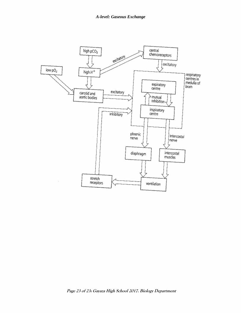
CONTROL OF BREATHING IN MAN
Breathing is usually automatic, controlled subconsciously by the respiratory center at the base of
the brain. People can also control their breathing when they wish, for example during speech,
singing, or voluntary breath holding.
The rate of breathing is controlled by the respiratory centre, which is in the lower part of the
brain stem, in the medulla oblongata. The respiratory centre comprises of the inspiratory and
expiratory centres.
Normally, an increased concentration of carbon dioxide is the strongest stimulus to the
depth and frequency of breathing.
Central chemoreceptors (cells that respond to chemical stimuli) in the medulla oblongata of
the brain detect changes in the concentration of carbon dioxide (CO2) in blood by monitoring the
PH of cerebrospinal fluid while Peripheral chemoreceptors in the carotid and aortic bodies
monitor both carbon dioxide and oxygen concentrations in blood.
High CO2 lowers the pH (an acid is a solution with a high H+ concentration).
CO2 + H2O H2CO3 HCO3- + H+
Eliminating CO2 is usually a bigger problem for terrestrial vertebrates than obtaining O2. The
body is therefore more sensitive to high CO2 concentration than low O2 concentration.
Aquatic vertebrates are more sensitive to low O2 because O2 is more limited in aquatic
environments.
Page 22
A-level: Gaseous Exchange
Page 22 of 23: Gayaza High School 2017. Biology Department
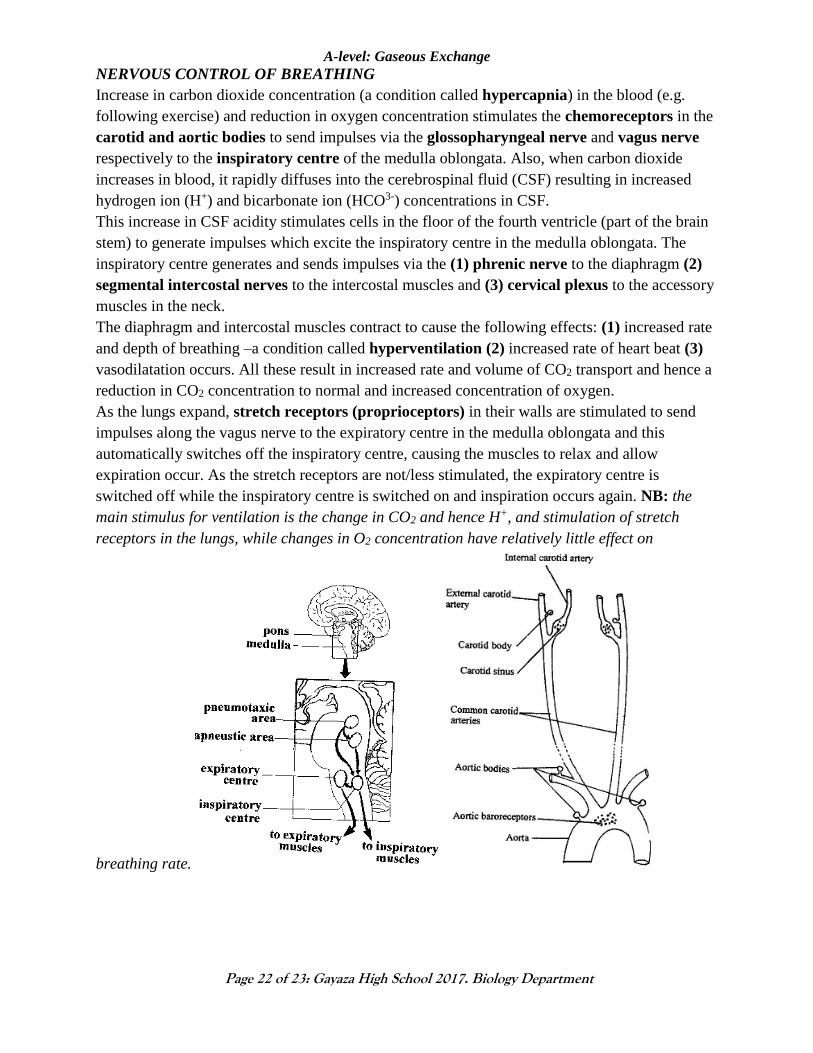
NERVOUS CONTROL OF BREATHING
Increase in carbon dioxide concentration (a condition called hypercapnia) in the blood (e.g.
following exercise) and reduction in oxygen concentration stimulates the chemoreceptors in the
carotid and aortic bodies to send impulses via the glossopharyngeal nerve and vagus nerve
respectively to the inspiratory centre of the medulla oblongata. Also, when carbon dioxide
increases in blood, it rapidly diffuses into the cerebrospinal fluid (CSF) resulting in increased
hydrogen ion (H+) and bicarbonate ion (HCO3-) concentrations in CSF.
This increase in CSF acidity stimulates cells in the floor of the fourth ventricle (part of the brain
stem) to generate impulses which excite the inspiratory centre in the medulla oblongata. The
inspiratory centre generates and sends impulses via the (1) phrenic nerve to the diaphragm (2)
segmental intercostal nerves to the intercostal muscles and (3) cervical plexus to the accessory
muscles in the neck.
The diaphragm and intercostal muscles contract to cause the following effects: (1) increased rate
and depth of breathing –a condition called hyperventilation (2) increased rate of heart beat (3)
vasodilatation occurs. All these result in increased rate and volume of CO2 transport and hence a
reduction in CO2 concentration to normal and increased concentration of oxygen.
As the lungs expand, stretch receptors (proprioceptors) in their walls are stimulated to send
impulses along the vagus nerve to the expiratory centre in the medulla oblongata and this
automatically switches off the inspiratory centre, causing the muscles to relax and allow
expiration occur. As the stretch receptors are not/less stimulated, the expiratory centre is
switched off while the inspiratory centre is switched on and inspiration occurs again. NB: the
main stimulus for ventilation is the change in CO2 and hence H+, and stimulation of stretch
receptors in the lungs, while changes in O2 concentration have relatively little effect on
breathing rate.
Page 23
A-level: Gaseous Exchange
Page 23 of 23: Gayaza High School 2017. Biology Department


![Gaseous exchange [all]](https://static.documents.pub/doc/80x56/54bdd34f4a79592e1a8b463d/gaseous-exchange-all.jpg)


