Purpose: The Schirmer’s test is commonly used in the clinic for the diagnosis of dry eye disease by measuring tear volume.This report describes a procedure which can be used to recover tears from the Schirmer strip for the measurement ofmultiple tear cytokines as well as matrix metalloproteinases (MMPs) by Luminex technology.Methods: Cytokine and MMP recovery was determined by using spiked Schirmer strips presoaked with known cytokinesor MMPs prepared in PBS with 1% BSA. In a clinical study, tears were collected from 5 subjects using Schirmer strips.Strips were stored on ice immediately after removal from the subject and stored dry at −20 °C for 16–24 h. Cytokineswere extracted from the Schirmer strip in 0.5 M NaCl with 0.5% Tween-20. Concentrations of cytokines and MMPs incollected tear samples were analyzed by Luminex using both a 10-cytokine and a 5-MMP kit.Results: The standard curves for the assay in both the kit assay buffer and extraction buffer were identical for 9 of the 10cytokines and all 5 MMPs. In the clinical sample all the cytokines (interleukin 1α [IL-1α], IL-1β, IL-1ra, IL-4, IL-6, IL-8,IL-10, IL-13, monocyte chemotactic protein-1 [MCP-1], and tumor necrosis factor-α [TNF-α]) and 5 MMPs (MMP-1,MMP-2, MMP-7, MMP-9, and MMP-10) tested were detected in at least 50% of the 10 subject samples. Recoveries fromextracted Schirmer strips were >60% for 8 of the 10 cytokines and all MMPs.Conclusions: Numerous cytokines and MMPs were detected in the tear samples collected using the Schirmer strip,including many that have been implicated in ocular surface disease. This procedure may be used to evaluate the cytokineand MMP content in tear samples in clinical studies, especially for the evaluation of dry eye therapeutics. Because theSchirmer test is routine in the assessment of dry eye, this method offers the opportunity to evaluate both the quantity andquality of the tears.
Tear samples are being used with increasing frequency todetect biomarkers of normal and diseased states of the ocularsurface [1-3], including allergy [4], and dry eye [5,6]. Whileuse of ELISA is common, the employment of multiplex bead[7,8], multi-array [9], and proteomic [10-12] technology hasenhanced the use of collected tears by allowing for analysisof small sample volumes while increasing the number ofdetectable targets.
The most commonly reported method to collect tearsamples for biomarker analysis is by capillary tube [13-15].In addition, it has been reported that cellulose acetateabsorbent filters [16], a Weck Cell Sponge [4], and an watereye wash [17] have been used to collect tear samples.However, Schirmer strips are routinely used to measure thetear volume for the clinical assessment of dry eye disease. Itwould be of added value if, rather than discarding the stripsafter the assessment, the tear components could be eluted fromthese strips and used for the measurement of knownbiomarkers, such as proinflammatory cytokines and otherinflammatory mediators.
Cytokines, chemokines, and matrix metalloproteinases(MMPs) have been considered as potential biomarkers for
ocular surface inflammation. Indeed, increased levels of pro-inflammatory cytokines such as interleukin 1 (IL-1), IL-6, andtumor necrosis factor-α (TNF-α), chemokines such as IL-8and monocyte chemotactic protein-1 (MCP-1), and matrixmetalloproteinase-9 (MMP-9) have been demonstrated to beassociated with ocular surface diseases [1,4,18,19] includingdry eye [20,21].
The Luminex multi-analyte profiling assay system is atechnology based on the principle of flow cytometry. Thesystem allows one to simultaneously measure numerousanalytes in a single microplate well, using very small samplevolumes [7] while achieving excellent correlations toindividual ELISA for many cytokines [22]. This system hasbeen successfully used to study the effect of anti-inflammatory agents on cytokine release profiles of culturedhuman ocular cells [23,24]. Recently, Luminex has also beenused to measure the cytokine content in tear samples collectedwith capillary tubes [5]. The results from the current studydemonstrate that numerous cytokines and MMPs can bedetected by Luminex from the tear samples collected using aSchirmer strip.
METHODSReagents: TearFlo™ Schirmer filter paper strips with an inkedruler were obtained from Contacare Ophthalmics &Diagnostics (Gujarat, India). Human multiplex-cytokine and
Molecular Vision 2011; 17:1056-1063 <http://www.molvis.org/molvis/v17/a118>Received 7 March 2011 | Accepted 20 April 2011 | Published 27 April 2011
MMP kits were from Millipore (Billerica, MA). All otherreagents were purchased from standard commercial sourcesand were of the highest available purity.
Standard curve of solution volume to Schirmer strip reading:To calculate millimeters of wetting with volume of tearscollected, a standard curve was developed. Phosphate-buffered saline (PBS; 2.5, 5, 7.5, 10, 15, 20, 25, or 30 µl) wastransferred to the rounded end of a Schirmer strip and the stripwas placed in a 2-ml eppendorf tube lying flat on the benchsurface. After 1 min the measurement as millimeter (mm)wetting was recorded. This measurement was repeated a totalof 3 times for each volume tested, and a standard curve ofvolume to Schirmer strip reading was generated.
Clinical subjects: The clinical study protocol was approvedby the Southwest Independent Institutional Review Board andwas conducted in accordance with 21 Code of FederalRegulations for Clinical Trials (CFR) Parts 812, 50, 54, and56, applicable Bausch + Lomb Standard OperatingProcedures, and the Declaration of Helsinki. All five healthyvolunteers gave informed consent and they were assessed foreligibility. Inclusion criteria were as follows: be 18 years orolder and have full legal capacity to volunteer, have no allergicconjunctivitis, not be using any topical ocular medications, nocontact lens wear or ophthalmic drop use 8 h before their studyvisit, be willing and able to follow instructions, and havesigned a statement of informed consent. Subjectdiscontinuation criteria were: adverse effects, other ocularcomplications, subject non-compliance, subject request, orsubject found to be ineligible during study participation. Theage, gender, Ocular Surface Disease Index (OSDI), andSchirmer strip measurements for the five subjects are shownin Table 1.
Tear sample collection: The Investigator, with gloves, placeda Schirmer strip over the lid margin at the junction of thelateral and middle thirds of the lower eyelids and kept in placefor 5 min while subjects closed their eyes without ananesthetic. The Schirmer strips were removed with gloves andtear volume in millimeters was recorded. Each Schirmer stripwas placed into a sterile 2-ml centrifuge tube, stored on icefor 20 min to 1 h, and then stored at −20 °C until processed.
Preparation of spiked cytokine and MMP Schirmer strips: Todetermine the feasibility and sensitivity of the kits forrecovering cytokines and MMPs from the Schirmer strips, thelow internal quality controls (QC-1) from each cytokine andMMP kit were prepared as described in the kit assay protocol.Twenty µl of each QC-1 was aliquotted to a Schirmer strip,allowed to flow for 1 min and the strip was transferred to a 2-ml eppendorf tube and frozen at −20 °C (24 h). For percentrecovery, 20 µl of each QC was simultaneously aliquotted intoa separate eppendorf tube and frozen at −20 °C (24 h). Eachexperiment for the strips and diluted frozen samples wereprepared in triplicate.Extraction of cytokines and MMPs from Schirmer strips:Assay buffer (200 µl) of the analyte kit containing 1% BSA(BSA) in phosphate buffered saline (PBS) with sodium azideas a preservative or extraction buffer containing 0.5 M NaCland 0.5% Tween-20 [25] was added to each 2-ml centrifugetube and incubated for 3 h at ambient temperature on a rocker(VWR, West Chester, PA), and then stored on ice uponcompletion. The strip was transferred to a new 2-ml tube andresidual liquid was removed by pinching the strip at the 25-mm mark in the sealed tube cap, and the sample was thencentrifuged (Microfuge R, Beckman, Palo Alto, CA) at ~100×g for 10 s. This liquid was combined with stored extractionbuffer. The Schirmer strips were discarded. Each 20-µl frozensample was diluted in 180 µl assay or extraction buffer andtreated to the same extraction regimen as described for thestrips.Standard curve comparison: To ensure there was nointerference in the assay from the extraction buffer, standardcurves were prepared in parallel in the kit assay buffer andextraction buffer and were assessed on the same assay plate.Standard curves were generated using the kit internalstandards vs maximum fluorescence intensity (MFI) and weregraphically represented using Sigmaplot software (Systat, SanJose, CA).Statistical analysis of cytokine and MMP standard curves inboth the assay buffer and extraction buffer: Comparison ofeach individual standard concentration points prepared in boththe kit assay buffer and extraction buffer was performed usinga one-way ANOVA-Tukey-Kramer test (JMP 7 software;
TABLE 1. CLINICAL SUBJECT DATA.
Subjectnumber
Age Gender OSDI* Severity based onOSDI
Schirmer OD Schirmer OS
1 54 F 37.5 1 10 152 41 F 25 1 6 73 26 M 0 0 28 324 53 F 20.8 1 6 145 25 F 0 0 35 35
*OSDI scale [34]: Normal (0) 0–20.7; Mild (1) 20.8–41.6; Moderate (2) 41.7–72.9; Severe (3) 73–100.
SAS Institute, Cary, NC) and for each standard concentrationpoint p<0.05 was pre-determined to be statisticallysignificant.Luminex multiplex cytokine and MMP analysis: Cytokine andMMP content in both the spiked and tear extracted strips wereanalyzed using multiplex Luminex technology [7,26] andperformed according to the manufacturer's instructions withall kit reagents, assay and wash buffers. Analysis wasperformed on cytokines and MMPs listed in Table 2 and Table3. Briefly, 25 µl of each sample extract was incubated withantibody-coated capture beads overnight at 4 °C on a TiterPlate Shaker (Lab-Line Instrument, Park, IL). Washed beadswere further incubated with biotin-labeled anti-humancytokine antibodies for 1 h at room temperature followed byincubation with streptavidin-phycoerythrin for 30 min andfollowed by an additional wash step. Beads were resuspendedin Luminex flow buffer for 5 min and samples were analyzedusing Luminex 200™ (Luminex, Austin, TX).
Determination of percent recovery of cytokine from spikedSchirmer strips: For percent recovery analysis the medianfluorescence intensity (MFI) was used to obtain theconcentration of each cytokine in pg/ml based on the standardcurve of each cytokine assayed by Luminex. Concentrationswere estimated using the Statlia software (BrendanTechnologies, Inc., Carlsbad, CA). Each sample was tested induplicate and resulting concentrations were averaged.Samples with concentrations of cytokines outside the assaylimits, as determined from the Statlia analysis, were excludedfrom further analysis. Cytokines recovered from both thediluted samples and spiked Schirmer strips were compared tothe nominal concentrations of the QC-1 control. Percentrecovery for each sample was determined by the equation:Percent recovery=(Mean of recovered sample analyteconcentration/nominal analyte concentration)*100. Thenominal analyte concentration was determined by performingthe QC-1 control according to the kit instructions.
TABLE 2. PERCENT RECOVERY OF CYTOKINES DILUTED IN BUFFER OR EXTRACTED FROM SPIKED SCHIRMER STRIPS.
Recovery in assay buffer Recovery in extraction buffer Cytokine Diluted sample
*p<0.05 versus cytokine diluted in same assay buffer and calculated from same buffer standard curve. BLD: Below the limit of detection. n=10 for all data.
TABLE 3. PERCENT RECOVERY OF MMPS DILUTED IN BUFFER OR EXTRACTED FROM SPIKED SCHIRMER STRIPS.
Statistical analysis of spiked cytokine from Schirmer strips:Comparison of the recovered sample from spiked Schirmerstrips to the sample diluted in buffer was performed using aone-way ANOVA-Tukey-Kramer test (JMP 7) and a p<0.05was pre-determined to be statistically significant.Determination of pg/mlmeasured in tears obtained viaSchirmer strips: The amount of each cytokine or MMPmeasured in the tears extracted from Schirmer strips wasexpressed as total recovery in picograms per milliliter(pg/ml) as follows: First, the calculated sample in pg/ml wasmultiplied by the total extraction sample volume (0.2 ml) togive total pg in the extracted sample. Final pg/ml based uponSchirmer volume was calculated by dividing total pgextracted by the calculated Schirmer strip volume (pg/µl)and multiplying by 1,000. Means and standard errors (SE)of the 10 samples were determined for each cytokine andMMP.
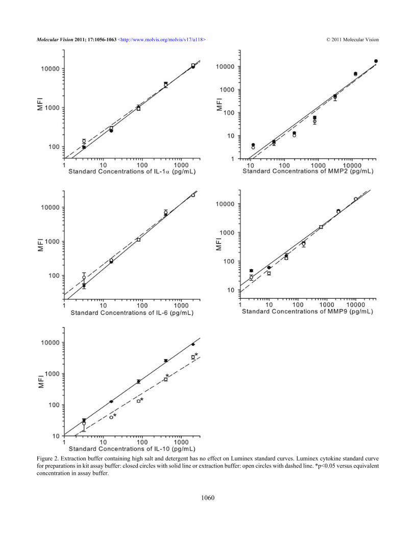
RESULTSStandard curve of solution volume to Schirmer strip reading:This experiment was designed to determine the relationshipbetween the reading on the Schirmer strip as millimeter (mm)and actual volume (µl) of the PBS solution absorbed onto thestrip over a period of one minute. Although a linearrelationship was found, the tear volume measurement inmillimeters did not equate to the same number for microliters(Figure 1). This standard curve was used to determine theactual volume of the tears on the clinical Schirmer strips.Luminex standard curves in standard assay and elutionbuffers: All the standard concentration points on curvesgenerated in the extraction buffer for all cytokines and MMPs,except for IL-10, were statistically comparable to theconcentration points generated in the kit assay buffer. ForIL-10, all standard concentrations except for the lowest
Figure 1. Graph of volume versus Schirmer strip measurement. Eachdata point represents the mean and standard deviation from 3individual experiments. The line was determined by linear regressionand the correlation coefficient was 0.999.
concentration were significantly lower in the extractionbuffer. Graphs for IL-10, 2 major pro-inflammatorycytokines, IL-1α, and IL-6, and MMPs 2 and 9 are shown inFigure 2.
Recovery of cytokines and MMPs from spiked Schirmer strips:For these experiments, and the clinical Schirmer stripextractions, identical 10 cytokine and 5 MMP kits were used.In these experiments the recovery for each 20-µl sampleextracted from a Schirmer strip was compared to the recoveryequal volume in solution. The percent recoveries represent theamount of diluted or extracted sample as compared to thetheoretical recovery for each cytokine with each buffer. Forthe assays performed with kit assay buffer, the recoveries ofIL-1β, IL-8, IL-10, and IL-13 were significantly lower in theextracted strips as compared to the diluted samples (Table 3).For the assays performed with the extraction buffer both IL-13and TNF-α had significantly lower extracted strip recoveries,while IL-10 was below the limit of detection in this buffer andcould not be analyzed. Of the extracted MMP strip samples,only MMP7, extracted in assay buffer had a significantlylower recovery.Recovery of cytokines and MMPs from clinical Schirmerstrips: Of the 10 samples tested in this study, allconcentrations for the 10 cytokines tested were determined tobe within the maximum reportable concentration except for 3of the samples tested for IL-1ra (Figure 3). These 3 sampleswere re-tested at a 10-fold dilution in extraction buffer forIL-1ra only and the determined concentrations were found tobe within the reportable range. Only 50% of samples for TNF-α were above the minimum detectable concentration for thisassay, while all samples contained IL-1α, IL-1ra, IL-8 andMCP-1. Cytokines IL-6 and IL-13 were recovered in 80% ofsample, IL-4 and IL-10 in 70% and IL-1β in 60%. Of the 5MMPs tested in this study MMP-7, MMP-9, and MMP-10were recovered in all the samples, while MMP-1 (70%) andMMP-2 (80%) were recovered in most of the samples (Figure4). Only the samples within the reportable range of the assaywere used for determination of calculated recoveries for thesecytokines.
DISCUSSIONResearchers are using ever-expanding platforms of multiplextechnology, which allow for the use of minimal samplevolumes from tears to identify biomarkers associated with dryeye disease (DED) [6,9]. Schirmer’s test has been considered,rightly or wrongly [27,28], as a “gold standard” for theassessment of dry eye conditions, and is therefore a key testin both the clinical office setting and clinical trials for varioustreatment modalities. In addition to its traditional use ofdetermining tear volume, this test also provides clinicalsamples which, until recently, have not been commonlyassessed for comprehensive biologic alterations to the tearfilm in ocular surface disease. In part, this has been because
Figure 2. Extraction buffer containing high salt and detergent has no effect on Luminex standard curves. Luminex cytokine standard curvefor preparations in kit assay buffer: closed circles with solid line or extraction buffer: open circles with dashed line. *p<0.05 versus equivalentconcentration in assay buffer.
of the relatively small amount of tears obtained as comparedto the amount needed for biologic assays. However, with theadvance of technologies which facilitate meaningfulassessments in small-volume clinical samples, the potentialfor additional assessment from this standard test is veryattractive. Recently, methods employed to collect tears fromsubjects include microcapillary tubes [8], polyvinyl acetatesponges [4], and acetate filters [16]. While each of thesemethods has its advantages, they would be an additional stepin the clinical setting, costing additional time to both patientand clinical researcher. Furthermore, as both the Schirmerstrip and the other techniques remove tears from the ocularsurface, if both were performed; either procedure followed by
the other would compromise the second test. Therefore, theuse of Schirmer strips for additional biomarker analysis,including cytokines, chemokines, and MMPs, would be ofimmense benefit. Schimer strips have been used tosuccessfully recover single analytes including eotaxins [25],cystatins [29], secretory IgA [30], and Vitamin C [31].However, a comprehensive evaluation using Schirmer stripsfor the measurement of multiple cytokines as well as MMPshas not been previously described.
The Luminex multiplex platform is ideally suited for thedetection of biomarkers from tear samples [6,8,28]. In thestudy presented here, Luminex kits were tested for capabilityof each kit to detect 10 cytokines and 5 MMPs recovered from
Figure 3. Concentrations of 10cytokines extracted from clinicalSchirmer strips. All data are expressedas mean±SEM.
Figure 4. Concentrations of 5 MMPsextracted from clinical Schirmer strips.All data are expressed as mean±SEM.
spiked Schirmer strips. Initial experiments indicate that theprototype extraction buffer containing high salt and detergentdoes not interfere with the reproducible generation of standardcurves for 14 of the 15 analytes tested. When the actualrecovered sample from the spiked Schirmer strip wascompared to the quantity recovered from the diluted control,both IL-10 and IL-13 were poorly recovered in both the assayand extraction buffers. It is unlikely that the observed poorrecoveries of IL-10 and IL-13 were due to the buffer systemsince the two buffers tested in the current study were verydifferent, one contains 0.5% Tween-20 and other one does not.Furthermore, cytokine kits from two other vendors were alsotested and generated somewhat less efficient recoveries formost cytokines (data not shown). These findings suggest thata simple wetting of the strips may be sufficient to recover thecytokines and MMPs, and as our goal was to maximize therecovery of the most relevant pro-inflammatory cytokines,including IL-1, IL-6 and IL-8, the methods employed and thekit used in our studies were highly suitable for this purpose.
Finally, clinical Schirmer strip tear samples obtainedfrom 5 subjects were tested for 10 cytokines of which 9 wererecovered in >50% of the samples. However, while ourclinical sample size was limited to 5 subjects, TNF-α was stillbelow the limit of detection for half of these samples,suggesting either an inability of this cytokine to be detectedusing our methodology or that minimal quantities of thiscytokine were present in our clinical test samples.Interestingly, IL-10, which was poorly recovered from spikedstrips, was recovered in detectable quantities in most clinicalsamples. Additional work would be needed to furtherunderstand this phenomenon. In addition, all 5 MMPs wereefficiently recovered from these samples, including MMP-9,which has been demonstrated to be involved in corneal barrierdisruption in experimental dry eye models [32,33]. Futureefforts will include the assessment of extraction buffercharacteristics with regard to recovery and detection ofcytokines, with emphasis on the primary pro- and anti-inflammatory cytokines and MMPs that may be affected indry eye conditions. In addition, kits from multiple vendorsmay be used to develop our ability to detect the key cytokinesin clinical samples.
Tear reflection is very common with Schirmer's test dueto the strong irritation by the strip. By looking at Table 1,subject 5 and 3 might have tear reflection during the test, astheir Schirmer's scores were abnormally high. With tearreflection, the cytokine and MMP can be easily diluted. Tearreflection was not considered in this study as the focus wasmore for the merits of the technical development on thecytokine recovery from the Schirmer strip than the actualconcentrations of the cytokines recovered and the tearcollection. However, this is a very important issue whichshould be considered when collecting tear samples andanalyzing the cytokine concentration.
In summary, results from the current study demonstratedthat numerous cytokines as well as several MMPs can berecovered from the Schirmer strip and quantitatively analyzedby Luminex technology. Because Schirmer’s test is routinelyused for the assessment of dry eye and inflammation isrecognized as a potential mechanism of dry eye or therapeutictarget, this method offers the opportunity to evaluate both thequantity and quality of the tears.
REFERENCES1. Pflugfelder SC, Jones D, Ji Z, Afonso A, Monroy D. Altered
cytokine balance in the tear fluid and conjunctiva of patientswith Sjogren's syndrome keratoconjunctivitis sicca. Curr EyeRes 1999; 19:201-11. [PMID: 10487957]
2. Zoukhri D. Effect of inflammation on lacrimal gland function.Exp Eye Res 2006; 82:885-98. [PMID: 16309672]
3. Lema I, Sobrino T, Duran JA, Brea D, Diez-Feijoo E.Subclinical keratoconus and inflammatory molecules fromtears. Br J Ophthalmol 2009; 93:820-4. [PMID: 19304583]
4. Acera A, Rocha G, Vecino E, Lema I, Duran JA. Inflammatorymarkers in the tears of patients with ocular surface disease.Ophthalmic Res 2008; 40:315-21. [PMID: 18688174]
5. Massingale ML, Li X, Vallabhajosyula M, Chen D, Wei Y,Asbell PA. Analysis of inflammatory cytokines in the tears ofdry eye patients. Cornea 2009; 28:1023-7. [PMID: 20162838]
6. Enríquez-de-Salamanca A, Castellanos E, Stern ME, FernándezI, Carreño E, García-Vázquez C, Herreras JM, Calonge M.Tear cytokine and chemokine analysis and clinicalcorrelations in evaporative-type dry eye disease. Mol Vis2010; 16:862-73. [PMID: 20508732]
8. LaFrance MW, Kehinde LE, Fullard RJ. Multiple cytokineanalysis in human tears: an optimized procedure forcytometric bead-based assay. Curr Eye Res 2008;33:525-44. [PMID: 18600485]
9. Li S, Sack R, Vijmasi T, Sathe S, Beaton A, Quigley D, GallupM, McNamara NA. Antibody protein array analysis of the tearfilm cytokines. Optom Vis Sci 2008; 85:653-60. [PMID:18677223]
10. Green-Church KB, Nichols KK, Kleinholz NM, Zhang L,Nichols JJ. Investigation of the human tear film proteomeusing multiple proteomic approaches. Mol Vis 2008;14:456-70. [PMID: 18334958]
11. Li K, Chen Z, Duan F, Liang J, Wu K. Quantification of tearproteins by SDS-PAGE with an internal standard protein: anew method with special reference to small volume tears.Graefes Arch Clin Exp Ophthalmol 2010; 248:853-62.[PMID: 20127108]
12. Zhou L, Beuerman RW, Chan CM, Zhao SZ, Li XR, Yang H,Tong L, Liu S, Stern ME, Tan D. Identification of tear fluidbiomarkers in dry eye syndrome using iTRAQ quantitativeproteomics. J Proteome Res 2009; 8:4889-905. [PMID:19705875]
13. Srinivasan S, Joyce E, Boone A, Simpson T, Jones L, SenchynaM. Tear lipocalin and lysozyme concentrations inpostmenopausal women. Ophthalmic Physiol Opt 2010;30:257-66. [PMID: 20444132]
14. Pokharel S, Shah DN, Joshi SN, Choudhary M. Tearfilmimmunoglobulin E (IgE) level in vernal keratoconjunctivitisby ELISA. Kathmandu Univ Med J (KUMJ) 2009;7:104-8.KUMJ
15. Sack RA, Sathe S, Beaton A, Kozinski M, Bogart B, Lew G,Sharma S, Upponi A. Is the cystatin-like domain of TSLfunctionally active in external ocular infections and during thenormal diurnal cycle? Exp Eye Res 2004; 78:371-8. [PMID:15106915]
16. Esmaeelpour M, Cai J, Watts P, Boulton M, Murphy PJ. Tearsample collection using cellulose acetate absorbent filters.Ophthalmic Physiol Opt 2008; 28:577-83. [PMID:19076560]
17. Argueso P, Balaram M, Spurr-Michaud S, Keutmann HT, DanaMR, Gipson IK. Decreased levels of the goblet cell mucinMUC5AC in tears of patients with Sjogren syndrome. InvestOphthalmol Vis Sci 2002; 43:1004-11. [PMID: 11923240]
18. Leonardi A, Sathe S, Bortolotti M, Beaton A, Sack R.Cytokines, matrix metalloproteases, angiogenic and growthfactors in tears of normal subjects and vernalkeratoconjunctivitis patients. Allergy 2009; 64:710-7.[PMID: 19220217]
19. Smith VA, Rishmawi H, Hussein H, Easty DL. Tear film MMPaccumulation and corneal disease. Br J Ophthalmol 2001;85:147-53. [PMID: 11159476]
20. Lam H, Bleiden L, de Paiva CS, Farley W, Stern ME,Pflugfelder SC. Tear cytokine profiles in dysfunctional tearsyndrome. Am J Ophthalmol 2009; 147:198-205. [PMID:18992869]
21. Narayanan S, Miller WL, McDermott AM. Conjunctivalcytokine expression in symptomatic moderate dry eyesubjects. Invest Ophthalmol Vis Sci 2006; 47:2445-50.[PMID: 16723455]
22. Dupont NC, Wang K, Wadhwa PD, Culhane JF, Nelson EL.Validation and comparison of luminex multiplex cytokineanalysis kits with ELISA: determinations of a panel of ninecytokines in clinical sample culture supernatants. J ReprodImmunol 2005; 66:175-91. [PMID: 16029895]
23. Cavet ME, Harrington KL, VanDerMeid KR, Ward KW, ZhangJZ. Comparison of the effect of multipurpose contact lenssolutions on the viability of cultured corneal epithelial cells.Cont Lens Anterior Eye 2009; 32:171-5. [PMID: 19540795]
24. Zhang JZ, Cavet ME, VanDerMeid KR, Salvador-Silva M,Lopez FJ, Ward KW. BOL-303242-X, a novel selectiveglucocorticoid receptor agonist, with full anti-inflammatory
properties in human ocular cells. Mol Vis 2009;15:2606-16. [PMID: 20011631]
25. Shoji J, Kitazawa M, Inada N, Sawa M, Ono T, Kawamura M,Kato H. Efficacy of tear eosinophil cationic protein levelmeasurement using filter paper for diagnosing allergicconjunctival disorders. Jpn J Ophthalmol 2003; 47:64-8.[PMID: 12586180]
26. Morgan E, Varro R, Sepulveda H, Ember JA, Apgar J, WilsonJ, Lowe L, Chen R, Shivraj L, Agadir A, Campos R, Ernst D,Gaur A. Cytometric bead array: a multiplexed assay platformwith applications in various areas of biology. Clin Immunol2004; 110:252-66. [PMID: 15047203]
27. Danjo Y. Diagnostic usefulness and cutoff value of Schirmer'sI test in the Japanese diagnostic criteria of dry eye. GraefesArch Clin Exp Ophthalmol 1997; 235:761-6. [PMID:9439968]
28. De Paiva CS, Hwang CS, Pitcher JD 3rd, Pangelinan SB,Rahimy E, Chen W, Yoon KC, Farley WJ, Niederkorn JY,Stern ME, Li DQ, Pflugfelder SC. Age-related T-cell cytokineprofile parallels corneal disease severity in Sjogren'ssyndrome-like keratoconjunctivitis sicca in CD25KO mice.Rheumatology (Oxford) 2009; 49:246-58. [PMID:20007286]
29. Barka T, Asbell PA, van der Noen H, Prasad A. Cystatins inhuman tear fluid. Curr Eye Res 1991; 10:25-34. [PMID:2029847]
30. Inada N, Shoji J, Hoshino M, Sawa M. Evaluation of total andallergen-specific secretory IgA in tears of allergicconjunctival disease patients. Jpn J Ophthalmol 2007;51:338-42. [PMID: 17926109]
31. Paterson CA, O'Rourke MC. Vitamin C levels in human tears.Arch Ophthalmol 1987; 105:376-7. [PMID: 3827714]
32. Corrales RM, Stern ME, De Paiva CS, Welch J, Li DQ,Pflugfelder SC. Desiccating stress stimulates expression ofmatrix metalloproteinases by the corneal epithelium. InvestOphthalmol Vis Sci 2006; 47:3293-302. [PMID: 16877394]
33. Pflugfelder SC, Farley W, Luo L, Chen LZ, de Paiva CS, OlmosLC, Li DQ, Fini ME. Matrix metalloproteinase-9 knockoutconfers resistance to corneal epithelial barrier disruption inexperimental dry eye. Am J Pathol 2005; 166:61-71. [PMID:15632000]
34. Schiffman RM, Christianson MD, Jacobsen G, Hirsch J, ReisBL. Reliability and validity of the Ocular Surface DiseaseIndex. Arch Ophthalmol 2000; 118:615-21. [PMID:10815152]
Articles are provided courtesy of Emory University and the Zhongshan Ophthalmic Center, Sun Yat-sen University, P.R. China.The print version of this article was created on 22 April 2011. This reflects all typographical corrections and errata to the articlethrough that date. Details of any changes may be found in the online version of the article.