191 Human cheek cells 7 a Model Membr ane D espite their variety, all cells have certain structures in common that perform essential functions. One such struc- ture is the cell membrane. The cell mem- brane is the outermost membrane and the barrier between the cell and its external environment. The cell membrane supports and protects the cell. If you compare a cell to a house, the membrane of a cell functions like the out- side walls, roof, and doors of the house. It separates the cell from its environment, and helps maintain the homeostasis that lets the structures and molecules in the cell func- tion. This includes regulating amounts of vital substances in the cell, such as salts and glucose, by controlling their movements into and out of the cell. Other key roles of the cell membrane are keeping out dangerous substances and organisms, such as disease-causing microbes, and sending and receiving signals from other cells. Both infectious and noninfectious diseases may involve the cell membrane. When the membrane is unable to keep out disease-causing microbes, infection results. HIV and Plasmodium are examples of microbes that must first interact with and enter cells in order to infect humans. Noninfectious diseases can disrupt normal membrane function. Some forms of the genetic disease muscular dystrophy, for example, prevent damaged muscle cell membranes from healing by interfering with their normal ability to reseal small tears. In this investigation you will explore a model that displays some of the features of a cell membrane. Challenge What structures and characteristics help the cell membrane perform its functions? 0 0
Transcript
191
Human cheek cells
7 aModelMembrane
Despite their variety, all cells have
certain structures in common that
perform essential functions. One such struc-
ture is the cell membrane. The cell mem-
brane is the outermost membrane and the
barrier between the cell and its external
environment.
The cell membrane supports and protects
the cell. If you compare a cell to a house, the
membrane of a cell functions like the out-
side walls, roof, and doors of the house. It
separates the cell from its environment, and
helps maintain the homeostasis that lets the
structures and molecules in the cell func-
tion. This includes regulating amounts of
vital substances in the cell, such as salts and
glucose, by controlling their movements into and out of the cell. Other key roles of
the cell membrane are keeping out dangerous substances and organisms, such as
disease-causing microbes, and sending and receiving signals from other cells.
Both infectious and noninfectious diseases may involve the cell membrane. When
the membrane is unable to keep out disease-causing microbes, infection results.
HIV and Plasmodium are examples of microbes that must first interact with and
enter cells in order to infect humans. Noninfectious diseases can disrupt normal
membrane function. Some forms of the genetic disease muscular dystrophy, for
example, prevent damaged muscle cell membranes from healing by interfering
with their normal ability to reseal small tears.
In this investigation you will explore a model that displays some of the features of
Procedure 1. To make the model, begin by filling the tray with a shallow layer of bubble
solution.
2. Each person in your group should unwrap a clean straw, and use a piece of
masking tape to label the straw with your initials. Keep track of your own
straw throughout the activity.
3. Allow each of you, one by one, to make a single large bubble by gently blow-
ing into the bubble solution. In your science notebook record your observa-
tions of the solution film—the membrane—that forms the bubble. If your
bubble pops before you can finish your observations, make a fresh bubble.
Let everyone in your group practice making a bubble. Take turns making the
additional bubbles when they are needed in the rest of the Procedure.
4. Make a fresh bubble. Try dropping a toothpick through the film. In your sci-
ence notebook record your observations. Remove the toothpick, and dry it
with a paper towel.
5. Make a fresh bubble. Then insert the plastic tube into the bubble. In your sci-
ence notebook, record your observations.
6. Now coat the plastic tube completely with the bubble solution in the tray. Make
a fresh bubble. Insert the coated tube through the bubble. Record your observa-
tions. While the tube is inserted in the membrane, drop a toothpick through
the plastic tube. In your science notebook, record your observations.
193
aModelMeMbrane • activity7
7. Move the plastic tube slowly and carefully from side to side, while it is in the
bubble. Insert a second solution-coated object, such as a straw, into the bub-
ble film, and move it around the bubble membrane also. In your science
notebook, record your observations.
8. Carefully remove the tube and straw from the bubble. In your science note-
book, record your observations.
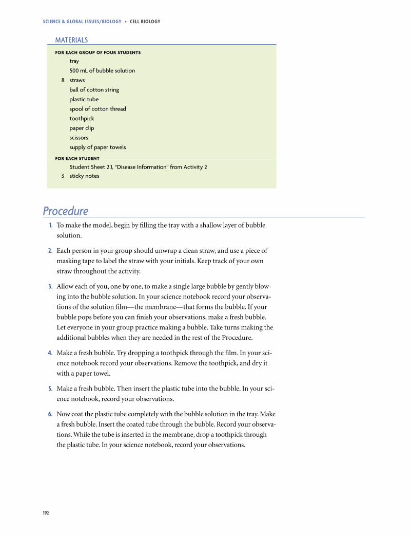
9. Thread the cotton string through four of the straws to make a square about
three-fourths the size of the tray. Knot the ends together. Then tie handles
onto two opposite sides of the square, as shown below.
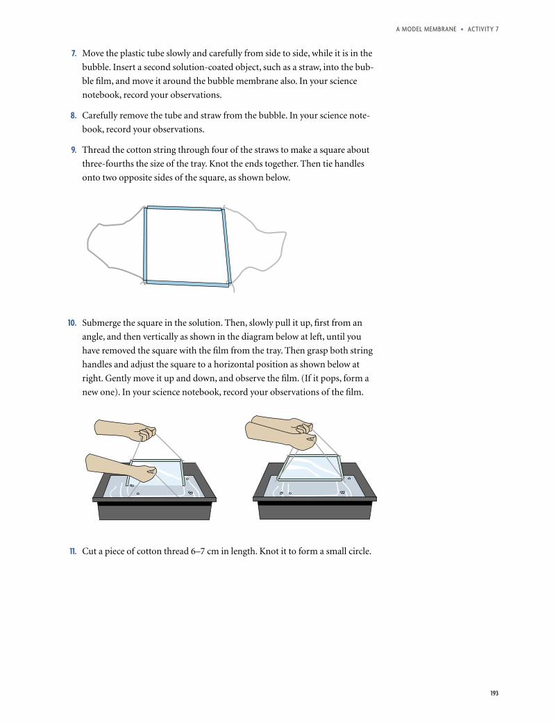
10. Submerge the square in the solution. Then, slowly pull it up, first from an
angle, and then vertically as shown in the diagram below at left, until you
have removed the square with the film from the tray. Then grasp both string
handles and adjust the square to a horizontal position as shown below at
right. Gently move it up and down, and observe the film. (If it pops, form a
new one). In your science notebook, record your observations of the film.
11. Cut a piece of cotton thread 6–7 cm in length. Knot it to form a small circle.
Science & Global iSSueS/bioloGy • cell bioloGy
194
12. Float the circle of thread on the film made with the straws, and form an open-
ing in the film by popping the inside of the circle with the end of a partially
unfolded paper clip. In your science notebook, record your observations.
Note: Be patient and gentle during Steps 12 and 13. If your film breaks, place
the square of straws back in the bubble solution to form another film.
13. Use the straight end of the paper clip to gently remove the circle of thread
from the film. In your science notebook, record your observations.
14. Now that you have worked with a simple model of the cell membrane, read
the box above about the actual structure of the cell membrane.
15. Follow your teacher’s directions for reading the case study about diabetes. As
you read, follow the “Read, Think, and Take Note” strategy.
16. Complete the information for diabetes on Student Sheet 2.1, “Disease Infor-
mation” after you read the case study.
Analysis 1. Based on your observations of the bubble film in Procedure Steps 3 and 7,
what do you think scientists mean when they say that the cell membrane is
fluid?
2. a. What did you have to do to make objects pass through the bubble mem-
brane without breaking the bubble?
b. A cell membrane is mostly made of phospholipids. Which would be more
likely to be able to move across a cell membrane: a structure made of
proteins, or a structure made of proteins coated with phospholipids?
Explain, based on the model.
c. The cell membrane can be described as a selective barrier. What does
that mean?
cell Membrane structure
The cell membrane is made mainly of proteins and phospholipids. The phospholipids form two layers—a bilayer—that gives the membrane both flexibility and strength. You saw this property with the detergent bubbles, which are also made of a type of lipid. The phospholipids in each layer of the cell membrane move from side to side in the cell membrane, trading places with each other and making the membrane a fluid structure.
195
aModelMeMbrane • activity7
3. A small break in a cell membrane sometimes closes back up. What properties of
the model that you just explored showed how the membrane can reseal itself?
4. In addition to the phospholipid bilayer, cell membranes also include special-
ized proteins. These proteins are embedded in the membrane and, like the
phospholipids, are able to move side to side in the membrane. Some of these
proteins function as transporters, allowing other molecules into the cell.
Explain how you modeled transport proteins in the Procedure.
5. From what you learned about diabetes in Case Study 3, explain the effect a
destroyed transport protein has on the membrane and the cell.
6. Based on the diabetes case study, what conclusions can you make about the
relationships between body weight, a country’s income level, and diabetes?