A neural theory of speech acquisition and production Frank H. Guenther a, b, c, d, * , Tony Vladusich a, d a Department of Cognitive and Neural Systems, Boston University, 677 Beacon Street, Boston, MA, 02215, USA b Division of Health Sciences and Technology, Harvard University – Massachusetts Institute of Technology, Cambridge, MA 02139, USA c Athinoula A. Martinos Center for Biomedical Imaging, Massachusetts General Hospital, Charlestown, MA 02129, USA d Center of Excellence for Learning in Education, Science and Technology, Boston University, 677 Beacon Street, Boston, MA, 02215, USA article info Article history: Received 25 November 2008 Received in revised form 19 August 2009 Accepted 20 August 2009 Keywords: Speech production Motor control Neural model fMRI Mirror system abstract This article describes a computational model, called DIVA, that provides a quantitative framework for understanding the roles of various brain regions involved in speech acquisition and produc- tion. An overview of the DIVA model is first provided, along with descriptions of the computations performed in the different brain regions represented in the model. Particular focus is given to the model’s speech sound map, which provides a link between the sensory representation of a speech sound and the motor program for that sound. Neurons in this map share with ‘‘mirror neurons’’ described in monkey ventral premotor cortex the key property of being active during both production and perception of specific motor actions. As the DIVA model is defined both computationally and anatomically, it is ideal for generating precise predictions concerning speech-related brain activation patterns observed during functional imaging experiments. The DIVA model thus provides a well-defined framework for guiding the interpretation of experimental results related to the putative human speech mirror system. Ó 2009 Elsevier Ltd. All rights reserved. 1. Introduction The production of speech sounds requires the integration of auditory, somatosensory, and motor information represented in the temporal, parietal, and frontal lobes of the cerebral cortex, respectively. Together with sub-cortical structuresdsuch as the cerebellum, basal ganglia and the brain stemdthese * Corresponding author. Department of Cognitive and Neural Systems, Boston University, 677 Beacon Street, Boston, MA, 02215, USA. Tel.: þ1 617 353 5765; fax: þ1 617 353 7755. E-mail address: [email protected](F.H. Guenther). Contents lists available at ScienceDirect Journal of Neurolinguistics journal homepage: www.elsevier.com/locate/ jneuroling 0911-6044/$ – see front matter Ó 2009 Elsevier Ltd. All rights reserved. doi:10.1016/j.jneuroling.2009.08.006 Journal of Neurolinguistics 25 (2012) 408–422
Transcript
Journal of Neurolinguistics 25 (2012) 408–422
Contents lists available at ScienceDirect
Journal of Neurolinguisticsjournal homepage: www.elsevier .com/locate/
jneurol ing
A neural theory of speech acquisition and production
Frank H. Guenther a,b,c,d,*, Tony Vladusich a,d
a Department of Cognitive and Neural Systems, Boston University, 677 Beacon Street, Boston, MA, 02215, USAb Division of Health Sciences and Technology, Harvard University – Massachusetts Institute of Technology, Cambridge, MA 02139, USAc Athinoula A. Martinos Center for Biomedical Imaging, Massachusetts General Hospital, Charlestown, MA 02129, USAd Center of Excellence for Learning in Education, Science and Technology, Boston University, 677 Beacon Street, Boston, MA,02215, USA
a r t i c l e i n f o
Article history:Received 25 November 2008Received in revised form 19 August 2009Accepted 20 August 2009
Keywords:Speech productionMotor controlNeural modelfMRIMirror system
* Corresponding author. Department of Cognitiv02215, USA. Tel.: þ1 617 353 5765; fax: þ1 617 35
0911-6044/$ – see front matter � 2009 Elsevier Ltdoi:10.1016/j.jneuroling.2009.08.006
a b s t r a c t
This article describes a computational model, called DIVA, thatprovides a quantitative framework for understanding the roles ofvarious brain regions involved in speech acquisition and produc-tion. An overview of the DIVA model is first provided, along withdescriptions of the computations performed in the different brainregions represented in the model. Particular focus is given to themodel’s speech sound map, which provides a link between thesensory representation of a speech sound and the motor programfor that sound. Neurons in this map share with ‘‘mirror neurons’’described in monkey ventral premotor cortex the key property ofbeing active during both production and perception of specificmotor actions. As the DIVA model is defined both computationallyand anatomically, it is ideal for generating precise predictionsconcerning speech-related brain activation patterns observedduring functional imaging experiments. The DIVA model thusprovides a well-defined framework for guiding the interpretationof experimental results related to the putative human speechmirror system.
� 2009 Elsevier Ltd. All rights reserved.
1. Introduction
The production of speech sounds requires the integration of auditory, somatosensory, and motorinformation represented in the temporal, parietal, and frontal lobes of the cerebral cortex, respectively.Together with sub-cortical structuresdsuch as the cerebellum, basal ganglia and the brain stemdthese
e and Neural Systems, Boston University, 677 Beacon Street, Boston, MA,3 7755.nther).
F.H. Guenther, T. Vladusich / Journal of Neurolinguistics 25 (2012) 408–422 409
cortical regions and their functional connections form a functional unit which we term the speechmotor control system. The speech motor control system is engaged during even the simplest speechtasks, such as babbling, imitating or reading single syllables and words (e.g., Fiez & Petersen, 1998;Guenther, 1995; Turkeltaub, Eden, Jones, & ZeffiroTurkeltaub, 2002). The present article describesa computational and neural framework, called DIVA, that provides a quantitative account of theinteractions between cortical motor, somatosensory, and auditory regions during speech output (seeGuenther, Ghosh, & Tourville, 2006, for computational details), thereby providing a ‘‘neural theory ofspeech production’’, in keeping with this special issue’s theme of a neural theory of language.
Our focus here is on the computations performed by the cerebral cortex, particularly the premotorregion of frontal cortex (see Barlow, 1999; Duffy, 1995; Kent, 1997; Zemlin, 1998, for detailed discus-sions of other speech-related brain regions). We discuss how the properties of a specific class of neuronin the DIVA model, known as speech sound map neurons (e.g., Guenther, 1992, 1994; Guenther et al.,2006), resemble ‘‘mirror neurons’’ described in the F5 region of monkey premotor cortex (Gallese,Fadiga, Fogassi, & Rizzolatti, 1996; Rizzolatti, Fadiga, Gallese, & Fogassi, 1996). As discussed in subse-quent detail, we hypothesize that the speech sound map resides in left ventral premotor cortex andposterior Broca’s area (BA 6, 44) of the human brain, and it is a crucial component of the networkresponsible for speech acquisition, as well as mature speech production. Some researchers (Rizzolatti &Arbib, 1998) have posited that monkey area F5, or the region immediately in front of area F5 (Petrides,Cadoret, & Mackey, 2005), is homologous to Broca’s area in humans, and that human speech evolved onthe basis of the functional properties of mirror neurons. Although this hypothesis appears plausible,research on the putative role of mirror neurons in human speech motor control would benefit greatlyfrom computational and neural frameworks with which to synthesize, interpret and predict empiricalresults (e.g., Bonaiuto, Rosta, & Arbib, 2007). Here we summarize data from several brain imaging andbehavioral studies which have provided support for key functional predictions of the DIVA model, andwe suggest how the DIVA model can be used to further examine the functional properties of mirrorneurons in the speech motor control system.
2. Overview of the DIVA model
Fig. 1 schematizes the cortical components of the DIVA model. Each box in the diagram correspondsto a set of neurons, or map, and arrows correspond to synaptic projections that transform one type ofneural representation into another. The outputs of the model control an articulatory synthesizer thatproduces an acoustic signal (Maeda, 1990). The articulator movements and acoustic signal produced bythe model have been quantitatively compared to the speech sounds and movements produced byhuman speakers, as detailed elsewhere (e.g., Callan, Kent, Guenther, & Vorperian, 2000; Guenther,1995; Guenther, Hampson, & Johnson, 1998; Guenther et al., 1999; Nieto-Castanon, Guenther, Perkell, &Curtin, 2005; Perkell, Guenther, et al., 2004; Perkell, Matthies, et al., 2004).
The production of a speech sound in the DIVA model starts with activation of neurons associatedwith that sound in the model’s speech sound map. A ‘‘speech sound’’ can be a phoneme, syllable, or evenshort syllable sequence, with the syllable being the most typical unit represented by a single ‘‘neuron’’in the speech sound map, with each model neuron assumed to correspond to a small population ofneurons in the cortex. Activation of the speech sound map neurons leads to motor commands thatarrive in primary motor cortex via two control subsystems. A feedforward control system projectsdirectly from the speech sound map to articulatory control units in the cerebellum and primary motorcortex. A feedback control systemdwhich is itself composed of an auditory feedback control subsystem,and a somatosensory feedback control subsystemdinvolves slower, indirect projections passing throughsensory brain areas. The functions of these various subsystems are described below.
The DIVA model provides a description of how a human infant learns to produce speech soundsthrough babbling and imitation processes. According to the model, a combination of motor, auditory,and somatosensory information generated during early random and reduplicated babbling is used totune the synaptic projections between the sensory error maps and motor cortex via a feedback controlmap in right ventral premotor cortex. Later in imitation learning, the error maps register thediscrepancy between the intended and the actual sensory states. The sensory-motor transformations
seta
Highlight
seta
Highlight
seta
Highlight
seta
Highlight
seta
Highlight
seta
Highlight
seta
Highlight
seta
Highlight
Fig. 1. Schematic of the cortical components of the DIVA model of speech acquisition and production. The mediating neuralrepresentation linking auditory and motor reference frames is the speech sound map, proposed to reside in the left posterior inferiorfrontal gyrus (Broca’s area) and adjoining ventral premotor cortex. Additional details of the model are described in the text.
F.H. Guenther, T. Vladusich / Journal of Neurolinguistics 25 (2012) 408–422410
learned during babbling allow detected sensory errors to be mapped into corrective motor commandsduring the imitation stage.
The imitation stage describes how syllable-specific learning occurs when an infant is presentedwith a new speech sound to learn, corresponding to an infant learning a sound from his/her nativelanguage. Detection of a novel sound leads to activation of previously unused speech sound mapneurons for that sound. The model first learns an auditory target for the new sound, represented asa time-varying acoustic signal. This auditory target is encoded in the synaptic projections from thespeech sound map to the auditory error map in Fig. 1. The target encodes the allowable variability of theacoustic signal throughout the duration of the syllable. The use of target regions, rather than points, isan important aspect of the DIVA model that provides a unified explanation for a wide range of speechproduction phenomena (see Guenther, 1995, for details). The speech sound map neurons that representthe new sound will also be used to produce the sound, as described next. Neurons in the speech soundmap are therefore active both when perceiving a sound and when producing the same sound. Thisprediction (Guenther, 1992, 1994) is supported by data from a recent functional MRI experiment onspeech production and perception (Wilson, Saygin, Sereno, & Iacoboni, 2004).
In the next step of the imitation learning process, the model attempts to produce the sound byactivating the speech sound map neurons corresponding to the sound. This leads to readout ofa feedforward command (represented by the arrow from the speech sound map to the articulatorvelocity and position maps in Fig. 1). On the first attempt to produce a novel sound, no tuned feed-forward command for the sound will exist. Thus, the model predicts that readout of the feedforwardcommand will result in auditory errors, and the system must employ the auditory feedback controlsubsystem to help shape the ongoing attempt to produce the sound by transforming auditory errorsinto corrective motor commands via the feedback control map in right ventral premotor cortex. Audi-tory error cell activity represents error in formant frequency space; this error must be transformed intoa corrective command in a motoric, or articulatory, representation. The transformation from auditoryerrors to corrective motor commands (i.e., the mapping from Directions in sensory space Into Velocities
seta
Highlight
seta
Highlight
seta
Highlight
seta
Highlight
seta
Highlight
seta
Highlight
seta
Highlight
seta
Highlight
seta
Highlight
seta
Highlight
seta
Highlight
seta
Highlight
seta
Highlight
F.H. Guenther, T. Vladusich / Journal of Neurolinguistics 25 (2012) 408–422 411
of Articulators from which the model gets its name) is learned during the model’s babbling cycle, andmathematically this transformation corresponds to the pseudoinverse of the Jacobian matrix relatingthe auditory and motor spaces (see Guenther et al., 2006 for details). Previous computer simulationshave shown how such a mapping can be learned by a biologically plausible neural network (e.g.,Guenther et al., 1998).
On each attempt to produce the sound, command signals in the feedforward control subsystem areupdated to incorporate the refined commands generated by the auditory feedback control subsystemon that attempt. This results in a more accurate feedforward command for the next attempt. Eventuallythe feedforward command by itself is sufficient to produce the sound in normal circumstances. That is,the feedforward command is accurate enough that it generates little or no auditory error duringproduction of the sound and thus does not invoke the auditory feedback control subsystem. At thispoint, the new sound can be produced fluently. As production of the speech sound is repeated,a somatosensory target region for the sound is learned, analogous to the auditory target regionmentioned above. This target represents the expected tactile and proprioceptive sensations associatedwith the sound and is used in the somatosensory feedback control subsystem to detect somatosensoryerrors.
3. The mirror neuron system in monkeys and humans
The hypothesized speech sound map neurons share the key properties of ‘‘mirror neurons’’ iden-tified in the F5 region of monkey frontal premotor cortex. Mirror neurons exhibit the remarkableproperty of spiking during both the active production and passive observation of certain motor actions(Ferrari, Gallese, Rizzolatti, & Fogassi, 2003; Gallese et al., 1996; Kohler, Keysers, Umilta, Fogassi, Galleseand Rizzolatti, 2002; di Pellegrino et al., 1992; Rizzolatti et al., 1996). Mirror neurons encode complexactions, such as grasping, rather than the individual movements that comprise an action. A givenmirror neuron may fire spikes, for example, when a monkey grasps a piece of fruit with the hand orwhen the monkey observes a human grasping fruit in a similar fashion. Mirror neurons related tocommunicative mouth movements (Ferrari et al., 2003) have been found in the region of monkeypremotor cortex immediately lateral to the region for grasping movements (di Pellegrino, Fadiga,Fogassi, Gallese and Rizzolatti, 1992). It has been proposed that this area corresponds to BA 44 ofBroca’s area in the human brain (Rizzolatti & Arbib, 1998).
Some functional MRI studies in humans support the notion that Broca’s area plays a central role inthe mirror representation of hand and finger gestures (Iacoboni & Dapretto, 2006; Iacoboni et al., 1999),in addition to its classical association with speech motor control. Based on these and related data, theputative mirror representation in Broca’s area has been implicated in imitation learning and theproduction and perception of human speech (Rizzolatti & Arbib, 1998; Iacoboni & Dapretto, 2006;Rizzolatti et al., 1996). Other fMRI studies (Lingnau, Gesierich, & Caramazza, 2009) and theoreticalanalyses (Hickok, 2009; Lotto, Hickok, & Holt, 2009) have, however, questioned the functional role ofthe putative human mirror system in relation to the claim that ‘‘we understand action because themotor representation of that action is activated in our brain’’ (Rizzolatti, Fogassi, & Gallese, 2001, p.661). As Mahon and Caramazza (2008, p. 62) ask: ‘‘Do mirror neurons become activated only afterperceptual analysis and recognition of the sensory stimulus, or is the activation of mirror neuronsdirectly and causally implicated in that perceptual analysis?’’ We claim that answers to functionalquestions concerning the roles of putative speech mirror representations are best considered withinthe broader context of how humans acquire and produce speech utterances. Below we emphasizesome key functional properties of the speech sound map (Section 4) and summarize empiricalevidence supporting various associated components of the DIVA model of speech acquisition andproduction (Section 5), before returning to a discussion of the functional issues in (Sections 6, 7 & 8).
4. The speech sound map: DIVA’S mirror
According to the DIVA model, higher-level prefrontal cortex regions involved in phonologicalencoding of an intended utterance sequentially activate speech sound map neurons that correspond tothe syllables to be produced. Activation of these neurons leads to the readout of feedforward motor
seta
Highlight
seta
Highlight
seta
Highlight
seta
Highlight
seta
Highlight
seta
Highlight
seta
Highlight
seta
Highlight
seta
Highlight
seta
Highlight
seta
Highlight
seta
Highlight
seta
Highlight
seta
Highlight
seta
Highlight
seta
Highlight
seta
Highlight
F.H. Guenther, T. Vladusich / Journal of Neurolinguistics 25 (2012) 408–422412
commands to the primary motor cortex. It is this feedforward control of speech sounds via neurons inthe speech sound map that we liken to the activity of mirror neurons during action production. Inaddition to driving the complex articulator movements required to produce speech sounds, neurons inthe speech sound map learn the expected pattern of acoustic stimulation associated with a specificsyllable, represented as trajectories of the formant frequencies (or ratios of formant frequencies; seeGuenther et al., 1998) defining a target speech sound. As described above, this learning is rapid andtakes place along the pathways between the speech sound map and the auditory error map during theperception of sample speech sounds (Fig. 1).
In order to commit a new neuron in the speech sound map to a particular auditory target region, thespeech sound map must first be activated by the new speech sound along pathways from the auditoryregions of superior temporal cortex (pathways not shown in Fig. 1). These pathways are generallyomitted from current implementations of the DIVA model for simplicity, although they are necessaryfor a more complete description of speech acquisition and production. The inputs along these pathwaysmay come from either the auditory state map or some higher-order categorical representation ofspeech sounds in the temporal cortex (see Guenther & Gjaja, 1996; Guenther, Nieto-Castanon, Ghosh, &Tourville, 2004, for discussions of categorical speech sound representation in auditory cortical maps).Indeed, it is this driving input from auditory cortex which we liken to activation of mirror neuronsduring action observation. Due to its capacity to mediate between auditory and motor speech repre-sentations, the speech sound map plays a pivotal role in imitation learning in the DIVA model. Asdiscussed below, the functional connectivity between the speech sound map and the auditory andmotor cortices predicted by the DIVA model opens up new avenues for future fMRI studies of thespeech-mirroring hypothesis. The next section describes previous studies that have successfully testedkey functional predictions of the DIVA model.
5. Testing the model with behavioral and brain imaging experiments
An important feature of the DIVA model that differentiates it from other computational models ofspeech production is that all of the model’s components have been associated with specific anatomicallocations in the brain. These locations, specified in the Montreal Neurological Institute (MNI) coordi-nate frame, are based on the results of neurophysiological and neuroanatomical studies of speechproduction and articulation (see Guenther et al., 2006, for details). Since the model’s componentscorrespond to groups of neurons at specific anatomical locations, it is possible to generate simulatedfMRI activations to compare against actual data. The relationship between the signal measured in bloodoxygen level dependent (BOLD) fMRI and electrical activity of neurons has been studied intensively inrecent years (e.g., Heeger, Huk, Geisler, & Albrecht, 2000; Logothetis, Pauls, Augath, Trinath, & Oel-termann, 2001; Rees, Friston, & Koch, 2000). It is well-known that the BOLD signal is relatively sluggishcompared to electrical neural activity. That is, for a very brief burst of neural activity, the BOLD signalwill begin to rise and continue rising well after the neural activity stops, peaking about 4–6 s after theneural activation burst before falling down to the starting level. Such a hemodynamic responsefunction (HRF), is used to transform activities from the model neurons into simulated fMRI activity.
In our modeling work, each model neuron is meant to correspond to a small population of neuronsthat fire together. The output of a neuron corresponds to the average number of action potentials persecond of the population of neurons. This output is sent to other neurons in the network, where it ismultiplied by synaptic weights to form synaptic inputs to these neurons. The activity level of a neuronis calculated as the sum of all the synaptic inputs to the neuron (both excitatory and inhibitory), and ifthe net activity is above zero, the neuron’s output is equal to this activity level. If the net activity isbelow zero, the neuron’s output is zero. It has been shown that the magnitude of the BOLD signaltypically scales proportionally with the average firing rate of the neurons in the region where the BOLDsignal is measured (e.g., Heeger et al., 2000; Rees et al., 2000). It has been noted elsewhere, however,that the BOLD signal actually correlates more closely with local field potentials, which are thought toarise primarily from averaged postsynaptic potentials (corresponding to the inputs of neurons), than itdoes to the average firing rate of an area (Logothetis et al., 2001). In accord with this finding, the fMRIactivations that we generate from our models are determined by convolving the total inputs to ourmodeled neurons, rather than the firing-rate outputs, with an idealized hemodynamic response
seta
Highlight
seta
Highlight
seta
Highlight
seta
Highlight
seta
Highlight
seta
Highlight
seta
Highlight
seta
Highlight
seta
Highlight
seta
Highlight
seta
Highlight
Fig. 2. Top. Lateral surfaces of the brain indicating locations of significant activations (random effects; statistics controlled at a falsediscovery rate of 0.05) measured in an fMRI experiment of single syllable production (speech – baseline contrast, where the baselinetask consisted of silently viewing the letters YYY on the video screen). Middle right. Lateral surface of the brain indicating locationsof the DIVA model components in the left hemisphere. Medial regions (superior paravermal cerebellum and deep cerebellar nuclei)are not visible. Unless otherwise noted, labels along the central sulcus correspond to the motor (anterior) and somatosensory(posterior) representation for each articulator. Bottom. Simulated fMRI activations from the DIVA model when performing the samespeech task as the subjects in the fMRI experiment. [Abbreviations: Aud¼ auditory state neurons; DA¼ auditory error neurons;DS¼ somatosensory error neurons; Lat Cbm¼ superior lateral cerebellum; Resp¼motor respiratory region; SSM¼ speech soundmap. *Palate representation is somatosensory only. yRespiratory representation is motor only].
F.H. Guenther, T. Vladusich / Journal of Neurolinguistics 25 (2012) 408–422 413
function (see Guenther et al., 2006, for details). Fig. 2 shows fMRI activations measured in a syllableproduction fMRI experiment (top) and simulated activations from the DIVA model when producing thesame speech sounds (bottom). Also shown are the hypothesized locations of each of the model’sneuron types (middle). Comparison of the experimental and simulated activation patterns indicatesthat the model qualitatively accounts for most of the activation found during syllable activation. Belowwe discuss experimental findings that support the key functional components of the model: theauditory and somatosensory feedback control subsystems and the feedforward control subsystems.
5.1. Auditory feedback control
It is well established that auditory feedback plays an important role in tuning the speech motorcontrol system. According to the DIVA model, axonal projections from speech sound map neurons inthe left ventral premotor cortex and posterior inferior frontal gyrus to higher-order auditory corticalareas embody the auditory target region for the speech sound currently being produced. That is, theyrepresent the auditory feedback that should arise when the speaker hears himself/herself producing
seta
Highlight
seta
Highlight
F.H. Guenther, T. Vladusich / Journal of Neurolinguistics 25 (2012) 408–422414
the current sound. This target is compared to incoming auditory information from the auditoryperiphery, and if the current auditory feedback is outside the target region, neurons in the auditoryerror map of the posterior superior temporal gyrus and planum temporale become active. These errorsignals are then transformed into corrective motor commands through projections from the auditoryerror map to motor cortex. The auditory target projections from the speech sound map to the auditorycortical areas inhibit auditory error map neurons. If the incoming auditory signal is within the targetregion, this inhibition cancels the excitatory effects of the incoming auditory signal. If the incomingauditory signal is outside the target region, the inhibitory target region will not completely cancel theexcitatory input from the auditory periphery, resulting in activation of auditory error neurons. The useof an inhibitory target region linked to projections from the speech sound map to auditory errorneurons constitutes a unique functional prediction of the DIVA model in relation to mirror neurons (seeSection 5).
Once the model has learned appropriate feedforward commands for a speech sound as described inthe preceding section, it can correctly produce the sound using just those feedforward commands. Thatis, no auditory error will arise during production, and thus the auditory feedback control subsystemwill not be activated. However, if an externally imposed perturbation occurs, such as a real-time‘‘warping’’ of the subject’s auditory feedback so that he hears himself/herself producing the wrongsound (c.f. Houde & Jordan, 1998), the auditory error neurons will become active and attempt to correctfor the perturbation. Due to neural transmission delays and the delay between muscle activation andthe resulting movement, these corrective commands will be delayed by approximately 75–150 msrelative to the onset of an unexpected perturbation.
These hypotheses were tested in an fMRI study involving real-time perturbation of the first formantfrequency (F1) of the speaker’s acoustic signal (Tourville, Reilly, & Guenther, 2008). In this study,subjects produced one-syllable words (e.g., ‘‘bet’’, ‘‘head’’) in the scanner. On 1 in 4 trials (randomlydispersed), the subject’s auditory feedback was perturbed by shifting F1 of his/her own speech upwardor downward by 30% in real time (18 ms delay). This frequency shift is not typically noticeable to thesubject. Subjects were scanned on a 3-Tesla Siemens Trio scanner using a sparse sampling, event-triggered fMRI protocol. Each trial was 12 s long. At the beginning of a trial, a word was projected ona video screen for 2 s, and the subject produced the word during this period. 2 s after the word dis-appeared, two whole-brain scans were collected. These scans were timed to occur during the peak ofthe hemodynamic response due to speaking the word (noting that the hemodynamic response toa brief burst of neural activity takes approximately 4–6 s to peak). This protocol, schematized in Fig. 3,allows the subject to speak in silence (other than the sound of his/her own speech) and avoids artifactsthat can arise if scanning occurs during movement of the speech articulators (e.g., Munhall, 2001).
According to the DIVA model, auditory error neurons should be active in the perturbed trials but notthe unperturbed trials; thus one should see activation of the auditory error map in the perturbed speech– unperturbed speech contrast. Fig. 4 shows the areas with significant activation (fixed effects analysis,statistics controlled for a false discovery rate of 0.05) in this contrast. As predicted by the model,auditory error activation is evident in the posterior superior temporal gyrus and planum temporale.The activation peak was located in the posterior end of the left planum temporale (crosshairs in Fig. 4);
Fig. 3. Timeline for a single trial in the fMRI speech perturbation protocol. The subject reads the stimulus out loud during stimuluspresentation, when the scanner is not collecting images and is thus quiet. Images are acquired approximately 2 s after articulationceases. [Abbreviations: HR¼ estimated hemodynamic response; A1,A2¼ acquisition periods of two full brain scans.].
seta
Highlight
seta
Highlight
seta
Highlight
seta
Highlight
seta
Highlight
seta
Highlight
Fig. 4. Regions of significant activation in the perturbed speech – unperturbed speech contrast of an fMRI speech perturbationexperiment investigating the effects of unexpected perturbation of auditory feedback (30% shift of the first formant frequency duringsingle word reading. Peak activations were found in the superior temporal gyrus bilaterally and right hemisphere ventral premotorcortex/inferior frontal gyrus).
F.H. Guenther, T. Vladusich / Journal of Neurolinguistics 25 (2012) 408–422 415
this area has been implicated as an auditory-motor interface for speech (Buchsbaum, Hickok, &Humphries, 2001; Hickok, Buchsbaum, Humphries, & Muftuler, 2003). Furthermore, the modelpredicts that auditory error map activity will lead to corrective motor commands in the motor corticalareas. The previous version of the DIVA model (Guenther et al., 2006) predicted that this motor corticalactivity should be bilateral and focused in primary motor cortex. The experimental results, however,indicated right-lateralized premotor activity rather than bilateral primary motor activity. For thisreason, the model as presented in Fig. 1 contains a new component, termed the feedback control map,located in right ventral premotor cortex. Functionally, this map is hypothesized to contain neuronscoding corrective motor commands for detected sensory errors. A second experiment involvinga somatosensory perturbation (Section 5.2) provided further support for the existence of this map.
The DIVA model also produces sound output that can be quantitatively compared to the vocaliza-tions of human subjects in the perturbation experiment. The speech of subjects in the fMRI study was,therefore, recorded and analyzed to identify whether they were compensating for the perturbationduring the perturbed trials (as predicted by the model), and to estimate the delay of such compen-sation. The gray shaded areas in Fig. 5 represent the 95% confidence interval for normalized F1 valuesduring the vowel for upward perturbation trials (dark shading) and downward perturbation trials(light shading). Subjects showed clear compensation for the perturbations, starting approximately
Fig. 5. Comparison of first formant frequency (F1) trajectories produced by the DIVA model (lines) and human subjects (shadedregions) when F1 is unexpectedly perturbed during production of a syllable. Utterances were perturbed by shifting F1 upward ordownward by 30% throughout the syllable. Traces are shown for 300 ms starting from the onset of the perturbation at the beginningof vocalization. Shaded areas denote the 95% confidence interval for normalized F1 values during upward (dark) and downward(light) perturbations in the experimental study. Lines indicate values obtained from a DIVA model simulation of the auditoryperturbation experiment. Both the model and the experimental subjects show compensation for the perturbation startingapproximately 75–150 ms after perturbation onset.
seta
Highlight
seta
Highlight
seta
Highlight
F.H. Guenther, T. Vladusich / Journal of Neurolinguistics 25 (2012) 408–422416
100–130 ms after the start of the vowel. Simulation results from the DIVA model are indicated by thedashed line (upward perturbation) and solid line (downward perturbation). The model’s productionsfall within the 95% confidence interval of the subjects’ productions, indicating that the model canquantitatively account for compensation seen in the fMRI subjects.
The results of this study supports several key aspects of the DIVA model’s account of auditoryfeedback control in speech production: (a) the brain contains auditory error neurons that signal thedifference between a speaker’s auditory target and the incoming auditory signal; (b) these errorneurons are located in the posterior superior temporal gyrus and supratemporal plane, particularly inthe planum temporale of the left hemisphere; and (c) unexpected perturbation of a speaker’s auditoryfeedback results in a compensatory articulatory response within approximately 75–150 ms of theperturbation onset. In addition, they suggested modification of the model to include a right-lateralizedventral premotor feedback control map.
5.2. Somatosensory feedback control
Like auditory information, somatosensory information has long been known to be important forspeech production. The DIVA model posits a somatosensory feedback control subsystem operatingalongside the auditory feedback control subsystem described above. The model’s somatosensory statemap corresponds to the representation of tactile and proprioceptive information from the speecharticulators in primary and higher-order somatosensory cortical areas in the postcentral gyrus andsupramarginal gyrus. The model’s somatosensory error map is hypothesized to reside in the supra-marginal gyrus, a region that has been implicated in phonological processing for speech perception(e.g., Caplan, Gow, & Makris, 1995; Celsis et al., 1999) and production (Damasio & Damasio, 1980;Geschwind, 1965). According to the model, neurons in this map become active during speech if thespeaker’s tactile and proprioceptive feedback from the vocal tract deviates from the somatosensorytarget region for the sound being produced. The output of the somatosensory error map then propa-gates to motor cortex through synapses that are tuned during babbling to encode the transformationfrom somatosensory errors into motor commands that correct those errors. Analogous to the trans-formation of auditory errors into motor commands described above, this transformation correspondsmathematically to the pseudoinverse of the Jacobian matrix relating the somatosensory and motorspaces (see Guenther et al., 2006 for details).
To test the model’s prediction of a somatosensory error map in the supramarginal gyrus, we per-formed an fMRI study that involved unexpected blocking of the jaw during speech production (Tourvilleet al., 2008). This intervention should activate somatosensory error neurons since it creates a mismatchbetween the desired and actual somatosensory state. Subjects read two-syllable pseudo-words shownon a screen (e.g., ‘‘abi’’, ‘‘agi’’). In 1 of 7 productions (randomly dispersed), a small, stiff balloon lyingbetween the molars was rapidly inflated (within 100 ms) to a diameter of 1–1.5 cm during the first vowelof the utterance. The balloon has the effect of blocking upward jaw movement for the start of the secondsyllable. A pilot articulometry study confirmed that subjects compensate for the balloon inflation byproducing more tongue raising to overcome the effects of the immobilized jaw. The remainder of theexperimental paradigm was similar to that described above for the auditory perturbation experiment.
Compared to unperturbed speech, perturbed speech caused significantly more activation in a widearea of the cerebral cortex, including portions of the frontal, temporal, and parietal lobes. The strongestactivations were found in the supramarginal gyrus bilaterally (left half of Fig. 6); this is consistent withthe location of the hypothesized somatosensory error map in the DIVA model (see model simulationresult in right half of Fig. 6). Another activation peak was found in right hemisphere ventral motor/premotor cortex. This right-lateralized frontal activity was not predicted by the previous version of theDIVA model, and it provides further support for the right-lateralized feedback control map that hasbeen added to the latest version of the model (Fig. 1).
5.3. Feedforward control
According to the DIVA model, projections from the speech sound map in left ventral premotor areasto primary motor cortex, supplemented by cerebellar projections, constitute feedforward motor
seta
Highlight
seta
Highlight
seta
Highlight
seta
Highlight
seta
Highlight
seta
Highlight
seta
Highlight
seta
Highlight
seta
Highlight
seta
Highlight
seta
Highlight
Fig. 6. The left half of the figure shows regions of significant activation in the perturbed speech – unperturbed speech contrast of anfMRI experiment investigating the effects of unexpected jaw perturbation during single word reading. The right half of the figureshows the results of simulations of the DIVA model during jaw-perturbed speech made prior to the experiment.
F.H. Guenther, T. Vladusich / Journal of Neurolinguistics 25 (2012) 408–422 417
commands for syllable production (dark shaded portion of Fig. 1). These projections might be inter-preted as constituting a gestural score (see Browman & Goldstein, 1989) or mental syllabary (see Levelt &Wheeldon 1994). The primary motor and premotor cortices are well-known to be strongly inter-connected (e.g., Krakauer & Ghez, 1999; Passingham, 1993). Furthermore, the cerebellum is known toreceive input via the pontine nuclei from premotor cortical areas, as well as higher-order auditory andsomatosensory areas that can provide state information important for choosing motor commands (e.g.,Schmahmann & Pandya, 1997), and projects heavily to the primary motor cortex (e.g., Middleton &Strick, 1997). Damage to the superior paravermal region of the cerebellar cortex results in ataxicdysarthria, a motor speech disorder characterized by slurred, poorly coordinated speech (Ackermann,Vogel, Petersen, & Poremba, 1992). This finding is in accord with the view that this region is involved inproviding the precisely-timed feedforward commands necessary for fluent speech.
Early in development, infants do not possess accurate feedforward commands for all speech sounds.Only after they practice producing the sounds of their language can feedforward commands be tuned.In the DIVA model, feedforward commands for a syllable are tuned on each production attempt. Themodel predicts that, on the first attempt to produce a new sound, infants will rely very heavily onauditory feedback control to produce the sound. The corrective commands issued by the auditoryfeedback control subsystem during the current attempt to produce the sound become stored in thefeedforward command pathway for use on the next attempt. We hypothesize that the superior par-avermal region of the cerebellum is involved in this process (see Ghosh, 2004, for details). Eachsubsequent attempt to produce the sound results in a better feedforward command and less auditoryerror. This cycle continues until the feedforward command is capable of producing the sound withoutproducing any auditory error, at which point the auditory feedback subsystem no longer contributes tospeech motor output unless speech is perturbed in some way or the sizes and shapes of the articulatorschange. As the speech articulators grow, the auditory feedback control subsystem continues to providecorrective commands that are subsumed into the feedforward controller, thus allowing the feedfor-ward controller to stay properly tuned despite changes in the sizes and shapes of the speech articu-lators over the course of a lifetime. Computer simulations of the DIVA model’s adaptation to changes invocal tract shape during infancy and childhood are provided in Callan et al. (2000).
The model’s account of feedforward control leads to the following predictions. If a speaker’sauditory feedback of his/her own speech is perturbed for an extended period (e.g., over manyconsecutive productions of a syllable), corrective commands issued by the auditory feedback controlsubsystem will eventually become incorporated into the feedforward commands. If the perturbation isthen removed, the speaker will show ‘‘after-effects’’. The speaker’s first few productions after normalfeedback is restored will therefore show signs of the adaptation of the feedforward command thatoccurred when the feedback was perturbed. Effects of this type have been reported in speech senso-rimotor adaptation experiments (e.g., Houde & Jordan, 1998).
We investigated these effects more closely in a sensorimotor adaptation experiment involvingsustained perturbation of the first formant frequency during speech. In this study (Villacorta, Perkell, &Guenther, 2007), subjects performed a speech production experiment that involved four phases: (a)a baseline phase in which the subject produced 15 repetitions of a short list of words with normal
seta
Highlight
seta
Highlight
seta
Highlight
seta
Highlight
seta
Highlight
seta
Highlight
seta
Highlight
seta
Highlight
seta
Highlight
seta
Highlight
seta
Highlight
seta
Highlight
seta
Highlight
seta
Highlight
seta
Highlight
F.H. Guenther, T. Vladusich / Journal of Neurolinguistics 25 (2012) 408–422418
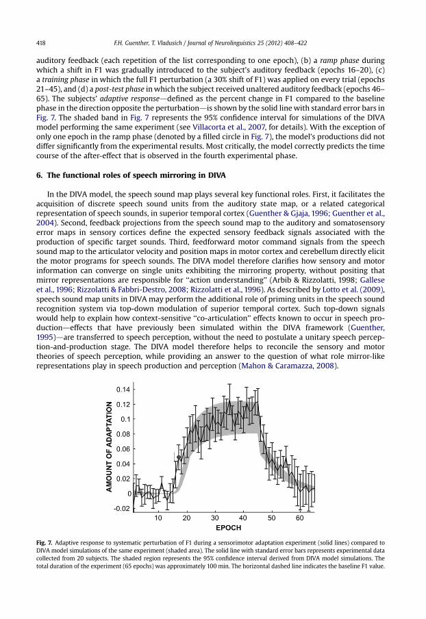
auditory feedback (each repetition of the list corresponding to one epoch), (b) a ramp phase duringwhich a shift in F1 was gradually introduced to the subject’s auditory feedback (epochs 16–20), (c)a training phase in which the full F1 perturbation (a 30% shift of F1) was applied on every trial (epochs21–45), and (d) a post-test phase in which the subject received unaltered auditory feedback (epochs 46–65). The subjects’ adaptive responseddefined as the percent change in F1 compared to the baselinephase in the direction opposite the perturbationdis shown by the solid line with standard error bars inFig. 7. The shaded band in Fig. 7 represents the 95% confidence interval for simulations of the DIVAmodel performing the same experiment (see Villacorta et al., 2007, for details). With the exception ofonly one epoch in the ramp phase (denoted by a filled circle in Fig. 7), the model’s productions did notdiffer significantly from the experimental results. Most critically, the model correctly predicts the timecourse of the after-effect that is observed in the fourth experimental phase.
6. The functional roles of speech mirroring in DIVA
In the DIVA model, the speech sound map plays several key functional roles. First, it facilitates theacquisition of discrete speech sound units from the auditory state map, or a related categoricalrepresentation of speech sounds, in superior temporal cortex (Guenther & Gjaja, 1996; Guenther et al.,2004). Second, feedback projections from the speech sound map to the auditory and somatosensoryerror maps in sensory cortices define the expected sensory feedback signals associated with theproduction of specific target sounds. Third, feedforward motor command signals from the speechsound map to the articulator velocity and position maps in motor cortex and cerebellum directly elicitthe motor programs for speech sounds. The DIVA model therefore clarifies how sensory and motorinformation can converge on single units exhibiting the mirroring property, without positing thatmirror representations are responsible for ‘‘action understanding’’ (Arbib & Rizzolatti, 1998; Galleseet al., 1996; Rizzolatti & Fabbri-Destro, 2008; Rizzolatti et al., 1996). As described by Lotto et al. (2009),speech sound map units in DIVA may perform the additional role of priming units in the speech soundrecognition system via top-down modulation of superior temporal cortex. Such top-down signalswould help to explain how context-sensitive ‘‘co-articulation’’ effects known to occur in speech pro-ductiondeffects that have previously been simulated within the DIVA framework (Guenther,1995)dare transferred to speech perception, without the need to postulate a unitary speech percep-tion-and-production stage. The DIVA model therefore helps to reconcile the sensory and motortheories of speech perception, while providing an answer to the question of what role mirror-likerepresentations play in speech production and perception (Mahon & Caramazza, 2008).
Fig. 7. Adaptive response to systematic perturbation of F1 during a sensorimotor adaptation experiment (solid lines) compared toDIVA model simulations of the same experiment (shaded area). The solid line with standard error bars represents experimental datacollected from 20 subjects. The shaded region represents the 95% confidence interval derived from DIVA model simulations. Thetotal duration of the experiment (65 epochs) was approximately 100 min. The horizontal dashed line indicates the baseline F1 value.
F.H. Guenther, T. Vladusich / Journal of Neurolinguistics 25 (2012) 408–422 419
7. Testing the speech mirroring hypothesis
The DIVA model makes some unique functional predictions that set it apart from other theories ofthe possible role(s) of mirror representations in speech production and perception (Arbib & Rizzolatti,1998; see also Hickok, 2009; Lotto et al., 2009; Wilson et al., 2004) and motor control (Bonaiuto et al.,2007; Gallese et al., 1996; Rizzolatti & Fabbri-Destro, 2008; Rizzolatti et al., 1996). According to DIVA,when an infant, or an adult learning an unfamiliar-sounding language, listens to a speaker producinga new speech sound, previously unused speech sound map neurons become active. Projections fromthese neurons to the auditory cortex rapidly become tuned to the acoustic characteristics of that sound.These projections thus represent a target auditory trace for that sound. Additional projections from thespeech sound map neurons to the primary motor cortex (both directly and via a cerebellar loop)represent (initially poorly tuned) feedforward commands for producing the newly learned sound.These feedforward command pathways become tuned over repeated attempts to produce the sound,with each attempt initiated by activating these same speech sound map neurons. This learning isdriven by the initial mismatch between the newly acquired sound target and the infant’s ownproduction attempt as represented in the auditory state map. These auditory error signals are thentransformed into a corrective motor command, and this corrective command is added to the feed-forward command for the next attempt. As the feedforward commands improve, fewer error signalsare generated and thus the contribution of the feedback control system gradually diminishes. The DIVAmodel thus predicts that mirror neurons emerge as a consequence of imitation learning, rather thandriving the imitation-learning process themselves (e.g., see Iacoboni et al., 1999).
The DIVA model also sheds light on the issue of how perceptual and motor reference frames become‘‘aligned’’ via the development of mirror neurons (Gallese et al., 1996; Iacoboni & Dapretto, 2006;Rizzolatti et al., 1996). As learning progresses in DIVA, speech sound map neurons gradually ‘‘acquire’’the feedforward motor command programs corresponding to the rapidly-acquired auditory targetsounds. The link between perception and action thus arises in the DIVA model because the motorreference frame is brought into register with the auditory reference frame (see Guenther et al., 1998, fora discussion of reference frames in speech production). The model therefore predicts a causal rela-tionship between the speech sounds acquired in auditory coordinates and their associated motorprograms: Individuals with more distinctive auditory speech representationsdthose people betterable to discriminate between similar speech soundsdshould produce more distinctive speech utter-ances than those with poorer auditory discrimination. Data from several studies of speech productionsupport this prediction (Perkell, Guenther, et al., 2004; Perkell, Matthies, et al., 2004; Villacorta et al.,2007). The DIVA model also makes the currently untested prediction that individuals with moredistinctive auditory and motor speech representations will exhibit statistically more separable patternsof fMRI activation in both Broca’s area and higher-order auditory cortex during speech perception andproduction experiments (Kriegeskorte, Goebel, & Bandettini, 2006).
Another prediction of the DIVA model is that projections from the speech sound map to the auditoryand somatosensory errors maps have the effect of inhibiting expected auditory inputs from one’s ownspeech. Evidence of inhibition in auditory areas of the superior temporal gyrus during one’s ownspeech comes from several different sources, including recorded neural responses during open brainsurgery (Creutzfeldt, Ojemann, & Lettich, 1989a, 1989b), magnetoencephalography (MEG) studies(Houde, Nagarajan, Sekihara, & Merzenich, 2002; Numminen & Curio, 1999; Numminen, Salmelin, &Hari, 1999), and positron emission tomography (PET) studies (Wise, Greene, Buchel, & Scott, 1999).Data regarding the prediction of an inhibitory effect of the speech sound map on the supramarginalgyrus during speech production is currently lacking, although this brain region has been implicated inphonological processing for speech perception (e.g., Caplan et al., 1995; Celsis et al., 1999) and speechproduction (Damasio & Damasio, 1980; Geschwind, 1965).
The DIVA model also predicts that the tuning of projections from the speech sound map to theauditory and somatosensory error maps will exhibit different time courses. Due to the necessity torapidly acquire new auditory targets during imitation learning, the projections from the speech soundmap to the auditory error map need to learn quickly and remain stable over long time periods. Thisargument does not, however, apply to projections to the somatosensory error map, which need to learntargets slowly over multiple production attempts. Since somatosensory target information cannot be
seta
Highlight
seta
Highlight
F.H. Guenther, T. Vladusich / Journal of Neurolinguistics 25 (2012) 408–422420
gained entirely from listening or viewing a speaker producing a new sound, somatosensory targetsmust instead be learned by monitoring one’s own correct self-productions after the speaker haslearned adequate feedforward commands for producing the sound.
The final set of predictions we will discuss here concerns the modulation of efferent and afferentpathways centered on the speech sound map. In particular, the DIVA model predicts that pathways fromthe speech sound map to motor cortex are modulated by a GO signal, computed by the SMA and basalganglia, which controls speaking rate (e.g., Guenther,1995). This signal is represented by the arrow fromthe Initiation Map to the Articulator Velocity and Position Maps in Fig. 1. Speaking rate varies propor-tionally with the magnitude of the GO signal (which itself varies between the normalized values of zeroand one): a larger GO signal is associated with a higher speaking rate. When the GO signal is zero, outputsfrom the speech sound map to primary motor cortex are gated off. This gating mechanism plays a crucialrole in preventing the obligatory imitation of perceived speech sounds. As far as we are aware, inves-tigations of the functional properties of mirror neurons have not yet addressed such a gating function.
8. Concluding remarks
This article has described a quantitative neural theory of speech acquisition and production thatprovides a unified account for a wide range of speech acoustic, kinematic, and neuroimaging data. Themodel posits three interacting subsystems for the neural control of speech production: an auditoryfeedback control subsystem, a somatosensory feedback control subsystem, and a feedforward controlsubsystem. The feedforward control subsystem is proposed to involve cortico-cortical projections frompremotor to motor cortex, as well as contributions from the cerebellum. The auditory feedback controlsubsystem involves projections from premotor cortex to higher-order auditory cortex that encodeauditory targets for speech sounds, as well as projections from higher-order auditory cortex to motorcortex that transform auditory errors into corrective motor commands. The somatosensory feedbackcontrol subsystem involves projections from premotor cortex to higher-order somatosensory cortexthat encode somatosensory targets for speech sounds, as well as projections from somatosensory errorneurons to motor cortex that encode corrective motor commands. The speech sound map coordinatesthe activities of these various maps during normal speech acquisition and production, providinga conduit between the perceptual and motor aspects of speech control. We expect that the quantitativenature of the DIVA formulationdas it applies to fMRI studies of the human mirror systemdwill helpfacilitate rapid advances in understanding the speech sound map in Broca’s area and its functionalconnectivity with related brain regions.
Acknowledgements
F.H.G. supported in part by the National Institute on Deafness and other Communication Disorders(R01 DC02852, F. Guenther PI). T.V. supported in part by the National Science Foundation (NSF SBE-0354378).
References
Ackermann, H., Vogel, M., Petersen, D., & Poremba, M. (1992). Speech deficits in ischaemic cerebellar lesions. Journal ofNeurology, 239, 223–227.
Barlow, S. M. (1999). Handbook of clinical speech physiology. San Diego: Singular.Bonaiuto, J., Rosta, E., & Arbib, M. (2007). Extending the mirror neuron system model, I. Audible actions and invisible grasps.
Biological Cybernetics, 96, 9–38.Browman, C. P., & Goldstein, L. (1989). Articulatory gestures as phonological units. Phonology, 6, 201–251.Buchsbaum, B. R., Hickok, G., & Humphries, C. (2001). Role of left posterior superior temporal gyrus in phonological processing
for speech perception and production. Cognitive Science, 25, 663–678.Callan, D. E., Kent, R. D., Guenther, F. H., & Vorperian, H. K. (2000). An auditory-feedback-based neural network model of speech
production that is robust to developmental changes in the size and shape of the articulatory system. Journal of Speech,Language, and Hearing Research, 43, 721–736.
Caplan, D., Gow, D., & Makris, N. (1995). Analysis of lesions by MRI in stroke patients with acoustic-phonetic processing deficits.Neurology, 45, 293–298.
F.H. Guenther, T. Vladusich / Journal of Neurolinguistics 25 (2012) 408–422 421
Celsis, P., Boulanouar, K., Doyon, B., Ranjeva, J. P., Berry, I., Nespoulous, J. L., et al. (1999). Differential fMRI responses in the leftposterior superior temporal gyrus and left supramarginal gyrus to habituation and change detection in syllables and tones.NeuroImage, 9, 135–144.
Creutzfeldt, O., Ojemann, G., & Lettich, E. (1989a). Neuronal-activity in the human lateral temporal-lobe.1. Responses to speech.Experimental Brain Research, 77, 451–475.
Creutzfeldt, O., Ojemann, G., & Lettich, E. (1989b). Neuronal-activity in the human lateral temporal-lobe.2. Responses to thesubjects own voice. Experimental Brain Research, 77, 476–489.
Damasio, H., & Damasio, A. R. (1980). The anatomical basis of conduction aphasia. Brain, 103, 337–350.di Pellegrino, G., Fadiga, L., Fogassi, L., Gallese, V., Rizzolatti., G. (1992). Understanding motor events: a neurophysiological study.
Experimental Brain Research, 91, 176–180.Duffy, J. R. (1995). Motor speech disorders: Substrates, differential diagnosis, and management. St. Louis: Mosby.Ferrari, P. F., Gallese, V., Rizzolatti, G., & Fogassi, L. (2003). Mirror neurons responding to the observation of ingestive and
communicative mouth actions in the monkey ventral premotor cortex. European Journal of Neuroscience, 17, 1703–1714.Fiez, J. A., & Petersen, S. E. (1998). Neuroimaging studies of word reading. Proceedings of the National Academy Sciences USA, 95, 914–921.Gallese, V., Fadiga, L., Fogassi, L., & Rizzolatti, G. (1996). Action recognition in the premotor cortex. Brain, 119, 593–609.Geschwind, N. (1965). Disconnexion syndromes in animals and man, II. Brain, 88, 585–644.Ghosh, S. S. (2004). Understanding cortical and cerebellar contributions to speech production through modeling and functional
imaging. Boston University Ph.D. dissertation. Boston, MA: Boston University.Guenther, F. H. (1992). Neural models of adaptive sensory-motor control for flexible reaching and speaking. Boston University
Ph.D. dissertation.Guenther, F. H. (1994). A neural network model of speech acquisition and motor equivalent speech production. Biological
Cybernetics, 72, 43–53.Guenther, F. H. (1995). Speech sound acquisition, coarticulation, and rate effects in a neural network model of speech
production. Psychological Review, 102, 594–621.Guenther, F. H., Espy-Wilson, C. Y., Boyce, S. E., Matthies, M. L., Zandipour, M., & Perkell, J. S. (1999). Articulatory tradeoffs reduce
acoustic variability during American English/r/production. Journal of the Acoustical Society of America, 105, 2854–2865.Guenther, F. H., Ghosh, S. S., & Tourville, J. A. (2006). Neural modeling and imaging of the cortical interactions underlying
syllable production. Brain and Language, 96, 280–301.Guenther, F. H., & Gjaja, M. N. (1996). The perceptual magnet effect as an emergent property of neural map formation. Journal of
the Acoustical Society of America, 100, 1111–1121.Guenther, F. H., Hampson, M., & Johnson, D. (1998). A theoretical investigation of reference frames for the planning of speech
movements. Psychological Review, 105, 611–633.Guenther, F. H., Nieto-Castanon, A., Ghosh, S. S., & Tourville, J. A. (2004). Representation of sound categories in auditory cortical
maps. Journal of Speech, Language, and Hearing Research, 47, 46–57.Heeger, D. J., Huk, A. C., Geisler, W. S., & Albrecht, D. G. (2000). Spikes versus BOLD: what does neuroimaging tell us about
neuronal activity? Nature Neuroscience, 3, 631–633.Hickok, G. (2009). Eight problems for the mirror neuron theory of action understanding in monkeys and humans. Journal of
Cognitive Neuroscience, 21, 1229–1243.Hickok, G., Buchsbaum, B., Humphries, C., & Muftuler, T. (2003). Auditory-motor interaction revealed by fMRI: speech, music,
and working memory in area Spt. Journal of Cognitive Neuroscience, 15, 673–682.Houde, J. F., & Jordan, M. I. (1998). Sensorimotor adaptation in speech production. Science, 279, 1213–1216.Houde, J. F., Nagarajan, S. S., Sekihara, K., & Merzenich, M. M. (2002). Modulation of the auditory cortex during speech: an MEG
study. Journal of Cognitive Neuroscience, 14, 1125–1138.Iacoboni, M., & Dapretto, M. (2006). The mirror neuron system and the consequences of its dysfunction. Nature Reviews
Neuroscience, 7, 942–951.Iacoboni, M., Woods, R. P., Brass, M., Bekkering, H., Mazziotta, J. C., & Rizzolatti, G. (1999). Cortical mechanisms of human
imitation. Science, 286, 2526–2528.Kent, R. D. (1997). The speech sciences. San Diego: Singular.Kohler, E., Keysers, C., Umilta, M. A. Fogassi, L., Gallese, V., Rizzolatti. G. (2002). Hearing sounds, understanding actions: action
representation in Mirror Neurons. Science 297, 846–848.Krakauer, J., & Ghez, C. (1999). Voluntary movement. In E. R. Kandel, J. H. Schwartz, & T. M. Jessell (Eds.), Principles of neural
science (4th ed.). (pp. 756–781) New York: McGraw Hill.Kriegeskorte, N., Goebel, R., & Bandettini, P. (2006). Information-based functional brain mapping. Proceedings of the National
Academy of Sciences USA, 103, 3863–3868.Levelt, W. J., & Wheeldon, L. (1994). Do speakers have access to a mental syllabary? Cognition, 50, 239–269.Lingnau, A., Gesierich, B., & Caramazza, A. (2009). Asymmetric fMRI adaptation reveals no evidence for mirror neurons in
humans. Proceedings of the National Academy of Sciences USA, 106, 9925–9930.Logothetis, N. K., Pauls, J., Augath, M., Trinath, T., & Oeltermann, A. (2001). Neurophysiological investigation of the basis of the
fMRI signal. Nature, 412, 150–157.Lotto, A. J., Hickok, G. S., & Holt, L. L. (2009). Reflections on mirror neurons and speech perception. Trends in Cognitive Sciences,
13, 110–114.Maeda, S. (1990). Compensatory articulation during speech: evidence from the analysis and synthesis of vocal tract shapes
using an articulatory model. In W. J. Hardcastle, & A. Marchal (Eds.), Speech production and speech modeling (pp. 131–149).Boston: Kluwer Academic Publishers.
Mahon, B. Z., & Caramazza, A. (2008). A critical look at the embodied cognition hypothesis and a new proposal for groundingconceptual content. Journal of Physioliology (Paris), 102, 59–70.
Middleton, F. A., & Strick, P. L. (1997). Cerebellar output channels. International Review of Neurobiology, 41, 61–82.Munhall, K. G. (2001). Functional imaging during speech production. Acta Psychologica, 107, 95–117.Nieto-Castanon, A., Guenther, F. H., Perkell, J. S., & Curtin, H. (2005). A modeling investigation of articulatory variability and
acoustic stability during American English/r/production. Journal of the Acoustical Society of America, 117, 3196–3212.
F.H. Guenther, T. Vladusich / Journal of Neurolinguistics 25 (2012) 408–422422
Numminen, J., & Curio, G. (1999). Differential effects of overt, covert and replayed speech on vowel-evoked responses of thehuman auditory cortex. Neuroscience Letters, 272, 29–32.
Numminen, J., Salmelin, R., & Hari, R. (1999). Subject’s own speech reduces reactivity of the human auditory cortex. Neuro-science Letters, 265, 119–122.
Passingham, R. E. (1993). The frontal lobes and voluntary action. Oxford: Oxford University Press.Perkell, J. S., Guenther, F. H., Lane, H., Matthies, M. L., Stockmann, E., Tiede, M., et al. (2004). Cross-subject correlations between
measures of vowel production and perception. Journal of the Acoustical Society of America, 116, 2338–2344.Perkell, J. S., Matthies, M. L., Tiede, M., Lane, H., Zandipour, M., Marrone, N., et al. (2004). The distinctness of speakers’/s-sh/
contrast is related to their auditory discrimination and use of an articulatory saturation effect. Journal of Speech, Language,and Hearing Research, 47, 1259–1269.
Petrides, M., Cadoret, G., & Mackey, S. (2005). Orofacial somatomotor responses in the macaque monkey homologue of broca’sarea. Nature, 435, 1235–1238.
Rees, G., Friston, K., & Koch, C. (2000). A direct quantitative relationship between the functional properties of human andmacaque V5. Nature Neuroscience, 3(7), 716–723.
Rizzolatti, G., & Arbib, M. A. (1998). Language within our grasp. Trends in Neurosciences, 21, 188–194.Rizzolatti, G., & Fabbri-Destro, M. (2008). The mirror system and its role in social cognition. Current Opinion in Neurobiology, 18,
179–184.Rizzolatti, G., Fadiga, L., Gallese, V., & Fogassi, L. (1996). Premotor cortex and the recognition of motor actions. Cognitive Brain
Research, 3, 131–141.Rizzolatti, G., Fogassi, L., & Gallese, V. (2001). Neurophysiological mechanisms underlying the understanding and imitation of
action. Nature Reviews Neuroscience, 2, 661–670.Schmahmann, J. D., & Pandya, D. N. (1997). The cerebrocerebellar system. International Review of Neurobiology, 41, 31–60.Tourville, J. A., Reilly, K. J., & Guenther, F. H. (2008). Neural mechanisms underlying auditory feedback control of speech.
NeuroImage, 39, 1429–1443.Turkeltaub, P. E., Eden, G. F., Jones, K. M., & Zeffiro, T. A. (2002). Meta-analysis of the functional neuroanatomy of single-word
reading: method and validation. NeuroImage, 16, 765–780.Villacorta, V. M., Perkell, J. S., & Guenther, F. H. (2007). Sensorimotor adaptation to feedback perturbations of vowel acoustics
and its relation to perception. Journal of the Acoustical Society of America, 122, 2306–2319.Wilson, S. M., Saygin, A. P., Sereno, M. I., & Iacoboni, M. (2004). Listening to speech activates motor areas involved in speech
production. Nature Neuroscience, 7, 701–702.Wise, R. J., Greene, J., Buchel, C., & Scott, S. K. (1999). Brain regions involved in articulation. Lancet, 353, 1057–1061.Zemlin, W. R. (1998). Speech and hearing science: Anatomy and physiology (4th ed.). Boston: Allyn and Bacon.