A new approach for the evaluation of the severity of coarctation of the aorta using Doppler velocity index and effective orifice area: In vitro validation and clinical implications Z. Keshavarz-Motamed a , J. Garcia b , N. Maftoon c , E. Bedard b , P. Chetaille d , L. Kadem a,n a Mechanical and Industrial Engineering, Concordia University, Montre´al, Canada b Que´bec Heart and Lung Institute, Laval University, Que´bec, Canada c BioMedical Engineering, McGill University, Montre ´al, Canada d Mother and Child Center, Laval University Medical Center, Que´bec, Canada article info Article history: Accepted 29 January 2012 Keywords: Aortic stenosis Coarctation of the aorta In vitro study Hemodynamics abstract Early detection and accurate estimation of COA severity are the most important predictors of successful long-term outcome. However, current clinical parameters used for the evaluation of the severity of COA have several limitations and are flow dependent. The objectives of this study are to evaluate the limitations of current existing parameters for the evaluation of the severity of coarctation of the aorta (COA) and suggest two new parameters: COA Doppler velocity index and COA effective orifice area. Three different severities of COAs were tested in a mock flow circulation model under various flow conditions and in the presence of normal and stenotic aortic valves. Catheter trans-COA pressure gradients and Doppler echocardiographic trans-COA pressure gradients were evaluated. COA Doppler velocity index was defined as the ratio of pre-COA to post-COA peak velocities measured by Doppler echocardiography. COA Doppler effective orifice area was determined using continuity equation. The results show that peak-to-peak trans-COA pressure gradient significantly increased with flow rate (from 83% to 85%). Peak Doppler pressure gradient also significantly increased with flow rate (80–85%). A stenotic or bicuspid aortic valve increased peak Doppler pressure gradient by 20–50% for a COA severity of 75%. Both COA Doppler velocity index and COA effective orifice area did not demonstrate significant flow dependence or dependence upon aortic valve condition. As a conclusion, COA Doppler velocity index and COA effective orifice area are flow independent and do not depend on aortic valve conditions. They can, then, more accurately predict the severity of COA. & 2012 Elsevier Ltd. All rights reserved. 1. Introduction Coarctation of the aorta is a congenital heart disease char- acterized by narrowing of the isthmus zone, the section of the descending aorta distal to the left subclavian artery. COA is encountered in 0.1% of newborns (De Mey et al., 2001) and is the third most prevailing defect in infants and children (5–8% of all congenital heart disorders) (Rao, 1995). COA often coexists with aortic stenosis (AS) (between 30% and 50%) (Brickner et al., 2000; Braverman et al., 2005). Untreated COA, in adults, can result in serious complications such as left ventricular hypertrophy, rupture of the aorta and premature coronary artery disease. The most important predictor of successful long-term outcome in patients with COA is age at time of initial repair (Cohen et al., 1989). Early detection and accurate estimation of COA severity are then of primary importance. However, arm-to-leg blood pressure difference may not accurately represent COA severity and may significantly change with flow rate (Araoz et al., 2003; Swan et al., 2003). Doppler echocardiography and MRI trans-coarctation pres- sure gradients (TCPGs) are also highly dependent on flow rate and on collateral blood supply (Steffens et al., 1994; Caravalho et al., 1990). Doppler echocardiography diastolic runoff, the magnitude of the antegrade diastolic flow, has also been suggested to evaluate the severity of COA. However, it is highly dependent on aortic com- pliance (DeGroff et al., 2003; Tacy et al., 1999). Invasively, catheter TPCGs are highly influenced by the flow rate and pressure recovery phenomena, and peak-to-peak pressure gradient also depends on compliant properties of the aorta (Kadem et al., 2006). Furthermore, using invasive cardiac catheterization might be problematic if multiple follow-up examinations after surgical repair are required knowing that recoarctation is a common occurrence (up to 40%) Contents lists available at SciVerse ScienceDirect journal homepage: www.elsevier.com/locate/jbiomech www.JBiomech.com Journal of Biomechanics 0021-9290/$ - see front matter & 2012 Elsevier Ltd. All rights reserved. doi:10.1016/j.jbiomech.2012.01.039 n Correspondence to: Laboratory of Cardiovascular Fluid Dynamics, Department of Mechanical and Industrial Engineering, Concordia University, 1455 de Maison- neuve Blvd W, Montreal, QC, Canada H3G 1M8. Tel.: 514 848 2424x3143; fax: 514 848 3175. E-mail address: [email protected] (L. Kadem). Journal of Biomechanics 45 (2012) 1239–1245
Transcript
Journal of Biomechanics 45 (2012) 1239–1245
Contents lists available at SciVerse ScienceDirect
A new approach for the evaluation of the severity of coarctation of the aortausing Doppler velocity index and effective orifice area: In vitro validation andclinical implications
Z. Keshavarz-Motamed a, J. Garcia b, N. Maftoon c, E. Bedard b, P. Chetaille d, L. Kadem a,n
a Mechanical and Industrial Engineering, Concordia University, Montreal, Canadab Quebec Heart and Lung Institute, Laval University, Quebec, Canadac BioMedical Engineering, McGill University, Montreal, Canadad Mother and Child Center, Laval University Medical Center, Quebec, Canada
a r t i c l e i n f o
Article history:
Accepted 29 January 2012Early detection and accurate estimation of COA severity are the most important predictors of successful
long-term outcome. However, current clinical parameters used for the evaluation of the severity of COA
have several limitations and are flow dependent. The objectives of this study are to evaluate the
limitations of current existing parameters for the evaluation of the severity of coarctation of the aorta
(COA) and suggest two new parameters: COA Doppler velocity index and COA effective orifice area.
Three different severities of COAs were tested in a mock flow circulation model under various flow
conditions and in the presence of normal and stenotic aortic valves. Catheter trans-COA pressure
gradients and Doppler echocardiographic trans-COA pressure gradients were evaluated. COA Doppler
velocity index was defined as the ratio of pre-COA to post-COA peak velocities measured by Doppler
echocardiography. COA Doppler effective orifice area was determined using continuity equation. The
results show that peak-to-peak trans-COA pressure gradient significantly increased with flow rate
(from 83% to 85%). Peak Doppler pressure gradient also significantly increased with flow rate (80–85%).
A stenotic or bicuspid aortic valve increased peak Doppler pressure gradient by 20–50% for a COA
severity of 75%. Both COA Doppler velocity index and COA effective orifice area did not demonstrate
significant flow dependence or dependence upon aortic valve condition. As a conclusion, COA Doppler
velocity index and COA effective orifice area are flow independent and do not depend on aortic valve
conditions. They can, then, more accurately predict the severity of COA.
& 2012 Elsevier Ltd. All rights reserved.
1. Introduction
Coarctation of the aorta is a congenital heart disease char-acterized by narrowing of the isthmus zone, the section of thedescending aorta distal to the left subclavian artery. COA isencountered in 0.1% of newborns (De Mey et al., 2001) and isthe third most prevailing defect in infants and children (5–8% ofall congenital heart disorders) (Rao, 1995). COA often coexistswith aortic stenosis (AS) (between 30% and 50%) (Brickner et al.,2000; Braverman et al., 2005). Untreated COA, in adults, can resultin serious complications such as left ventricular hypertrophy,rupture of the aorta and premature coronary artery disease.
ll rights reserved.
Fluid Dynamics, Department
University, 1455 de Maison-
514 848 2424x3143;
em).
The most important predictor of successful long-term outcomein patients with COA is age at time of initial repair (Cohen et al.,1989). Early detection and accurate estimation of COA severity arethen of primary importance. However, arm-to-leg blood pressuredifference may not accurately represent COA severity and maysignificantly change with flow rate (Araoz et al., 2003; Swan et al.,2003). Doppler echocardiography and MRI trans-coarctation pres-sure gradients (TCPGs) are also highly dependent on flow rate andon collateral blood supply (Steffens et al., 1994; Caravalho et al.,1990). Doppler echocardiography diastolic runoff, the magnitude ofthe antegrade diastolic flow, has also been suggested to evaluate theseverity of COA. However, it is highly dependent on aortic com-pliance (DeGroff et al., 2003; Tacy et al., 1999). Invasively, catheterTPCGs are highly influenced by the flow rate and pressure recoveryphenomena, and peak-to-peak pressure gradient also depends oncompliant properties of the aorta (Kadem et al., 2006). Furthermore,using invasive cardiac catheterization might be problematic ifmultiple follow-up examinations after surgical repair are requiredknowing that recoarctation is a common occurrence (up to 40%)
Z. Keshavarz-Motamed et al. / Journal of Biomechanics 45 (2012) 1239–12451240
after COA repair (Araoz et al., 2003; Boxer et al., 1986; Parks et al.,1995).
In summary, the existing parameters to evaluate the severityof COA have significant limitations. It is, then, difficult to accu-rately compare different patients with different COA severities ora same patient between different follow ups. Therefore, there is acrucial need to introduce new parameters capable of accuratelypredicting the severity of COA and clinical outcomes. Our hypoth-esis is that a parameter like COA velocity index defined as theratio between pre-COA velocity and COA jet velocity and defininga COA effective orifice area using continuity equation measuredby Doppler echocardiography can accurately predict the severityof COA. In order to validate our hypothesis, an original in-vitrostudy was performed using a mock flow circulation model withdifferent COA severities, and different aortic valve conditionsunder different total flow rates.
Table 1Distribution of the flow rate directed toward aortic arch arteries and through COA
for different severities of COA used in this study.
Flow through COA (L/min) Flow through aortic archarteries (L/min)
COA 50% 70% of total flow rate 30% of total flow rate
COA 75% 60% of total flow rate 40% of total flow rate
COA 90% 45% of total flow rate 55% of total flow rate
2. Methods
We designed and constructed a mock flow circulation model which consisted
of a fluid reservoir, a gear pump, realistic elastic three-dimensional models of the
aorta with out-of-plane curvature (including: ascending aorta, aortic branches and
descending aorta), an adjustable systemic arterial resistance and compliance
(Fig. 1). We fabricated elastic models of an aorta by using a multi-silicone layer
method from an anatomically shaped mold reconstructed based on a data set
obtained in an adult patient by magnetic resonance imaging. With the use of this
technique, successive layers of silicone were applied on the mold until both radial
dilatation of the proximal aorta and total arterial compliance (determined by the
ratio of pulse arterial pressure over stroke volume) match physiological values.
The elastic model of the aorta used in this study has a radial dilation of the
proximal aorta of 8% (physiological value around 10% (O0Rourke et al., 2008;
Herment et al., 2011)) and a total arterial compliance of 1.75 ml/mmHg (physio-
logical value 1.8470.76 ml/mmHg (Chemla et al., 1998)). The aorta does not have
tapering and its diameter is 2972 mm. In this study, COA was simulated in vitro
using thin rigid circular orifices (with length of 471 mm) correctly representing a
discrete COA which is one of the most common configurations of COA (Stern et al.,
1991). The fluid (a mixture of 60% water and 40% glycerol, dynamic viscosity of
4 cP) is pumped from an open tank (reservoir), crosses the model of the aortic
valve (bioprosthetic valve or silicone models of bicuspid and tricuspid stenoses
(Blais et al., 2006)) and directed towards the arterial module. Under normal
conditions (no COA) a small portion of the total flow rate (15%) is directed towards
aortic arch branches. However, when a COA is present, depending on its severity,
Fig. 1. Schematic diagram of
a larger portion of the total flow rate bypasses the COA (forwarded towards the
aortic branches and potential collaterals) (Markl et al., 2009; Hope et al., 2010).
Including aortic arch branches is essential for the investigation of COA hemody-
namics and represents a significant advantage compared to previous in vitro
setups dedicated to COA (Seifert et al., 1999; De Mey et al., 2001). In this study, the
proportion of the total flow directed towards aortic arch arteries was adjusted
following a mathematical modeling of the flow through COA (Table 1) (Keshavarz-
Motamed et al., 2011). Then, the flow in aortic arch arteries is redirected towards
the main reservoir, while the flow in the descending aorta is directed towards the
model of the arterial system. The compliance and the resistance of the systemic
arterial system can be adjusted to ensure physiological aortic pressure waveforms.
Instantaneous flow rates were measured by two electromagnetic flowmeters
(Carolina Medical Electronics, East Bend, NC, USA, 600 series, accuracy of 1% full
scale) at the level of the ascending aorta and aortic arch arteries.
The pressures in the left ventricle, aorta, upstream from the COA and down-
stream of COA were measured using Millar catheters (Millar Instruments,
Houston, TX, USA, SPC 360S, accuracy 0.5% full scale) located 20 mm upstream
of the valve, 20 mm downstream of the valve, 20 mm upstream of the COA and
20 mm downstream of COA, respectively. Pressure measurements were used to
determine: peak-to-peak, mean and maximal catheter TCPGs.
Doppler echocardiographic measurements were performed using a HP Sonos
5500 ultrasound machine (Philips healthcare, Best, The Netherlands) with a probe of
2.5 MHz. The probe was positioned on the elastic aorta and the ultrasound beam was
oriented towards the COA. Both pre-COA and post-COA instantaneous velocities were
measured. The measurements were performed three times and averaged. Doppler
echocardiographic measurements included mean (TCPGmean) and maximal (TCPGmax)
trans-COA pressure gradients using simplified energy equation (the unsteady flow
component and the energy losses by turbulence and friction are neglected), with
considering pre-COA velocity (TCPG¼ 0:5r½V2�V2
P � � 4½V2�V2
P �) and without con-
sidering pre-COA velocity (TCPG¼ 0:5rV2� 4V2) (De Mey et al., 2001). Where V is
the velocity at COA vena contracta, VP is the velocity proximal to COA, r is the blood
density and the number 4 (mmHg s2/m2) is obtained by replacement of the blood
density and unit conversion from Pa to mmHg. COA Doppler velocity index was
defined as DVIjCOA ¼ Vmaxjpre�COA=Vmaxjpost�COA; i.e., the ratio between upstream COA
peak velocity (measured with pulsed-wave Doppler) and downstream COA peak
the in vitro flow model.
Z. Keshavarz-Motamed et al. / Journal of Biomechanics 45 (2012) 1239–1245 1241
velocity (measured with continuous-wave Doppler) (Fig. 1). EOACOA ¼ SVCOA=VTICOA
¼ SVCOA=R Ts
0 VCOAdt. Where SVCOA , VTICOA , VCOA and TS are stroke volume crossing
the COA, velocity-time integral downstream of COA, instantaneous velocity measured
by echo Doppler and systolic duration, respectively. Sensitivity and specificity
analysis were performed to evaluate the accuracy of current and proposed para-
meters to predict COA severity (Table 2).
2.1. Experimental conditions
First, we validated the model under physiological conditions (total stroke
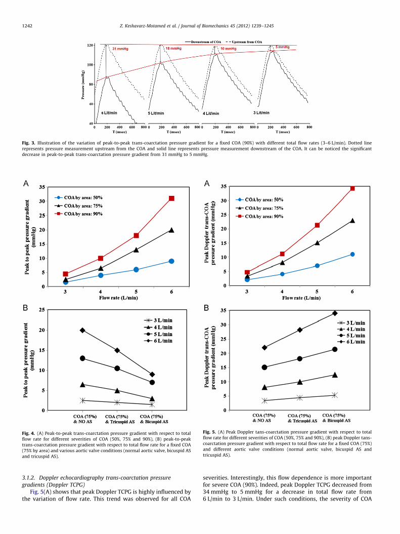
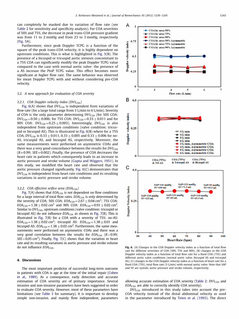
Fig. 3 demonstrates that PtoP TCPG is significantly affected bythe variation of flow rate. Indeed, for a severe COA (90%), PtoPTCPG can almost vanish at low flow rate conditions (PtoP TCPGat 6 L/min: 31 mmHg vs. PtoP TCPG at 3 L/min: 5 mmHg). Undersuch conditions, the severe COA (90%) will almost completely bemasked by a decrease in flow rate. These findings were alsoobserved with COA severities of 50% (decrease from 9 mmHg to1.5 mmHg) and 75% (decrease from 20 mmHg to 2.5 mmHg)(Fig. 4A).
Fig. 4(B) shows how aortic valve condition can affect PtoPTCPG (for simplicity, only a COA with severity of 75% is dis-played). It can be noticed that whatever is aortic valve condition(normal, tricuspid AS or bicuspid AS), PtoP TCPG is significantlyreduced when the total flow rate is decreased from 6 L/min to3 L/min. Furthermore, it appears that at a specific flow rate, aorticvalve condition interacts with the COA and modifies PtoP TCPG:the presence of an AS reduces the PtoP TCPG value. This effect ismore significant at higher flow rate. To these effects, PtoP TCPGcannot provide an accurate evaluation of the COA severity whichcan also be concluded from sensitivity and specificity analysis(Table 2).
Continuous wave Doppler measurement(downstream of COA)
5.19m/s
(without COA and/or AS): (A) left ventricle and ascending aorta (B) upstream and
oppler measurements (upstream from the COA), (D) continuous wave Doppler
Fig. 3. Illustration of the variation of peak-to-peak trans-coarctation pressure gradient for a fixed COA (90%) with different total flow rates (3–6 L/min). Dotted line
represents pressure measurement upstream from the COA and solid line represents pressure measurement downstream of the COA. It can be noticed the significant
decrease in peak-to-peak trans-coarctation pressure gradient from 31 mmHg to 5 mmHg.
Fig. 4. (A) Peak-to-peak trans-coarctation pressure gradient with respect to total
flow rate for different severities of COA (50%, 75% and 90%), (B) peak-to-peak
trans-coarctation pressure gradient with respect to total flow rate for a fixed COA
(75% by area) and various aortic valve conditions (normal aortic valve, bicuspid AS
and tricuspid AS).
Fig. 5. (A) Peak Doppler tans-coarctation pressure gradient with respect to total
flow rate for different severities of COA (50%, 75% and 90%), (B) peak Doppler tans-
coarctation pressure gradient with respect to total flow rate for a fixed COA (75%)
and different aortic valve conditions (normal aortic valve, bicuspid AS and
tricuspid AS).
Z. Keshavarz-Motamed et al. / Journal of Biomechanics 45 (2012) 1239–12451242
Fig. 5(A) shows that peak Doppler TCPG is highly influenced bythe variation of flow rate. This trend was observed for all COA
severities. Interestingly, this flow dependence is more importantfor severe COA (90%). Indeed, peak Doppler TCPG decreased from34 mmHg to 5 mmHg for a decrease in total flow rate from6 L/min to 3 L/min. Under such conditions, the severity of COA
Fig. 6. (A) Changes in the COA Doppler velocity index as a function of total flow
Z. Keshavarz-Motamed et al. / Journal of Biomechanics 45 (2012) 1239–1245 1243
can completely be masked due to variation of flow rate (seeTable 2 for sensitivity and specificity analysis). For COA severitiesof 50% and 75%, the decrease in peak trans-COA pressure gradientwas from 11 to 2 mmHg and from 23 to 3 mmHg, respectively(Fig. 5A).
Furthermore, since peak Doppler TCPG is a function of thesquare of the peak trans-COA velocity, it is highly dependent onupstream conditions. This is what is highlighted in Fig. 5(B). Thepresence of a bicuspid or tricuspid aortic stenosis concomitant toa 75% COA can significantly modify the peak Doppler TCPG valuecompared to the case with normal aortic valve: the presence ofa AS increase the PtoP TCPG value. This effect becomes moresignificant at higher flow rate. The same behavior was observedfor mean Doppler TCPG with and without considering pre-COAvelocity.
3.2. A new approach for evaluation of COA severity
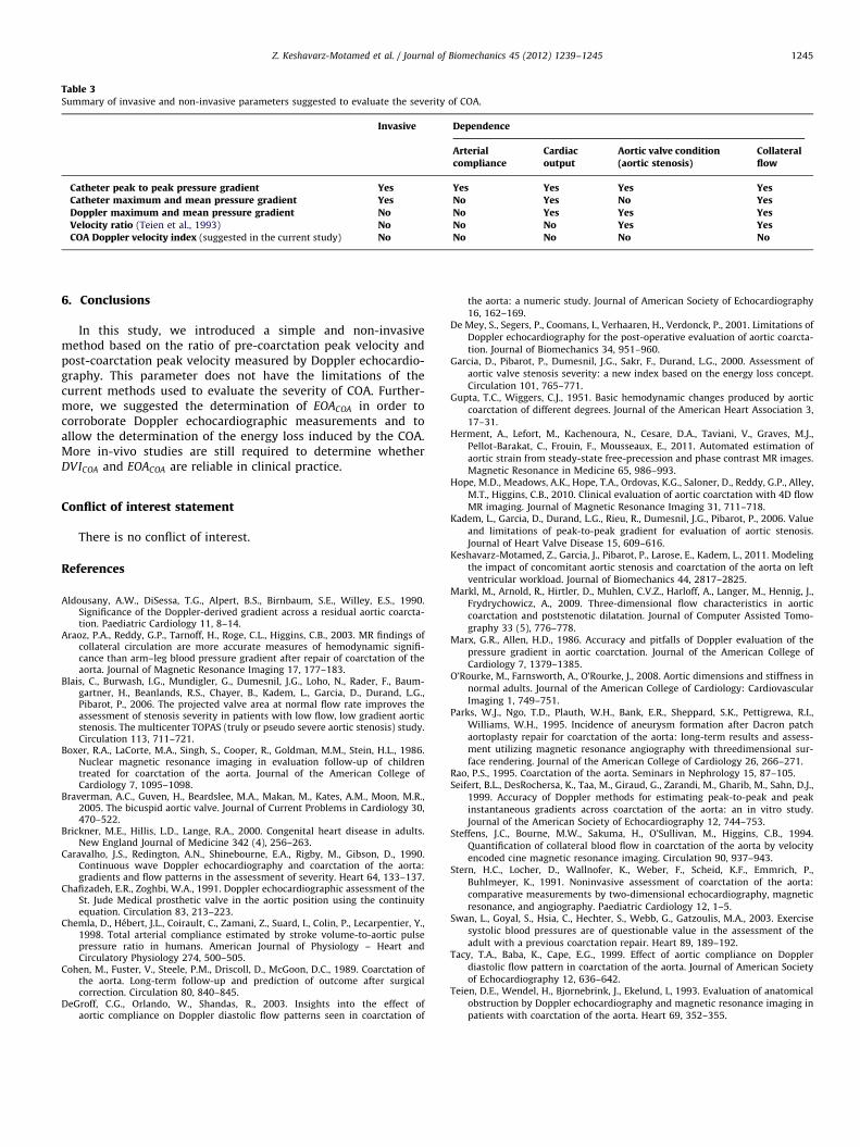
3.2.1. COA Doppler velocity index (DVICOA)
Fig. 6(A) shows that DVICOA is independent from variations offlow rate (for a large total range from 3 L/min to 6 L/min). Severityof COA is the only parameter determining DVICOA (for 50% COA:DVICOA¼0.5070.006; for 75% COA: DVICOA¼0.3370.011 and for90% COA: DVICOA¼0.2570.003). Interestingly, DVICOA is alsoindependent from upstream conditions (valve condition: tricus-pid or bicuspid AS). This is illustrated in Fig. 6(B) where for a 75%COA, DVICOA is 0.3370.011, 0.3370.005 and 0.3370.006 for no-AS, tricuspid AS, and bicuspid AS, respectively. Moreover, thesame measurements were performed on asymmetric COAs andthere was a very good concordance between the results for DVICOA
(R¼0.99; SEE¼0.002). Finally, the presence of COA decreases theheart rate in patients which consequently leads to an increase inaortic pressure and stroke volume (Gupta and Wiggers, 1951). Inthis study, we modified the heart rate and observed that theaortic pressure changed significantly. Fig. 6(C) demonstrates thatDVICOA is independent from heart rate conditions and its resultingvariations in aortic pressure and stroke volume.
3.2.2. COA effective orifice area (EOACOA)
Fig. 7(A) shows that EOACOA is not dependent on flow conditionsfor a large interval of total flow rates. EOACOA is only determined bythe severity of COA; 50% COA: EOACOA¼2.6770.04 cm2; 75% COA:EOACOA¼1.3870.02 cm2 and 90% COA: EOACOA¼0.9170.02 cm2.Similar to DVICOA, upstream conditions (valve condition: tricuspid orbicuspid AS) do not influence EOACOA as shown in Fig. 7(B). This isillustrated in Fig. 7(B) for a COA with a severity of 75%: no-AS:EOACOA¼1.3870.02 cm2; tricuspid AS: EOACOA¼1.3870.01 andbicuspid AS: EOACOA¼1.3870.02 cm2. Furthermore, the same mea-surements were performed on asymmetric COAs and there was avery good correlation between the results for EOACOA (R¼0.99;SEE¼0.05 cm2). Finally, Fig. 7(C) shows that the variation in heartrate and its resulting variations in aortic pressure and stroke volumedo not influence EOACOA.
rate for different severities of COA (50%, 75% and 90%), (B) changes in the COA
Doppler velocity index as a function of total flow rate for a fixed COA (75%) and
different aortic valve conditions (normal aortic valve, bicuspid AS and tricuspid
AS), (C) changes in the COA Doppler velocity index as a function of heart rate for a
fixed COA (75%), total flow rate (5 L/min) with normal aortic valve. Note that SAP
and SV are systolic aortic pressure and stroke volume, respectively.
4. Discussions
The most important predictor of successful long-term outcomein patients with COA is age at the time of the initial repair (Cohenet al., 1989). As a consequence, early detection and accurateestimation of COA severity are of primary importance. Severalinvasive and non-invasive parameters have been suggested in orderto evaluate COA severity. However, most of these parameters havelimitations (see Table 3 for summary). It is important to developsimple non-invasive, and mainly flow independent, parameters
allowing accurate estimation of COA severity (Table 2: DVICOA andEOACOA are able to correctly identify COA severity).
DVICOA introduced in this study takes into account the pre-COA velocity instead of the distal abdominal velocity as usedin the parameter introduced by Teien et al. (1993). The direct
Fig. 7. (A) Changes in COA effective orifice area as a function of total flow rate for
different severities of COA (50%, 75% and 90%), (B) changes in COA effective orifice
area as a function of total flow rate for a fixed COA (75%) and various aortic valve
conditions (normal aortic valve, bicuspid AS and tricuspid AS), (C) changes in the
COA effective orifice area as a function of heart rate for a fixed COA (75%), total
flow rate (5 L/min) with normal aortic valve. Note that SAP and SV are systolic
aortic pressure and stroke volume, respectively.
Z. Keshavarz-Motamed et al. / Journal of Biomechanics 45 (2012) 1239–12451244
consequence of this choice is that DVICOA is independent from thedevelopment of collateral flow, a common occurrence in patientswith COA. It is also important to note that: (1) DVICOA is analogousin its definition to the velocity ratio (peak LVOT velocity/peak
aortic velocity; LVOT: left ventricle outflow tract) introduced byChafizadeh and Zoghbi (1991) in order to evaluate AS severity andprosthetic heart valves; (2) DVICOA correlates very well (in thisstudy: R¼0.98) with Euler number (ratio of the pressure lossinduces by the COA and the inertial force upstream from the COA)used by De Mey et al. (2001) to investigate the limitationsof Doppler echocardiography in the evaluation of COA severity.The major advantage of DVICOA is that it does not rely on thedetermination of the aortic area upstream from COA, sincemeasuring this area using Doppler echocardiography might bedifficult in vivo.
The results of this study are based on in vitro experiments; thishas the advantage of allowing a closer control of the differentparameters involved in the determination of COA severity. To beapplicable in vivo both pre-COA and post-COA velocities have tobe measured using Doppler echocardiography. Measuring post-COA velocity using continuous wave Doppler is now a clinicalroutine. Pre-COA velocity is less commonly measured in patientswith COA, except when correcting Doppler trans-COA pressuregradients using pre-COA velocity. For this purpose, two differentapproaches can be considered: (1) using continuous wave Dopplermeasurements: by optimizing the gain and the gray scale, it ispossible to obtain a Doppler signal including both pre-COA andpost-COA velocities (double envelope) (Marx and Allen, 1986;Aldousany et al., 1990); (2) using pulsed wave Doppler measure-ments upstream from the COA (Marx and Allen, 1986; Aldousanyet al., 1990).
In order to evaluate the performance of DVICOA in vivo, weused the data published in two previous studies: (1) Marx andAllen (1986): prospective study of 32 patients (pre-COA velocitywas not measured in 6 patients), catheter trans-COA pressuregradient included only peak-to-peak pressure gradient;(2) Aldousany et al. (1990): retrospective study of 11 patients,catheter trans-COA gradient included peak-to-peak pressuregradient, maximal instantaneous pressure gradient (not in2 patients) and mean gradient (not in 1 patient). There was agood correlation between DVICOA and peak-to-peak transvalvularpressure gradient: Aldousany et al. (R¼�0.78); Marx and Allen(R¼�0.79); both studies: (R¼�0.78). There was a moderatecorrelation between DVICOA and catheter mean pressure gradient(R¼�0.62). This moderate correlation can be explained by thefact that DVICOA is an instantaneous parameter, while meancatheter pressure gradient is a time-averaged parameter. Thisargument is further reinforced by considering the very goodcorrelation between DVICOA and maximal catheter instantaneouspressure gradient (R¼�0.89).
Although DVICOA and EOACOA behave in the same manner todetermine the severity of COA, DVICOA does not inform clinicianson the energy loss induced by the presence of the COA. This canbe done using EOACOA and aortic post-COA area. These twoparameters can be used to determine an energy loss coefficient(Garcia et al., 2000).
5. Limitations of the study
The model does not consider collateral flows or aortic valveregurgitation. This however should not modify the findings sinceboth DVICOA and EOACOA have been showed in this study to beflow independent. It also should be mentioned that the determi-nation of EOACOA, in vitro, using Doppler echocardiography wasfeasible because the aortic area in the model was known. It mightnot be the case, in vivo, since measuring aortic area using Dopplerechocardiography upstream of the COA is challenging. Moreaccurate results for EOACOA should be obtained using magneticresonance imaging.
Table 3Summary of invasive and non-invasive parameters suggested to evaluate the severity of COA.
Invasive Dependence
Arterialcompliance
Cardiacoutput
Aortic valve condition(aortic stenosis)
Collateralflow
Catheter peak to peak pressure gradient Yes Yes Yes Yes YesCatheter maximum and mean pressure gradient Yes No Yes No YesDoppler maximum and mean pressure gradient No No Yes Yes YesVelocity ratio (Teien et al., 1993) No No No Yes YesCOA Doppler velocity index (suggested in the current study) No No No No No
Z. Keshavarz-Motamed et al. / Journal of Biomechanics 45 (2012) 1239–1245 1245
6. Conclusions
In this study, we introduced a simple and non-invasivemethod based on the ratio of pre-coarctation peak velocity andpost-coarctation peak velocity measured by Doppler echocardio-graphy. This parameter does not have the limitations of thecurrent methods used to evaluate the severity of COA. Further-more, we suggested the determination of EOACOA in order tocorroborate Doppler echocardiographic measurements and toallow the determination of the energy loss induced by the COA.More in-vivo studies are still required to determine whetherDVICOA and EOACOA are reliable in clinical practice.
Conflict of interest statement
There is no conflict of interest.
References
Aldousany, A.W., DiSessa, T.G., Alpert, B.S., Birnbaum, S.E., Willey, E.S., 1990.Significance of the Doppler-derived gradient across a residual aortic coarcta-tion. Paediatric Cardiology 11, 8–14.
Araoz, P.A., Reddy, G.P., Tarnoff, H., Roge, C.L., Higgins, C.B., 2003. MR findings ofcollateral circulation are more accurate measures of hemodynamic signifi-cance than arm–leg blood pressure gradient after repair of coarctation of theaorta. Journal of Magnetic Resonance Imaging 17, 177–183.
Blais, C., Burwash, I.G., Mundigler, G., Dumesnil, J.G., Loho, N., Rader, F., Baum-gartner, H., Beanlands, R.S., Chayer, B., Kadem, L., Garcia, D., Durand, L.G.,Pibarot, P., 2006. The projected valve area at normal flow rate improves theassessment of stenosis severity in patients with low flow, low gradient aorticstenosis. The multicenter TOPAS (truly or pseudo severe aortic stenosis) study.Circulation 113, 711–721.
Boxer, R.A., LaCorte, M.A., Singh, S., Cooper, R., Goldman, M.M., Stein, H.L., 1986.Nuclear magnetic resonance imaging in evaluation follow-up of childrentreated for coarctation of the aorta. Journal of the American College ofCardiology 7, 1095–1098.
Braverman, A.C., Guven, H., Beardslee, M.A., Makan, M., Kates, A.M., Moon, M.R.,2005. The bicuspid aortic valve. Journal of Current Problems in Cardiology 30,470–522.
Brickner, M.E., Hillis, L.D., Lange, R.A., 2000. Congenital heart disease in adults.New England Journal of Medicine 342 (4), 256–263.
Caravalho, J.S., Redington, A.N., Shinebourne, E.A., Rigby, M., Gibson, D., 1990.Continuous wave Doppler echocardiography and coarctation of the aorta:gradients and flow patterns in the assessment of severity. Heart 64, 133–137.
Chafizadeh, E.R., Zoghbi, W.A., 1991. Doppler echocardiographic assessment of theSt. Jude Medical prosthetic valve in the aortic position using the continuityequation. Circulation 83, 213–223.
Chemla, D., Hebert, J.L., Coirault, C., Zamani, Z., Suard, I., Colin, P., Lecarpentier, Y.,1998. Total arterial compliance estimated by stroke volume-to-aortic pulsepressure ratio in humans. American Journal of Physiology – Heart andCirculatory Physiology 274, 500–505.
Cohen, M., Fuster, V., Steele, P.M., Driscoll, D., McGoon, D.C., 1989. Coarctation ofthe aorta. Long-term follow-up and prediction of outcome after surgicalcorrection. Circulation 80, 840–845.
DeGroff, C.G., Orlando, W., Shandas, R., 2003. Insights into the effect ofaortic compliance on Doppler diastolic flow patterns seen in coarctation of
the aorta: a numeric study. Journal of American Society of Echocardiography16, 162–169.
De Mey, S., Segers, P., Coomans, I., Verhaaren, H., Verdonck, P., 2001. Limitations ofDoppler echocardiography for the post-operative evaluation of aortic coarcta-
tion. Journal of Biomechanics 34, 951–960.Garcia, D., Pibarot, P., Dumesnil, J.G., Sakr, F., Durand, L.G., 2000. Assessment of
aortic valve stenosis severity: a new index based on the energy loss concept.
Circulation 101, 765–771.Gupta, T.C., Wiggers, C.J., 1951. Basic hemodynamic changes produced by aortic
coarctation of different degrees. Journal of the American Heart Association 3,17–31.
Herment, A., Lefort, M., Kachenoura, N., Cesare, D.A., Taviani, V., Graves, M.J.,
Pellot-Barakat, C., Frouin, F., Mousseaux, E., 2011. Automated estimation ofaortic strain from steady-state free-precession and phase contrast MR images.Magnetic Resonance in Medicine 65, 986–993.
M.T., Higgins, C.B., 2010. Clinical evaluation of aortic coarctation with 4D flowMR imaging. Journal of Magnetic Resonance Imaging 31, 711–718.
Kadem, L., Garcia, D., Durand, L.G., Rieu, R., Dumesnil, J.G., Pibarot, P., 2006. Value
and limitations of peak-to-peak gradient for evaluation of aortic stenosis.Journal of Heart Valve Disease 15, 609–616.
Keshavarz-Motamed, Z., Garcia, J., Pibarot, P., Larose, E., Kadem, L., 2011. Modelingthe impact of concomitant aortic stenosis and coarctation of the aorta on left
ventricular workload. Journal of Biomechanics 44, 2817–2825.Markl, M., Arnold, R., Hirtler, D., Muhlen, C.V.Z., Harloff, A., Langer, M., Hennig, J.,
Frydrychowicz, A., 2009. Three-dimensional flow characteristics in aorticcoarctation and poststenotic dilatation. Journal of Computer Assisted Tomo-
graphy 33 (5), 776–778.Marx, G.R., Allen, H.D., 1986. Accuracy and pitfalls of Doppler evaluation of the
pressure gradient in aortic coarctation. Journal of the American College ofCardiology 7, 1379–1385.
O’Rourke, M., Farnsworth, A., O’Rourke, J., 2008. Aortic dimensions and stiffness in
normal adults. Journal of the American College of Cardiology: CardiovascularImaging 1, 749–751.
Williams, W.H., 1995. Incidence of aneurysm formation after Dacron patchaortoplasty repair for coarctation of the aorta: long-term results and assess-ment utilizing magnetic resonance angiography with threedimensional sur-
face rendering. Journal of the American College of Cardiology 26, 266–271.Rao, P.S., 1995. Coarctation of the aorta. Seminars in Nephrology 15, 87–105.Seifert, B.L., DesRochersa, K., Taa, M., Giraud, G., Zarandi, M., Gharib, M., Sahn, D.J.,
1999. Accuracy of Doppler methods for estimating peak-to-peak and peakinstantaneous gradients across coarctation of the aorta: an in vitro study.
Journal of the American Society of Echocardiography 12, 744–753.Steffens, J.C., Bourne, M.W., Sakuma, H., O’Sullivan, M., Higgins, C.B., 1994.
Quantification of collateral blood flow in coarctation of the aorta by velocity
encoded cine magnetic resonance imaging. Circulation 90, 937–943.Stern, H.C., Locher, D., Wallnofer, K., Weber, F., Scheid, K.F., Emmrich, P.,
Buhlmeyer, K., 1991. Noninvasive assessment of coarctation of the aorta:comparative measurements by two-dimensional echocardiography, magnetic