82 IJCID A New Innovative Approach for Treatment of Gingival Recession: A Clinical Case 1 Himanshu Deswal, 2 Amit Bhardwaj, 3 Harpreet Singh Grover, 4 Yogender Singh Deswal CASE REPORT 1,4 Postgraduate Student, 2 Reader, 3 Professor 1-4 Department of Periodontology, SGT Dental College, Gurgaon Haryana, India Corresponding Author: Amit Bhardwaj, Reader, Department of Periodontology, SGT Dental College, Gurgaon, Haryana, India Phone: 9818718872, e-mail: [email protected]10.5005/jp-journals-10004-1039 ABSTRACT Gingival recession is characterized by the apical migration of tissue margin apical to cement enamel junction. These defects give rise to both functional and esthetic challenges. A new innovative technique of periodontal plastic surgery was used in which periosteum was incised and mobilized at the apical part of the mucoperiosteum flap to cover the defect. The purpose of this case report was to clinically evaluate the use of periosteum eversion technique to cover gingival recession defect. Keywords: Eversion technique, Gingival recession, Periodontal flap. How to cite this article: Deswal H, Bhardwaj A, Grover HS, Deswal YS. A New Innovative Approach for Treatment of Gingival Recession: A Clinical Case. Int J Clin Implant Dent 2015;1(2):82-84. Source of support: Nil Conflict of interest: None INTRODUCTION Gingival recession is the apical migration of the gin- gival margin to the cementoenamel junction, either coagnated with inflammatory periodontal disease 1 and mechanical trauma 2 or with the presence of a host of predisposing factors that include tooth malposition and root prominence, gingival phenotype, 3 underlying alveolar dehiscence, 4,5 orthodontic tooth movement, 4,6 aberrant frenulum attachment 7 and iatrogenic restorative and periodontal treatment-related factors (surgical recession). 8 For the correction and management of mucogingival deformities and defects, variety of perio- dontal plastic surgery procedures have been described in the literature, with the aim to set up and preserve the dentition and periodontium, healthy and functional with most favorable esthetics throughout the life time. Since, the addition of finest esthetics to the goal of periodontal therapy parallels a great revolutionationary step in speciality of periodontology. This is where the periodontal plastic therapy as an active contributor in the field of esthetic dentistry. Root coverage techniques can be broadly divided into transposition of free tissue graft or transposition of periodontal flaps or application of membrane, although combination procedures, with or without adjunctive regenerative and root biomodification procedures, using citric acid, tetracycline, and ethylenediaminetetraacetic acid. 9 Therefore, the best clinical results for covering extended denuded root surfaces are achieved by free soft tissue graft surgical techniques but this leads to donor site morbidity. 10 To exploit the advantages of a connective tissue graft on the one hand and to avoid donor site-related problems on the other, a new surgical technique for root coverage has been developed over the last few years. The bases of this technique transposition of the local periosteum combined with a mucoperiosteum transpositional flap. Herein, the new periosteum eversion technique used in this study is described in this case report. CASE REPORT A 38 years old male patient reported to the Department of Periodontology, Faculty of Dental Sciences, Sri Guru Gobind Tricentenary University, with the chief complaint of unesthetic appearance in the upper front tooth region. A 3 mm class I gingival recession defect (Miller, 1985) was diagnosed on examination in relation to maxillary left premolars (i.e. 24,25) (Fig. 1). After the approval by the ethical committee for the surgical procedure, informed Fig. 1: Preoperative photograph
Transcript
Himanshu Deswal et al
82
ijcid
A New Innovative Approach for Treatment of Gingival Recession: A Clinical Case1Himanshu Deswal, 2Amit Bhardwaj, 3Harpreet Singh Grover, 4Yogender Singh Deswal
case report
1,4Postgraduate Student, 2Reader, 3Professor1-4Department of Periodontology, Sgt Dental College, gurgaon Haryana, India
Corresponding Author: Amit Bhardwaj, Reader, Department of Periodontology, Sgt Dental College, gurgaon, Haryana, India Phone: 9818718872, e-mail: [email protected]
10.5005/jp-journals-10004-1039
ABSTRACTgingival recession is characterized by the apical migration of tissue margin apical to cement enamel junction. these defects give rise to both functional and esthetic challenges. A new innovative technique of periodontal plastic surgery was used in which periosteum was incised and mobilized at the apical part of the mucoperiosteum flap to cover the defect. The purpose of this case report was to clinically evaluate the use of periosteum eversion technique to cover gingival recession defect.
How to cite this article: Deswal H, Bhardwaj A, grover HS, Deswal YS. A New Innovative Approach for treatment of gingival Recession: A Clinical Case. Int J Clin Implant Dent 2015;1(2):82-84.
Source of support: Nil
Conflict of interest: None
INTRoDuCTIoN
Gingival recession is the apical migration of the gingival margin to the cementoenamel junction, either coagnated with inflammatory periodontal disease1
and mechanical trauma2 or with the presence of a host of predisposing factors that include tooth malposition and root prominence, gingival phenotype,3 underlying alveolar dehiscence,4,5 orthodontic tooth movement,4,6
aberrant frenulum attachment7 and iatrogenic restorative and periodontal treatmentrelated factors (surgical recession).8 For the correction and management of muco gingival deformities and defects, variety of periodontal plastic surgery procedures have been described in the literature, with the aim to set up and preserve the dentition and periodontium, healthy and functional with most favorable esthetics throughout the life time. Since, the addition of finest esthetics to the goal of periodontal therapy parallels a great revolutionationary step in speciality of periodontology. This is where the
periodontal plastic therapy as an active contributor in the field of esthetic dentistry. Root coverage techniques can be broadly divided into transposition of free tissue graft or transposition of periodontal flaps or application of membrane, although combination procedures, with or without adjunctive regenerative and root biomodification procedures, using citric acid, tetracycline, and ethylenediaminetetraacetic acid.9
Therefore, the best clinical results for covering extended denuded root surfaces are achieved by free soft tissue graft surgical techniques but this leads to donor site morbidity.10 To exploit the advantages of a connective tissue graft on the one hand and to avoid donor siterelated problems on the other, a new surgical technique for root coverage has been developed over the last few years. The bases of this technique transposition of the local periosteum combined with a mucoperiosteum transpositional flap. Herein, the new periosteum eversion technique used in this study is described in this case report.
CASe RepoRT
A 38 years old male patient reported to the Department of Periodontology, Faculty of Dental Sciences, Sri Guru Gobind Tricentenary University, with the chief complaint of unesthetic appearance in the upper front tooth region. A 3 mm class I gingival recession defect (Miller, 1985) was diagnosed on examination in relation to maxillary left premolars (i.e. 24,25) (Fig. 1). After the approval by the ethical committee for the surgical procedure, informed
Fig. 1: Preoperative photograph
A New Innovative Approach for Treatment of Gingival Recession: A Clinical Case
International Journal of Clinical Implant Dentistry, May-August 2015;1(2):82-84 83
ijcid
consent was obtained from the patient after thorough explanation of the risks and benefits of the clinical procedure planned. Patient was a nonsmoker, systemically healthy and had no contraindications for periodontal surgery. For the root coverage, periodontal plastic surgery was planned with transposition of periosteum in combination with a mucoperiosteum transpositional flap.
Surgical Technique
Following careful debridement of exposed root surfaces, The surgical area was anesthetized with 2% lignocaine hydrochloride containing adrenaline at a concentration of 1:80,000. An intrasulcular incision was made by using Bard parker number 15 blade (BP Blade) at the buccal aspect of the involved tooth. Two oblique vertical incisions were extended beyond the mucogingival junction to relieve muscle tension and a trapezoidal full thickness flap was raised and extended apically beyond the mucogingival junction, releasing the tension (Fig. 2).
Root surface was mechanically cleaned by scaling in combination with the conditioning of the root surface. Then prepare the everted periosteum flap. With the baseline incision (Fig. 3), the periosteum was separated from the underlying submucous connective tissue up to attached gingival border. The crestally pedicled periosteum was everted and transpositioned coronally where it was fixed with interdental sutures (Fig. 4). The mucoperiosteum was coronally transpositioned and then mucoperiosteum flap was also sutured interdentally (Fig. 5).
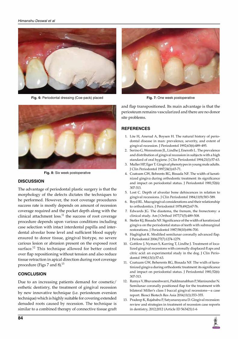
Periodontal pack (Coe pack) was placed (Fig. 6). Patient was instructed to refrain from mechanical plaque control measures particularly in the surgical area for 4 weeks.
However, active chemical control was maintained by 0.12% chlorhexidine mouthwash in dosage of 15 ml for 30 seconds to 1 minute twice a day. Furthermore, the patient was instructed to take tablet ibuprofen 400 mg twice a day for 3 days. Clinical followup was performed after 1 week and 6 weeks postoperatively (Figs 7 and 8).
Fig. 2: Full thickness flap elevated Fig. 3: Baseline incision
Himanshu Deswal et al
84
DISCuSSIoN
The advantage of periodontal plastic surgery is that the morphology of the defects dictates the techniques to be performed. However, the root coverage procedures success rate is mostly depends on amount of recession coverage required and the pocket depth along with the clinical attachment loss.11 the success of root coverage procedure depends upon various conditions including case selection with intact interdental papilla and interdental alveolar bone level and sufficient blood supply ensured to donor tissue, gingival biotype, no severe carious lesion or abrasion present on the exposed root surface.12 This technique allowed for better control over flap repositioning without tension and also reduce tissue retraction in apical direction during root coverage procedure (Figs 7 and 8).13
CoNCluSIoN
Due to an increasing patients demand for cosmetic/esthetic dentistry, the treatment of gingival recession by new innovative technique (i.e. periosteum eversion technique) which is highly suitable for covering extended denuded roots caused by recession. The technique is similar to a combined therapy of connective tissue graft
and flap transpositioned. Its main advantage is that the periosteum remains vascularized and there are no donor site problems.
RefeReNCeS
1. Löe H, Anerud A, Boysen H. The natural history of periodontal disease in man: prevalence, severity, and extent of gingival recession. J Periodontol 1992;63(6):489495.
2. Serino G, Wennstrom JL, Lindhe J, Eneroth L. The prevalence and distribution of gingival recession in subjects with a high standard of oral hygiene. J Clin Periodontol 1994;21(1):5763.
3. Muller HP, Eger T. Gingival phenotypes in young male adults. J Clin Periodontol 1997;24(1):6571.
4. Coatoam GW, Behrents RG, Bissada NF. The width of keratinized gingiva during orthodontic treatment: its significance and impact on periodontal status. J Periodontol 1981;52(6): 307313.
5. Lost C. Depth of alveolar bone dehiscences in relation to gin gival recessions. J Clin Periodontol 1984;11(9):583589.
6. Boyd RL. Mucogingival considerations and their relationship to orthodontics. J Periodontol 1978;49(2):6776.
7. Edwards JG. The diastema, the frenum, the frenectomy: a clinical study. Am J Orthod 1977;71(5):489508.
8. Stetler KJ, Bissada NF. Significance of the width of keratinized gingiva on the periodontal status of teeth with submarginal restorations. J Periodontol 1987;58(10):696700.
10. Gottlow J, Nyman S, Karring T, Lindhe J. Treatment of localized gingival recessions with coronally displaced fl aps and citric acid: an experimental study in the dog. J Clin Periodontol 1990;13(1):5763.
11. Coatuam GW, Beherents RG, Bissada NF. The widh of keratinized gingiva during orthodontic treatment: its significance and impact on periodontal status. J Periodontal 1981;52(6): 307313.
12. Ramya V, Bhuvaneshwarri, Paddmanabhan P, Manisundar N. Semilunar coronally positioned flap for the treatment with bilateral Miller’s class I buccal gingival recessions—a case report. Biosci Biotech Res Asia 2014;11(1):353355.
13. Pradeep K, Rajababu P, Satyanarayana D. Gingival recession: review and strategies in treatment of recession case reports in dentistry, 2012;2012 (Article ID 563421):16