A new integrated behavioural interventionfor knee osteoarthritis: development andpilot studyStephen J. Preece1*, Nathan Brookes1,2, Anita E. Williams1, Richard K. Jones1, Chelsea Starbuck1,Anthony Jones3 and Nicola E. Walsh4
Abstract
Background: Exercise-based approaches have been a cornerstone of physiotherapy management of kneeosteoarthritis for many years. However, clinical effects are considered small to modest and the need for continuedadherence identified as a barrier to clinical efficacy. While exercise-based approaches focus on musclestrengthening, biomechanical research has identified that people with knee osteoarthritis over activate theirmuscles during functional tasks. Therefore, we aimed to create a new behavioural intervention, which integratedpsychologically informed practice with biofeedback training to reduce muscle overactivity, and which was suitablefor delivery by a physiotherapist.
Methods: Through literature review, we created a framework linking theory from pain science with emergingbiomechanical concepts related to overactivity of the knee muscles. Using recognised behaviour change theory, wethen mapped a set of intervention components which were iteratively developed through ongoing testing andconsultation with patients and physiotherapists.
Results: The underlying framework incorporated ideas related to central sensitisation, motor responses to painand also focused on the idea that increased knee muscle overactivity could result from posturalcompensation. Building on these ideas, we created an intervention with five components: making sense ofpain, general relaxation, postural deconstruction, responding differently to pain and functional muscleretraining. The intervention incorporated a range of animated instructional videos to communicate conceptsrelated to pain and biomechanical theory and also used EMG biofeedback to facilitate visualization of musclepatterns. User feedback was positive with patients describing the intervention as enabling them to “create anew normal” and to be “in control of their own treatment.” Furthermore, large reductions in pain wereobserved from 11 patients who received a prototype version of the intervention.
* Correspondence: [email protected] for Health Sciences Research, University of Salford, Manchester M66PU, UKFull list of author information is available at the end of the article
Preece et al. BMC Musculoskeletal Disorders (2021) 22:526 https://doi.org/10.1186/s12891-021-04389-0
Conclusion: We have created a new intervention for knee osteoarthritis, designed to empower individualswith capability and motivation to change muscle activation patterns and beliefs associated with pain. Werefer to this intervention as Cognitive Muscular Therapy. Preliminary feedback and clinical indications arepositive, motivating future large-scale trials to understand potential efficacy. It is possible that this newapproach could bring about improvements in the pain associated with knee osteoarthritis without the needfor continued adherence to muscle strengthening programmes.
BackgroundKnee osteoarthritis (OA) is a chronic long-term condi-tion that results in pain, disability and reduced quality oflife [1]. This condition affects a large proportion of indi-viduals, with a global age-standardised prevalence forknee OA estimated to be 3.8% [2]. Indeed, it has beenestimated that 10% of the population over the age of 55will be diagnosed with knee OA [3]. For many patients,conservative treatments do not provide sufficient long-term relief and they choose to undergo total kneereplacement. However, as populations age and rates ofobesity (a known risk factor [4]) rise, the increasing needfor surgical management is putting healthcare systemsunder considerable strain. Given this huge societal cost,along with the individual burden associated with the dis-ease, there is an urgent need to explore new conservativemethods to manage knee OA.The universally recommended first line of clinical
management for knee OA is a physiotherapist-deliveredexercise programme. These programmes typically con-sist of muscle strengthening, advice to remain active [5]along with coping skills [6] and education about self-management. While this approach is supported by large-scale trials [7] and incorporated into national guidelines[8], the magnitude of clinical effect is considered moder-ate to small [9] and is known to diminish over time [10].Exercise programmes which consist of two strengtheningsessions per week [11], the minimum recommended bythe ACSM [12], typically provide a 25–30% reduction inpain and/or function [7]. Furthermore, research hasdemonstrated that for approximately 40% of patients,exercise-based approaches do not provide any meaning-ful clinical [13] improvement in symptoms [7]. Whileadherence has been identified as an issue which maylower the true effectiveness of exercise-based approaches[14], it is unlikely to explain why, for a relatively largenumber of people, exercise provides no relief from kneeOA pain.While current guidelines focus on the use of exercises
to improve strength, there is clear evidence that peoplewith knee OA over activate their muscles during
functional tasks [15–17]. This overactivity is charac-terised by both increased amplitude [18] and prolongedduration [16] of the knee flexor and extensor muscles.Biomechanical studies have investigated the potential ef-fects of muscle overactivity, typically quantifying thisphenomenon using a co-contraction index [19]. In-creased co-contraction has been linked to increased pain[20], elevated joint load [21], a more rapid rate of cartil-age loss [22] and an increase in the likelihood that pa-tients will opt for a knee replacement at 5-year follow up[23]. Given these findings, muscle overactivity is likely toincrease the mechanical stress on the articular surface,the bone, joint capsule and periarticular structures andtherefore may increase nociceptive input, exacerbatingpain [24]. It is therefore important to understand the po-tential of conservative management techniques whichfocus on reducing muscle overactivity.Psychosocial factors have been linked with clinical
pain/disability in knee OA. For example, catastrophising[25] and anxiety [26] have been associated with pain in-tensity and kinesiophobia linked to physical function[27]. Given these links, a number of physiotherapy inter-ventions have been developed which integrate psycho-logical techniques [28, 29] with muscle retraining. Thisapproach is in line with the use of a holistic approachaddressing both biomedical and psychosocial factors forthe management of chronic low back pain [30, 31]. Forexample, integrated interventions for knee OA have in-corporated a behavioural graded activity programme[32] or have included self-management components toprovide reassurance about the value of exercise in OA[11, 33]. However, these interventions have focused pri-marily on muscle strength training. Therefore, it is un-clear whether improved clinical outcomes would beobtained if psychological techniques were integratedwith training to reduce muscle overactivity.This paper describes the development of a new behav-
ioural intervention for knee OA. This intervention inte-grates psychosocial concepts with emerging biomechanicaltheory relating to potential drivers of muscle overactivity.The overall aim was to create an intervention that was
Preece et al. BMC Musculoskeletal Disorders (2021) 22:526 Page 2 of 14
appropriate for facilitation by a suitably trained physiother-apist and was deliverable within UK NHS resources. Inaddition to describing the development process and finalintervention, we also include some preliminary clinicalfindings.
MethodsThe structure of the results section follows the guide-lines for reporting intervention development studies setout by Duncan et al. [34]. Firstly, we report on the con-text, purpose, setting and target population (Section 1)after which we provide an overview of how publishedintervention development approaches contributed to ourthinking (Section 2). In Section 3, we describe stake-holder contributions, and, in Section 4, we outline thetheoretical ideas which underpin the new intervention.We then outline guiding principles which were priori-tised during development (Section 5) and describe in de-tail the five components of the final intervention(Section 6). Section 7 provides insight into the evolutionof the intervention after which we describe potentialmodifications for subgroups as well as uncertainties(Section 8). At the end of the results section, we presentpreliminary clinical findings (Section 9) and user percep-tions (Section 10).In order to develop our new intervention, we recruited
21 patients (10 female) with knee OA (mean (SD) age 61(10) years), who received at least two face-to-face clinicalsessions. Of these 21 patients, 11 received five or six ses-sions of a fully formed version of the intervention. Pa-tients were included if they satisfied the ACR criteria[35] at the time of participation and had experiencedknee OA pain for at least 6 months duration. All patientswere competent users of the internet. In addition to thepatients with knee OA, we recruited 45 healthy individ-uals in order to create a database of healthy EMG tem-plates. All participants provided informed consent toparticipate and ethical approval was obtained from a UKNHS research ethics committee (18/NW/0282). All pro-cedures were performed in accordance with the Declar-ation of Helsinki.
ResultsContext, purpose, setting and target populationThe remit was to create a behaviour change interventionfor knee OA which was suitable for delivery by an ap-propriately trained physiotherapist within a UK NHSoutpatient clinic. As the UK NHS is a resource-limitedhealthcare setting, a total of six face-to-face clinical ses-sions was considered the maximum feasible. The aimwas to create an intervention that would be appropriatefor any level of knee OA severity, provided there was nosignificant impairment in mobility, defined as an inabil-ity to walk at least 100m unaided.
Overview of the intervention development processWe combined a range of different approaches in develop-ing our new intervention [36]. Following the framework ofintervention mapping [37], we followed a process thatallowed us to define specific changeable determinants ofbehaviour that had the potential to exacerbate pain inpeople with knee OA. This process consisted of a reviewof biomechanical concepts relating to muscle overactivity,a review of psychosocial theory related to chronic muscu-loskeletal pain and an exploration of patient beliefs. Ouraim was to develop an intervention which was consistentwith the COM-B (capability, opportunity and motivation)model [38] which has been recommended for individual-level behaviour change interventions [39]. Throughoutintervention development, we adopted a co-design/part-nership approach to ensure that the views of patients andphysiotherapists were fully represented.Figure 1 illustrates the stages of intervention develop-
ment. Following an in-depth literature review (Stage 1),we presented our findings to a group of four patientswith knee OA and also a group of four physiotherapists.
Fig. 1 Schematic diagram to show the stages ofintervention development
Preece et al. BMC Musculoskeletal Disorders (2021) 22:526 Page 3 of 14
This consultation (Stage 2) allowed us to explore userperceptions of the theory and to understand beliefs andbehaviours which were related to knee OA pain. An ini-tial prototype of the intervention was then created (Stage3). Between two and six sessions of this prototype inter-vention were delivered to 10 patients with knee OA bythe lead physiotherapist (NB) (Stage 4). User feedbackon this initial prototype (Stage 5) was obtained via threemechanisms: feedback directly to the physiotherapistafter each session; interviewing of patients by a qualita-tive researcher; and through co-design workshops in-volving both physiotherapists and patients (see sectionbelow).In order to respond to user feedback, the intervention
was again refined/developed (Stage 6). This seconditeration of the intervention was delivered to a furthersix patients (5–6 sessions), again by the lead physiother-apist (Stage 4). Following this delivery, we used the samethree mechanisms to obtain user feedback (Stage 5),again refining the intervention as appropriate. At theend of this second iteration, the intervention was deliv-ered to a further five patients (5–6 sessions). During thisfinal period of testing, only minor refinements weremade in response to feedback made directly to thephysiotherapist.
Stakeholders contribution to intervention developmentThrough our initial user consultation (Stage 2, Fig. 1),we explored patient’s perceptions of their knee condi-tion. This exploration was carried out following a pres-entation of the theory, allowing patients to contextualisetheir own experiences and to reflect on possible explana-tions for pain that were hitherto unknown to them.Through consultation with physiotherapists, we wereable to understand potential barriers and facilitators fordelivery within the UK NHS, for example, the need tocreate an intervention which could be delivered throughsix clinical sessions. Discussions were analysed using aframework developed to understand the acceptability ofhealthcare interventions [40] and the findings used tospecify changeable determinants of behaviour. The out-puts from these discussions were also used to inform theguiding principles which were prioritised during inter-vention development. Through this process, we createda specification for the initial intervention prototype.At the end of each physiotherapy session, we recorded
the patient’s view of the different aspects of the interven-tion, such as educational materials, and whether therehas been any change in their pain-related beliefs. Inaddition, a subset of three patients were interviewed by aqualitative researcher (NW or AW) to gain furtherinsight into user perspectives and potential healthbenefits. With both these approaches, thematic analysis[41] was used to specify how the intervention could be
improved. Three co-design workshops were held duringintervention development (Stage 5, Fig. 1) involving atleast four physiotherapists and at least four patients. Fol-lowing a presentation of the theory and demonstrationof the intervention, we ran separate and combined focusgroups (with patients/physiotherapists) to understanduser perspectives.We consulted with a patient advisory committee on
various aspects of intervention development and re-search design. This group consisted of four individualswith a history (>5 y) of knee OA. The group providedinput on aspects such as the format of the co-designworkshops, participant information resources and speci-fications for subsequent iterations of the intervention.No PPI members were included in the final 11 partici-pants who received a fully formed version of the inter-vention and for whom we report clinical outcomes.
Theoretical components and patient beliefsThe theoretical framework for the intervention was cre-ated from three separate components. These compo-nents were postural mechanisms which could underliemuscle overactivity, motor responses to pain and alteredcentral pain processing. As the aim was to create a com-pletely new intervention, we drew on emerging evidenceand theory, ideas from other chronic musculoskeletaldisease, e.g. low back pain, and also incorporated thefindings of ongoing biomechanics research in our lab.
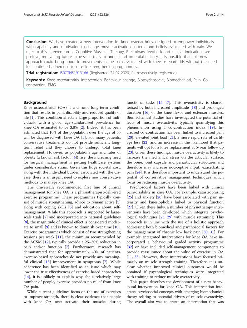
Muscle overactivity through postural mechanismsThere is clear evidence of altered postural alignment inpeople with knee OA. This is characterised by a flexedposture [42], altered lumbo-pelvic alignment [43] and anincrease in forward spinal inclination [44, 45]. Given thepotential link between intersegmental muscle length andposture [46, 47], these findings may indicate that peoplewith knee OA have some form of muscle imbalance ofthe hip/trunk muscles. This idea is consistent withresearch showing that people with knee OA have in-creased passive stiffness of the hip flexor muscles [48].Such increased stiffness will limit posterior pelvic rota-tion (pelvic tilt) [49], preventing the pelvis from return-ing to a neutral position in upright standing (Fig. 2a-b).Without any biomechanical compensation, a passivelystiff hip flexor will increase forward spinal inclination,shifting the centre of mass anteriorly (Fig. 2b). Increasedhip extensor (e.g. hamstring) activity will then be re-quired to maintain upright standing. However, we sug-gest that such an extremely flexed position is unlikely tobe adopted. Instead, it is likely that an individual with apassively stiff hip flexor will “biomechanically compen-sate”, by flexing the hip, knee and ankle [42] and by in-creasing lumbar lordosis in an attempt to stand uprightand maintain gaze alignment (Fig. 2c). This compensation
Preece et al. BMC Musculoskeletal Disorders (2021) 22:526 Page 4 of 14
will require an increase in quadriceps muscle activity tomaintain a flexed knee in standing.As well as influencing muscle activity in standing, pos-
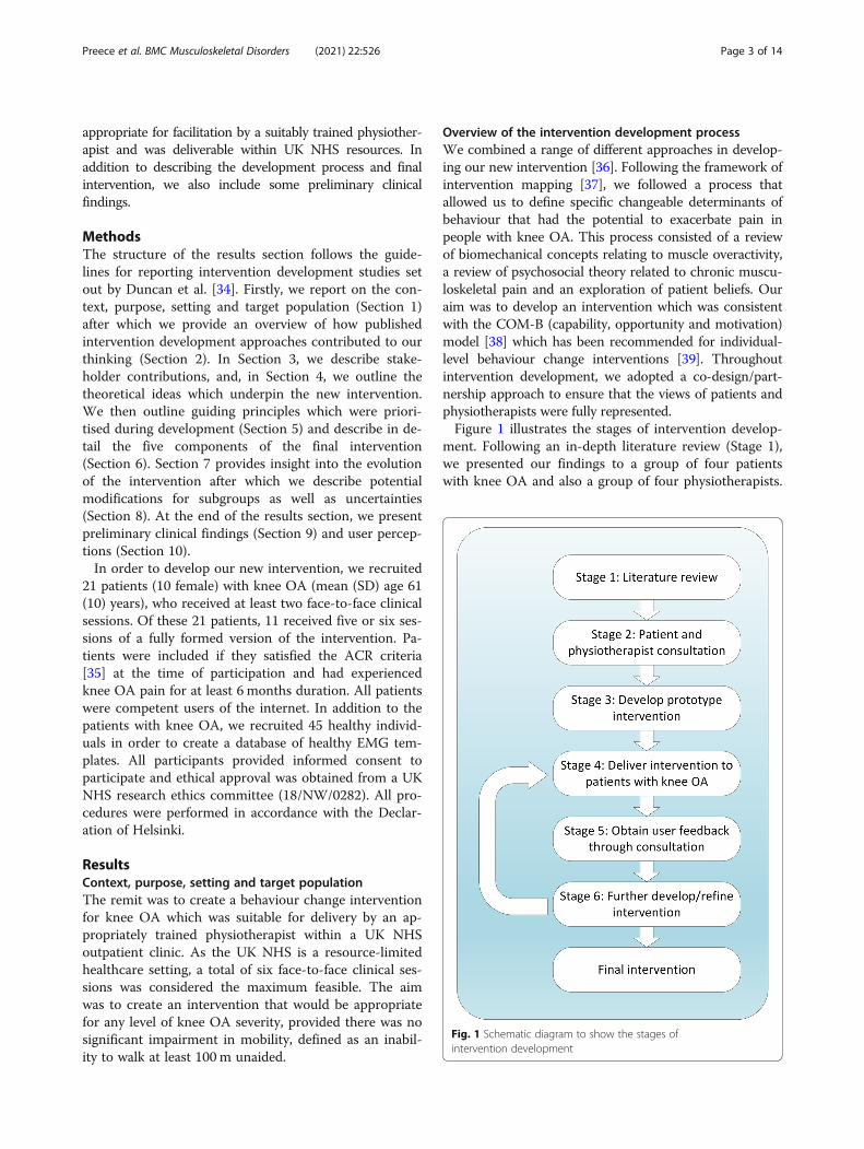
tural mechanisms may also underlie, to some degree,muscle overactivity during walking, which is exhibitedby people with knee OA [50]. Emerging research fromour lab supports this idea, showing a link between trunkflexion in walking and passive stiffness of the hip flexormuscles [48] and higher knee muscle activity in healthypeople who walk with more trunk inclination [51]. Im-portantly, we have observed that people with knee OAwalk with an increased flexion (forward lean) of thetrunk [52]. Critically, when we instruct healthy people toincrease their trunk flexion by only 5°, knee flexormuscle patterns become similar to those associated withknee OA [50, 53] (Fig. 3).Central to the postural mechanisms outline above is
the idea that overactivity of the knee muscles could re-sult from increased passive stiffness of hip/trunk mus-cles. It has been suggested that chronic understretch [54,55] can lead to increased passive stiffness of muscles.This is consistent with our research showing limitedpassive hip extension in healthy people who sit for pro-longed periods and who are physically inactive [56]. Itmay also explain the observation of reduced transverseplane motion of the thoracic spine which would resultfrom increased stiffness of the abdominal muscles [57].Figure 2 depicts how passive stiffness of hip flexor mus-cles could trigger compensatory changes in knee muscleactivity. Similarly, increased stiffness of the abdominal
muscles will reduce the capacity of the rib cage to movesuperiorly, relative to the pelvis [58], and could there-fore affect postural alignment in standing, potentiallytriggering knee muscle overactivity. Given these ideas,we integrated the idea of a link between sedentarybehaviour, increased passive stiffness of hip/trunkmuscles, biomechanical compensation and knee muscleoveractivity (Fig. 4).
Fig. 2 (a, b) A passively stiff hip flexor (illustrated as a rope) prevents the pelvis returning to a neutral position in standing. (c) Biomechanicalcompensation for a passively stiff hip flexor, consisting of a flexed hip, knee and ankle and an increased lumbar lordosis. Note there is still a slightflexion of the trunk. A full animation of this pattern can be viewed at: www.cogmustherapy.com/BMC_example_1
Fig. 3 Medial hamstring EMG during walking in people with kneeOA (blue), in healthy people (green) and in healthy people afterinstruction to increase trunk flexion by 5° (red). Note how themuscle pattern in the healthy people changes dramatically,becoming similar to the OA pattern, with increased trunk flexion.MVIC refers to maximal voluntary isometric contraction
Preece et al. BMC Musculoskeletal Disorders (2021) 22:526 Page 5 of 14
Central modulation of the pain experienceThere is strong evidence to support the idea that peoplewith knee OA are oversensitive to pain in general [59,60]. This so called central sensitisation [61] can occurthrough a range of mechanisms, such as amplification ofafferent nociceptive impulses from peripheral receptorsor alteration of sensory processing in the brain. Whileintense and continued nociceptive input is known tocause central sensitisation [62], it is also possible thatemotional responses to pain [63] or pain expectationscan influence sensitisation. This idea is consistent withresearch which has demonstrated that psychosocial fac-tors can mediate the association between hyperalgesiaand knee pain [64]. Characteristics such as pain catastro-phising [65], kinesiophobia (fear of movement) [25],helplessness [66], reduced self-efficacy [67], anxiety [26]and depression [26] have all been shown to be associatedwith knee OA pain and are likely to play a role in shap-ing the pain experience. Therefore, we integrated theidea of a relationship between central modulation, thepain experience and emotional responses to pain.
Motor adaptation to painAdaptation of the motor system to pain [68] or antici-pated pain [69] is a well-recognised phenomenon [70].Given the consistent observation of longer duration andincreased amplitude of EMG in people with knee OAacross different tasks [16, 18], it would appear thatmotor adaptation in this disease is characterised by anoveractivity of the knee muscles. Although this strategymay enhance joint stability following acute injury [71], itwill increase joint loading [72] and is likely to increasenociceptor input, exacerbating pain, if maintained in thelong-term. There is evidence that muscle overactivity inlow back pain is related to pain-related fear [73] andpain catastrophising [74]. This indicates that emotionalresponses and expectations are likely to shape long-termmotor adaptation to musculoskeletal pain. While this re-lationship has not been explored in knee OA, a recentstudy demonstrated a link between central sensitisationto pain and muscle overactivity [75] in people with thisdisease. This may suggest a link between motor adaptionand central modulation, which as explained above, islikely to be mediated by emotional responses to pain.
Given this likely interdependence, we integrated the ideaof a relationship between motor adaption, the pain ex-perience, central modulation and emotional responses topain.
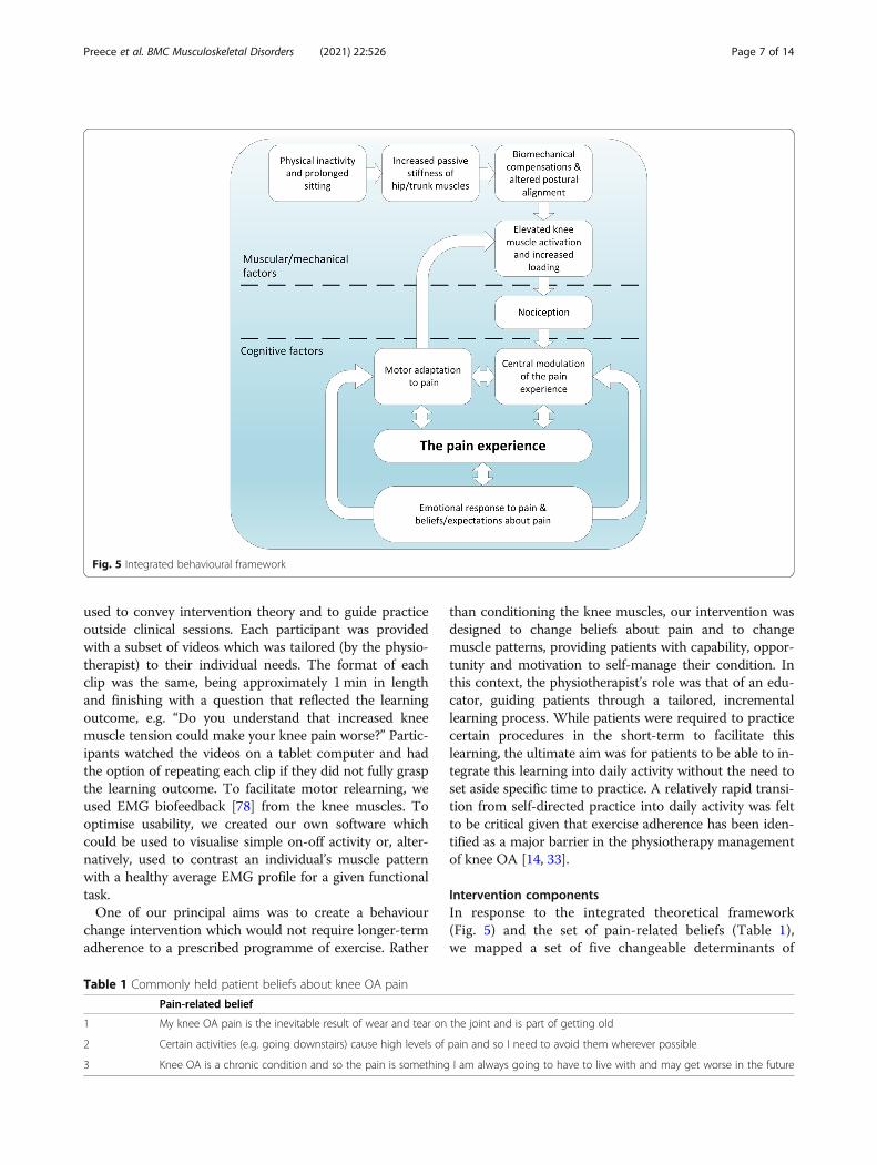
The integrated frameworkFigure 5 shows the fully integrated behavioural frame-work, obtained by combining the different mechanismsdescribed above. This framework can be divided intomuscular/mechanical factors which stimulate nociceptiveinput and cognitive factors which shape responses to painand the pain experience. It is important to stress that ourproposed framework is not a comprehensive model toexplain the onset of knee OA. Clearly, OA pain may havemany origins, such as ligament rupture or other traumaticinjury [76, 77]. Instead, we have attempted to includefactors which could exacerbate knee pain, may relate topatient beliefs, and that could be targeted through aneffective behaviour change intervention.
Exploration of patient beliefsFollowing theoretical development, we consulted with pa-tients (Stage 2, Fig. 1), encouraging them to contextualisetheir own pain experiences. A range of beliefs were identi-fied which were subsequently confirmed during interven-tion testing (Stage 5, Fig. 1). Table 1 lists the three beliefswhich were universally held amongst patients. In additionto these three beliefs, we identified beliefs relating to thedegree in which patients were fearful or anxious abouttheir pain. These beliefs were often markedly different be-tween individuals and this highlighted the need for ourfinal intervention to be individually tailored based on apatient's need. By combining our understanding of patientbeliefs with the theoretical framework (Fig. 5), we devel-oped a set of changeable determinants of behaviour. Be-fore we describe this development, we highlight guidingprinciples for intervention development which were estab-lished as part of our consultation.
Guiding principles prioritised during interventiondevelopmentTo facilitate patient learning, we used digital technologieswhere possible, working with a local animation studio tocreate a range of instructional videos. These videos were
Fig. 4 Postural framework to explain elevated knee muscle activation from increased passive stiffness of hip/trunk muscles
Preece et al. BMC Musculoskeletal Disorders (2021) 22:526 Page 6 of 14
used to convey intervention theory and to guide practiceoutside clinical sessions. Each participant was providedwith a subset of videos which was tailored (by the physio-therapist) to their individual needs. The format of eachclip was the same, being approximately 1min in lengthand finishing with a question that reflected the learningoutcome, e.g. “Do you understand that increased kneemuscle tension could make your knee pain worse?” Partic-ipants watched the videos on a tablet computer and hadthe option of repeating each clip if they did not fully graspthe learning outcome. To facilitate motor relearning, weused EMG biofeedback [78] from the knee muscles. Tooptimise usability, we created our own software whichcould be used to visualise simple on-off activity or, alter-natively, used to contrast an individual’s muscle patternwith a healthy average EMG profile for a given functionaltask.One of our principal aims was to create a behaviour
change intervention which would not require longer-termadherence to a prescribed programme of exercise. Rather
than conditioning the knee muscles, our intervention wasdesigned to change beliefs about pain and to changemuscle patterns, providing patients with capability, oppor-tunity and motivation to self-manage their condition. Inthis context, the physiotherapist’s role was that of an edu-cator, guiding patients through a tailored, incrementallearning process. While patients were required to practicecertain procedures in the short-term to facilitate thislearning, the ultimate aim was for patients to be able to in-tegrate this learning into daily activity without the need toset aside specific time to practice. A relatively rapid transi-tion from self-directed practice into daily activity was feltto be critical given that exercise adherence has been iden-tified as a major barrier in the physiotherapy managementof knee OA [14, 33].
Intervention componentsIn response to the integrated theoretical framework(Fig. 5) and the set of pain-related beliefs (Table 1),we mapped a set of five changeable determinants of
Fig. 5 Integrated behavioural framework
Table 1 Commonly held patient beliefs about knee OA pain
Pain-related belief
1 My knee OA pain is the inevitable result of wear and tear on the joint and is part of getting old
2 Certain activities (e.g. going downstairs) cause high levels of pain and so I need to avoid them wherever possible
3 Knee OA is a chronic condition and so the pain is something I am always going to have to live with and may get worse in the future
Preece et al. BMC Musculoskeletal Disorders (2021) 22:526 Page 7 of 14
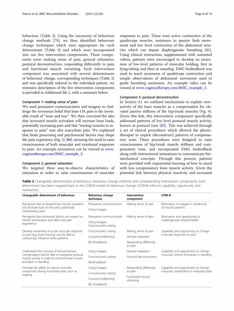
behaviour (Table 2). Using the taxonomy of behaviourchange methods [79], we then identified behaviourchange techniques which were appropriate for eachdeterminant (Table 3) and which were incorporatedinto our five intervention components. These compo-nents were: making sense of pain, general relaxation,postural deconstruction, responding differently to painand functional muscle retraining. Each interventioncomponent was associated with several determinantsof behaviour change, corresponding techniques (Table 2)and was specifically tailored to the individual patient. Anextensive description of the five intervention componentsis provided in Additional file 1, with a summary below.
Component 1: making sense of painWe used persuasive communication and imagery to chal-lenge the erroneous belief that knee OA pain is the inevit-able result of “wear and tear”. We then conveyed the ideathat increased muscle activation will increase knee loads,potentially increasing pain and that “tensing muscles in re-sponse to pain” may also exacerbate pain. We explainedthat brain processing and psychosocial factors may shapethe pain experience (Fig. 5) [80], stressing the need to raiseconsciousness of both muscular and emotional responsesto pain. An example animation can be viewed at www.cogmustherapy.com/BMC_example_2.
Component 2: general relaxationWe targeted three easy-to-observe characteristics ofrelaxation in order to raise consciousness of muscular
responses to pain. These were active contraction of thequadriceps muscles, resistance to passive limb move-ment and low level contraction of the abdominal mus-cles which can impair diaphragmatic breathing [81].Using clinical instruction, supplemented with animatedvideos, patients were encouraged to develop an aware-ness of low-level patterns of muscular holding, first inlying/sitting and then in standing. EMG biofeedback wasused to teach awareness of quadriceps contraction andsimple observations of abdominal movement used toguide breathing awareness. An example video can beviewed at www.cogmustherapy.com/BMC_example_3.
Component 3: postural deconstructionIn Section 4.1 we outlined mechanisms to explain over-activity of the knee muscles as a compensation for ele-vated passive stiffness of the hip/trunk muscles (Fig. 4).Given this link, this intervention component specificallyaddressed patterns of low level postural muscle activity,known as postural tone [82]. This was achieved througha set of clinical procedures which allowed the physio-therapist to unpick (deconstruct) patterns of compensa-tory tone. These procedures were designed to raiseconsciousness of hip/trunk muscle stiffness and com-pensatory tone, and incorporated EMG biofeedbackalong with instructional animations to communicate bio-mechanical concepts. Through this process, patientswere provided with experiential learning of how to standwith less compensatory knee muscle activity. Given thepotential link between physical inactivity and increased
Table 2 Changeable determinants of behaviour, behaviour change methods and corresponding intervention components. Eachdeterminant has been mapped back to the COM-B model of behaviour change. (COM-B refers to capability, opportunity andmotivation)
Changeable determinant of behaviour Behaviour changetechnique
Interventioncomponent
COM-B
Recognise that increased knee muscle activationwill increase load on the joint, potentiallyexacerbating pain.
Persuasive communication Making sense of pain Motivation to engage in re-learningof muscle patterns
Using imagery
Recognise that emotional factors can impact oncentral sensitisation and affect the painexperience.
Persuasive communication Making sense of pain Motivation and opportunity tochallenge pain-related beliefs
Using imageryConsciousness raising
Develop awareness of acute muscular responseto pain (e.g. knee bracing) and be able toconsciously influence these patterns.
Consciousness raising Making sense of pain Capability and opportunity to changemuscular responses to pain
Counterconditioning General relaxation
Bio (Feedback) Responding differentlyto pain
Understand the concept of biomechanicalcompensation and be able to reorganise posturalmuscle activity in order to minimise knee muscleactivation in standing.
Using imagery General relaxation Capability and opportunity to changemuscular control of posture in standing
Consciousness raising Postural deconstruction
(Bio)Feedback
Develop the ability to reduce muscularoveractivity during functional tasks, such aswalking.
Using imagery Responding differentlyto pain
Capability and opportunity to changemuscular coordination in everyday tasks
passive stiffness of hip/trunk muscles, patients were en-couraged to take regular walking exercise and break upperiods of prolonged sitting. The physiotherapist alsochallenged beliefs relating to exercise avoidance.
Component 4: responding differently to painThis intervention component used EMG biofeedback toraise consciousness of inappropriate contraction of theknee muscles which was triggered by pain expectations.Using biofeedback, the patient was taught to down regu-late (counter condition) anticipatory muscular contrac-tion, which occurred before initiation of functionalmovement, e.g. before stepping down. Such muscle pat-terns are likely to be connected to past experience andbeliefs about pain. Therefore, the clinician used this op-portunity to continue to explore patient’s beliefs aroundthe causes of pain and encouraged individuals to reflecton their own emotional responses to anticipated pain.
Component 5: functional muscle retrainingWe created software which facilitated the visualisation ofa patient’s EMG profile against a healthy template for dif-ferent functional tasks. Using this software, the clinicianidentified periods of inappropriate muscle activity andthen used motor imagery [83] to encourage downregula-tion of knee muscle activity. For example, many peoplewith knee OA exhibit prolonged quadriceps activity intomidstance of walking [84]. By using an instruction (for ex-ample “imagine a rope pulling the leg forwards as youwalk”), the patient learned to associate the specific motorcommand with the healthy template, receiving continuousEMG biofeedback to guide learning. By working through arange of functional tasks, the clinician challenged beliefsthat certain movements should be avoided, providing ex-periential learning that these tasks could be performedwith less muscle activation.
Intervention scheduleThe final intervention was delivered as a course of sixone-to-one clinical sessions (one every two weeks), eachlasting 45–60min and which was augmented with spe-cific tasks that patients completed outside of contact ses-sions. The first clinical session typically covered makingsense of pain (component 1) and general relaxation
(component 2). In sessions 2–4, this material was re-vised, and the patient taught postural deconstruction(component 3) and responding differently to pain (com-ponent 4). In the final two sessions, there was morefocus on functional muscle retraining (component 5),however, this was determined on individual needs. Out-side clinical sessions, patients practised relaxation, pos-tural deconstruction and the use of specific motorcommands to influence muscle patterns. They were alsoencouraged to take regular exercise and to notice theiremotional and muscular responses to pain. The ultimateaim of the intervention was to create capability, oppor-tunity and motivation to change behaviour related toknee pain. In line with this philosophy, patients wereinstructed to gradually integrate as many of the ideasand practices into their activities of daily living, remov-ing the need to dedicate specific time each day topractice.
The evolution of the interventionDuring the two-year development process, we changedthe way the intervention was delineated into differentcomponents. The initial prototype contained componentsaligned with specific aspects of the technical developmentwork, such as instructional animations and biofeedbacksoftware. However, we later delineated the interventioninto components that aligned with learning objectives,such as making sense of pain, general relaxation and func-tional muscle retraining. We also moved from the originalconcept of an introductory video to explain psychologicaland biomechanical concepts, to the practice of producingseveral short clips (< 1min). As the intervention pro-gressed, the physiotherapist was able to add these clips toa playlist on a tablet computer (provided to the patient),gradually increasing the information that patients were re-quired to digest after each session.A major part of the development work was focused on
the clinical procedures which formed the basis of thepostural deconstruction component of the intervention.After experimenting with numerous strategies, we foundthat the idea of a “tension point” to be the most effectiveway to link our conceptual framework with a patient’skinaesthetic understanding. In line with the focus onpostural tone, this idea shifted the objective from that of
Table 3 The five primary behaviour change methods
Behaviour change method Definition
Persuasive communication Guiding individuals toward the adoption of an idea, attitude, or action by using arguments or other means.
Using imagery Presenting information in a pictorial format will aid the communication of conceptual ideas and facilitate the learningof new motor patterns
Consciousness raising Providing information and feedback about the causes, consequences, and alternatives for a problem behaviour.
Counter conditioning Encouraging the learning of healthier behaviours that can substitute for problem behaviours.
Feedback Giving information to individuals regarding the extent to which they are accomplishing learning or performance.
Preece et al. BMC Musculoskeletal Disorders (2021) 22:526 Page 9 of 14
achieving a distinct postural alignment to a focus on themuscles used to stand erect. Another part of thedevelopmental work focused on the functional muscleretraining. While our original idea had been to guidepatients through a set of incremental activities foreach task, it proved difficult to break down complexfunctional movements, such as walking. Therefore,following the preliminary balance training, the use ofguided imagery was found to be the most appropriatemethod for changing motor patterns.
Intervention modification for subgroups and potentialuncertaintiesGiven that our intervention is tailored to an individual’sneeds, we are confident that it would be appropriate formost patients with knee OA. However, we acknowledgethat further development would be required for peoplewho are unable to stand unaided or need to use a walk-ing aid. Our intervention was specifically designed for aUK NHS setting and therefore we did not investigate thepotential of adapting the number of intervention ses-sions on an individual basis. However, we suggest thatincreasing the number of clinical sessions might improveoutcomes. Another potential uncertainty is the traininga physiotherapist would require in order to deliver thisnew intervention. Wherever possible, we created supple-mentary material to facilitate patient learning, used estab-lished psychosocial techniques for the management ofchronic musculoskeletal pain and used existing clinicalphysiotherapy assessment methods. Therefore, we suggestthat a relatively short training course should prove suffi-cient and we are currently exploring how to design such acourse.
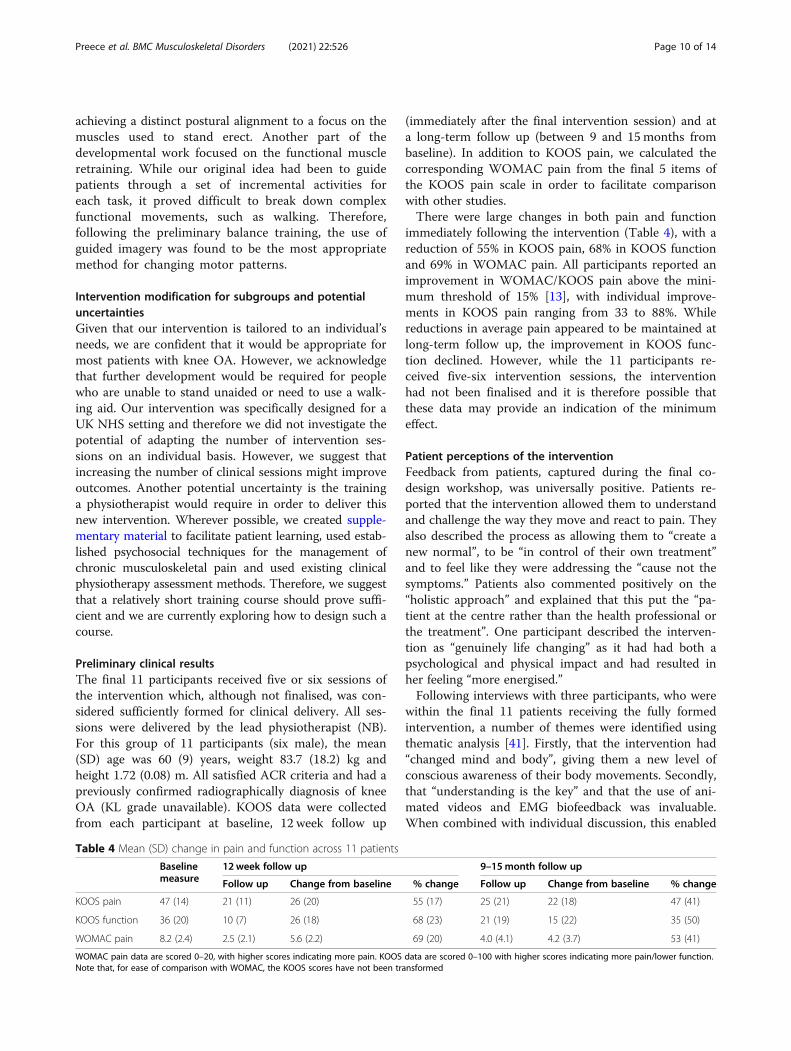
Preliminary clinical resultsThe final 11 participants received five or six sessions ofthe intervention which, although not finalised, was con-sidered sufficiently formed for clinical delivery. All ses-sions were delivered by the lead physiotherapist (NB).For this group of 11 participants (six male), the mean(SD) age was 60 (9) years, weight 83.7 (18.2) kg andheight 1.72 (0.08) m. All satisfied ACR criteria and had apreviously confirmed radiographically diagnosis of kneeOA (KL grade unavailable). KOOS data were collectedfrom each participant at baseline, 12 week follow up
(immediately after the final intervention session) and ata long-term follow up (between 9 and 15months frombaseline). In addition to KOOS pain, we calculated thecorresponding WOMAC pain from the final 5 items ofthe KOOS pain scale in order to facilitate comparisonwith other studies.There were large changes in both pain and function
immediately following the intervention (Table 4), with areduction of 55% in KOOS pain, 68% in KOOS functionand 69% in WOMAC pain. All participants reported animprovement in WOMAC/KOOS pain above the mini-mum threshold of 15% [13], with individual improve-ments in KOOS pain ranging from 33 to 88%. Whilereductions in average pain appeared to be maintained atlong-term follow up, the improvement in KOOS func-tion declined. However, while the 11 participants re-ceived five-six intervention sessions, the interventionhad not been finalised and it is therefore possible thatthese data may provide an indication of the minimumeffect.
Patient perceptions of the interventionFeedback from patients, captured during the final co-design workshop, was universally positive. Patients re-ported that the intervention allowed them to understandand challenge the way they move and react to pain. Theyalso described the process as allowing them to “create anew normal”, to be “in control of their own treatment”and to feel like they were addressing the “cause not thesymptoms.” Patients also commented positively on the“holistic approach” and explained that this put the “pa-tient at the centre rather than the health professional orthe treatment”. One participant described the interven-tion as “genuinely life changing” as it had had both apsychological and physical impact and had resulted inher feeling “more energised.”Following interviews with three participants, who were
within the final 11 patients receiving the fully formedintervention, a number of themes were identified usingthematic analysis [41]. Firstly, that the intervention had“changed mind and body”, giving them a new level ofconscious awareness of their body movements. Secondly,that “understanding is the key” and that the use of ani-mated videos and EMG biofeedback was invaluable.When combined with individual discussion, this enabled
Table 4 Mean (SD) change in pain and function across 11 patients
Baselinemeasure
12 week follow up 9–15month follow up
Follow up Change from baseline % change Follow up Change from baseline % change
WOMAC pain data are scored 0–20, with higher scores indicating more pain. KOOS data are scored 0–100 with higher scores indicating more pain/lower function.Note that, for ease of comparison with WOMAC, the KOOS scores have not been transformed
Preece et al. BMC Musculoskeletal Disorders (2021) 22:526 Page 10 of 14
patients to “reset expectations” about their knee painand give them a new feeling of “responsibility” for theircondition. A third theme related to the need to “keepgoing with the new me”, recognising the importance ofcontinued awareness in daily life. Finally, the importanceof an empathic and positive attitude of the therapist wasrecognised as being crucial in changing patient’s beliefsabout pain and guiding them through the learningprocess.
DiscussionOur intervention is novel because of the integration ofpsychological techniques with muscle biofeedback train-ing, specifically designed to target muscle overactivity.Given this integration, we propose a label for the interven-tion of “Cognitive Muscular Therapy.” EMG biofeedbacktechniques have been used extensively in rehabilitation[85]. However, our approach is especially unique becauseof the use of biofeedback to raise consciousness of muscleoveractivity related to pain expectations (component 4)and the use of postural deconstruction to reduce muscleoveractivity in standing (component 3). We acknowledgethat some of the biomechanical underpinnings for theintervention are based on emerging concepts rather thanon unequivocal evidence. Nevertheless, our preliminaryclinical findings and positive patient feedback motivatefurther research to understand the links between muscleoveractivity, motor adaption to pain and central sensitisation.People with knee OA are known to exhibit weakness
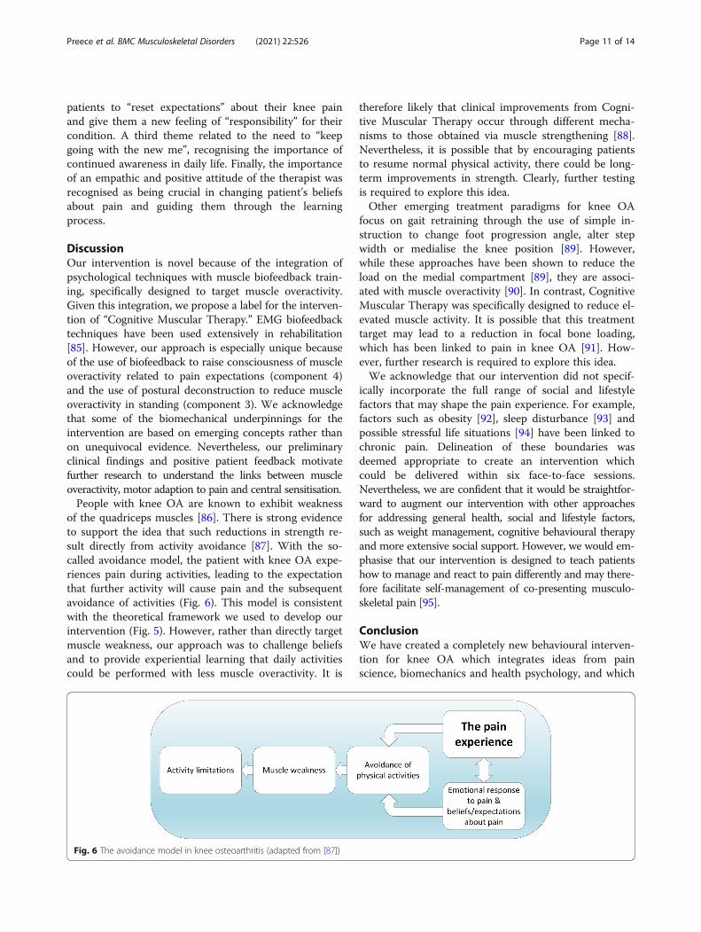
of the quadriceps muscles [86]. There is strong evidenceto support the idea that such reductions in strength re-sult directly from activity avoidance [87]. With the so-called avoidance model, the patient with knee OA expe-riences pain during activities, leading to the expectationthat further activity will cause pain and the subsequentavoidance of activities (Fig. 6). This model is consistentwith the theoretical framework we used to develop ourintervention (Fig. 5). However, rather than directly targetmuscle weakness, our approach was to challenge beliefsand to provide experiential learning that daily activitiescould be performed with less muscle overactivity. It is
therefore likely that clinical improvements from Cogni-tive Muscular Therapy occur through different mecha-nisms to those obtained via muscle strengthening [88].Nevertheless, it is possible that by encouraging patientsto resume normal physical activity, there could be long-term improvements in strength. Clearly, further testingis required to explore this idea.Other emerging treatment paradigms for knee OA
focus on gait retraining through the use of simple in-struction to change foot progression angle, alter stepwidth or medialise the knee position [89]. However,while these approaches have been shown to reduce theload on the medial compartment [89], they are associ-ated with muscle overactivity [90]. In contrast, CognitiveMuscular Therapy was specifically designed to reduce el-evated muscle activity. It is possible that this treatmenttarget may lead to a reduction in focal bone loading,which has been linked to pain in knee OA [91]. How-ever, further research is required to explore this idea.We acknowledge that our intervention did not specif-
ically incorporate the full range of social and lifestylefactors that may shape the pain experience. For example,factors such as obesity [92], sleep disturbance [93] andpossible stressful life situations [94] have been linked tochronic pain. Delineation of these boundaries wasdeemed appropriate to create an intervention whichcould be delivered within six face-to-face sessions.Nevertheless, we are confident that it would be straightfor-ward to augment our intervention with other approachesfor addressing general health, social and lifestyle factors,such as weight management, cognitive behavioural therapyand more extensive social support. However, we would em-phasise that our intervention is designed to teach patientshow to manage and react to pain differently and may there-fore facilitate self-management of co-presenting musculo-skeletal pain [95].
ConclusionWe have created a completely new behavioural interven-tion for knee OA which integrates ideas from painscience, biomechanics and health psychology, and which
Fig. 6 The avoidance model in knee osteoarthritis (adapted from [87])
Preece et al. BMC Musculoskeletal Disorders (2021) 22:526 Page 11 of 14
can be delivered by a physiotherapist. We propose torefer to this intervention as Cognitive Muscular Therapy.The intervention contains a focus on changing musclepatterns and teaching patients about how their beliefsand behaviours can shape the pain experience. Theintervention is consistent with the COM-B model of be-haviour change. User feedback was incredibly positive.However, while encouraging, our preliminary clinicaldata does not constitute proof of effectiveness. There-fore, larger trials are now required to understandwhether this intervention could bring about long-termimprovements in the pain associated with knee OAwhen delivered either within the UK NHS or otherhealthcare settings.
Supplementary InformationThe online version contains supplementary material available at https://doi.org/10.1186/s12891-021-04389-0.
Additional file 1. Full description of the final intervention.
AcknowledgementsThe authors would like to thank Dr. Daniela Ghio for her feedback on apreliminary draft of the manuscript.
Authors’ contributionsSJP conceived the original study idea, performed the literature review,synthesised the underlying concepts, consulted with patients andphysiotherapists to develop the final intervention and drafted the finalmanuscript. NB synthesised the underlying concepts, collected laboratorydata, delivered successive iterations of the intervention to the participants,consulted with patients and physiotherapists and commented on the finalmanuscript. AEW consulted with patients and physiotherapists, interpretedqualitative data, refined the intervention and contributed to the drafting ofthe final manuscript. RKJ synthesised the underlying concepts, assisted withdata interpretation and made substantial revisions to the final manuscript. CScreated biofeedback software, collected laboratory data and contributed todata interpretation. AJ synthesised the underlying concepts, assisted withdata interpretation and contributed towards drafting of the final manuscript.NEW consulted with patients and physiotherapists, contributed to thesynthesis of underlying concepts, interpreted qualitative data andcontributed to the drafting of the final manuscript. The author(s) read andapproved the final manuscript.
FundingThis paper presents independent research funded by the National Institutefor Health Research (NIHR), UK, under its Research for Patient Benefit (RfPB)Programme (Grant Reference Number PB-PG-0816-20024. The viewsexpressed are those of the author(s) and not necessarily those of the NIHR orthe Department of Health and Social Care.
Availability of data and materialsFull KOOS data collected from the final 11 participants can be downloaded at thefollowing link: https://doi.org/10.17866/rd.salford.14709276.
Declarations
Ethics approval and consent to participateThe following statement was provided on ethical approval in the methodssection “… ethical approval was obtained from a UK NHS research ethicscommittee (18/NW/0282).” A statement was also provided in the methodssection on consent to participate, “All participants provided informedconsent to participate.”
Consent for publicationNot applicable.
Competing interestsThe authors declare that they have no competing interests.
Author details1Centre for Health Sciences Research, University of Salford, Manchester M66PU, UK. 2Physiotherapy Department, Salford Royal NHS Foundation Trust,Salford M6 8HD, UK. 3Human Pain Research Group, University of Manchester,Clinical Sciences Building, Salford Royal NHS Foundation Trust, Salford M68HD, UK. 4Faculty of Health and Applied Sciences, University of the West ofEngland, Bristol BS16 1DD, UK.
Received: 9 December 2020 Accepted: 30 April 2021
References1. Woolf AD, Pfleger B. Burden of major musculoskeletal conditions. Bull World
Health Organ. 2003;81(9):646–56.2. Cross M, Smith E, Hoy D, Nolte S, Ackerman I, Fransen M, et al. The global
burden of hip and knee osteoarthritis: estimates from the global burden ofdisease 2010 study. Ann Rheum Dis. 2014;73(7):1323–30. https://doi.org/10.1136/annrheumdis-2013-204763.
3. Peat G, McCarney R, Croft P. Knee pain and osteoarthritis in older adults: areview of community burden and current use of primary health care. AnnRheum Dis. 2001;60(2):91–7. https://doi.org/10.1136/ard.60.2.91.
4. King LK, March L, Anandacoomarasamy A. Obesity & osteoarthritis. Indian JMed Res. 2013;138(2):185–93.
5. Osteoarthritis: care and management (2014). Clinical guideline [CG177].National Institute for Clinical Excellence.
6. Bennell KL, Ahamed Y, Jull G, Bryant C, Hunt MA, Forbes AB, et al. Physicaltherapist-delivered pain coping skills training and exercise for kneeosteoarthritis: randomized controlled trial. Arthritis Care Res. 2016;68(5):590–602. https://doi.org/10.1002/acr.22744.
7. Hurley MV, Walsh NE, Mitchell H, Nicholas J, Patel A. Long-term outcomesand costs of an integrated rehabilitation program for chronic knee pain: apragmatic, cluster randomized, controlled trial. Arthritis Care Res. 2012;64(2):238–47. https://doi.org/10.1002/acr.20642.
8. Osteoarthritis: care and management [CG177]: NICE guidelines. 2014.9. Fransen M, McConnell S, Harmer AR, Van der Esch M, Simic M, Bennell KL.
Exercise for osteoarthritis of the knee. Cochrane Database Syst Rev. 2015;1.https://doi.org/10.1002/14651858.CD004376.pub3.
10. Goh SL, Persson MSM, Stocks J, Hou Y, Lin J, Hall MC, et al. Efficacy andpotential determinants of exercise therapy in knee and hip osteoarthritis: asystematic review and meta-analysis. Ann Phys Rehabil Med. 2019;62(5):356–65. https://doi.org/10.1016/j.rehab.2019.04.006.
11. Hurley MV, Walsh NE, Mitchell HL, Pimm TJ, Patel A, Williamson E, et al.Clinical effectiveness of a rehabilitation program integrating exercise, self-management, and active coping strategies for chronic knee pain: a clusterrandomized trial. Arthritis Rheum. 2007;57(7):1211–9. https://doi.org/10.1002/art.22995.
12. Garber CE, Blissmer B, Deschenes MR, Franklin BA, Lamonte MJ, Lee IM,et al. American College of Sports Medicine position stand. Quantity andquality of exercise for developing and maintaining cardiorespiratory,musculoskeletal, and neuromotor fitness in apparently healthy adults:guidance for prescribing exercise. Med Sci Sports Exerc. 2011;43(7):1334–59.https://doi.org/10.1249/MSS.0b013e318213fefb.
13. Angst F, Aeschlimann A, Stucki G. Smallest detectable and minimal clinicallyimportant differences of rehabilitation intervention with their implicationsfor required sample sizes using WOMAC and SF-36 quality of lifemeasurement instruments in patients with osteoarthritis of the lowerextremities. Arthritis Rheum. 2001;45(4):384–91. https://doi.org/10.1002/1529-0131(200108)45:4<384::AID-ART352>3.0.CO;2-0.
14. Bennell KL, Dobson F, Hinman RS. Exercise in osteoarthritis: moving fromprescription to adherence. Best Pract Res Clin Rheumatol. 2014;28(1):93–117.https://doi.org/10.1016/j.berh.2014.01.009.
15. Lyytinen T, Liikavainio T, Bragge T, Hakkarainen M, Karjalainen PA, ArokoskiJPA. Postural control and thigh muscle activity in men with kneeosteoarthritis. J Electromyogr Kinesiol. 2010;20(6):1066–74. https://doi.org/10.1016/j.jelekin.2010.05.005.
Preece et al. BMC Musculoskeletal Disorders (2021) 22:526 Page 12 of 14
16. Childs JD, Sparto PJ, Fitzgerald GK, Bizzini M, Irrgang JJ. Alterations in lowerextremity movement and muscle activation patterns in individuals withknee osteoarthritis. Clin Biomech (Bristol, Avon). 2004;19(1):44–9.
17. Hubley-Kozey CL, Deluzio KJ, Landry SC, McNutt JS, Stanish WD.Neuromuscular alterations during walking in persons with moderate kneeosteoarthritis. J Electromyogr Kinesiol. 2006;16(4):365–78. https://doi.org/10.1016/j.jelekin.2005.07.014.
18. Hortobagyi T, Westerkamp L, Beam S, Moody J, Garry J, Holbert D, et al.Altered hamstring-quadriceps muscle balance in patients with kneeosteoarthritis. Clin Biomech. 2005;20(1):97–104. https://doi.org/10.1016/j.clinbiomech.2004.08.004.
19. Winby CR, Gerus P, Kirk TB, Lloyd DG. Correlation between EMG-based co-activation measures and medial and lateral compartment loads of the kneeduring gait. Clin Biomech. 2013;28(9):1014–9. https://doi.org/10.1016/j.clinbiomech.2013.09.006.
20. Heiden TL, Lloyd DG, Ackland TR. Knee joint kinematics, kinetics and muscleco-contraction in knee osteoarthritis patient gait. Clin Biomech. 2009;24(10):833–41. https://doi.org/10.1016/j.clinbiomech.2009.08.005.
21. Brandon SCE, Miller RH, Thelen DG, Deluzio KJ. Selective lateral muscleactivation in moderate medial knee osteoarthritis subjects does not unloadmedial knee condyle. J Biomech. 2014;47(6):1409–15. https://doi.org/10.1016/j.jbiomech.2014.01.038.
22. Hodges PW, van den Hoorn W, Wrigley TV, Hinman RS, Bowles K-A, CicuttiniF, et al. Increased duration of co-contraction of medial knee muscles isassociated with greater progression of knee osteoarthritis. Man Ther. 2016;21:151–8. https://doi.org/10.1016/j.math.2015.07.004.
23. Hatfield GL, Costello KE, Astephen Wilson JL, Stanish WD, Hubley-Kozey CL.Baseline gait muscle activation patterns differ for osteoarthritis patients whoundergo total knee arthroplasty 5–8 years later from those who do not.Arthritis Care Res. 2020;73:549–58. 101002/acr24143.
24. Preece SJ, Jones RK, Brown CA, Cacciatore TW, Jones AK. Reductions in co-contraction following neuromuscular re-education in people with kneeosteoarthritis. BMC Musculoskelet Disord. 2016;17(1):372. https://doi.org/10.1186/s12891-016-1209-2.
25. Odole A, Ekediegwu E, Ekechukwu END, Uchenwoke C. Correlates andpredictors of pain intensity and physical function among individuals withchronic knee osteoarthritis in Nigeria. Musculoskelet Sci Pract. 2019;39:150–6. https://doi.org/10.1016/j.msksp.2018.11.014.
26. Sharma A, Kudesia P, Shi Q, Gandhi R. Anxiety and depression in patients withosteoarthritis: impact and management challenges. Open Access Rheumatol.2016;8:103–13. https://doi.org/10.2147/OARRR.S93516.
27. Scopaz KA, Piva SR, Wisniewski S, Fitzgerald GK. Relationships of fear,anxiety, and depression with physical function in patients with kneeosteoarthritis. Arch Phys Med Rehabil. 2009;90(11):1866–73. https://doi.org/10.1016/j.apmr.2009.06.012.
28. Silva Guerrero AV, Maujean A, Campbell L, Sterling M. A systematic reviewand meta-analysis of the effectiveness of psychological interventionsdelivered by physiotherapists on pain, disability and psychologicaloutcomes in musculoskeletal pain conditions. Clin J Pain. 2018;34(9):838–57.https://doi.org/10.1097/AJP.0000000000000601.
29. Denneny D, Frijdal A, Bianchi-Berthouze N, Greenwood J, McLoughlin R,Petersen K, Singh A, Williams ACC. The application of psychologicallyinformed practice: observations of experienced physiotherapists workingwith people with chronic pain. Physiotherapy. 2020;106:163–73. https://doi.org/10.1016/j.physio.2019.01.014.
30. O'Sullivan PB, Caneiro JP, O'Keeffe M, Smith A, Dankaerts W, Fersum K, et al.Cognitive functional therapy: an integrated behavioral approach for thetargeted Management of Disabling low Back Pain. Phys Ther. 2018;98(5):408–23. https://doi.org/10.1093/ptj/pzy022.
31. Koes BW, van Tulder M, Lin C-WC, Macedo LG, McAuley J, Maher C. Anupdated overview of clinical guidelines for the management of non-specificlow back pain in primary care. Eur Spine J. 2010;19(12):2075–94. https://doi.org/10.1007/s00586-010-1502-y.
32. Veenhof C, Köke AJ, Dekker J, Oostendorp RA, Bijlsma JW, van Tulder MW,et al. Effectiveness of behavioral graded activity in patients withosteoarthritis of the hip and/or knee: a randomized clinical trial. ArthritisRheum. 2006;55(6):925–34. https://doi.org/10.1002/art.22341.
33. Hurley M, Dickson K, Hallett R, Grant R, Hauari H, Walsh N, et al. Exerciseinterventions and patient beliefs for people with hip, knee or hip and kneeosteoarthritis: amixedmethods review. Cochrane Database Syst Rev. 2018;4.https://doi.org/10.1002/14651858.CD004376.pub3.
34. Duncan E, O'Cathain A, Rousseau N, Croot L, Sworn K, Turner KM, et al.Guidance for reporting intervention development studies in health research(GUIDED): an evidence-based consensus study. BMJ Open. 2020;10(4):e033516. https://doi.org/10.1136/bmjopen-2019-033516.
35. Altman R, Alarcon G, Appelroth D. The American College of Rheumatologycriteria for the classification and reporting of osteoarthritis of the knee.Arthritis Rheum. 1986;29(8):1039–49. https://doi.org/10.1002/art.1780290816.
36. O'Cathain A, Croot L, Duncan E, Rousseau N, Sworn K, Turner KM, et al.Guidance on how to develop complex interventions to improve health andhealthcare. BMJ Open. 2019;9(8):e029954. https://doi.org/10.1136/bmjopen-2019-029954.
38. Michie S, van Stralen MM, West R. The behaviour change wheel: a newmethod for characterising and designing behaviour change interventions.Implement Sci. 2011;6(1):42. https://doi.org/10.1186/1748-5908-6-42.
39. Behaviour change: individual approaches: Public health guideline [PH49]:NICE guidelines. 2014.
40. Sekhon M, Cartwright M, Francis JJ. Acceptability of healthcareinterventions: an overview of reviews and development of a theoreticalframework. BMC Health Serv Res. 2017;17(1):88. https://doi.org/10.1186/s12913-017-2031-8.
41. Braun V, Clarke V. Using thematic analysis in psychology. Qual Res Psychol.2006;3(2):77–101. https://doi.org/10.1191/1478088706qp063oa.
42. Turcot K, Sagawa Y Jr, Hoffmeyer P, Suvà D, Armand S. Multi-joint posturalbehavior in patients with knee osteoarthritis. Knee. 2015;22(6):517–21.https://doi.org/10.1016/j.knee.2014.09.001.
43. Yasuda T, Togawa D, Hasegawa T, Yamato Y, Kobayashi S, Yoshida G, et al.Relationship between knee osteoarthritis and Spinopelvic sagittal alignmentin volunteers over 50 years of age. Asian Spine J. 2020;14(4):495–501.https://doi.org/10.31616/asj.2018.0266.
44. Wang WJ, Liu F, Zhu YW, Sun MH, Qiu Y, Weng WJ. Sagittal alignment ofthe spine-pelvis-lower extremity axis in patients with severe kneeosteoarthritis: a radiographic study. Bone Joint Res. 2016;5(5):198–205.https://doi.org/10.1302/2046-3758.55.2000538.
45. Tauchi R, Imagama S, Muramoto A, Tsuboi M, Ishiguro N, Hasegawa Y.Influence of spinal imbalance on knee osteoarthritis in community-livingelderly adults. Nagoya J Med Sci. 2015;77(3):329–37.
46. Janda V, Jull G. Muscles and motor control in low back pain: assessmentand management. Physical Therapy of the Low Back. T Twomey, Churchilllivingstone New York. 1987:p253–78.
47. Kendall F, McCreary E, Provance P, Rodgers M, Romani W: Testing andfunction with posture and pain: Lippincoll Williams & Wilkins; 2005.
48. Preece SJ, Alghamdi W. Inter-individual variation in hip flexor length mayexplain differences in trunk flexion during walking in people with kneeosteoarthritis. Osteoarthr Cartil. 2020;28:S244–5. https://doi.org/10.1016/j.joca.2020.02.392.
49. Preece SJ, Tan YF, Alghamdi TD, Frances A: Comparison of pelvic tilt beforeand after hip flexor stretching in healthy adults Journal of Manipulative andPhysiological Therapeutics (accepted) 2020.
50. Preece SJ, Alghamdi W. The effect of increasing trunk flexion during normalwalking. Gait & Posture. 2021;83:250–5. https://doi.org/10.1016/j.gaitpost.2020.10.021.
51. Alghamdi W, Preece SJ. How does normal variability in trunk flexion affectlower limb muscle activity during walking? Hum Mov Sci. 2020;72:102630.https://doi.org/10.1016/j.humov.2020.102630.
52. Preece SJ, Algarni AS, Jones RK. Trunk flexion during walking in people withknee osteoarthritis. Gait Posture. 2019;72:202–5. https://doi.org/10.1016/j.gaitpost.2019.06.012.
53. Preece SJ, Alghamdi W, Jones R. Could increased trunk flexion underliealterations in knee muscle activity in people with knee OA? OsteoarthrCartil. 2019;27:S116–7. https://doi.org/10.1016/j.joca.2019.02.172.
55. Wisdom KM, Delp SL, Kuhl E. Use it or lose it: multiscale skeletal muscleadaptation to mechanical stimuli. Biomech Model Mechanobiol. 2015;14(2):195–215. https://doi.org/10.1007/s10237-014-0607-3.
56. Boukabache A, Preece SJ, Brookes N. Prolonged sitting and physicalinactivity are associated with limited hip extension: a cross-sectional study.
Preece et al. BMC Musculoskeletal Disorders (2021) 22:526 Page 13 of 14
57. Heneghan NR, Baker G, Thomas K, Falla D, Rushton A. What is the effect ofprolonged sitting and physical activity on thoracic spine mobility? Anobservational study of young adults in a UK university setting. BMJ Open.2018;8(5):e019371. https://doi.org/10.1136/bmjopen-2017-019371.
58. Mier A, Brophy C, Estenne M, Moxham J, Green M, De Troyer A. Action ofabdominal muscles on rib cage in humans. J Appl Physiol (Bethesda, Md :1985). 1985;58(5):1438–43.
59. Lluch E, Torres R, Nijs J, Van Oosterwijck J. Evidence for central sensitizationin patients with osteoarthritis pain: a systematic literature review. Eur J Pain(London, England). 2014;18(10):1367–75.
60. Finan PH, Buenaver LF, Bounds SC, Hussain S, Park RJ, Haque UJ, et al.Discordance between pain and radiographic severity in knee osteoarthritis:findings from quantitative sensory testing of central sensitization. ArthritisRheum. 2013;65(2):363–72. https://doi.org/10.1002/art.34646.
61. Girbes EL, Nijs J, Torres-Cueco R, Cubas CL. Pain treatment for patients withosteoarthritis and central sensitization. Phys Ther. 2013;93(6):842–51. https://doi.org/10.2522/ptj.20120253.
62. Martindale JC, Wilson AW, Reeve AJ, Chessell IP, Headley PM. Chronic secondaryhypersensitivity of dorsal horn neurones following inflammation of the kneejoint. Pain. 2007;133(1–3):79–86. https://doi.org/10.1016/j.pain.2007.03.006.
63. Lumley MA, Cohen JL, Borszcz GS, Cano A, Radcliffe AM, Porter LS, et al.Pain and emotion: a biopsychosocial review of recent research. J ClinPsychol. 2011;67(9):942–68. https://doi.org/10.1002/jclp.20816.
64. Mason KJ, O'Neill TW, Lunt M, Jones AKP, McBeth J. Psychosocial factorspartially mediate the relationship between mechanical hyperalgesia andself-reported pain. Scand J Pain. 2018;18(1):59–69. https://doi.org/10.1515/sjpain-2017-0109.
65. Lopez-Bravo MD, Zamarron-Cassinello MD, Touche RL, Munoz-Plata R,Cuenca-Martinez F, Ramos-Toro M. Psychological factors associated withfunctional disability in patients with hip and knee osteoarthritis. Behav Med.2020:1–11. https://doi.org/10.1080/08964289.2020.1813682.
66. Creamer P, Lethbridge-Cejku M, Hochberg MC. Determinants of painseverity in knee osteoarthritis: effect of demographic andpsychosocial variables using 3 pain measures. J Rheumatol. 1999;26(8):1785–92.
67. Degerstedt Å, Alinaghizadeh H, Thorstensson CA, Olsson CB. Highself-efficacy – a predictor of reduced pain and higher levels ofphysical activity among patients with osteoarthritis: an observationalstudy. BMC Musculoskelet Disord. 2020;21(1):380. https://doi.org/10.1186/s12891-020-03407-x.
68. Hodges PW, Tucker K. Moving differently in pain: a new theory to explainthe adaptation to pain. Pain. 2011;152(3):S90–8. https://doi.org/10.1016/j.pain.2010.10.020.
69. Hodges PW, Tsao H, Sims K. Gain of postural responses increases inresponse to real and anticipated pain. Exp Brain Res. 2015;233(9):2745–52.https://doi.org/10.1007/s00221-015-4347-0.
70. Butera KA, Fox EJ, George SZ. Toward a transformed understanding: frompain and movement to pain with movement. Phys Ther. 2016;96(10):1503–7. https://doi.org/10.2522/ptj.20160211.
71. Lewek MD, Ramsey DK, Snyder-Mackler L, Rudolph KS. Knee stabilization inpatients with medial compartment knee osteoarthritis. Arthritis Rheum.2005;52(9):2845–53. https://doi.org/10.1002/art.21237.
72. Sritharan P, Lin YC, Richardson SE, Crossley KM, Birmingham TB, Pandy MG.Musculoskeletal loading in the symptomatic and asymptomatic knees ofmiddle-aged osteoarthritis patients. J Orthop Res. 2016;35:321–30.
73. Geisser ME, Haig AJ, Wallbom AS, Wiggert EA. Pain-related fear, lumbarflexion, and dynamic EMG among persons with chronic musculoskeletallow back pain. Clin J Pain. 2004;20(2):61–9. https://doi.org/10.1097/00002508-200403000-00001.
74. Pakzad M, Fung J, Preuss R. Pain catastrophizing and trunk muscleactivation during walking in patients with chronic low back pain. GaitPosture. 2016;49:73–7. https://doi.org/10.1016/j.gaitpost.2016.06.025.
75. Stefanik JJ, Frey-Law L, Segal NA, Niu J, Lewis CE, Nevitt MC, et al. TheRelation of Peripheral and Central Sensitization to Muscle Co-contraction:The MOST Study. Osteoarthritis Cartil. 2020, https://doi.org/10.1016/j.joca.2020.06.002;28(9):1214–9.
76. Andriacchi TP, Mündermann A, Smith RL, Alexander EJ, Dyrby CO, Koo S. Aframework for the in vivo pathomechanics of osteoarthritis at the knee. Ann BiomedEng. 2004;32(3):447–57. https://doi.org/10.1023/B:ABME.0000017541.82498.37.
77. Andriacchi TP, Mundermann A. The role of ambulatory mechanics in theinitiation and progression of knee osteoarthritis. Curr Opin Rheumatol. 2006;18(5):514–8. https://doi.org/10.1097/01.bor.0000240365.16842.4e.
78. Giggins OM, Persson UM, Caulfield B. Biofeedback in rehabilitation. JNeuroeng Rehabil. 2013;10(1):60. https://doi.org/10.1186/1743-0003-10-60.
79. Kok G, Gottlieb NH, Peters GJ, Mullen PD, Parcel GS, Ruiter RA, et al. Ataxonomy of behaviour change methods: an intervention mappingapproach. Health Psychol Rev. 2016;10(3):297–312. https://doi.org/10.1080/17437199.2015.1077155.
80. Louw A, Diener I, Butler DS, Puentedura EJ. The effect of neuroscienceeducation on pain, disability, anxiety, and stress in chronic musculoskeletalpain. Arch Phys Med Rehabil. 2011;92(12):2041–56. https://doi.org/10.1016/j.apmr.2011.07.198.
81. De Troyer A. Mechanical role of the abdominal muscles in relation toposture. Respir Physiol. 1983;53(3):341–53. https://doi.org/10.1016/0034-5687(83)90124-X.
82. Ivanenko Y, Gurfinkel VS. Human Postural Control. Front Neurosci. 2018;12:171.
83. Jeannerod M. Mental imagery in the motor context. Neuropsychologia.1995;33(11):1419–32. https://doi.org/10.1016/0028-3932(95)00073-C.
84. Rutherford DJ, Hubley-Kozey CL, Stanish WD, Dunbar MJ. Neuromuscularalterations exist with knee osteoarthritis presence and severity despitewalking velocity similarities. Clin Biomech. 2011;26(4):377–83. https://doi.org/10.1016/j.clinbiomech.2010.11.018.
85. Huang H, Wolf SL, He J. Recent developments in biofeedback forneuromotor rehabilitation. J Neuroeng Rehabil. 2006;3(1):11. https://doi.org/10.1186/1743-0003-3-11.
86. van der Esch M, Holla JF, van der Leeden M, Knol DL, Lems WF, Roorda LD,et al. Decrease of muscle strength is associated with increase of activitylimitations in early knee osteoarthritis: 3-year results from the cohort hipand cohort knee study. Arch Phys Med Rehabil. 2014;95(10):1962–8. https://doi.org/10.1016/j.apmr.2014.06.007.
87. Holla JFM, Sanchez-Ramirez DC, Van Der Leeden M, Ket JCF, Roorda LD,Lems WF, et al. The avoidance model in knee and hip osteoarthritis: asystematic review of the evidence. J Behav Med. 2014;37(6):1226–41. https://doi.org/10.1007/s10865-014-9571-8.
88. Beckwée D, Vaes P, Cnudde M, Swinnen E, Bautmans I. Osteoarthritis of theknee: why does exercise work? A qualitative study of the literature. AgeingRes Rev. 2013;12(1):226–36. https://doi.org/10.1016/j.arr.2012.09.005.
89. Richards RE, van den Noort JC, van der Esch M, Booij MJ, Harlaar J. Effect ofreal-time biofeedback on peak knee adduction moment in patients withmedial knee osteoarthritis: Is direct feedback effective? Clin Biomech (Bristol,Avon). 2018;57:150–8.
90. Booij MJ, Richards R, Harlaar J, van den Noort JC. Effect of walking with amodified gait on activation patterns of the knee spanning muscles inpeople with medial knee osteoarthritis. Knee. 2020;27(1):198–206. https://doi.org/10.1016/j.knee.2019.10.006.
92. Anandacoomarasamy A, Caterson I, Sambrook P, Fransen M, March L. Theimpact of obesity on the musculoskeletal system. Int J Obes (2005). 2008;32(2):211–22.
93. Gerhart JI, Burns JW, Post KM, Smith DA, Porter LS, Burgess HJ, et al.Relationships between sleep quality and pain-related factors forpeople with chronic low Back pain: tests of reciprocal and time ofday effects. Ann Behav Med. 2017;51(3):365–75. https://doi.org/10.1007/s12160-016-9860-2.
94. McCracken LM. Social context and acceptance of chronic pain: the role ofsolicitous and punishing responses. Pain. 2005;113(1–2):155–9. https://doi.org/10.1016/j.pain.2004.10.004.
95. Suri P, Morgenroth DC, Kwoh CK, Bean JF, Kalichman L, Hunter DJ. Low Backpain and other musculoskeletal pain comorbidities in individuals withsymptomatic osteoarthritis of the knee: data from the osteoarthritis initiative.Arthritis Care Res. 2010;62(12):1715–23. https://doi.org/10.1002/acr.20324.
Publisher’s NoteSpringer Nature remains neutral with regard to jurisdictional claims inpublished maps and institutional affiliations.
Preece et al. BMC Musculoskeletal Disorders (2021) 22:526 Page 14 of 14


![Knee orthoses for treating patellofemoral pain syndrome · 2016-01-06 · [Intervention Review] Knee orthoses for treating patellofemoral pain syndrome Toby O Smith 1, Benjamin T](https://static.documents.pub/doc/80x56/5f0bca237e708231d4323979/knee-orthoses-for-treating-patellofemoral-pain-syndrome-2016-01-06-intervention.jpg)

![Knee arthroplasty: growing trends and future problems...knee [1]. To our knowledge this was the first report of a surgical intervention to treat arthri- ... the mechanics of the knee](https://static.documents.pub/doc/80x56/5f47e18577eb0450be40dff2/knee-arthroplasty-growing-trends-and-future-problems-knee-1-to-our-knowledge.jpg)