Case ReportA New Intraoral Appliance for Trismus in OralSubmucous Fibrosis
Nallan C. S. K. Chaitanya ,1 C. M. S. Krishna Prasad,2 Reshma Priyanka Danam,1
Madireddy Nithika,1 Chintada Suvarna,1 Jampala Nancypriyanka,1 and Rajkumar Badam2
1Department of Oral Medicine and Radiology, Panineeya Institute of Dental Sciences, Hyderabad, India2Department of Orthodontics and Dentofacial Orthopedics, Panineeya Institute of Dental Sciences, Hyderabad, India
Correspondence should be addressed to Nallan C. S. K. Chaitanya; [email protected]
Received 18 April 2018; Revised 21 June 2018; Accepted 31 July 2018; Published 9 September 2018
Trismus is the most common sequelae of various pathological processes leading to compromised nutritional state in addition tophysical and psychological disabilities. Therapeutic interventions are available to relieve trismus, which range from oral usage ofpharmacological agents to intralesional steroid therapy. Intraoral appliance therapy can be employed as an alternative oradjuvant treatment for radiotherapy-induced fibrosis and autoimmune disorders such as scleroderma, psychogenic trismus, andoral submucous fibrosis, decreasing the adverse effects associated with other pharmacological interventions. A novel intraoralappliance—“Nallan C-H”—has been developed and tried for trismus producing better results. A report on three such caseshaving trismus due to a premalignancy has been presented. It is hypothesized that the same appliance can be used for treatinginoperable trismus in palliative care setting additionally or as an adjuvant to pharmacological approach.
1. Introduction
Trismus refers to the condition where an individual is unableto open the mouth, occurs due to various causes rangingfrom simple, nonprogressive to potentially life-threatening[1]. It is often considered as a common complication ofdental treatment. It has further implications on both mastica-tion and speech [2]. Studies also had shown that trismus isassociated with a significant impact on health-related qualityof life (HRQOL) [3]. Normal mouth opening in individualsranges from 40 to 60mm; in patients with trismus, its restric-tion varies from a few millimetres (mm) to even severalcentimetres (cm). For its successful treatment, recognitionof its cause followed by initiation of effective managementis vital or it may lead to permanent functional impairment[1]. Complications due to radiotherapy include osteoradio-necrosis which results in pain, trismus, suppuration, andwound [2]. Most commonly encountered noninfectious
causes of trismus are oral submucous fibrosis andradiation-induced fibrosis.
Oral submucous fibrosis (OSMF) refers to chronic,premalignant condition of the oral mucosa. It is prevalentin India affecting 0.2% to 0.5% of the general population withgender variation of 0.2–2.3% in males and 1.2–4.57% infemales. Age distribution of patients with OSMF is wide,ranging between 20 and 40 years. The initial presentation ofOSMF is inflammation, followed by hypovascularity andfibrosis. Moderate stage of OSMF (group II and group IIIby Khanna et al.) is characterised by irreversible fibrosiswhich is progressive, invariably leading to trismus withvariable mouth opening. Trismus in oral submucous fibrosisis due to fibrosis of the lamina propria resulting in loss ofelasticity and stiffness. It is different in other conditions suchas space infections, muscle spasm due to tetanus, maxillo-mandibular factures in which collagen fibres are unaffected,and no loss of elasticity. OSMF-related trismus has further
HindawiCase Reports in DentistryVolume 2018, Article ID 1039391, 5 pageshttps://doi.org/10.1155/2018/1039391
bearing on oral hygiene, speech, mastication, and possiblyswallowing. Moreover, the risk for its malignant transforma-tion is found to be varied from 7% to 30% [4].
Along with other treatment modalities such as intrale-sional corticosteroids with and without combination ofplacentrex, surgical release of the fibrous bands is anothertreatment modality for this condition. Relapse is thecommon complication during and after treatment, and thecompliance with treatment is a major detrimental factor forsuccessful outcome. Alternative options in treating arepentoxifylline 400mg thrice daily medication, vitamin Asupplements, heat short-wave diathermy, antioxidants,immunised milk, turmeric, and aloe vera oral applications.All of these modalities have limitations and relapse [5].
An intraoral appliance which would not be cumbersomeand bulkier in oral cavity and also comfortable for the patientto wear would be beneficial. The appliance should notdemonstrate functional changes in the teeth as well as inthe occlusion. It should cause desirable mouth opening withminimal side effects. This case series focuses on the devel-opment and fabrication of a novel intraoral appliancecalled “Nallan C-H appliance” for the improvement ofmouth opening in patients with trismus. It is noninvasive,economical, and had fair compliance with the patients.
2. Case Reports
2.1. Case 1. A male patient, aged 39 years, presented to aprivate clinic with a chief complaint of difficulty in mouthopening since one and half years. The patient had a habit ofchewing gutka for the past eight years. It was observed thatthere is noticeable decline in mouth opening of 17mm(intercanine distance) and tongue protrusion of 10mm. Onintraoral examination, generalized blanching of the oralmucosa with grayish black pigmentation was seen. And also,multiple vertical palpable fibrous bands with loss of elasticityand leathery in texture were noticed. OSMF was diagnosed,and the patient was treated with conventional intralesionalsteroid injections. Since the patient has been under similar
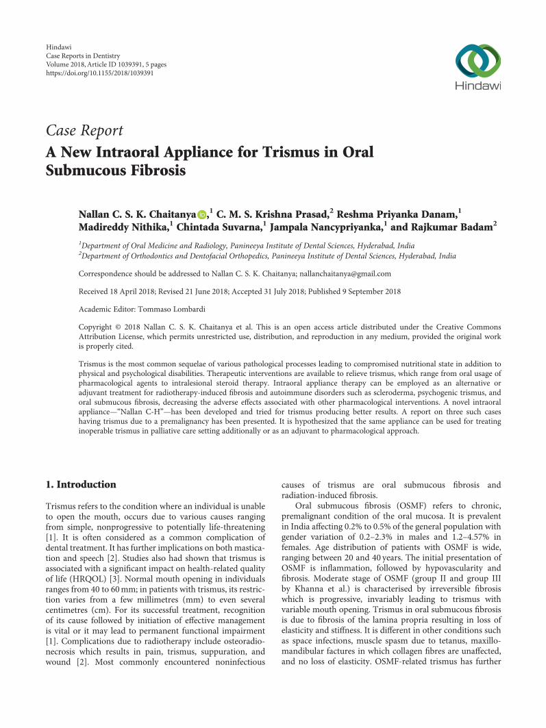
treatment for over a period of time with no recognisablechange or relief, he requested for an alternative therapy.Intraoral appliance therapy was considered, and priorconsent was obtained from the patient. The patient was dulyprovided with necessary precautions regarding the usage ofappliance and weekly follow-up without discontinuing thetreatment. The treatment was carried out for a total periodof 8 weeks and a follow-up of two months after completionof the therapy (Figure 1).
2.2. Case 2. A female patient, aged 56 years, presented to theprivate clinic with chief complaint of difficulty in mouthopening since one month. During her first visit, i.e., approx-imately a year back, she reported about the treatment that shereceived for trismus (due to OSMF) using intralesionalinjections. At that time, the patient had marginal relief fromthe symptoms. Again, she started developing trismus sinceone month and also had burning sensation in the oral cavity.Patient had restricted mouth opening of 30mm (canine-canine distance) and tongue protrusion of 12mm with allsigns of OSMF (group 2 by Khanna et al.) in the oral cavity.As she was not able to tolerate any more pain from intrale-sional steroid injections, she was advised intraoral appliancetherapy for 8 weeks. She was also instructed for weeklyfollow-ups with precautions during appliance position inthe oral cavity.
2.3. Case 3. A male patient, aged 40 years, with a history ofchewing betel quid for the past 15 years, presented to theprivate clinic with reduction in mouth opening since oneyear. Patient had a restricted mouth opening of 35mm(canine-canine distance) and tongue protrusion of 12mmwith all signs of OSMF (group 2 by Khanna et al.) in the oralcavity. The patient was then started with intralesionalcorticosteroids, which showed improved mouth opening till42mm (canine-canine distance), and then this treatmentmodality was discontinued due to pain arising from repeatedpunctures. The patient then requested for alternative ther-apy. He was advised appliance therapy and was instructed
Figure 1: Pre and post appliance therapy.
2 Case Reports in Dentistry
for weekly follow-ups for 8 weeks with precautions inpositioning and usage of the appliance in the oral cavity.
3. Clinical Procedure and Fabrication ofthe Appliance
For all patients, maximum mouth opening was recordedusing appropriate measuring device at baseline prior to theinitiation of the appliance therapy. Necessary precautionswere taken during the fabrication of the appliance such asthe following:
(1) The appliance should not impinge gingival margins
(2) It should be easy to manipulate by the patient
(3) Should be comfortable to use and also rigid enough toresist masticatory forces
Alginate impression was taken for both upper and lowerarches with stock metal trays. Impression was poured usingdental stone. The obtained casts were then articulated withan apex articulator. Fabrication of appliance was done byusing self-cure acrylic resin and sprinkle on technique cover-ing the sulcus area in the anterior region, which broadensposteriorly to cover the buccal area and occlusal surface ofthe lower arch. On the upper arch, only the molar areacovered the teeth both occlusally and buccally. Mounting ofcast in the occlusion was done in a hinge articulator so thatocclusal relation was maintained. The wax was then adaptedon the buccal surface of the lower arch to keep the distance of2mm from the gingiva, so that it did not impinge thesoft tissues. Hyrax screws of 12mm gauge were adapted
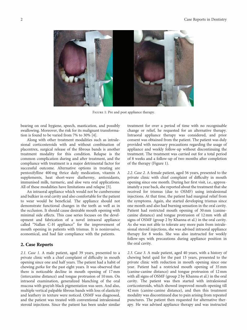
bilaterally on the buccal aspects of the molars on thewax. Precaution was taken to avoid blocking the activationhole of the screws. Once the acrylic had begun to set, theappliance was removed cleaned, trimmed, and polished. Alower labial extension was given for the appliance in orderto prevent accidental breakage and subsequent aspiration ofthe appliance (Figure 2).
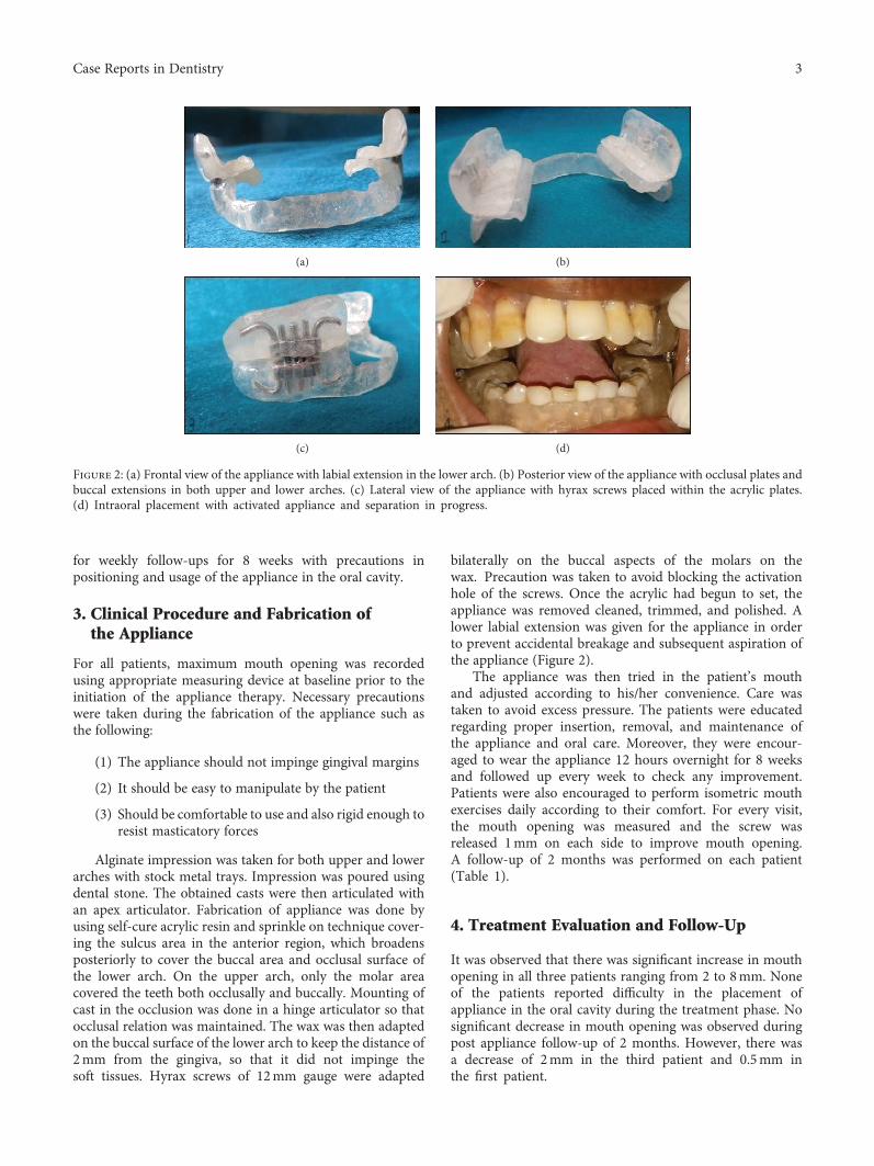
The appliance was then tried in the patient’s mouthand adjusted according to his/her convenience. Care wastaken to avoid excess pressure. The patients were educatedregarding proper insertion, removal, and maintenance ofthe appliance and oral care. Moreover, they were encour-aged to wear the appliance 12 hours overnight for 8 weeksand followed up every week to check any improvement.Patients were also encouraged to perform isometric mouthexercises daily according to their comfort. For every visit,the mouth opening was measured and the screw wasreleased 1mm on each side to improve mouth opening.A follow-up of 2 months was performed on each patient(Table 1).
4. Treatment Evaluation and Follow-Up
It was observed that there was significant increase in mouthopening in all three patients ranging from 2 to 8mm. Noneof the patients reported difficulty in the placement ofappliance in the oral cavity during the treatment phase. Nosignificant decrease in mouth opening was observed duringpost appliance follow-up of 2 months. However, there wasa decrease of 2mm in the third patient and 0.5mm inthe first patient.
(a) (b)
(c) (d)
Figure 2: (a) Frontal view of the appliance with labial extension in the lower arch. (b) Posterior view of the appliance with occlusal plates andbuccal extensions in both upper and lower arches. (c) Lateral view of the appliance with hyrax screws placed within the acrylic plates.(d) Intraoral placement with activated appliance and separation in progress.
3Case Reports in Dentistry
5. Discussion
Trismus is defined as a prolonged tonic spasm of the muscles,which results in restricted mouth opening. OSMF is a poten-tially malignant, chronic, progressive disorder seen mostly inpeople from Asia and is found to affect most of the parts ofthe oral cavity that includes the lips, tongue, palate, pharynx,and even the upper third of the oesophagus. In later stages,further stiffening occurs due to myofibrosis of the subepithe-lial and submucosal tissues, thereby resulting in limitationsin the mouth opening and tongue protrusion causingdifficulty in eating, swallowing, and also phonation-relatedissues [6]. Various treatment modalities such as physical oraltherapy, intralesional corticosteroids, ultrasound therapy,and surgical modalities were tried till date [4].
In the present case series, a newer treatment procedure istried on patients suffering fromOSMF. An appliance that canbe easily fabricated was designed and used in patients withtrismus due to any noninfectious pathology. For patientswho are not comfortable or given consent for treatment withintralesional steroids, this appliance therapy could be analternative treatment modality. Moreover, cost and theadverse effects are involved in this treatment when comparedwith steroid therapy. Yadav et al. believed that protectingsurgically reconstructed defects using flaps is vital, and theauthors fabricated an appliance in order to avoid trauma tothe flap in the postoperative period [6]. It is further believedthat physiotherapeutic effect is a probable mechanism behindappliance therapy, which causes remodelling of the tissuesfor improving mouth opening [7].
From design, the appliance works by causing mechanicalforce which then induces the stretching of the elevator anddepressor muscles. Based on the design, these appliancesare classified into externally and internally activated types.Externally activated appliances exert force by stretching theelevator muscles and depressing the mandible whereasinternally activated appliances employ the force on thedepressor muscles to stretch the elevator muscles. Theyimpart forces which are continuous or intermittent, elasticor nonelastic, and light or heavy. The force generated bythe elevator muscles is greater than that by the depressormuscles. The amount of force delivered depends on thestrength, frequency, duration of stretching, and motivationof the patient [8].
The activation cycle is unique to the appliance. The keywhich is provided for opening up the hyrax screws duringrapid palatal expansion is used for activation purposes inrelieving the trismus. Each full turn is equal to approximately0.2mm and total number of 4 full turns is given whichaccount to 0.8mm per week. The patient is followed up everyweek for 8 weeks. The approximate mouth opening hypoth-esized ranges from 5mm to 1.5 cm.
In the present case series, the appliance emits intermit-tent and bilateral forces, which help to depress the mandibleand make the maxillary and mandibular teeth apart therebyrelieving the trismus. Physical therapy improves the rangeof motion of temporomandibular joint, reduces pain, pre-vents hypomobility, avoids fibrosis formation, strengthensthe musculature, and improves flexibility, tissue elasticity,and blood circulation.
The appliance can be fabricated in patients who arecompletely edentulous and also in those partially edentulouspatients. As the appliance is passive over the teeth and doesnot cause functional tooth movements, it can be comfortablyworn in patients with missing dentition. As the occlusion isundisturbed, there is elimination of alteration in theocclusion sequence. Caution has to be maintained whenthere are periodontal compromised teeth. Excessive verticalforces may further breakdown the periodontium. Periodon-tal assessment has to be carried out before the appliancefabrication. Teeth with excessive mobility and poorprognosis should be managed prior to appliance insertion.Mild-to-moderate periodontitis is not a contraindicationfor appliance fabrication.
Patil et al. from their study concluded that the use ofmouth exercising device appears to be effective for theseparation of collagen fibers and increased the subcutaneousmatrix area leading to improved blood circulation [9]. Oswalet al. fabricated an oral screen prosthesis to stabilize thesecured flaps and to prevent it from being bitten into occlu-sion, and the same can also be used as an oral stent to preventrelapse [10]. Similarly, Li et al. fabricated a EZBite openmouth device and conducted a 12-week structured openmouth training program and stated a marked improvementin mouth opening [11].
Till date, there are two jaw-exercising devices used inpalliative care. “TheraBite” is a mechanical device with leversystem which assists mouth opening by squeezing the handleof the device and is able to control the extent of the stretch to
Table 1: Treatment evaluation at various stages for all 3 patients.
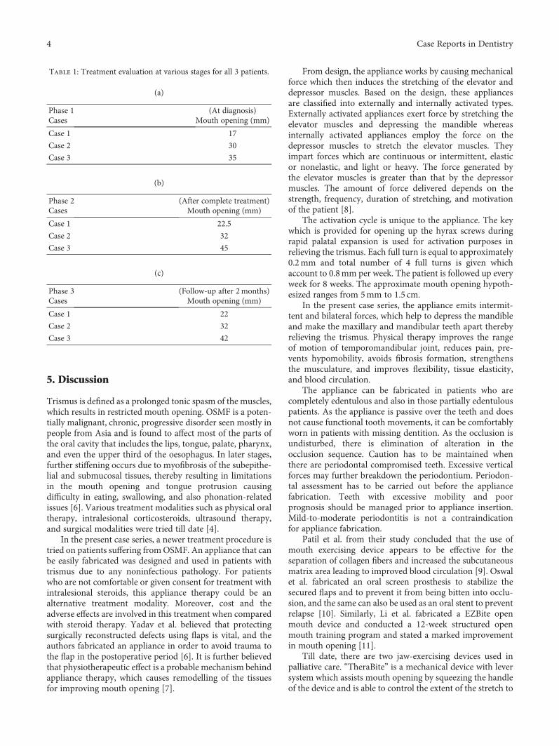
Phase 3 (Follow-up after 2months)Cases Mouth opening (mm)
Case 1 22
Case 2 32
Case 3 42
4 Case Reports in Dentistry
the tissues. Another appliance is “Dynasplint trismus system”which is used with a low-torque and prolonged durationstretch designed to lengthen connective tissue [12, 13].Both of these appliances are used effectively to relievetrismus due to various causes. TheraBite is a lever systemwhich is patient dependent, and the maximum openingclaimed is almost 41mm. Dynasplint trismus system isbulkier compared to the presently described appliance.The present appliance is not visible outside the oral cavityunlike the two above-described systems. The mandibularrange of motion may not be achieved with the present model,and modifications may be required to assess the same infurther fabrication.
6. Biocompatibility of the Appliance
The appliance is made up of acrylic resin material whichis routinely used in the fabrication of partial or completeremovable dentures and hyrax screw which may be usedfor palatal expansion in orthodontic treatment. The com-ponents have been proven to be biocompatible in thepatients. The labial extension of the appliance is a safetymeasure which enables the appliance to stay fit in the oralcavity without breakage and accidental slip into the oralmucosa and esophagus.
7. Limitations and Adverse Effects
The adverse effects with present appliance were as follows:
(1) Difficulty to insert intraorally during the initialphases of treatment
(2) Excessive salivation
(3) Reduced strength of appliance after weeks of usagemay be due to fabrication errors
8. Conclusion and Future Recommendations
The present appliance can also be successfully intervened intrismus arising from any of the abovementioned causeswithout much adverse effects. Long-term studies are requiredto evaluate the effectiveness of the appliance over a largegroup of population and its compliance among the patients.Follow-up at regular intervals may be required for effectivemanagement of the patients with trismus and relapse.
Conflicts of Interest
The authors declare that they have no conflicts of interest.
Acknowledgments
We acknowledge the staff of the Department of OralMedicine and Radiology, Panineeya Institute of DentalSciences, and Dr. Shivaram, senior lecturer, Departmentof Orthodontics and Dentofacial Orthopaedics, for theirsupport in each step of this case study.
References
[1] M. Marien Jr, “Trismus: causes, differential diagnosis, andtreatment,” General Dentistry, vol. 45, no. 4, pp. 350–355,1997.
[2] P. J. Dhanrajani and O. Jonaidel, “Trismus: aetiology, differen-tial diagnosis and treatment,” Dental Update, vol. 29, no. 2,pp. 88–94, 2002.
[3] J. Johnson, M. Johansson, A. Ryden, E. Houltz, and C. Finizia,“Impact of trismus on health-related quality of life and mentalhealth,” Head & Neck, vol. 37, no. 11, pp. 1672–1679, 2015.
[4] U. Wollina, S. B. Verma, F. M. Ali, and K. Patil, “Oralsubmucous fibrosis: an update,” Clinical, Cosmetic andInvestigational Dermatology, vol. 8, pp. 193–204, 2015.
[5] U. Dayanarayana, N. Doggalli, K. Patil, J. Shankar, K. Mahesh,and Sanjay, “Non surgical approaches in treatment of OSF,”IOSR Journal of Dental and Medical Sciences, vol. 13, no. 11,pp. 63–69, 2014.
[6] A. O. Yadav, B. H. Vanza, R. M. Borle, and K. A. Joglekar,“Custom made protective appliance for oral submucousfibrosis,” Journal of Maxillofacial & Oral Surgery, vol. 12,no. 4, pp. 472–474, 2013.
[7] S. Cox and H. Zoellner, “Physiotherapeutic treatmentimproves oral opening in oral submucous fibrosis,” Journalof Oral Pathology & Medicine, vol. 38, no. 2, pp. 220–226,2009.
[8] V. Mehrotra, K. Garg, Z. Sajid, and P. Sharma, “The saviors:appliances used for the treatment of trismus,” InternationalJournal of Preventive & Clinical Dental Research, vol. 1,no. 3, pp. 62–67, 2014.
[9] P. Patil, V. Hazarey, R. Chaudhari, and S. Nimbalkar-Patil,“Clinical efficacy of a mouth-exercising device adjunct to localointment intra-lesional injections and surgical treatment fororal submucous fibrosis: a randomized controlled trial,” AsianPacific Journal of Cancer Prevention, vol. 17, no. 3, pp. 1255–1259, 2016.
[10] C. Oswal, P. Gandhi, and A. Sabane, “Prosthodontic manage-ment of surgically treated oral sub mucous fibrosis using theoral screen prosthesis,” IOSR Journal of Dental and MedicalSciences, vol. 10, no. 4, pp. 33–36, 2013.
[11] Y.-H. Li, C. C. Liu, T. E. Chiang, and Y. W. Chen, “EZBiteopen-mouth device: a new treatment option for oral submu-cous fibrosis-related trismus,” Journal of Dental Sciences,vol. 13, no. 1, pp. 80-81, 2018.
[12] J. I. Kamstra, J. L. N. Roodenburg, C. H. G. Beurskens,H. Reintsema, and P. U. Dijkstra, “TheraBite exercises to treattrismus secondary to head and neck cancer,” Supportive Carein Cancer, vol. 21, no. 4, pp. 951–957, 2013.
[13] J. I. Kamstra, H. Reintsema, J. L. N. Roodenburg, and P. U.Dijkstra, “Dynasplint trismus system exercises for trismussecondary to head and neck cancer: a prospective explor-ative study,” Supportive Care in Cancer, vol. 24, no. 8,pp. 3315–3323, 2016.