ORIGINAL PAPER A new mathematical model to simulate AVA cold-induced vasodilation reaction to local cooling Mohamad Rida & Wafaa Karaki & Nesreen Ghaddar & Kamel Ghali & Jamal Hoballah Received: 24 June 2013 /Revised: 6 January 2014 /Accepted: 7 January 2014 # ISB 2014 Abstract The purpose of this work was to integrate a new mathematical model with a bioheat model, based on physiol- ogy and first principles, to predict thermoregulatory arterio- venous anastomoses (AVA) and cold-induced vasodilation (CIVD) reaction to local cooling. The transient energy balance equations of body segments constrained by thermoregulatory controls were solved numerically to predict segmental core and skin temperatures, and arterial blood flow for given met- abolic rate and environmental conditions. Two similar AVA – CIVD mechanisms were incorporated. The first was activated during drop in local skin temperature (<32 °C). The second mechanism was activated at a minimum finger skin tempera- ture, T CIVD, min , where the AVA flow is dilated and constricted once the skin temperature reached a maximum value. The value of T CIVD,min was determined empirically from values reported in literature for hand immersions in cold fluid. When compared with published data, the model predicted accurately the onset time of CIVD at 25 min and T CIVD,min at 10 °C for hand exposure to still air at 0 °C. Good agreement was also obtained between predicted finger skin temperature and ex- perimentally published values for repeated immersion in cold water at environmental conditions of 30, 25, and 20 °C. The CIVD thermal response was found related to core body tem- perature, finger skin temperature, and initial finger sensible heat loss rate upon exposure to cold fluid. The model captured central and local stimulations of the CIVD and accommodated observed variability reported in literature of onset time of CIVD reaction and T CIVD,min . Keyword Cold-induced vasodilation model . Thermal response due to finger cooling . Blood flow in fingers . Arterio-venous anastomoses Nomenclature A Area, m 2 AVA Arterio-venous anastomoses c Specific heat of blood, J/kg⋅K C Thermal capacitance for each body segment, J/K CIVD Cold-induced vasodilation h c Convection heat transfer coefficient, W/K·m 2 h e Evaporation heat transfer coefficient between the skin and the air, W/kPa·m 2 M Basal metabolic rate, W ˙ m Blood flow rate, kg/s n Number of data points P Vapor pressure, kPa q Sensible heat loss , W Q Heat loss or exchange, W t Time, s T Temperature, °C TMSD Root Mean Square Deviation W Mechanical work done by the body, W Subscripts a Artery adjacent Adjacent node in the previous body segment amb Ambient artery Artery AVA Arterio-venous anastomoses bl Blood CIVD Cold-induced vasodilation c Reference to skin and air convection cr Core cr − sk Core and skin contact exp Experimental value M. Rida : W. Karaki : N. Ghaddar (*) : K. Ghali Mechanical Engineering Department, American University of Beirut, P.O. Box 11-0236, Beirut 1107-2020, Lebanon e-mail: [email protected]J. Hoballah Department of Surgery, American University of Beirut Medical Center, P.O. Box 11-0236, Beirut 1107-2020, Lebanon Int J Biometeorol DOI 10.1007/s00484-014-0792-x
Transcript
ORIGINAL PAPER
A new mathematical model to simulate AVA cold-inducedvasodilation reaction to local cooling
Mohamad Rida & Wafaa Karaki & Nesreen Ghaddar &
Kamel Ghali & Jamal Hoballah
Received: 24 June 2013 /Revised: 6 January 2014 /Accepted: 7 January 2014# ISB 2014
Abstract The purpose of this work was to integrate a newmathematical model with a bioheat model, based on physiol-ogy and first principles, to predict thermoregulatory arterio-venous anastomoses (AVA) and cold-induced vasodilation(CIVD) reaction to local cooling. The transient energy balanceequations of body segments constrained by thermoregulatorycontrols were solved numerically to predict segmental coreand skin temperatures, and arterial blood flow for given met-abolic rate and environmental conditions. Two similar AVA–CIVDmechanisms were incorporated. The first was activatedduring drop in local skin temperature (<32 °C). The secondmechanism was activated at a minimum finger skin tempera-ture, TCIVD, min, where the AVA flow is dilated and constrictedonce the skin temperature reached a maximum value. Thevalue of TCIVD,min was determined empirically from valuesreported in literature for hand immersions in cold fluid. Whencompared with published data, the model predicted accuratelythe onset time of CIVD at 25 min and TCIVD,min at 10 °C forhand exposure to still air at 0 °C. Good agreement was alsoobtained between predicted finger skin temperature and ex-perimentally published values for repeated immersion in coldwater at environmental conditions of 30, 25, and 20 °C. TheCIVD thermal response was found related to core body tem-perature, finger skin temperature, and initial finger sensibleheat loss rate upon exposure to cold fluid. The model capturedcentral and local stimulations of the CIVD and accommodatedobserved variability reported in literature of onset time ofCIVD reaction and TCIVD,min.
Keyword Cold-inducedvasodilationmodel .Thermalresponsedue to finger cooling . Blood flow in fingers . Arterio-venousanastomoses
NomenclatureA Area, m2
AVA Arterio-venous anastomosesc Specific heat of blood, J/kg⋅KC Thermal capacitance for each body segment, J/KCIVD Cold-induced vasodilationhc Convection heat transfer coefficient, W/K·m2
he Evaporation heat transfer coefficient between theskin and the air, W/kPa·m2
M Basal metabolic rate, Wm Blood flow rate, kg/sn Number of data pointsP Vapor pressure, kPaq Sensible heat loss , WQ Heat loss or exchange, Wt Time, sT Temperature, °CTMSD Root Mean Square DeviationW Mechanical work done by the body, W
Subscriptsa Arteryadjacent Adjacent node in the previous body segmentamb Ambientartery ArteryAVA Arterio-venous anastomosesbl BloodCIVD Cold-induced vasodilationc Reference to skin and air convectioncr Corecr−sk Core and skin contactexp Experimental value
M. Rida :W. Karaki :N. Ghaddar (*) :K. GhaliMechanical Engineering Department, American University ofBeirut, P.O. Box 11-0236, Beirut 1107-2020, Lebanone-mail: [email protected]
J. HoballahDepartment of Surgery, American University of Beirut MedicalCenter, P.O. Box 11-0236, Beirut 1107-2020, Lebanon
Int J BiometeorolDOI 10.1007/s00484-014-0792-x
finger Fingermin Minimummodel Modelo Initial value of parameter upon immersion in cold
fluidperf Perfusionr Radiativeres Respirationsk Skinsk, exp Exposed skinshiv Shiveringsur Surfacevein Veinvein, s The superficial vein
Greek symbolsα Coefficient, which is 1 for the chest and zero for all other
body partsτ Time constant of the AVA response (s)
Introduction
When fingers are exposed locally to cold environment, thefinger skin temperature has been reported for some humans tooscillate such that the local finger skin temperature does notgo below a minimum threshold value (Lewis 1930; Castellaniand O’Brien 2005; Daanen and van der Struijs 2005a). Uponfinger exposure to cold environment, vasoconstriction occursinitially to prevent heat loss causing a sharp decrease to theskin temperature threshold value after which a cycle of vaso-dilation and vasoconstriction occurs, believed to protectagainst local cold injury (Castellani et al. 2006; Wilson andGoldman 1970, Iida, 1949; Daanen 2003; Daanen and van derStruijs 2005b; DuCharme and Brajkovic 2005). The vasodi-lation during the CIVD response is caused by dilation of thearterio-venous anastomoses (AVA) in the finger (Daanen 1991;Bergersen et al. 1999).CIVD is reported to be much weaker orabsent in African-Americans and female gender, and it is morepronounced in people working in cold surroundings(Kaciuba-Uscilko and Greenleaf 1989; DeGroot et al. 2003).There is a definite variation in the occurrence, magnitude,and duration of the CIVD (Teichner 1966). However,when the CIVD exists in a normal individual, then thequestion is how this phenomenon influences the periph-eral skin temperature during cold exposure.
Numerous experimental studies have documented CIVDresponse through measurements on finger skin temperatureand body core temperature in experiments on humans atthermally neutral state or slightly warm state who were subjectto local finger or hand cooling for different durations (Daanen1991, 2003; Daanen and Ducharme 1999; Daanen et al. 1997;Sawada 1996; Miura et al. 1977; Chen et al. 1996; Van der
Struijs et al. 2008; Cheung and Mekjavic 2007). Cheung andMekjavic (2007) reported variation of onset finger skin tem-perature of CIVD reaction. Daanen et al. (1997) demonstratedthat the colder the core temperature, the smaller and moredelayed the CIVD response. Sendowski et al. (1997) showedthat cooling of the index finger led to earlier and faster CIVDwithout significant cardiovascular change and thatCIVDonsetcould be influenced by local cooling. Daanen et al. (1997)concluded that the characteristic of the finger skin temperatureresponse during cold water hand immersion depended on coretemperature and the mean skin temperature. Geurts et al.(2005) reported that CIVD was a local phenomenon isolatedto the fingers. However, Sawada et al. (2000) noticed that atambient temperature of 20 °C, the CIVD response after re-peated immersion in water at 8 °C became weak and thesubject core temperature dropped. Clearly, the central temper-ature of the body core and the body local skin temperatureplay a major role in CIVD thermoregulation.
Flouris et al. (2008) reported experimental data on tenadults indicating that when CIVD occurred, the mean bodytemperature decreased while CIVD ceased when body tem-perature fell below a threshold of 36.65 °C. Bergersen et al.(1999) reported that CIVD in the human finger is a localvascular phenomenon triggered by dilatation of the arterio-venous anastomoses (AVA). Flouris and Cheung (2009) report-ed that CIVDwas a centrally originating phenomenon depen-dent on excess heat. A general observation from literaturesurveyed is that there is almost an agreement that the CIVDis centrally controlled by the core temperature as well as by thelocal finger skin temperature. However, there is no agreementon the finger temperature at which this mechanism is trig-gered, and researchers reported different triggering valuesdepending on the environmental conditions of theirexperiments.
The current literature models are incapable of explainingthese discrepancies in CIVD reaction. CIVD modeling ap-proaches have limitation and have not accommodated varia-tions in thresholds of skin and core temperatures for onset ofthe CIVD reaction. There is a need to develop a model basedon physiology that can predict CIVD response with associatedAVA function and blood flow to finger/s taking into consider-ation all the reported parameters that influence this phe-nomenon. The modeling of CIVD response cannot beachieved, independent of accurate modeling AVA thermo-regulatory function during transients where AVAs play akey role with regard to the thermal comfort of extremities(Koscheyev et al. 1998).
Twomodeling approaches were used in literature to predictthe finger and hand thermal responses. One approach was tomodel the hand/finger as independent segment from the body(Shitzer et al. 1997, 1998). These models require empiricalinputs on the arterial and venous blood flow into and from theextremities and on the temperature of the lower arm tissues
Int J Biometeorol
(core and skin) at the connecting interface to the hand. Thesecond modeling approach relied on bioheat modeling of thewhole human to predict skin and core segmental temperaturesusing physiology and thermoregulatory functions. Fewbioheat models incorporated accurate modeling of the extrem-ities AVA functions. Iyoho et al. (2009) used Takemori (1995)to improve the digits thermal response and accounted for theCIVDmechanism by increasing limb blood flow to about 16times the finger nutritional blood flow for a specified period oftime (2 min) when finger skin temperature was below aminimum threshold of 12 °C.
Karaki et al. (2013) extended the multi-branched arterialtree of Avolio (1980) to include the hand artery branching intothe five fingers while adjusting terminal impedances of thesmaller fingers’ arteries. They introduced a new AVAmodel forthe fingers relating to the arterial tree radii as well as the AVAcontrol mechanism to cardiac output. According to Karakiet al. (2013), the maximum vasodilation occurs when bodytemperature is 37.2 °C, although it started at core value above36.7 °C, and the maximum vasoconstriction occurs when theskin temperature reaches 27.8 °C, although it started whenskin temperature is below 33.7 °C. However, their thermalcontrol mechanism did not consider CIVD integration withAVA response.
It is of interest to use an accurate physiological model thatstems from the arterial tree flow modeling to improve modelability for predicting the CIVD reaction. This work aims todevelop a new and robust CIVDmodel and improve thermo-regulatory control equations for both AVA and CIVD in fingersduring cold exposure taking into consideration the variabilityin parameters that trigger CIVD reaction. The model will bevalidated by published experimental data for single and re-peated finger and hand local exposure to cold environment.
Mathematical formulation
Karaki et al. (2013) divided the human body into 25 segments:head, upper and lower chest/trunk, upper arms, forearms,palms, hand fingers, thighs, calves, and feet. Each segmentis composed of a core node, skin node, artery node, vein node,and when applicable a superficial vein node. The modelingof blood flow was based on a modified multi-branchedarterial tree model of Avolio (1980) to include the fivefingers for each hand.
This work extends the bioheat model of Karaki et al. (2013)to include the CIVDobserved in fingers after local exposure tocold in the extremities. In this model, blood flow to the hand isnot needed as an input from empirical data, but is determinedby a model which uses a modified Avolio (1980) multi-branched circulatory system to determine the blood flow intoeach individual body segment, including each of the tenfingers. CIVD occurs when the fingers are exposed to a cold
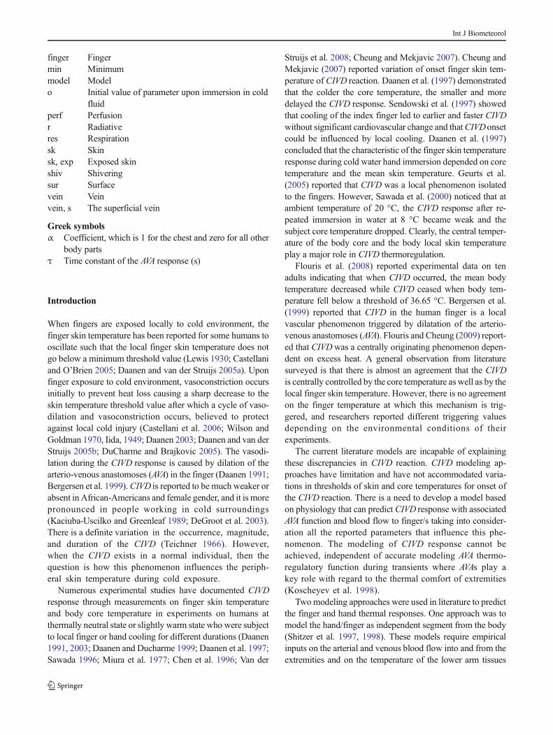
environment, while the rest of the body is subject to warm orcomfortable conditions. Figure 1 shows the AVA and CIVDmechanisms and associated parameters that will be included inthis transient phenomena modeling during local exposure ofthe finger to cold environment. The first AVA mechanism isactivated during drops in local skin temperature below athreshold value, which cause the constriction of AVA flow tothe fingers (Karaki et al. 2013, Shitzer et al. 1998). This AVAmechanism is valid as long as the finger skin temperature isabove a certain minimum limit TCIVD,min. If the body is subjectto warm conditions and the fingers are exposed to cold con-ditions where the local skin temperature crosses the TCIVD,minlimit, CIVD response is triggered and a second CIVD AVAmechanism is activated. The periodic constriction and dilationof the AVA in the fingers result in the periodicCIVD finger skintemperature response observed in experimental literature. TheCIVD response period is noted by tCIVD, and to1 is the onsettime of the CIVD response triggered by a threshold finger skintemperature, while any subsequent to2 …to5 is the referencetime when a change in AVA closing or opening takes place.
The CIVD response, presented through local skin temper-ature variation, is controlled by blood flow to the finger skin.During this CIVD phenomenon, AVA periodically constrictsand dilates causing the temperature in the extremities to followsuit. In our model, this periodic change in blood flow to theskin node due to the CIVD response, similar to the actualphysiological response, is modeled by constricting and dilat-ing the blood flow through the AVAmechanism in the fingersperiodically. Thus, in addition to the normal AVAmechanism,another CIVD specific mechanism was added to the bioheatmodel of Karaki et al. (2013).
This section presents the following: (1) energy balanceequations of the hand, and finger segments for core, skin,and superficial vein nodes; (2) the revised model of AVA andassociated thermoregulatory vasomotor response; (3) new
Fig. 1 Schematic of finger skin temperature curve showingCIVD responseand its associated parameters
Int J Biometeorol
CIVD model based on physiology for predicting finger andbody thermal response under single and repetitive finger/hand exposure to cold environment; and (4) numericalmethodology used in the transient simulations.
Multi-node energy balances of nude segments
Energy balances are performed for the four nodes, core,skin, artery, and vein, of each body segment followingthe formulation of Karaki et al. (2013) while introduc-ing minor modifications on skin node and superficialvein node. In non-periphery segments, the blood comesin through the artery while the blood returning throughcore vein is the combination of blood from the previousvein (mprev;vein ) and the blood coming in throughperfusion from the core. In terminal body segments suchas hands and fingers where there are superficial veins,the blood returning through the superficial vein is theblood from the previous superficial vein (mprev;s;vein )plus the blood perfusion through the skin and alsothrough the AVA. The body segments, which containthe superficial vein node, are the ten digits of the hand,hands, and forearms. Using lumped capacitance, thereare five nodes for the energy balance in each peripheralsegment as shown in Fig. 2a for the forearm segmentand in Fig. 2b for the finger segment. The finger seg-ment shows the AVA path between the artery and theshallow superficial vein.
The standard energy balance for the core node (Karaki et al.2013) is given by
CcrdT cr
dt¼ M cr þM shiv−W−αQres−Qcr‐sk−
Xarteries
harteryAartery T cr−Tbl;a
� �
−Xveins
hveinAvein T cr−Tbl;v
� �þ mperf ;cr þmperf ;sk
� �cbl T bl;a−T crð Þ
ð1Þ
where Ccr is the core thermal capacitance, Mcr and Mshiv
are the basal and segmental thermoregulatory shiveringmetabolic rates, respectively, W is the mechanical workdone by the body, α is a coefficient, which is 1.0 for theupper chest and zero for all other body parts, and Qres isthe heat dissipated through respiration, hartery and hveinare the convection coefficients of the blood in the arteryand core vein, respectively, Aartery and Avein are the cross-sectional area of the artery and core vein, respectively,Tbl,a and Tbl,v are the temperatures of the blood in theartery and core vein, respectively, mperf ;cr is the bloodperfusion in the core only, which is obtained bysubtracting the skin blood perfusion and the AVA bloodflow from the total blood flow into the finger, cbl is theblood specific heat, and Qcr−sk=K (Tcr−Tsk) is the heatexchange between the core and skin at conductance K.
The skin node energy balance is given by
CskdT sk
dt¼ M sk þ Qcr‐sk−Ask;exp
hhc Tsk;sur−T amb
� �þ hr Tsk;sur−T amb
� �þhe Psk−Pambð Þ
iþ mperf ;sk þmAVA
� �cp T cr−T skð Þ
þXveins
hvein;sAvein;s Tbl;v;s−T skð Þð2Þ
where Csk is the skin thermal capacitance, Msk is the skinmetabolic rate, Ask,exp is the exposed skin surface area, hc isthe convection heat transfer coefficient between the skin andthe air, hr is the linearized radiation heat transfer coefficient(ASHRAE 2009), he is the evaporation coefficient deductedfrom hc by Lewis formula, Tsk,sur is the skin surface tempera-ture (House et al. 2002), Pamb is the ambient vapor pressure,Psk is the vapor pressure at Tsk, hvein,s is the convection heattransfer coefficient of blood in the superficial vein, Avein,s is thecross-sectional area of the superficial vein, andmperf ;sk is theblood perfusion in the skin layer andmAVA is the blood flowthrough AVA to the skin layer. The heat transfer coefficientbetween the skin and the air, hc, is calculated using the corre-lations of Ishigaki et al. (1993) for forced and mixed convec-tion around local body segments as function of air velocity andtemperature difference between skin and environment. In
Fig. 2 a–b Schematic of the fivenodes (core, skin, artery, vein, andsuperficial vein) in the energybalances of a the forearm segmentand b the finger segment
Int J Biometeorol
addition, if the segment is locally exposed to waterinstead of air, then radiative and evaporative heat trans-fer coefficients are set to zero.
The artery and vein nodes energy balances for each bodysegment are similar to those used in the bioheat model ofSalloum et al. (2007). Superficial vein nodes exist in distallimbs and digit segments and the energy balance for a super-ficial vein is given by
where Tbl,v,adjacent is the temperature of the blood in the adja-cent core vein of the previous body segment, Cbl.v,s is thesuperficial vein blood capacitance, andmv;s is the blood flowrate through the superficial vein. The last term is direct heattransfer by AVA directly from the artery of the segment to thesuperficial vein.
AVAmechanism model
The AVA mechanism was modified over the model of Karakiet al. (2013) to extend its applicability to conditions when lowskin temperature in extremities triggers CIVD. To create a
unified model of the AVA mechanism during conditions ofCIVD, two main modifications were introduced in the AVAduring CIVD reaction. The first modification was separatingthe total skin blood flow into two distinct parts: skin bloodperfusion mperf ;sk and AVA blood flow to the skinmAVA (seeenergy balance Eqs. (2) and (3) of the skin node and thesuperficial vein node). The second modification was makingthe AVA flow, mAVA , a function of local skin temperature.Previously, under conditions where skin temperature is greaterthan a threshold value below 31.8–32.0 °C, the partition of theskin blood flow was mostly AVA for dissipation of heat. Theskin perfusion flow was very small portion rendering theeffect on thermal response of the extremities to be insignifi-cant. Whereas at lower temperature where AVAmechanism isOFF, accurate estimation of skin blood flow is needed todetermine the thermal and physiologic response, which re-quires modification of the model during these conditions.
Skin perfusion mperf ;sk varies between a minimum and amaximum based on local core and skin temperature. Themodel of Smith (1991) is used to find skin perfusion asfollows:
mperf ;sk ¼ mperf ;sk;dil⋅mperf ;sk;con
mperf ;sk;basal
ð4Þ
where
mperf ;sk;dil ¼ T cr−36:837:2−36:8
mperf ;sk;basal for T cr≤36:8 �C
mperf ;sk;max−mperf ;sk;basal
� �þmperf ;sk;basal for 36:8 �C≤T cr≤37:2 �C
mperf ;sk;max for T cr≤37:2 �C
8>><>>:
and
mperf ;sk;con ¼ T sk−10:733:7−10:7
mperf ;sk;min for T sk≤10:7 �C
mperf ;sk;basal−mperf ;sk;min
� �þmperf ;sk;min for 10:7 �C≤T sk≤33:7 �C
mperf ;sk;basal for T sk≤33:7 �C
8>><>>:
Rubinstein and Sessler (1990) reported that blood flowthrough thermoregulatory arterio-venous shunts can rangefrom negligible up to 80 % of the total cutaneous circulation.They also reported that these AVA shunts have a larger diam-eter (100 μm) compared with capillaries, which gives themthe capability of allowing much greater blood passage, yet at
the same time, they are located deeper in the skin layer thusdissipating less heat than superficial capillaries. To incorporateRubinstein and Sessler’s findings, the minimum constrictedblood flow as reported by Smith (1991) was kept the same andwas considered to be the minimum required nutritional skinperfusionmperf ;sk;min . However, the basal and maximum skin
Int J Biometeorol
perfusion in our model (mperf ;sk;basal and mperf ;sk;max ) wereconservatively taken to be 30 %, of the basal and maximumskin blood flow reported for each body segment by Smith(1991), respectively. In our model, the blood flow flowing intothe hand reported by Smith (1991) was equally divided be-tween the fingers and the palm and dorsal hand, and the flowinto the fingers was divided equally amongst the five fingers.This is consistent with the findings by Johnson et al. (1995)who reported Laser Doppler Flow measurements of bloodflow in the hand to be split between the fingers and the palmby 42 and 58 %, respectively, at standard deviation of ±26 %.
The AVA control mechanism in the ten digits in our modeldepends on the local skin temperature. The exact temperatureat which the AVA turns on and off under total body heating orcooling was derived from comparing multiple experimentalvalues of finger skin temperature at which the sharp decline ofskin temperature begins at Tskin=32 °C (Kuklane et al. 2011;Vanggaard et al. 2012; Shitzer et al. 1997; Karaki et al. 2013).The AVA blood flow is activated through integrating a rate ofchange at finger skin condition that triggers this change. TheAVA blood flow model during ON-mode (when Tskin of fingeris above 32 °C and Tco-av>36.7 °C) is given in deferential andintegrated form in Eqs. (6a) and (6b), respectively, as follows:
dmAVA
dt¼ mfinger−mperf ;sk−mAVA;0
� � 1
τe−
t−t0ð Þτ ð5aÞ
mAVA tð Þ ¼ mfinger−mperf ;sk−mAVA;0
� �1−e−
t−t0ð Þτ
� �ð5bÞ
while the AVA blood flow model during OFF mode (whenTcore_av<36.7 °C) is given in deferential and integrated form inEq. 7, respectively, as follows:
dmAVA
dt¼mAVA;0
1
τe−
t−t0ð Þτ ð6aÞ
mAVA ¼mAVA;0⋅e−t−t0ð Þτ ð6bÞ
where τ is a time constant control parameter for the exponen-tial rate of change of the AVA flow and to is the reference timeto2…to5 when a change in AVA closing or opening takes place.This parameter was determined empirically to match pub-lished all experimental data of single and multiple exposuresto air or water with a maximum interval 30–60 min (Shitzeret al. 1998, Sawada et al. 2000). The value of τ is accordinglyfound to be equal to 500 s. When the AVA turns ON, itincreases the AVA flow, mAVA , exponentially to a maximumvalue equal to the total amount of blood flow in the finger(mfinger ) minus the skin perfusion and minus the initial AVAflow present mAVA;0 when the AVA turned on at time t0,
[(mAVA;max ¼mfinger−mperf ;sk−mAVA;0 ) as in Eq. (5a)]. Thevalue of the maximum AVA flow, mAVA;max , does changesignificantly in comparison with the changes in the AVA flowmAVA and can be considered constant during the period inwhich AVA turns ON or OFF. If the AVA turns OFF, the AVAblood flow decreases exponentially from the initial AVA flowpresent when the AVAwas turned off to a minimum value ofzero. This effectively makes the AVA flow range from a min-imum of zero to a maximum possible value equal to 70 % ofthe total skin blood flow at maximum dilation.
CIVDmechanism model
When the local finger skin temperature drops below 32 °C,constriction of AVA flow to the fingers occur (see Fig. 1). ThisAVA OFF threshold of finger skin temperature is determinedempirically as mentioned earlier from published experimentaldata of (Shitzer et al. 1998, Kuklane et al. 2011, Vanggaardet al. 2012). TheCIVDmechanism is valid for the temperaturerange of TCIVD,min to TCIVD,max. The regular AVA modelEqs. (5a–5b) and (6a–6b) are valid when the finger tempera-ture is above TCIVD,max and the ON/OFF threshold being forfinger skin temperature below 32 °C. The value of theTCIVD,min and TCIVD,max are determined empirically by com-parison to published experimental results of Shitzer et al.(1998), Cheung and Mekjavic (2007), and Sawada et al.(2000), as will be shown in the results section.
If the body is subject to warm conditions (Tcore>36.7 °C)and the fingers are exposed to cold conditions where the localskin temperature crosses the TCIVD,min limit, CIVD response istriggered and a second CIVD–AVA mechanism is activated.The two mechanisms follow the same AVA equation with thedifference being the time constant that is adjusted empiricallyto better predict the CIVD response. The response path of theCIVD–AVA flow is described by the AVA flow being dilatedbelow a finger skin temperature TCIVD,min where it remainsdilated until it reaches a skin temperature TCIVD,max. Then theAVA is constricted again once above a temperature TCIVD,maxand remains constricted while the skin temperature isdropping till TCIVD,min. The periodic constriction and dilationof the AVA in the fingers results in the periodic CIVD fingerskin temperature response observed in experimental literature.The AVAandCIVD triggering conditions for ON andOFF stateare summarized in Table 1.
The values of TCIVD,min, TCIVD,max are not universal and havebeen observed to depend on body core temperature and environ-mental conditions (Cheung and Mekjavic 2007; Sawada et al.2000). The response is central in the sense that it depends on theaverage core temperature of the body determined by the thresh-old set at 36.7 °C because if the core temperature is less than thatvalue, no matter what the local conditions are, theAVAwill not beactivated. However, the local control and the degree of openness
Int J Biometeorol
of the AVA is dependent on environmental cooling conditions,which affect the local rate of heat loss, and this might explainsome of the difference for onset of CIVD when exposure takesplace in water or air. The local heat loss rate from the fingercontrols the openness of the AVAand the onset temperature of theCIVD activation. The magnitude of CIVD, defined asΔTCIVD=(TCIVD,max−TCIVD,min), is reported to be subject depen-dent where each human body responds differently when theextremity is exposed to cold environment. This led us to assumean average difference ΔTCIVD of 4 °C, which occurred for themajority of subjects (Shitzer et al. 1998, Sawada et al. 2000).
The threshold minimum temperature for the CIVD changesnot only from one subject to another for the same conditionbut also for the same subject at different ambient conditions ofbody environment (affecting body core temperature) and localcooling environment (Sawada et al. 2000). The observeddependence of TCIVD,min on the initial drop in skin tempera-ture is incorporated by calculating the initial heat loss in thefinger immediately after immersion and comparing reportedvalues of TCIVD,min in literature at different local environmenttemperature surrounding the immersed finger in water and theexposed finger to air.
Table 2 summarizes the conditions for the adoptedTCIVD,min for each range of finger initial sensible heat loss,qo,finger. The CIVDminimum temperature TCIVD,min decreases
almost linearly with initial heat loss upon exposure. It is fittedwith the following correlation:
TCIVD;min ¼ 0:4429� qo;finger
þ 9:2916 Valid for 1:59 W < qo;finger < 10:48 W
ð7Þ
where TCIVD,min is in °C. When the initial finger heat loss ishigh, it triggers earlier CIVD reaction at higher TCIVD,min. Theresults section will show comparisons with various experi-mental published data on finger skin temperature under singleand repeated exposure of finger or whole hand to air or waterat different local and global environment temperatures. Notethat previous models have used a fixed temperature thresholdof 12 °C irrespective of exposure local conditions and envi-ronment global conditions (Iyoho et al. 2009).
Numerical methods
The numerical methodology is based on Euler-Forward first-order scheme of Salloum et al. (2007) with a time stepΔt. Thetransient coupled blood flow model and the thermal bioheatmodel introduced by Karaki et al. (2013) was extended toinclude the interdependent AVA and CIVDmodels when localcooling of extremities take place to predict nodal temperaturesof core, skin, artery, vein, and peripheral vein nodes or eachsegment including fingers during CIVD. For given initialthermal state of the human body, metabolic rate, ambientconditions, and the physiological and physical parametersinherent in the bioheat model of the human body, the simula-tion program will update at every time step, i, the core andregional blood flow rates, thermoregulatory responses, skinvapor pressure, and temperatures of the core, skin, artery, vein,and superficial vein nodes. The discrete model of the AVA flowrate during ON and OFF periods given in Eqs. (5b) and (6b) isalso incorporated in the numerical model to calculate the AVAblood flow at any instant of time at time step i.
At the start of any simulation, the AVA is initialized to zeroand the associated bioheat model of Karaki et al. (2013) isallowed to reach steady state at the initial conditions of anysimulated experiment (metabolic rate and environment condi-tions of the room) before beginning any transient analysisassociated with finger or hand exposure to cold conditionsor any other form of transient imposed either by changes inroom conditions or activity level of the human. In other words,suitable initial conditions were determined starting from theneutral conditions of Tcr=36.7 °C and Tsk=33.7 °C to simulatea relatively long exposure to any pre-conditioning environ-ment. The obtained steady state values were then used asinitial conditions for other unsteady calculations of variousnode temperatures for all segments.
A time stepΔt of 0.02 s is used over the desired simulationperiod given that a complete cycle of the heart beat
Table 1 The AVA and CIVD triggering conditions for ON or OFF state
Thermal conditions for AVA and CIVD mechanisms AVA CIVD
Average bodycore temperature
Finger skintemperature
Rate of change ofskin temperature
Tcr_av<36.7 °C OFF OFF
Tcr_av>36.7 °C Tskin≥32 °C ON OFF
TCIVD,max≤Tskin<32 °C
OFF OFF
TCIVD,min≤Tskin<TCIVD,max
dT skindt < 0 OFF OFF
TCIVD,min≤Tskin<TCIVD,max
dT skindt > 0 OFF ON
Tskin<TCIVD,min OFF ON
Table 2 The conditions for the adopted TCIVD,min for each range of fingerinitial heat loss
qo,finger (W) TCIVD,min (°C) (Reference)
<1.59 10 –
1.59 10 Shitzer et al. (1998)
6.08 12 Sawada et al. (2000) at Tamb=20 °C
9.25 13.3 Sawada et al. (2000) at Tamb=25 °C
10.48 14 Sawada et al. (2000) at Tamb=30 °C
>10.48 14 -
Int J Biometeorol
approximately takes 0.8 s at the neutral state of the body andcan vary depending on the metabolic rate and core tempera-ture (Salloum et al. 2007). Numerical tests were performed foruniform time step sizes of 0.02, 0.01, and 0.005 s. The timestep of 0.02 s was found to be of sufficient accuracy at relativeerror of less than 0.008 % in values of blood flow ratescompared with the lower time step and an error of less than0.01 °C in predicted temperature.
Results and discussion
In this section, the extended bioheat model for AVA–CIVDresponse in hand and fingers based on thermal modelingphysiology is validated by comparing simulated finger skin
and body core temperatures with several published experi-mental data of other researchers.
Model validation with published experimental datafor continuous hand exposure to cold environment
Shitzer et al. (1998) published experimental data of Sharpand Hamlet (1988/1989) on finger skin temperature for abare hand exposed for 1 h to still air at 0 °C while theparticipants were sitting indoor. In addition, Shitzer et al.(1998) used the experimental data to validate their handmodel assuming a constant arterial blood temperature of30 °C and AVA blood flow variations that were symmetricand triangular waves allowing for gradual opening–closingcycles of the blood flow to the fingers. The results from
Fig. 3 a–cSchematic of variationin time of a, the reportedmeasurements of Sharp andHamlet (1988/9) by Shitzer et al.(1998) and predicted values offinger skin temperature uponexposed to still air at 0 °C and bfinger AVA and skin perfusionblood flow rate during CIVDreaction, and c finger sensible heatloss during CIVD reaction
Int J Biometeorol
their model compared well to measurements recorded viainfrared camera of bare fingertip temperature after handexposure for 1 h to still air at 0 °C. We simulated experi-ments reported by Shitzer et al. (1998) to predict finger skintemperature using our current proposed new CIVD model.The global environmental conditions were not specified inthe work but are assumed to be at 28 °C for the purpose ofthe simulation since we are modeling the whole humanbody and not just the hand segment.
Figure 3a presents a plot of the reported measurements byShitzer et al. (1998) and predicted values of finger skin tem-perature as a function of time. It is clear that our presentedmodel based on physiology was able to predict experimentalCIVD thermal response of the hand and fingers as well as theonset time within acceptable accuracy. The associated AVAandCIVD mechanisms are shown in Fig. 3b where the corre-sponding AVA blood flow mAVA into the finger is shownduring local cooling in addition to the skin perfusion bloodflow rate (see scale on the right hand axis of the graph) thatreflects our assumption of almost 30 % of the total blood flowinto the finger with its value ranged between an initialmaximum of 4.92×10⁻⁵ and a minimum value of 4.83×10⁻⁵ kg/s during the 1 h of simulation period. Fingerblood perfusion is periodic and follows AVA’s cyclic flow.Figure 3c shows the corresponding total sensible heat lossfrom the finger. The AVA is closed during cooling until ithits the TCIVD,min of 10 °C triggered by high heat lossexperienced immediately after the immersion of finger incold fluid. The finger heat loss and AVA blood flow are insynchronization with a short delay of 2.2 s in heat losswhere the warm blood heats the finger and when AVAcloses, the finger skin temperature goes down.
Another validation is done by comparing the model pre-dictions with the experimental data published by Cheung andMekjavic (2007) for hand immersion in cold water. Theexperiments were conducted in an ambient environmentbetween 24 and 26 °C with relative humidity of 40–45 %, and seated subjects wore T-shirt and shorts.They started the experiment by immersing the handfirstly in hot water of 35 °C for 5 min, then the partic-ipants placed their hands in the circulating water bath of8 °C. Figure 4 shows the measurements of Cheung andMekjavic (2007) and predicted values of finger skintemperature by simulation as a function of time. Theagreement is excellent, and the model was able to accu-rately predict the onset time and the threshold minimumCIVD temperature for the CIVD reaction to start. Inaddition, the CIVD reaction was seen to diminish afterthe first CIVD cycle as also seen in the simulations dueto drop of the body core temperature to value below36.7 °C (22.23 min after immersion), which caused con-tinued vasoconstriction to prevent core heat loss.According to the simulations, when only one finger isimmersed in water of 8 °C and the rest of the body wasexposed to 26 °C air, the decrease in core temperatureafter 20 min of exposure was of the order of 0.02 °C.However, when the whole hand is immersed in water forthe same duration, core temperature decreases from 36.8to 36.75 °C.
Cold water immersion resulted in faster drop in fingerskin temperature and also shorter CIVD period where inhalf an hour, two hunting reactions are detected while ittook one complete hour to notice the two hunting CIVDreactions in still air.
Fig. 4 A plot of the reportedmeasurements of Cheung andMekjavic (2007) and predictedvalues of finger skin temperatureby simulation as a function oftime for finger and handimmersion in cold water at 8 °C
Int J Biometeorol
Model validation for repeated finger immersion in water
Sawada et al. (2000) reported experimental results on fingerskin temperature of six participants who immersed their leftindex finger in cold water of 10 °C for 10min six times, whereafter each cold water immersion, a recovery period of 5 min inair is imposed. The experiments of Sawada et al. (2000) weredone for three different ambient conditions of 30, 25, and20 °C with a relative humidity of 50 %. Subjects wore T-shirt and short pants during the experiment. The reported
finger skin temperature was based on values recorded every10 s using a thermistor probes. They observed that the CIVDresponse did not change upon repetition of immersion attemperatures of 25 and 30 °C, but became weak and delayedat 20 °C ambient conditions. During the repeated cold waterimmersion, they also found that the lowest threshold temper-ature for the CIVD is at the lower ambient condition 20 °C andhighest at 30 °C. These experimental data were instrumentalin developing the CIVDmodel of this work and estimating thethreshold temperature TCIVD,min as a function of finger
Fig. 5 a–c Plots of the reportedmeasurements by Sawada et al.(2000) on finger skin temperatureand the predicted values bysimulation model at environmenttemperature of a 20, b 25, and c30 °C
Int J Biometeorol
sensible heat loss rate immediately after immersion in coldenvironment (see Table 2).
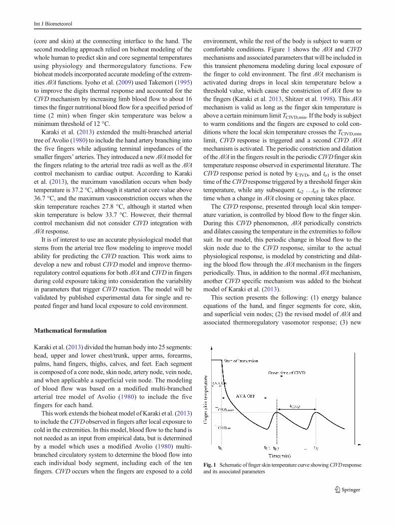
The environmental conditions of experiments conductedby Sawada et al. (2000) and human metabolic rate and cloth-ing parameters were simulated using our modified bioheatmodel with the new AVA–CIVD model to predict finger skintemperature in addition to finger heat loss and body coretemperature. Figure 5a–c shows plots of the reported mea-surements by Sawada et al. (2000) on finger skin temperatureand the predicted values by simulation model at environmenttemperature of (a) 20 °C, (b) 25 °C, and (c) 30 °C. The
predicted skin temperature has captured accurately the CIVDresponse in all three ambient conditions. We calculated theabsolute maximum error between reported mean measuredexperimental values based on six subjects by Sawada et al.(2000) and predicted values of finger skin temperature in ourmodel, and it was less than 0.7 °C. The maximum error iswithin the standard error reported by Sawada et al. (2000) onvariations of subjects’ finger skin temperatures from the meanvalues. In addition, the Root Mean Square Deviation,
Fig. 6 a–c Plots of the predictedsensible heat by the model offinger skin temperature and thepredicted values by simulationmodel at environmenttemperature of a 20, b 25, and c30 °C of Sawada et al. (2000)published experimentalconditions
Int J Biometeorol
the ambient temperature experiments of Sawada et al. (2000)at 20, 25, and 30 °C and of our model resulting in RMSDvalues of 0.61, 0.71, and 0.66 °C, respectively. The RMSDvalues for our model are also acceptable as they are withinexperimental variability of measured value from differentsubjects.
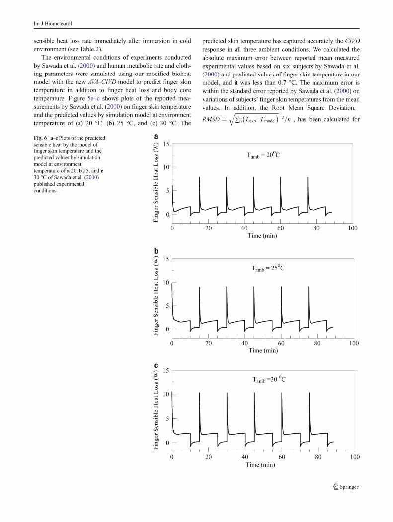
The mean core temperatures predicted by the model were36.82±0.01 °C, 36.74±0.01 °C, and 36.715±0.005 °C atambient temperature 30, 25, and 20 °C, respectively. Sawadaet al. (2000) reported that during the experiments at 30 and25 °C ambient temperature, oral temperature was almostconstant throughout at 36.9 °C±0.23 °C whereas oral temper-ature was lower at ambient of 20 °C. The higher core temper-ature is predicted at the higher ambient temperature cases with
values within the range of reported measured values and theirstandard deviation. Upon examining the predicted initial fin-ger heat loss immediately after immersion (see Fig. 6a–c), it isfound that the highest initial heat loss rate occurs at Tamb=30 °C where we have the highest temperature differencebetween initial skin temperature and the temperature of waterin which the finger is immersed. The heat loss from the fingerdrops by almost 40 % within the first 10 s of the coldimmersion (Fig. 6a), while the change in core temperature issmall during CIVD reaction. Sawada et al. (2000) have notprovided measurements of the core temperature, but our mod-el has captured this effect and has accounted for the overallthermal state of the body affected by environmental condi-tions. Figure 7a, b shows the predicted total blood flow rate,
Fig. 7 a–b Plots of the predictedtotal blood flow rate, AVA bloodflow rate, and skin perfusion ofthe finger in kg/s by simulationmodel at environmenttemperature of a 20, b 25, and c30 °C of the publishedexperimental conditions ofSawada et al. (2000)
Int J Biometeorol
AVAblood flow rate, and skin perfusion of the finger in kg/s bysimulation model at environment temperature of (a) 20 °C, (b)25 °C, and (c) 30 °C of the published experimental conditionsof Sawada et al. (2000). The finger blood flow directly influ-ences the finger heat loss (see Fig. 6a–c).
Conclusions
The segmental bioheat model of Karaki et al. (2013) has beenmodified to accurately predict the transient CIVD response infingers after local exposure to cold conditions. The modelresulted in accurate predictions of CIVD thermal responseand its control parameters stemmed from physiology relatingthe response to the core body-average temperature, the fingerskin local temperature, and the initial skin heat loss rate fromthe finger.
Future research will focus on implementingCIVDmodel inapplications such as using of hand cooling as a mechanism toefficiently cool the body after high metabolic activity(Giesbrecht et al. 2007; Kuennen et al. 2010) and designingheating vests (Brajkovic and Ducharme 2001) that can im-prove CIVD response in hands and increase functionality ofthe hands during cold.
References
ASHRAE Handbook of Fundamentals (2009) American Society ofHeating Refrigerating and Air-Conditioning Engineer. ASHRAEInc, Atlanta
Avolio AP (1980) Multi-branched model of the human arterial system.Med Biol Eng Comp 18:709–718
Bergersen TK, Hisdal J, Walløe L (1999) Perfusion of the human fingerduring cold-induced vasodilatation. Am J Physiol 276(3, Pt. 2):R731–R739
Brajkovic D, Ducharme MB (2001) Maintaining finger dexterity in thecold: a comparison of passive, direct and indirect hand heatingmethods. RTO HFM Symposium on Blowing Hot and Cold:Protecting Against Climatic Extremes, held in Dresden, Germany,8–10 October 2001, and published in RTO-MP-076
Castellani JW, O’Brien C (2005) Peripheral vasodilation responses toprevent local cold injuries. In Prevention of Cold Injuries (pp. KN2–1 – KN2–14). Meeting Proceedings RTO-MP-HFM-126, May 2,2005, Keynote 2. Neuilly-sur-Seine, France: RTO
Castellani JW, Young AJ, Ducharme MB, Giesbrecht GG, Glickman E,Sallis RE (2006) Prevention of cold injuries during exercise. MedSci Sports Exerc 38(11):2012–2029
Chen F, Liu ZY, Holmer I (1996) Hand and Wnger skin temperatures inconvective and contact cold exposure. Eur J Appl Physiol OccupPhysiol 72:372–379
Cheung SS, Mekjavic IB (2007) Cold-induced vasodilatation is nothomogenous or generalizable across the hand and feet. Eur J ApplPhysiol 99:701–705
Daanen HAM (1991) Arterio-venous anastomoses and thermoregulation.Report No. IZF 1991 B-12, TNO Institute for Perception Group:Thermophysiology, Soesterberg, Netherland, 1991
Daanen HAM (2003) Finger cold-induced vasodilation: a review. EurJAppl Physiol 89:411–426
Daanen HA, Ducharme MB (1999) Finger cold-induced vasodilationduring mild hypothermia, hyperthermia and at thermoneutrality.Aviat Space Environ Med 70:1206–1210
Daanen HAM, van der Struijs N (2005a) The Risk Index for frostbite. InPrevention of Cold Injuries. Meeting Proceedings RTO-MP-HFM-126, Paper 13: 13-1 – 13-10. Neuilly-sur-Seine, France: RTO. May2005
Daanen HAM, van der Struijs NR (2005b) Resistance index of frostbiteas a predictor of cold injury in arctic operations. Aviat SpaceEnviron Med 76(12):1119–1122
Daanen HAM, van de Linde FJG, Romet TT, Ducharme MB (1997) Theeffect of body temperature on the hunting response of the middlefinger skin temperature. Eur J Appl Physiol 76:538–543
DeGroot DW, Castellani JW, Williams JO, Amoroso PJ (2003)Epidemiology of U.S. Army cold weather injuries, 1980-1999.Aviat Space Environ Med 74:564–570
DuCharme MB, Brajkovic D (2005) Risk of frostbite and CIVD on theface during cold wind exposure. Defense Research & Development,Canada, RTO-MP-HFM-168
Flouris AD, Westwood DA, Mekjavic IB, Cheung SS (2008) Effect ofbody temperature on cold induced vasodilation. Eur J Appl Physiol104:491–499
Flouris AD, Cheung SS (2009) Influence of thermal balance on cold-induced vasodilation. J Appl Physiol 106:1264–1271
Geurts CLM, Sleive G, Cheung SS (2005) Effect of cold-induced vaso-dilatation in the index finger on temperature and contractile charac-teristics of the first dorsal interosseus muscle during cold-waterimmersion. Eur J Appl Physiol 93:524–529
Giesbrecht GG, Jamieson C, Cahill F (2007) Cooling hyperthermic firefighters by immersing forearms and hands in 10°c and 20°c water.Aviat Space Environ Med 78(6):561–567
House JR, Michael AE, Tipton J (2002) Using skin temperature gradientsor skin heat flux measurements to determine thresholds of vasocon-striction and vasodilatation. Eur J Appl Physiol 88:141–145
Iida T (1949) Studies concerning vascular reaction to cold (Part 1),Physiological significance of vascular reaction to cold. J PhysiolSoc Jpn 11:73–78
Ishigaki H, Horikoshi IT, Uematsu T, SahashiM, Tsuchikawa T,MochidaST, Hieda T, Isoda N, Kuno SH (1993) The convective heat transfercoefficient of the human body is essential to predict convective heatloss from the body. J Therm Biol 18(5/6):455–458
Iyoho AE, Jang T, Nair SS (2009) Human thermal model with extremitiesfor asymmetric environments. ASHRAE Trans 115(1):484–495
Johnson JM, Pergola PE, Liao FK, Kellogg DL Jr, Crandall CG (1995)Skin of the dorsal aspect of human hands and fingers possesses anactive vasodilator system. J Appl Physiol 78(3):948–954
Kaciuba-Uscilko H, Greenleaf GE (1989) Acclimatization to cold inhumans, National Aeronautics and Space Administration (NASA),EASA-TB- 10 10 12
Karaki W, Ghaddar N, Ghali K, Kalev K, Holmer I, Vanguard LL (2013)Human thermal response with improved AVA modeling of thedigits. Int J Therm Sci 67:41–52
Koscheyev VS, Paul S, Leon GR, Tanchida D, Taylor TJ, Koscheyev IV(1998) Body surface temperature tuning as a comfort support systemin space and other extreme environments. SAE Technical PaperSeries 981723. In Proceedings of the 28th International Conferenceon Environmental Systems, July 13–16, Danvers, MA, pp. 1–8, 1998
Kuklane K, Smolander J, Holmér I, Vanggaard L (2011) Does reducedheat production during mild whole body cooling override increasedheat generation by pre-shivering muscle tension? XIV International
Int J Biometeorol
Conference on Environmental Ergonomics, Nafplio, Greece, July10-15, 2011
Lewis T (1930) Observations upon the reactions of the vessels of thehuman skin to cold. Heart 15:177–208
Miura T, Kimotsuki K, Tominaga Y, Suzuki Y (1977) Effect of environ-mental conditions on the cold-induced vasodilation of officeworkers and forestry workers. J Science of Labor 53:75–81
Rubinstein EH, Sessler DI (1990) Skin-surface temperature gradientscorrelate with fingertip blood flow in humans. Journal ofAnesthesiology 73(3):541–545
Salloum M, Ghaddar N, Ghali K (2007) A new transient bio-heat modelof the human body and its integration to clothing models. Int JTherm Sci 46(4):371–384
Sawada S (1996) Cold-induced vasodilatation response of finger skinblood vessels in older men observed by using a modified local coldtolerance test. Ind Health 34:51–56
Sawada S, Araki S, Yokoyama K (2000) Changes in cold-inducedvasodilatation, pain and cold sensation in fingers caused byrepeated finger cooling in a cool environment. Ind Health 38:79–86
Sendowski I, Sarourey G, Besnard Y, Bittel J (1997) Cold inducedvasodilatation and cardiovascular responses in humans during coldwater immersion of various upper limb areas. Eur J Appl PhysiolOccup Physiol 75(6):471–477
Sharp MW, Hamlet MP (1988/9) Peripheral circulatory effects of coldstress and smoked or chewed tobacco products measured by infrared
thermography. US Army Research Institute of EnvironmentMedicine, Natick, MA (unpublished)
Shitzer A, Bellomo S, Stroschein LA, Gonzalez R, Pandolf KB(1998) Simulation of a cold-stressed finger including theeffects of wind, gloves, and cold-induced vasodilatation. JBiomed Eng 120:389–394
Shitzer A, Stoschein LA, Vital P, Gonzalez RR, Pandolf KB (1997)Numerical analysis of an extremity in a cold environment includingcountercurrent arterio-venous heat exchange. J Biomech Eng 119:179–186
Smith CE (1991) A transient three-dimensional model of the thermalsystem, PhD thesis, Kansas State University
Takemori T (1995) A fundamental model of the human thermal systemfor prediction of thermal comfort. Heat Transfer-Japanese Research24(2):147–165
Teichner W (1966) Individual thermal and behavioral factors in cold-induced vasodilatation. Psychophysiology 2(4):295–304
Van der Struijs NR, Van Es EM, Raymann RJEM, Daanen HAM (2008)Finger and toe temperatures on exposure to cold water and cold air.Aviat Space Environ Med 79:941–946
Vanggaard L, Kuklane K, Holmer I, Smolander J (2012) Thermal re-sponses to whole-body cooling in air with special reference toarteriovenous anastomoses in fingers. Clin Physiol Funct Imaging32(6):463–469
Wilson O, Goldman RF (1970) Role of air temperature and wind in thetime necessary for a finger to freeze. J Appl Physiol 29(5):658–664