This article was downloaded by: [American Public University System] On: 27 December 2013, At: 06:19 Publisher: Taylor & Francis Informa Ltd Registered in England and Wales Registered Number: 1072954 Registered office: Mortimer House, 37-41 Mortimer Street, London W1T 3JH, UK Journal of Natural History Publication details, including instructions for authors and subscription information: http://www.tandfonline.com/loi/tnah20 A new species of Remanea Klie, 1929 (Copepoda: Harpacticoida: Paramesochridae) with a redescription of the type species Jinwook Back a , Wonchoel Lee a & Rony Huys b a Department of Life Science , College of Natural Sciences, Hanyang University , Seoul, 133-791, Korea b Department of Zoology , Natural History Museum , Cromwell Road, London, SW7 5BD, UK Published online: 03 Nov 2011. To cite this article: Jinwook Back , Wonchoel Lee & Rony Huys (2011) A new species of Remanea Klie, 1929 (Copepoda: Harpacticoida: Paramesochridae) with a redescription of the type species, Journal of Natural History, 45:47-48, 2939-2964, DOI: 10.1080/00222933.2011.622057 To link to this article: http://dx.doi.org/10.1080/00222933.2011.622057 PLEASE SCROLL DOWN FOR ARTICLE Taylor & Francis makes every effort to ensure the accuracy of all the information (the “Content”) contained in the publications on our platform. However, Taylor & Francis, our agents, and our licensors make no representations or warranties whatsoever as to the accuracy, completeness, or suitability for any purpose of the Content. Any opinions and views expressed in this publication are the opinions and views of the authors, and are not the views of or endorsed by Taylor & Francis. The accuracy of the Content should not be relied upon and should be independently verified with primary sources of information. Taylor and Francis shall not be liable for any losses, actions, claims, proceedings, demands, costs, expenses, damages, and other liabilities whatsoever or howsoever caused arising directly or indirectly in connection with, in relation to or arising out of the use of the Content. This article may be used for research, teaching, and private study purposes. Any substantial or systematic reproduction, redistribution, reselling, loan, sub-licensing, systematic supply, or distribution in any form to anyone is expressly forbidden. Terms &
Transcript
This article was downloaded by: [American Public University System]On: 27 December 2013, At: 06:19Publisher: Taylor & FrancisInforma Ltd Registered in England and Wales Registered Number: 1072954 Registeredoffice: Mortimer House, 37-41 Mortimer Street, London W1T 3JH, UK
Journal of Natural HistoryPublication details, including instructions for authors andsubscription information:http://www.tandfonline.com/loi/tnah20
A new species of Remanea Klie,1929 (Copepoda: Harpacticoida:Paramesochridae) with a redescriptionof the type speciesJinwook Back a , Wonchoel Lee a & Rony Huys ba Department of Life Science , College of Natural Sciences,Hanyang University , Seoul, 133-791, Koreab Department of Zoology , Natural History Museum , CromwellRoad, London, SW7 5BD, UKPublished online: 03 Nov 2011.
To cite this article: Jinwook Back , Wonchoel Lee & Rony Huys (2011) A new species of RemaneaKlie, 1929 (Copepoda: Harpacticoida: Paramesochridae) with a redescription of the type species,Journal of Natural History, 45:47-48, 2939-2964, DOI: 10.1080/00222933.2011.622057
To link to this article: http://dx.doi.org/10.1080/00222933.2011.622057
PLEASE SCROLL DOWN FOR ARTICLE
Taylor & Francis makes every effort to ensure the accuracy of all the information (the“Content”) contained in the publications on our platform. However, Taylor & Francis,our agents, and our licensors make no representations or warranties whatsoever as tothe accuracy, completeness, or suitability for any purpose of the Content. Any opinionsand views expressed in this publication are the opinions and views of the authors,and are not the views of or endorsed by Taylor & Francis. The accuracy of the Contentshould not be relied upon and should be independently verified with primary sourcesof information. Taylor and Francis shall not be liable for any losses, actions, claims,proceedings, demands, costs, expenses, damages, and other liabilities whatsoever orhowsoever caused arising directly or indirectly in connection with, in relation to or arisingout of the use of the Content.
This article may be used for research, teaching, and private study purposes. Anysubstantial or systematic reproduction, redistribution, reselling, loan, sub-licensing,systematic supply, or distribution in any form to anyone is expressly forbidden. Terms &
Journal of Natural HistoryVol. 45, Nos. 47–48, December 2011, 2939–2964
A new species of Remanea Klie, 1929 (Copepoda: Harpacticoida:Paramesochridae) with a redescription of the type species
Jinwook Backa, Wonchoel Leea* and Rony Huysb
aDepartment of Life Science, College of Natural Sciences, Hanyang University, Seoul133-791, Korea; bDepartment of Zoology, Natural History Museum, Cromwell Road, LondonSW7 5BD, UK
(Received 25 March 2011; final version received 6 September 2011; printed 27 October 2011)
A new species of Remanea Klie, 1929 (Copepoda: Harpacticoida:Paramesochridae) is described from a brackish water habitat near Naksanbeach on the east coast of Korea, representing the third species of the genus. Thenew species was assigned to the genus Remanea on the basis of the well-developedrostrum, the eight-segmented female antennule, the two-segmented antennaryexopod and the three-segmented P1 exopod. Remanea naksanensis sp. nov. ismost closely related to the type species, Remanea arenicola Klie, 1929, sharingthe same armature formula of the swimming legs and the previously overlookedsexual dimorphism on the distal endopodal segment of P3. The new species canbe distinguished from its congeners primarily by the morphology of the P5 inboth sexes, and setal length differences in the P1 endopod and caudal rami.A brief redescription of R. arenicola is provided based on Klie’s original typeslide material. The distribution records of all three species are summarized andan updated identification key to species is presented. Partial sequences of themitochondrial gene COI (cytochrome c oxidase subunit I) of R. naksanensis wereobtained and submitted to GenBank.
Keywords: Remanea arenicola; Remanea naksanensis; Harpacticoida;Paramesochridae; barcode; Korea
Introduction
Although the family Paramesochridae (Copepoda: Harpacticoida) is known to havesuccessfully entered the deep sea (Becker 1979; Veit-Köhler 2004, 2005; Gheerardynand Veit-Köhler 2009; Plum and George 2009; Vasconcelos et al. 2009; Veit-Köhlerand Drewes 2009), the great majority of this group typically inhabits the mesopsam-mic environment of subtidal and intertidal sandy substrates (Boxshall and Halsey2004), which they colonized either by miniaturization (e.g. Paramesochra T. Scott,1892; Emertonia Wilson, 1932; Kunzia Wells, 1967) or by adopting a vermiform bodyshape (e.g. Biuncus Huys, 1996; Apodopsyllus Huys, 2009). Several ecological stud-ies have demonstrated that paramesochrids are frequently the dominant harpacticoidsin sandy sediments (Coull and Dudley 1985; Bodin and Jackson 1989; George andSchminke 2002). Nicholls (1945) proposed the family Remaneidae for the generaRemanea Klie, 1929, Leptopsyllus T. Scott, 1894, Paramesochra and Emertonia, which
he distinguished on the basis of general body shape, segmentation of the P1 exopod,and morphology of the mandibular palp. Nicholls’ (1945) family-group name is, how-ever, a junior synonym of Lang’s (1944) Paramesochridae, which was proposed forthe genera Paramesochra, Leptopsyllus, Remanea and Paraleptopsyllus Lang, 1944 [seepostscript in Lang (1948: 1621)]. Lang (1948) primarily used swimming leg segmenta-tion characters to differentiate these genera, considered Emertonia as a genus incertaesedis in the family, and divided the type genus Paramesochra into three species groups(dubia-, robertsoni- and intermedia-groups) based on the armature and segmentationof P2–P4. Kunz’s revision of the family (Kunz 1962) resulted in the removal of severalspecies from the genera Leptopsyllus and Paramesochra to four newly established gen-era: Apodopsyllus Kunz, 1962; Intermedopsyllus Kunz, 1962; Kliopsyllus Kunz, 1962;and Scottopsyllus Kunz, 1962. Huys (2009) pointed out that none of these genericnames was available from Kunz (1962) because they failed to meet the provisions ofICZN Art. 13.3 (mandatory type fixation for names proposed after 1930). Accordingto Huys (2009), the correct name, authorship and dates for the four genus-group nameslisted above are Apodopsyllus Huys, 2009; Intermediopsyllus Huys, 2009 (correctedspelling); Emertonia Wilson, 1932 (senior subjective synonym); and ScottopsyllusApostolov and Marinov, 1988, respectively. Based on the Principle of Priority (ICZNArt. 23.3.5) both Intermediopsyllus and Scottopsyllus are now considered subgenerain the genus Wellsopsyllus Kunz, 1981. The morphology-based phylogenetic schemeproposed by Kunz (1981) recognized three lineages within the family (Diarthrodella-,Paramesochra- and Scottopsyllus-groups). It was subsequently revised by Huys (1987),who proposed two subfamilies, Paramesochrinae and Diarthrodellinae, and identi-fied four species-groups within the type genus Paramesochra. The family currentlycomprises 13 valid genera (Wells 2007).
At present, the genus Remanea accommodates two valid species, Remanea areni-cola Klie, 1929 (type species by monotypy) and Remanea plumosa Pennak, 1942(known from the female only). During a study of the harpacticoid copepod faunaalong the east coast of Korea, a new species of the genus Remanea was discovered.Here we provide an illustrated description of both sexes and the partial sequence ofthe mitochondrial gene COI (cytochrome c oxidase subunit I) as a DNA barcode ofthe new species and discuss its relationships to the other two congeners. To confirmits differences with the new species, we also present a partial redescription of the typespecies, R. arenicola, based on its type slide material.
Materials and methods
Specimens and scanning electron microscopySamples were collected from a brackish water system near Naksan beach on theeast coast of South Korea. Sediment samples were fixed in 5% neutralized forma-lin. Specimens were dissected with the aid of an Olympus SZX12 stereomicroscope,and the dissected parts were mounted on slides in lactophenol mounting medium.Preparations were sealed with transparent nail varnish. All drawings were preparedwith the aid of a drawing tube mounted on an Olympus BX51 microscope equippedwith differential interference contrast optics.
For scanning electron microscopy, specimens were transferred to 70% ethanol,dehydrated through a graded ethanol series for observation in a Hitachi S-2380N
Dow
nloa
ded
by [
Am
eric
an P
ublic
Uni
vers
ity S
yste
m]
at 0
6:19
27
Dec
embe
r 20
13
Journal of Natural History 2941
scanning electron microscope (in Hanyang University) or through an acetone seriesfor a Philips XL-30 (in the Natural History Museum, London), critical-pointdried, mounted on stubs using double-sided tape, coated with gold, and then pho-tographed.
The descriptive terminology is adopted from Huys et al. (1996). Abbreviationsused in the text, figures and Table 1 are: ae, aesthetasc; exp, exopod; enp, endopod;P1–P6, first to sixth thoracopods; exp(enp)-1(2, 3) to denote the proximal (mid-dle, distal) segment of a three-segmented ramus. Specimens were deposited in theNational Institute of Biological Resources (NIBR), Korea. Scale bars in figures areindicated in µm.
DNA sequencingHarpacticoid samples were collected with a hand net (mesh size 63 µm) and sub-sequently rinsed with seawater on a 63-µm mesh size sieve and fixed in absoluteethanol. Mitochondrial cytochrome oxidase c subunit I (mtCOI) was amplified bypolymerase chain reaction (PCR) using PCR premix (BiONEER Co., Korea). Theamplification primers used were LCO-1490 (5′-GGT CAA CAA ATC ATA AAGATA TTG G-3′) and HCO-2198 (5′-TAA ACT TCA GGG TGA CCA AAA AATCA-3′) (Folmer et al. 1994). The thermocycling profile was 94◦C (1 min), 46◦C(2 min), 72◦C (3 min) and was carried out for 40 cycles. PCR products werepurified with a LAboPass PCR purification Kit (COSMO Co. Ltd, Korea) andsequenced in both directions using an Applied Biosystems 3730xl DNA Analyzer(COSMO Co. Ltd).
Systematics
Family PARAMESOCHRIDAE Lang, 1944Genus Remanea Klie, 1929
Remanea naksanensis sp. nov.(Figures 1–7)
Type locality
Naksan sandy beach, east coast of South Korea (38◦06′20′′ N, 128◦38′46′′ E); wash-ings of sandy sediments from a brackish water system (salinity 4–7 psu) near a smallunnamed stream.
Material examined
Holotype female (NIBRV0000238524) dissected on six slides, and paratype male(NIBRV0000238525) dissected on four slides. Additional paratypes represented by14 females (NIBRV0000238526) and three males (NIBRV0000238527) in ethanol. Allspecimens were deposited in the NIBR, Korea; seven females and four males were used
Dow
nloa
ded
by [
Am
eric
an P
ublic
Uni
vers
ity S
yste
m]
at 0
6:19
27
Dec
embe
r 20
13
2942 J. Back et al.
for scanning electron microscopy while 12 females were used for DNA extraction. Allspecimens were collected from the type locality by J. Back on 23 April 2010.
DNA-barcode (mtCOI) sequences and traces were submitted to GenBank.
Description of female
Body fusiform, slightly depressed dorsoventrally (Figure 1A, B), with sensilla as illus-trated. Total body length, 553 µm (n = 15, mean = 541 µm); largest width (153 µm)measured at posterior margin of cephalic shield. Body somites with well-developedarthrodial membranes.
Cephalothorax and somites bearing P2–P4 with crenulate posterior hyaline frill.Cephalothorax bell-shaped, with several sensilla; pleural areas weakly developed,posterolateral angles rounded.
Urosomites (Figure 1A–C), except for anal somite, with striations on hyalinefrills dorsally and laterally. Fifth urosomite and anal somite without sensilla;the latter small, without anal operculum, but with well-developed flimsy serratepseudoperculum arising from penultimate somite. Genital somite (second urosomite)and third urosomite completely fused forming genital double-somite; genital fieldlocated in posterior half of second urosomite (proximal half of genital double-somite),with copulatory pore positioned medially, and two small pores located on either sideof copulatory pore.
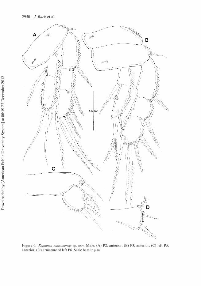
Caudal rami (Figures 1C, 2C, 7H) rectangular, about 1.6 times as long as wide;with three transverse spinular rows dorsally; with spinules at bases of setae III (ven-trally) and VII (dorsally), and around distal margin of rami (ventrally); secretory poreslocated dorsally near base of seta III and ventrally near base of seta IV; with sevensetae, setae I–II located halfway down the outer margin; setae III–VII located in dis-tal third of ramus; seta II short and pinnate; seta I bare; seta III plumose; seta IVwell developed and plumose, seta V longest and pinnate in distal half; seta VI pinnate;dorsal seta VII bi-articulate at base, naked.
Rostrum (Figure 1A) well developed, with rounded tip, defined at base; with twosmall lateral sensilla.
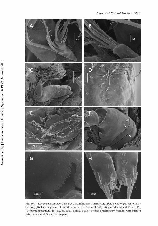
Antennule (Figure 2A) slender, eight-segmented; segment 1 with short row of longspinules along anterior margin and one pinnate seta; segments 1 and 2 similar inlength; segment 4 with sub-cylindrical process bearing one bare seta fused basallyto aesthetasc; armature formula: 1-[1 pinnate], 2-[7 pinnate + 1 spinulose + 3 bare],3-[5 pinnate + 3 bare], 4-[3 pinnate + (1 + ae)], 5-[1 pinnate + 1 bare], 6-[3 pin-nate + 1 spinulose + 1 bare], 7-[1 pinnate + 1 spinulose seta + bare], 8-[5 bare+ (2 + ae)]. Acrothek consisting of apical aesthetasc and two basally fused baresetae.
Antenna (Figures 2B, 7A): coxa and basis without surface ornamentation. Exopodtwo-segmented; exp-1 shorter than exp-2, the former with one pinnate seta distally;exp-2 with row of strong spinules apically and with four setae; armature formula:1-[1 pinnate], 2-[3 pinnate + 1 bare]. Endopod two-segmented; enp-1 with lateral seta,without surface ornamentation; lateral armature of enp-2 consisting of two bare setae,one small spinulose element, and one pinnate seta; distal armature of enp-2 consistingof four geniculate and two bare setae (one small bare seta laterally and one long bareseta fused at base to largest geniculate seta).
Figure 2. Remanea naksanensis sp. nov. Female: (A) Antennule, ventral; (B) antenna;(C) pseudoperculum, anal somite and caudal rami, dorsal (inset showing full length of setaeIV–V); (D) fifth pair of legs (P5); (E) P6 and genital field. Scale bars in µm.
Dow
nloa
ded
by [
Am
eric
an P
ublic
Uni
vers
ity S
yste
m]
at 0
6:19
27
Dec
embe
r 20
13
Journal of Natural History 2945
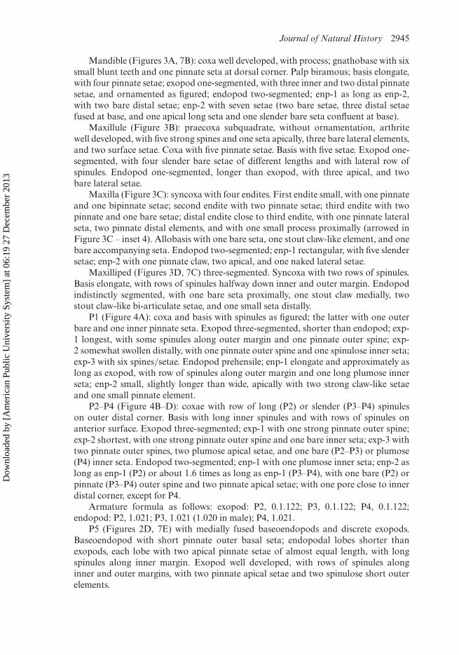
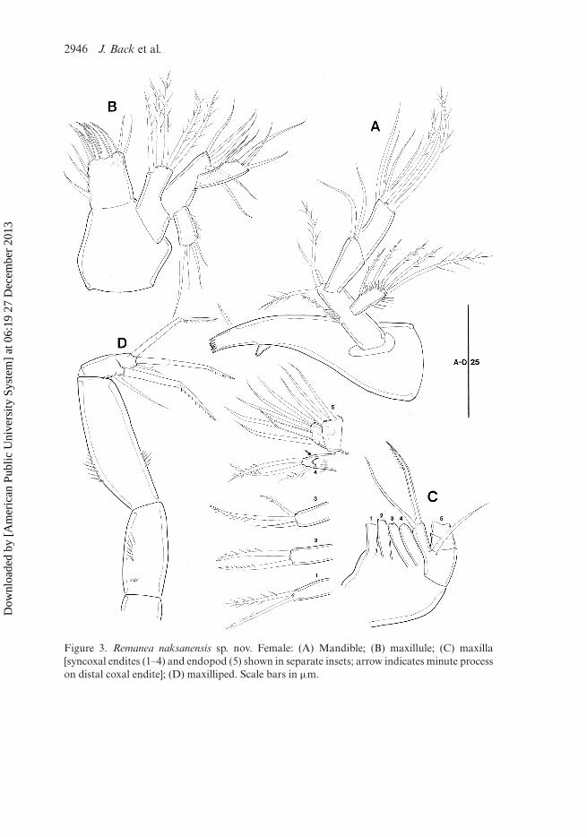
Mandible (Figures 3A, 7B): coxa well developed, with process; gnathobase with sixsmall blunt teeth and one pinnate seta at dorsal corner. Palp biramous; basis elongate,with four pinnate setae; exopod one-segmented, with three inner and two distal pinnatesetae, and ornamented as figured; endopod two-segmented; enp-1 as long as enp-2,with two bare distal setae; enp-2 with seven setae (two bare setae, three distal setaefused at base, and one apical long seta and one slender bare seta confluent at base).
Maxillule (Figure 3B): praecoxa subquadrate, without ornamentation, arthritewell developed, with five strong spines and one seta apically, three bare lateral elements,and two surface setae. Coxa with five pinnate setae. Basis with five setae. Exopod one-segmented, with four slender bare setae of different lengths and with lateral row ofspinules. Endopod one-segmented, longer than exopod, with three apical, and twobare lateral setae.
Maxilla (Figure 3C): syncoxa with four endites. First endite small, with one pinnateand one bipinnate setae; second endite with two pinnate setae; third endite with twopinnate and one bare setae; distal endite close to third endite, with one pinnate lateralseta, two pinnate distal elements, and with one small process proximally (arrowed inFigure 3C – inset 4). Allobasis with one bare seta, one stout claw-like element, and onebare accompanying seta. Endopod two-segmented; enp-1 rectangular, with five slendersetae; enp-2 with one pinnate claw, two apical, and one naked lateral setae.
Maxilliped (Figures 3D, 7C) three-segmented. Syncoxa with two rows of spinules.Basis elongate, with rows of spinules halfway down inner and outer margin. Endopodindistinctly segmented, with one bare seta proximally, one stout claw medially, twostout claw-like bi-articulate setae, and one small seta distally.
P1 (Figure 4A): coxa and basis with spinules as figured; the latter with one outerbare and one inner pinnate seta. Exopod three-segmented, shorter than endopod; exp-1 longest, with some spinules along outer margin and one pinnate outer spine; exp-2 somewhat swollen distally, with one pinnate outer spine and one spinulose inner seta;exp-3 with six spines/setae. Endopod prehensile; enp-1 elongate and approximately aslong as exopod, with row of spinules along outer margin and one long plumose innerseta; enp-2 small, slightly longer than wide, apically with two strong claw-like setaeand one small pinnate element.
P2–P4 (Figure 4B–D): coxae with row of long (P2) or slender (P3–P4) spinuleson outer distal corner. Basis with long inner spinules and with rows of spinules onanterior surface. Exopod three-segmented; exp-1 with one strong pinnate outer spine;exp-2 shortest, with one strong pinnate outer spine and one bare inner seta; exp-3 withtwo pinnate outer spines, two plumose apical setae, and one bare (P2–P3) or plumose(P4) inner seta. Endopod two-segmented; enp-1 with one plumose inner seta; enp-2 aslong as enp-1 (P2) or about 1.6 times as long as enp-1 (P3–P4), with one bare (P2) orpinnate (P3–P4) outer spine and two pinnate apical setae; with one pore close to innerdistal corner, except for P4.
Armature formula as follows: exopod: P2, 0.1.122; P3, 0.1.122; P4, 0.1.122;endopod: P2, 1.021; P3, 1.021 (1.020 in male); P4, 1.021.
P5 (Figures 2D, 7E) with medially fused baseoendopods and discrete exopods.Baseoendopod with short pinnate outer basal seta; endopodal lobes shorter thanexopods, each lobe with two apical pinnate setae of almost equal length, with longspinules along inner margin. Exopod well developed, with rows of spinules alonginner and outer margins, with two pinnate apical setae and two spinulose short outerelements.
Dow
nloa
ded
by [
Am
eric
an P
ublic
Uni
vers
ity S
yste
m]
at 0
6:19
27
Dec
embe
r 20
13
2946 J. Back et al.
Figure 3. Remanea naksanensis sp. nov. Female: (A) Mandible; (B) maxillule; (C) maxilla[syncoxal endites (1–4) and endopod (5) shown in separate insets; arrow indicates minute processon distal coxal endite]; (D) maxilliped. Scale bars in µm.
P6 (Figures 2E, 7D) represented by narrow transverse plate, armed with one bareinner seta and two pinnate outer elements of different lengths, the former longest.
Description of male
Slightly smaller and more slender than female, body length 498 µm (n = 4, mean =488 µm) (Figure 5A). Largest width (92 µm) measured at posterior margin of cephalicshield. Cephalothorax (Figure 5A) slightly more angular and with more sensilla thanin female. General body shape and ornamentation as in female except for separationof genital somite; additional sexual dimorphism in antennule, P2, P3, P5 and P6.
Antennule (Figures 5B, 7F): eight-segmented, subchirocer; segment 6 swollen,largest. Aesthetascs on sixth and eighth segments. Sixth segment with surface suturedorsoanteriorly (arrowed in Figure 7F). Armature formula: 1-[1 pinnate], 2-[4 bare +1 pinnate], 3-[5 bare + 3 pinnate], 4-[3 bare + 3 pinnate], 5-[2 bare + 1 pinnate],6-[5 bare + 6 pinnate + (1 + ae)], 7-[4 bare], 8-[7 bare + (2 + ae)].
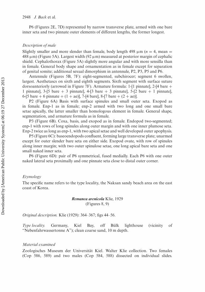
P2 (Figure 6A) Basis with surface spinules and small outer seta. Exopod asin female. Enp-1 as in female; enp-2 armed with two long and one small baresetae apically, the latter smaller than homologous element in female. General shape,segmentation, and armature formula as in female.
P3 (Figure 6B). Coxa, basis, and exopod as in female. Endopod two-segmented;enp-1 with rows of long spinules along outer margin and with one inner plumose seta.Enp-2 twice as long as enp-1, with two apical setae and well developed outer apophysis.
P5 (Figure 6C): baseoendopods confluent, forming large transverse plate; unarmedexcept for outer slender bare seta on either side. Exopod ovate, with row of spinulesalong inner margin; with two outer spinulose setae, one long apical bare seta and onesmall naked inner seta.
P6 (Figure 6D): pair of P6 symmetrical, fused medially. Each P6 with one outernaked lateral seta proximally and one pinnate seta close to distal outer corner.
Etymology
The specific name refers to the type locality, the Naksan sandy beach area on the eastcoast of Korea.
Remanea arenicola Klie, 1929(Figures 8, 9)
Original description. Klie (1929): 364–367; figs 44–56.
Type locality. Germany, Kiel Bay, off Bülk lighthouse (vicinity of“Nebenfahrwassertonne A”); clean coarse sand, 10 m depth.
Material examined
Zoologisches Museum der Universität Kiel. Walter Klie collection. Two females(Cop 586, 589) and two males (Cop 584, 588) dissected on individual slides.
Dow
nloa
ded
by [
Am
eric
an P
ublic
Uni
vers
ity S
yste
m]
at 0
6:19
27
Dec
embe
r 20
13
Journal of Natural History 2949
Figure 5. Remanea naksanensis sp. nov. Male: (A) Habitus, dorsal; (B) antennule, ventral(arrows indicate position of partial surface sutures on swollen segment 5). Scale bars in µm.
Dow
nloa
ded
by [
Am
eric
an P
ublic
Uni
vers
ity S
yste
m]
at 0
6:19
27
Dec
embe
r 20
13
2950 J. Back et al.
Figure 6. Remanea naksanensis sp. nov. Male: (A) P2, anterior; (B) P3, anterior; (C) left P5,anterior; (D) armature of left P6. Scale bars in µm.
Dow
nloa
ded
by [
Am
eric
an P
ublic
Uni
vers
ity S
yste
m]
at 0
6:19
27
Dec
embe
r 20
13
Journal of Natural History 2951
Figure 7. Remanea naksanensis sp. nov., scanning electron micrographs. Female: (A) Antennaryexopod; (B) distal segment of mandibular palp; (C) maxilliped; (D) genital field and P6; (E) P5;(G) pseudoperculum; (H) caudal rami, dorsal. Male: (F) fifth antennulary segment with surfacesutures arrowed. Scale bars in µm.
Dow
nloa
ded
by [
Am
eric
an P
ublic
Uni
vers
ity S
yste
m]
at 0
6:19
27
Dec
embe
r 20
13
2952 J. Back et al.
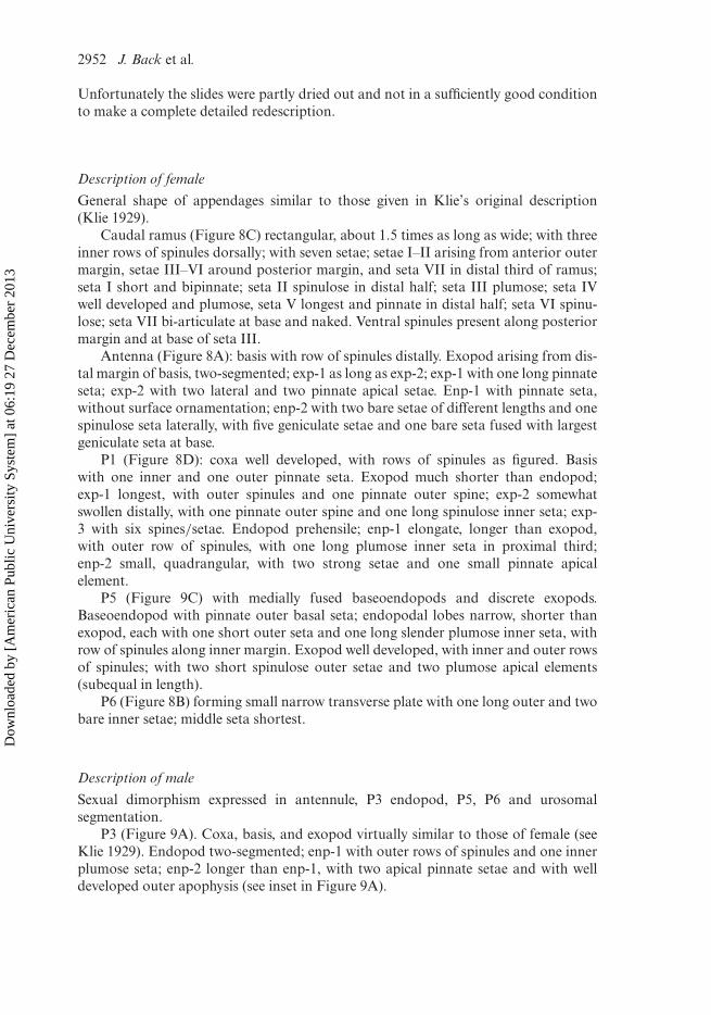
Unfortunately the slides were partly dried out and not in a sufficiently good conditionto make a complete detailed redescription.
Description of female
General shape of appendages similar to those given in Klie’s original description(Klie 1929).
Caudal ramus (Figure 8C) rectangular, about 1.5 times as long as wide; with threeinner rows of spinules dorsally; with seven setae; setae I–II arising from anterior outermargin, setae III–VI around posterior margin, and seta VII in distal third of ramus;seta I short and bipinnate; seta II spinulose in distal half; seta III plumose; seta IVwell developed and plumose, seta V longest and pinnate in distal half; seta VI spinu-lose; seta VII bi-articulate at base and naked. Ventral spinules present along posteriormargin and at base of seta III.
Antenna (Figure 8A): basis with row of spinules distally. Exopod arising from dis-tal margin of basis, two-segmented; exp-1 as long as exp-2; exp-1 with one long pinnateseta; exp-2 with two lateral and two pinnate apical setae. Enp-1 with pinnate seta,without surface ornamentation; enp-2 with two bare setae of different lengths and onespinulose seta laterally, with five geniculate setae and one bare seta fused with largestgeniculate seta at base.
P1 (Figure 8D): coxa well developed, with rows of spinules as figured. Basiswith one inner and one outer pinnate seta. Exopod much shorter than endopod;exp-1 longest, with outer spinules and one pinnate outer spine; exp-2 somewhatswollen distally, with one pinnate outer spine and one long spinulose inner seta; exp-3 with six spines/setae. Endopod prehensile; enp-1 elongate, longer than exopod,with outer row of spinules, with one long plumose inner seta in proximal third;enp-2 small, quadrangular, with two strong setae and one small pinnate apicalelement.
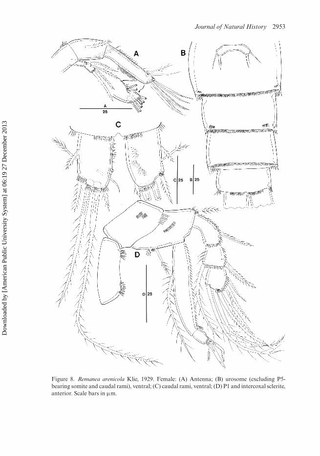
P5 (Figure 9C) with medially fused baseoendopods and discrete exopods.Baseoendopod with pinnate outer basal seta; endopodal lobes narrow, shorter thanexopod, each with one short outer seta and one long slender plumose inner seta, withrow of spinules along inner margin. Exopod well developed, with inner and outer rowsof spinules; with two short spinulose outer setae and two plumose apical elements(subequal in length).
P6 (Figure 8B) forming small narrow transverse plate with one long outer and twobare inner setae; middle seta shortest.
Description of male
Sexual dimorphism expressed in antennule, P3 endopod, P5, P6 and urosomalsegmentation.
P3 (Figure 9A). Coxa, basis, and exopod virtually similar to those of female (seeKlie 1929). Endopod two-segmented; enp-1 with outer rows of spinules and one innerplumose seta; enp-2 longer than enp-1, with two apical pinnate setae and with welldeveloped outer apophysis (see inset in Figure 9A).
P5 (Figure 9B). Baseoendopods confluent, each with one outer bare basal seta.Exopod ovate, discrete, with inner row of spinules, and with two spinulose outer setae,one pinnate apical element, and one pinnate inner seta.
Discussion
Review of previous descriptions and updated generic diagnosis of Remanea Klie, 1929Since its original description by Klie (1929) R. arenicola has been the subject ofa number of redescriptions (Nicholls 1945; Božic 1955; Mielke 1975; Arlt 1983)based on material from the west coast of Scotland, Brittany, the Isle of Sylt andthe Baltic. Comparison reveals a great deal of alleged variation in some charactersand what appear to be erroneous observations in others, hampering reliable speciesdiscrimination in the genus. These characters are reviewed below.
Ornamentation of antennulary setae
Pennak (1942a) noted that most setae in R. plumosa were plumose (hence the specificepithet) whereas in Klie’s (1929) description of R. arenicola they were clearly illustratedas naked. Nicholls (1945) showed pinnate setae on segments 1 and 2, however our re-examination has confirmed that – as in R. naksanensis – one or more pinnate/plumosesetae are present on all segments except for the apical one.
Armature of antennary exopod
Pennak (1942a) stated that the distal segment of the antennary exopod has six setaebut only figured five. The outermost apical element shown in his Plate I (fig. 21) isvery small and it is therefore highly conceivable that Pennak has misinterpreted one ofthe spinules typically found in that position (Figure 2B) as an additional armature ele-ment. Arlt (1983) claimed that his specimens from the Baltic had only three setae buthis illustration suggests that he has overlooked one of the lateral elements. Our obser-vations confirmed that the antennary exopod in both R. arenicola and R. naksanensishas a [1, 4] setal formula which is here considered as the basic pattern for the genus.
P1 enp-1 armature
One of the major morphological differences between R. plumosa and its congeners isthe absence of the long inner seta on enp-1. Within Paramesochridae the armaturepattern of P1 is typically conservative at generic level and it is therefore likely thatPennak (1942a) overlooked the inner seta, either because it was dislodged or concealedbehind the long proximal endopod segment. Similar observational errors were madein his description of Psammoleptastacus arenaridus Pennak, 1942 (cf. Sak et al. 2008).
P1 enp-2 armature
In both the original description of R. plumosa and all previous descriptions (Klie1929; Nicholls 1945; Arlt 1983 – note that the latter author mislabelled the P1 male
Dow
nloa
ded
by [
Am
eric
an P
ublic
Uni
vers
ity S
yste
m]
at 0
6:19
27
Dec
embe
r 20
13
2956 J. Back et al.
as P5 female and vice versa) of R. arenicola the distal segment of the P1 endopodhas consistently been illustrated with two apical elements. Mielke (1975) listed thesetal formulae for the swimming legs but did not include the P1. In both R. arenicola(Figure 4A) and R. naksanensis sp. nov., we observed an additional bipinnate setaarising from the inner distal corner. This seta is often concealed by the inner apicalgeniculate seta, raising the suspicion that it was overlooked in earlier descriptions.We speculate that it is probably the homologue of the minute element found at theinner distal corner of enp-2 in some Rossopsyllus species (Mielke 1985; Cottarelliand Baldari 1987) or of the well developed seta originating from the inner marginof enp-2 in Tisbisoma spinisetum Božic, 1964 (Božic 1964).
Sexual dimorphism P3 endopod
The sexual dimorphism on the P3 endopod has as yet been unnoticed. BothR. arenicola and R. naksanensis have a spiniform outer apophysis on the distalendopod segment of P3. Although this condition has yet to be confirmed in R.plumosa (male unknown) it is likely to be a diagnostic character for the genus. Themale apophysis is the positional homologue of the outer distal seta expressed in thefemale. Only the primitive genera Rossopsyllus Soyer, 1975, Tisbisoma Božic, 1964and some representatives of Diarthrodella Klie, 1949 possess an outer seta/spine onP3 enp-2 but none of these taxa displays sexual dimorphism on this ramus. We there-fore hypothesize that the presence of an apophysis on the male P3 endopod is anautapomorphy of Remanea rather than the ancestral condition of the family. Sexualdimorphism in the Paramesochridae (except for the conventional modifications in theantennule, P5, P6 and urosomal segmentation) is rare. Veit-Köhler (2000) reportedon the sexual dimorphism in the shape and length of caudal ramus setae in represen-tatives of the subgenus Wellsopsyllus (Scottopsyllus) while Mielke (1984, 1985) doc-umented modifications in the male antennary exopod of Rossopsyllus and somespecies of Diarthrodella. Swimming leg sexual dimorphism has only been reported forDiarthrodella galapagoensis Mielke, 1985 (P4 exp-3 without inner seta in the male),Kliopsyllus regulexstans Mielke, 1984 (inner distal corner of P2–P3 exp-3 forming smallspiniform projection) and Rossopsyllus kerguelenensis quellonensis Mielke, 1985 (innerseta of P3–P4 enp-1 with different ornamentation).
Female sixth legs
Neither Klie (1929) nor Pennak (1942a) figured or commented on the female genitalfield. The first author to refer to it was Nicholls (1945) who illustrated the P6 withtwo naked setae. Božic (1955) remarked that his specimens from the Roscoff areahad three naked setae, which was confirmed by our re-examination of the type mate-rial (Figures 1C, 2E) except that the middle and outer ones are pinnate rather thanunornamented.
Pseudoperculum
The presence of a pseudoperculum covering the anal opening is a family diagnostic forthe Paramesochridae. It consists of a posterior hyaline extension originating from the
Dow
nloa
ded
by [
Am
eric
an P
ublic
Uni
vers
ity S
yste
m]
at 0
6:19
27
Dec
embe
r 20
13
Journal of Natural History 2957
penultimate somite. Early workers have often overlooked such transparent structuresand none of Klie (1929), Pennak (1942a) or Nicholls (1945) made reference to it. Božic(1955) illustrated it as a deeply incised frill in R. arenicola, which was confirmed in ourmaterial of the type species and R. naksanensis.
Caudal ramus ornamentation
Kunz (1962) used the number of elements on P2–P3 exp-3 and the spinularornamentation on the caudal ramus to differentiate R. arenicola and R. plumosa.Pennak’s (1942a) description of R. plumosa showed multiple transverse spinule rowson the median half of the caudal ramus while Klie (1929) illustrated the caudal ramusof R. arenicola with only one dorsal spinule row (“ . . . auf der dorsalen Fläche, gle-ichlaufend mit dem Innenrande, eine Leiste aus feinen Haaren”). The redescription byNicholls (1945) adds further confusion because it does not show any ornamentationon the caudal ramus. Although his illustration of the urosome appears to suggest thatit was drawn in dorsal aspect because of the presence of a “dorsal” seta on the caudalrami, the spinule rows around the posterior margin of the anal somite indicate thatit is a ventral view, in which case it would explain the absence of the spinules in themedian half of the caudal ramus [see Figure 8C and Mielke (1975: Abb. 50B)]. The“dorsal” seta of Nicholls (1945) is located far proximally near the rear margin of theanal somite and hence cannot possibly be the real seta VII, which typically arises fromnear the posterior margin of the caudal ramus in each of the three congeners. Božic’s(1955) partial redescription and our observations (Figure 8C) confirmed that the spin-ular ornamentation on the dorsal surface of the caudal ramus is essentially identical inR. arenicola and R. plumosa and can no longer be used as a species discriminant.
Updated generic diagnosis of Remanea Klie, 1929Paramesochridae
Body broad anteriorly, rather flattened; with distinct separation between prosomeand urosome; dorsal and lateral hyaline frills of cephalothorax and body somiteswell developed, crenulate. Rostrum defined at base. Pseudoperculum well developed,incised. Caudal rami with seven setae, seta I well developed, seta VII positionednear posterior margin of ramus. Antennule eight-segmented in both sexes, subchi-rocer in male; with pinnate setae on segments 1–7 in female and segments 1–5 inmale. Antennary exopod two-segmented with one seta on exp-1 and four setae onexp-2. Mandible with four setae on basis; two-segmented endopod and one-segmentedexopod. Maxilla with four endites on syncoxa; endopod one- or two-segmented.Maxilliped with elongate basis; endopod one-segmented with three or four setae andclaws. P1–P4 biramous, with three-segmented exopods and two-segmented endopods;P2 endopod with weak sexual dimorphism (R. naksanensis); P3 endopod with outerapical spinous apophysis on enp-2 (unconfirmed in R. plumosa). P1–P4 armature for-mulae: exopods: P1, 0.1.222; P2, 0.1.[0–1]22; P3, 0.1.[0–1]22; P4, 0.1.122; endopods:P1, 1.030; P2, 1.02[0–1]; P3, 1.021 (1.020 in male); P4, 1.021.
P5 with medially fused baseoendopods in both sexes. Endopodal lobe well devel-oped in female, with two apical setae; not developed in male, unarmed. Exopod
Dow
nloa
ded
by [
Am
eric
an P
ublic
Uni
vers
ity S
yste
m]
at 0
6:19
27
Dec
embe
r 20
13
2958 J. Back et al.
discrete in both sexes; with five elements in female and four in male. P6 with threesetae in female and two in male.
Type species: Remanea arenicola Klie, 1929.
Additional species: R. plumosa Pennak, 1942a, R. naksanensis sp. nov.
Species discriminationRemanea naksanensis is placed in the genus Remanea on account of the three-segmented P1 exopod, the eight-segmented female antennule, the armature formulaeof the swimming legs, and the shape of the caudal rami. The new species resemblesR. arenicola in most characters except for the morphology of the P5 in both sexes:(a) the general shape of the endopodal lobe in the female (more robust and distinctlyshorter in R. naksanensis sp. nov.; compare Figures 2D and 9C), (b) the relative lengthof the endopodal setae in the female (inner seta slightly longer than exopod and outerseta in R. naksanensis sp. nov., but inner seta clearly longer than exopod and outer setavery small in R. arenicola), and (c) in R. naksanensis the exopod in the male is armedwith one small naked inner seta, one bare terminal seta, and two pinnate outer setae,while in R. arenicola it is armed with two pinnate outer setae, and one terminal and oneinner well-developed pinnate seta (compare Figures 6C and 9B). The new species hasthe distal segment of P1 endopod armed with three setae; however, R. arenicola alsohas the same number of setae (see Figure 8D). Additional morphometric differencesseparating both species are summarized in Table 1.
Key to species of Remanea Klie, 19291. P2–P3 exp-3 with four setae/spines; enp-2 with two elements . . . . . . . . . . . . . . . .
2. P5 of female endopodal inner seta 0.25 the length of outer seta; P5 of maleinner exopodal seta bipinnate . . . . . . . . . . . . . . . . . . . . . . . . . . . . . R. arenicolaP5 of female endopodal inner seta 0.85 the length of outer seta; P5 of maleinner exopodal seta naked . . . . . . . . . . . . . . . . . . . . . . . R. naksanensis sp. nov.
DistributionFrom a biogeographical point of view, it would appear that R. arenicola is confinedto western Europe, assuming a distribution along the Atlantic seaboard from Swedento Portugal. It is widely distributed in the Baltic but absent from the Mediterraneanbasin. Plum and George (2009) listed R. arenicola as a “cosmopolitan” species andcite Australia as one of the records. This is a mistake based on an erroneous inter-pretation of Nicholls (1945), which dealt with a new species of Paramesochra fromAustralia and included a redescription of R. arenicola; however, the author statedexplicitly (p. 94) that the latter was based on material collected from the Firth ofClyde in Scotland. Unlike its congeners, R. arenicola is found in sandy substrata in
Dow
nloa
ded
by [
Am
eric
an P
ublic
Uni
vers
ity S
yste
m]
at 0
6:19
27
Dec
embe
r 20
13
Journal of Natural History 2959
Table 1. Morphological comparison of the species of Remanea Klie, 1929.
Character R. arenicola R. plumosa R. naksanensissp. nov.
both intertidal beaches (e.g. Nicholls 1939; Noodt 1952; Harris 1972a, b) and sub-tidal localities (e.g. Klie 1929; Drzycimski 1974; Arlt 1983; Willems et al. 2009). Itsdistribution records are summarized below.
Sweden: Skåne County, Simrishamn (Noodt 1955a; Brinck et al. 1956); VästraGötaland County, Koster-area (Willems et al. 2009).
Denmark: southwest of the isle of Bornholm in the Baltic (Arlt 1983).Germany: Isle of Sylt (Noodt 1952, 1956, 1957; Mielke 1975) and Jade Bay (Rose and
Seifried 2006) along the North Sea coast; off Bülk lighthouse (Klie 1929),Weißenhaus and Bottsand (Noodt 1956, 1957), and Schilksee (Kunz 1935,1937) in the Kiel Bay (Kieler Bucht).
Russia: Kaliningrad oblast, Curonian Spit (Kurishe Nehrung) (Kunz 1939).Scotland: Isle of Cumbrae in the Firth of Clyde (Nicholls 1939, 1945).
Wales: Traeth Bychan, Anglesey (Geddes 1972).England: Isles of Scilly (Wells 1961, 1970; R. Huys unpublished data); Cornwall,
Whitsand Bay (Harris 1972a, b).Belgium: coastal area (Vandepitte et al 2010).
France: Brittany, Kernic (Božic 1955). Noodt (1955b, c) recorded a single juvenilefrom Contis-Plage (Landes) (as Remanea spec.) and remarked that it mostlikely belonged to R. arenicola.
Portugal: Porto District, Francelos and Cabedelo (Galhano 1970).
The only confirmed records of R. plumosa are those by Pennak (1942a, b) whofound numerous specimens just above the low tide mark at North Cape Cod beach andFalmouth beach near Woods Hole, Massachusetts. The record by Collette (1983) ofR. arenicola from Canoe beach in Nahant, Massachusetts, almost certainly refers toR. plumosa.
Baguley (2004) listed “Remanea texana” in a checklist of harpacticoid copepodsfrom the deep sea in the northern Gulf of Mexico but since no description has beenpublished so far this name is to be regarded a nomen nudum.
BarcodingSo far, the taxonomy of harpacticoid copepods has been based on morphologicalcharacters. For the identification of species, morphological data are of primary impor-tance; however, molecular sequence data are often required to confirm differencesamong species that closely resemble each other (e.g. Castro-Longoria et al. 2003), orto study phylogenetic relationships. Molecular methods can be a useful tool for therapid identification of species. Recent studies have demonstrated that a fragment ofthe mitochondrial cytochrome c oxidase subunit I (COI) gene can be used as a DNAbarcode for species-level resolution among crustacean lineages (Hebert et al. 2003;Costa et al. 2007; Bradford et al. 2010). Moreover, some researchers have alreadybegun adopting a DNA-based phylogenetic approach to study copepods, especiallylineages where morphological characters have not yielded resolution (e.g. Caudill andBucklin 2004; Eyun et al. 2007). Recently, Handschumacher et al. (2010) elucidatedthe genetic relationships of Tigriopus brevicornis (Müller, 1776), a harpacticoid witha worldwide distribution, using mitochondrial DNA sequencing. To date, partial orcomplete mtCOI sequences have been submitted to GenBank of only 11 harpacticoidspecies, representing six families: Tigriopus brevicornis, Tigriopus fulvus (Fischer, 1860),Tigriopus californicus (Baker, 1912), Tigriopus japonicus Mori, 1938, Tigriopus sp.,Macrosetella gracilis (Dana, 1847), Miracia efferata Dana, 1849, Cletocamptus deit-ersi (Richard, 1897), Cletocamptus helobius Fleeger, 1980, Coullana sp., Cletopsyllidaesp. and Clytemnestridae sp. Ideally, morphological descriptions of new species shouldbe accompanied by the submission of a barcode sequence to GenBank/EMBL andthe deposition in an appropriate institution (ICZN Recommendations 16C and 72F)of voucher specimens preserved in an adequate storage reagent (e.g. RNAlater
®) for
future molecular studies. The sequences of mtCOI of R. naksanensis was submitted toGenBank under accession numbers JN222346–JN222357.
Dow
nloa
ded
by [
Am
eric
an P
ublic
Uni
vers
ity S
yste
m]
at 0
6:19
27
Dec
embe
r 20
13
Journal of Natural History 2961
Acknowledgements
This research was supported by the Basic Science Research Programme of the National ResearchFoundation of Korea (NRF) funded by the Ministry of Education, Science and Technology(2009-0075502), and by the Discovery of Korean Indigenous Species Project, NIBR (NationalInstitute of Biological Resources).
References
Arlt G. 1983. Taxonomy and ecology of some harpacticoids (Crustacea, Copepoda) in the BalticSea and Kattegat. Zool Jb Syst 110:45–85.
Baguley JG. 2004. Meiofauna Community Structure and Function in the Northern Gulf ofMexico Deep Sea. [PhD thesis]. The University of Texas at Austin, i–xxi + 201 pp.
Becker K-H. 1979. Eidonomie und Taxonomie abyssaler Harpacticoida (Crustacea, Copepoda).Teil II. Paramesochridae, Cylindropsyllidae und Cletodidae. Meteor Forsch-Ergebn.(D)31:1–37.
Bodin P, Jackson DF. 1989. A comparison of the intertidal harpacticoid copepod assemblagesof sandy beaches in Galway Bay (Ireland) and Northern Brittany (France). J Mar BiolAssoc UK. 69:573–588.
Boxshall GA, Halsey SH. 2004. An introduction to copepod diversity. London: RaySociety.
Božic B. 1955. Copépodes harpacticoïdes des sables des environs de Roscoff. Description dequelques formes nouvelles. Arch Zool Exp Gén. 92, Notes et Revue 1:1–12.
Božic B. 1964. Tisbisoma spinisetum n. gen., n. sp., Copépode Harpacticoïde de la Réunion. BullSoc Zool Fr. 89:219–225.
Bradford T, Adams M, Humphreys WF, Austin AD, Cooper SJB. 2010. DNA barcoding ofstygofauna uncovers cryptic amphipod diversity in a calcrete aquifer in Western Australia’sarid zone. Mol Ecol Resour. 10:41–50.
Brinck P, Dahl E, Wieser W. 1956. On the littoral subsoil fauna of the Simrishamn beach ineastern Scania. Smärre Undersökningar över Öresund 19. K fysiogr Sällsk Lund Förh.25(14):109–129.
Castro-Longoria E, Alvarez-Borrego J, Rocha-Olivares A, Gómez S, Kober V. 2003. Power of amultidisciplinary approach: use of a morphological, molecular and digital methods in thestudy of harpacticoid cryptic species. Mar Ecol Progr Ser 249:297–303.
Caudill CC, Bucklin A. 2004. Molecular phylogeography and evolutionary history of theestuarine copepod, Acartia tonsa, on the Northwest Atlantic coast. Hydrobiologia.511:91–102.
Collette BB. 1983. Marine flora and fauna of Nahant. Northeastern University, Marine ScienceCenter, Nahant: 21 pp. [cited 2011 August 17]. Available from: http://www.northeastern.edu/marinescience/pdf/specieslist.pdf.
Costa FO, deWaard JR, Bouthillier J, Ratnasingham S, Dooh RT, Hajibabaei M, Hebert PDN.2007. Biological identification through DNA barcodes: the case of the Crustacea. Can JFish aquat Sci. 64:272–295.
Cottarelli V, Baldari F. 1987. Rossopsyllus obscurus n. sp. (Copepoda, Harpacticoida) fromMacquarie Island, South Pacific Ocean. Crustaceana. 53:181–185.
Coull BC, Dudley BW. 1985. Dynamics of meiobenthic copepod populations: a long-term study(1973–1983). Mar Ecol Prog Ser. 24:219–229.
Demel K. 1936. Uzupełnienie do wykazu bezkregowców i ryb Bałtyku polskiego. Archumhydrobiol i ryb, Suwałki 10:197–204.
Drzycimski I. 1967. Harpacticoida (Copepoda) nowe dla fauny Polski. Harpacticoida(Copepoda) new for the fauna of Poland. Fragm faun. 13:489–493.
Drzycimski I. 1974. Harpacticoida (Copepoda) polskich wód przybrzeznych Bałtyku. TheHarpacticoida (Copepoda) of the Baltic coastal waters. Fragm faun. 20:61–73.
Drzycimski I. 1975. Copepod harpacticoid species new for the southern Baltic fauna. In:Proceedings of the Third Baltic Symposium on Marine Biology, Helsinki/Helsingfors,11–17 June 1973. Merentutkimuslait Julk/Havsforskningsinst Skr. 239:41–44.
Drzycimski I. 1986. Changes in the species composition of Harpacticoida (Copepoda) in VistulaLagoon, Puck Bay, and southern Baltic. In Schriever, G., H.K. Schminke & C.-t. Shih (eds),Proceedings II. International Conference on Copepoda, Ottawa, Canada, 13–17 August,1984. Syllogeus. 58:552–555.
Eyun SI, Lee YH, Suh HL, Kim S, Soh HY. 2007. Genetic identification and molecular phy-logeny of Pseudodiaptomus species (Calanoida, Pseudodiaptomidae) in Korean waters.Zool Sci. 24:265–271.
Folmer O, Black M, Hoeh W, Lutz R, Vrijenhoek R. 1994. DNA primers for amplification ofmitochondrial cytochrome c oxidase subunit I from diverse metazoan invertebrates. MolMar Biol Biotech. 3:294–299.
Galhano MH. 1970. Contribução para o conhecimento da fauna intersticial em Portugal.Publções Inst Zool Dr Augusto Nobre 110:1–206.
Geddes DC. 1972. The Copepoda Harpacticoida of Anglesey and the North Wales coast.Naturalist, Hull. 921:61–76.
George KH, Schminke HK. 2002. Harpacticoida (Crustacea, Copepoda) of the Great MeteorSeamount, with first conclusions as to the origin of the plateau fauna. Mar Biol.141:887–895.
Gheerardyn H, Veit-Köhler G. 2009. Diversity and large-scale biogeography ofParamesochridae (Copepoda, Harpacticoida) in South Atlantic abyssal plains andthe deep Southern Ocean. Deep-Sea Res I. 56:1804–1815.
Handschumacher L, Steinarsdóttir MB, Edmands S, Ingólfsson A. 2010. Phylogeography of therock-pool copepod Tigriopus brevicornis (Harpacticoida) in the northern North Atlantic,and its relationship to other species of the genus. Mar Biol. 157:1357–1366.
Harris RP. 1972a. Seasonal changes in population density and vertical distributionof harpacticoid copepods on an intertidal sand beach. J Mar Biol Assoc UK.52:493–505.
Harris RP. 1972b. Reproductive activity of the interstitial copepods of a sandy beach. J MarBiol Assoc UK. 52:507–524.
Hebert PDN, Cywinska A, Ball SL, deWaard JR. 2003. Biological identifications through DNAbarcodes. Proc R Soc Lond B270:313–321.
Huys R. 1987. Paramesochra T. Scott 1892 (Copepoda, Harpacticoida): a revised key, includ-ing a new species from the SW Dutch coast and some remarks on the phylogeny of theParamesochridae. Hydrobiologia. 144:193–210.
Huys R. 2009. Unresolved cases of type fixation, synonymy and homonymy in harpacticoidcopepod nomenclature (Crustacea: Copepoda). Zootaxa. 2183:1–99.
Huys R, Gee JM, Moore CG, Hamond R. 1996. Marine and brackish water harpacticoidcopepods. Part 1. Synopses of the British Fauna (New Series). Shrewsbury: Field StudiesCouncil.
ICZN (International Commission on Zoological Nomenclature) (1999) International Code ofZoological Nomenclature, Fourth Edition. London: The International Trust for ZoologicalNomenclature, xxx + 306 pp.
Klie W. 1929. Die Copepoda Harpacticoida der südlichen und westlichen Ostsee mit besondererBerücksichtigung der Sandfauna der Kieler Bucht. Zool Jb Syst. 57:197–386.
Kunz H. 1935. Zur Oekologie der Copepoden Schleswig-Holsteins und der Kieler Bucht. Schrnaturw Ver Schlesw-Holst. 21:84–132.
Kunz H. 1937. Zur Kenntnis der Harpacticoiden des Küstengrundwassers der Kieler Förde.(Studien an marinen Copepoden. I). Kieler Meeresforsch. 2:95–115.
Kunz H. 1939. Harpacticoiden vom Sandstrande der Kurischen Nehrung. (Studien an marinenCopepoden. III). Kieler Meeresforsch. 3:148–157.
Dow
nloa
ded
by [
Am
eric
an P
ublic
Uni
vers
ity S
yste
m]
at 0
6:19
27
Dec
embe
r 20
13
Journal of Natural History 2963
Kunz H. 1962. Revision der Paramesochridae (Crust. Copepoda). Kieler Meeresforsch.18:245–257.
Kunz H. 1981. Beitrag zur Systematik der Paramesochridae (Copepoda, Harpacticoida) mitBeschreibung einiger neuer Arten. Mitt zool Mus Univ Kiel. 1(8):1–33.
Lang K. 1944. Monographie der Harpacticiden (Vorläufige Mitteilung). Uppsala: Almqvist &Wiksells Boktryckeri Ab.
Lang K. 1948. Monographie der Harpacticiden. Lund: Håkan Ohlsson.Mielke W. 1975. Systematik der Copepoda eines Sandstrandes der Nordseeinsel Sylt.
Mikrofauna Meeresboden. 52:1–134.Mielke W. 1984. Interstitielle Fauna von Galapagos. XXXI. Paramesochridae (Harpacticoida).
Microf Mar. 1:63–147.Mielke W. 1985. Diarthrodella chilensis sp. n. und Rossopsyllus kerguelensis quellonensis subsp. n.
(Copepoda, Paramesochridae) von Chile. Zool Scr. 14:45–53.Nicholls AG. 1939. Some new sand-dwelling copepods. J Mar Biol Assoc UK. 23:327–341.Nicholls AG. 1945. Marine Copepoda from Western Australia. V. A new species of
Paramesochra with an account of a new harpacticoid family, the Remaneidae, and itsaffinities. J Proc R Soc W Aust. 29:91–105.
Noodt W. 1952. Marine Harpacticiden (Cop.) aus dem eulitoralen Sandstrand der Insel Sylt.Abh mat-nat Kl Akad Wiss Mainz. 1952(3):105–142.
Noodt W. 1955a. Sandstrand-Copepoden von der schwedischen Ostküste. SmärreUndersökingar över Öresund. 17. K fysiogr Sällsk Lund Förh. 24(19):1–8.
Noodt W. 1955b. Harpacticides (Crust. Cop.) psammiques de la côte sud-ouest de la France.Vie Milieu. 6:151–153.
Noodt W. 1955c. Harpacticiden (Crust. Cop.) aus dem Sandstrand der französischen Biscaya-Küste. Kieler Meeresforsch. 11:86–109.
Noodt W. 1956. Verzeichnis der im Eulitoral der schleswig-holsteinischen Küsten angetroffenenCopepoda Harpacticoidea. Schr naturw Ver Schlesw-Holst. 28:42–64.
Noodt W. 1957. Zur Ökologie der Harpacticoidea (Crust. Cop.) des Eulitorals der deutschenMeeresküste und der angrenzenden Brackgewässer. Z Morph Ökol Tier. 46:149–242.
Pennak RW. 1942a. Harpacticoid copepods from some intertidal beaches near Woods Hole,Massachusetts. Trans Am Microsc Soc. 61:274–285.
Pennak RW. 1942b. Ecology of some copepods inhabiting intertidal beaches near Woods Hole,Massachusetts. Ecology. 23:446–456.
Plum C, George KH. 2009. The paramesochrid fauna of the Great Meteor Seamount (NortheastAtlantic) including the description of a new species of Scottopsyllus (Intermedopsyllus)Kunz (Copepoda: Harpacticoida: Paramesochridae). Mar Biodiv. 39:265–289.
Rose A, Seifried S. 2006. Small-scale diversity of Harpacticoida (Crustacea, Copepoda) froman intertidal sandflat in the Jade Bay (German Bight, North Sea). Senckenberg marit.36:109–122.
Sak S, Huys R, Karaytug S. 2008. Disentangling the subgeneric division of Arenopontia Kunz,1937: resurrection of Psammoleptastacus Pennak, 1942, re-examination of Neoleptastacusspinicaudatus Nicholls, 1945, and proposal of two new genera and a new generic classifica-tion (Copepoda, Harpacticoida, Arenopontiidae). Zool J Linn Soc. 152:409–458.
Sywula T. 1982. Notes on the Crustacea inhabiting the subterranean waters of the Baltic Seashore. Acta Hydrobiol Krakow. 24:53–61.
Vandepitte L, Decock W, Mees J (eds). 2010. Belgian Register of Marine Species, com-piled and validated by the VLIZ Belgian Marine Species Consortium. VLIZ Spec Publ46:1–86.
Vasconcelos DM, Veit-Köhler G, Drewes J, Parreira dos Santos PJ. 2009. First record ofthe genus Kliopsyllus Kunz, 1962 (Copepoda, Harpacticoida, Paramesochridae) fromNortheastern Brazil with description of the deep-sea species Kliopsyllus minor sp. nov. In:
Dow
nloa
ded
by [
Am
eric
an P
ublic
Uni
vers
ity S
yste
m]
at 0
6:19
27
Dec
embe
r 20
13
2964 J. Back et al.
Brökeland W, George KH, editors. Deep-sea taxonomy — a contribution to our knowledgeof biodiversity. Zootaxa 2096:327–337.
Veit-Köhler G. 2000. Habitat preference and sexual dimorphism in species of Scottopsyllus(Copepoda, Harpacticoida) with the description of Scottopsyllus (S.) praecipuus sp. n. fromthe Antarctic. Vie Milieu. 50:1–17.
Veit-Köhler G. 2004. Kliopsyllus andeep sp. n. (Copepoda: Harpacticoida) from the Antarcticdeep sea – a copepod closely related to certain shallow-water species. Deep-Sea Res II.51:1629–1641.
Veit-Köhler G. 2005. Results of the DIVA-1 expedition of RV “Meteor” (Cruise M48/1). Firstdeep-sea record of the genus Kliopsyllus Kunz, 1962 (Copepoda: Harpacticoida) with thedescription of Kliopsyllus diva sp. n. – the most abundant member of Paramesochridae attwo different sites of the Angola Basin. Org Div Evol. 5(Suppl. 1):29–41.
Veit-Köhler G, Drewes J. 2009. Kliopsyllus schminkei sp. n. (Copepoda, Harpacticoida,Paramesochridae) – a new copepod from the southeast Atlantic deep sea (Angola Basin).In: Brökeland W, George KH, editors. Deep-sea taxonomy — a contribution to ourknowledge of biodiversity. Zootaxa 2096:313–326.
Wells JBJ. 1961. Interstitial copepods from the Isles of Scilly. Crustaceana 2:262–274.Wells JBJ. 1970. The marine flora and fauna of the Isles of Scilly. Crustacea: Copepoda:
Harpacticoida. J Nat Hist. 4:255–268.Wells JBJ. 2007. An annotated checklist and keys to the species of Copepoda Harpacticoida
(Crustacea). Zootaxa. 1568:1–872.Willems WR, Curini-Galletti M, Ferrero TJ, Fontaneto D, Heiner I, Huys R, Ivanenko VN,
Kristensen RM, Kånneby T, MacNaughton MO, et al. 2009. Meiofauna of the Koster-area, results from a workshop at the Sven Lovén Centre for Marine Sciences (Tjärnö,Sweden). Meiofauna Mar. 17:1–34.