A newly established bovine intestinal epithelial cell line is effective for in vitro screening of potential antiviral immunobiotic microorganisms for cattle Eriko Chiba a,1 , Julio Villena a,1 , Shoichi Hosoya a , Naoya Takanashi a , Tomoyuki Shimazu b , Hisashi Aso c , Masanori Tohno d , Yoshihito Suda e , Yasushi Kawai a , Tadao Saito a , Kenji Miyazawa f , Fang He f , Haruki Kitazawa a,⇑ a Food Immunology Group, Laboratory of Animal Products Chemistry, Graduate School of Agricultural Science, Tohoku University, Sendai 981-8555, Japan b Laboratory of Animal Breeding and Genetics, Graduate School of Agricultural Science, Tohoku University, Sendai 981-8555, Japan c Cell Biology Laboratory, Graduate School of Agricultural Science, Tohoku University, Sendai 981-8555, Japan d National Agriculture and Food Research Organization, National Institute of Livestock and Grassland Science, Nasushiobara 329-2793, Japan e Department of Food, Agriculture and Environment, Miyagi University, Sendai 982-0215, Japan f Technical Research Laboratory, Takanashi Milk Products Co., Ltd, Yokohama, Kanagawa 241-0023, Japan article info Article history: Received 9 July 2011 Accepted 3 October 2011 Keywords: Antiviral immune response Bovine intestinal epithelial cells Immunobiotic Toll-like receptor 3 (TLR3) abstract We evaluated whether a bovine intestinal epithelial (BIE) cell line could serve as a useful in vitro model system for studying antiviral immune responses in bovine intestinal epithelial cells (IECs) and for the pri- mary screening of immunobiotic microorganisms with antiviral protective capabilities. Immunofluores- cent analyses revealed that toll-like receptor 3 (TLR3) was expressed in BIE cells, and the results of real- time quantitative PCR showed that these cells respond to stimulation with poly(I:C) by up-regulating pro-inflammatory cytokines and type I interferons. In addition, we demonstrated that BIE cells are useful for the primary screening of immunobiotic lactic acid bacteria strains which are able to beneficially mod- ulate antiviral immune responses triggered by TLR3 activation in bovine IECs. The characterization of BIE cells performed in the present study represents an important step towards the establishment of a valu- able bovine in vitro system that could be used for the development of immunomodulatory feed for bovine hosts. Ó 2011 Elsevier Ltd. All rights reserved. 1. Introduction Diarrhea is an important cause of morbidity and mortality in young calves, resulting in significant financial losses to cattle pro- ducers. In particular, bovine neonatal gastroenteritis is a multifac- torial disease that can be caused by a number of pathogens, including bovine rotavirus (BRV), bovine coronavirus (BCV), and bovine viral diarrhea viruses (BVDVs) (Aich et al., 2007; Lee et al., 2008). Although these viruses belong to different families and have distinct physical characteristics, they are all able to infect intestinal epithelial cells (IECs), and induce villous atrophy and diarrhea. IECs are able to distinguish between the diverse elements pres- ent in the intestinal environment through the expression of pattern recognition receptors, such as toll-like receptors (TLRs) (Westendorf et al., 2010). Among TLRs, TLR3 recognizes dsRNA and is therefore important for defense against viral infections. Upon recognition of viral dsRNA, TLR3 transmits signals that activate transcription fac- tors responsible for the induction of type I interferon (IFN; IFN-a/ b) and IFN-inducible genes, which play critical roles in antiviral host defense (Matsumoto et al., 2002). Thus, activation of TLR3 signaling is of great importance for defense against BRV, a dsRNA virus, and BCV and BVDV, which replicate via intermediary dsRNA. Recently, significant progress has been made in understanding the role of TLR3 in innate and adaptive immunity. The majority of studies aimed at dissecting the mechanisms of TLR3 function have been performed principally in mouse and human cell lines (Gauzzi et al., 2010). However, few studies have been conducted on cattle, despite growing interest in the bovine immune system due to the economic importance of cattle as livestock. Therefore, investigating how TLR3 mediates antiviral defenses in bovine IECs is important for understanding the activation and regulation of the intestinal immune system of cattle. Moreover, determining the underlying mechanisms of TLR3 activation and regulation in bo- vine IECs may aid in the development of effective therapies for the prevention and treatment of viral diseases, such as oral vac- cines and functional feed, which specifically target anti-viral im- mune responses in the bovine gut. 0034-5288/$ - see front matter Ó 2011 Elsevier Ltd. All rights reserved. doi:10.1016/j.rvsc.2011.10.002 ⇑ Corresponding author. Tel.: +81 22 717 8713; fax: +81 22 717 8715. E-mail address: [email protected](H. Kitazawa). 1 These authors equally contributed to this work. Research in Veterinary Science 93 (2012) 688–694 Contents lists available at SciVerse ScienceDirect Research in Veterinary Science journal homepage: www.elsevier.com/locate/rvsc
Transcript
Research in Veterinary Science 93 (2012) 688–694
Contents lists available at SciVerse ScienceDirect
Research in Veterinary Science
journal homepage: www.elsevier .com/locate / rvsc
A newly established bovine intestinal epithelial cell line is effective for in vitroscreening of potential antiviral immunobiotic microorganisms for cattle
Eriko Chiba a,1, Julio Villena a,1, Shoichi Hosoya a, Naoya Takanashi a, Tomoyuki Shimazu b, Hisashi Aso c,Masanori Tohno d, Yoshihito Suda e, Yasushi Kawai a, Tadao Saito a, Kenji Miyazawa f, Fang He f,Haruki Kitazawa a,⇑a Food Immunology Group, Laboratory of Animal Products Chemistry, Graduate School of Agricultural Science, Tohoku University, Sendai 981-8555, Japanb Laboratory of Animal Breeding and Genetics, Graduate School of Agricultural Science, Tohoku University, Sendai 981-8555, Japanc Cell Biology Laboratory, Graduate School of Agricultural Science, Tohoku University, Sendai 981-8555, Japand National Agriculture and Food Research Organization, National Institute of Livestock and Grassland Science, Nasushiobara 329-2793, Japane Department of Food, Agriculture and Environment, Miyagi University, Sendai 982-0215, Japanf Technical Research Laboratory, Takanashi Milk Products Co., Ltd, Yokohama, Kanagawa 241-0023, Japan
a r t i c l e i n f o
Article history:Received 9 July 2011Accepted 3 October 2011
We evaluated whether a bovine intestinal epithelial (BIE) cell line could serve as a useful in vitro modelsystem for studying antiviral immune responses in bovine intestinal epithelial cells (IECs) and for the pri-mary screening of immunobiotic microorganisms with antiviral protective capabilities. Immunofluores-cent analyses revealed that toll-like receptor 3 (TLR3) was expressed in BIE cells, and the results of real-time quantitative PCR showed that these cells respond to stimulation with poly(I:C) by up-regulatingpro-inflammatory cytokines and type I interferons. In addition, we demonstrated that BIE cells are usefulfor the primary screening of immunobiotic lactic acid bacteria strains which are able to beneficially mod-ulate antiviral immune responses triggered by TLR3 activation in bovine IECs. The characterization of BIEcells performed in the present study represents an important step towards the establishment of a valu-able bovine in vitro system that could be used for the development of immunomodulatory feed for bovinehosts.
� 2011 Elsevier Ltd. All rights reserved.
1. Introduction
Diarrhea is an important cause of morbidity and mortality inyoung calves, resulting in significant financial losses to cattle pro-ducers. In particular, bovine neonatal gastroenteritis is a multifac-torial disease that can be caused by a number of pathogens,including bovine rotavirus (BRV), bovine coronavirus (BCV), andbovine viral diarrhea viruses (BVDVs) (Aich et al., 2007; Leeet al., 2008). Although these viruses belong to different familiesand have distinct physical characteristics, they are all able to infectintestinal epithelial cells (IECs), and induce villous atrophy anddiarrhea.
IECs are able to distinguish between the diverse elements pres-ent in the intestinal environment through the expression of patternrecognition receptors, such as toll-like receptors (TLRs) (Westendorfet al., 2010). Among TLRs, TLR3 recognizes dsRNA and is thereforeimportant for defense against viral infections. Upon recognition of
ll rights reserved.
: +81 22 717 8715.awa).
viral dsRNA, TLR3 transmits signals that activate transcription fac-tors responsible for the induction of type I interferon (IFN; IFN-a/b) and IFN-inducible genes, which play critical roles in antiviral hostdefense (Matsumoto et al., 2002). Thus, activation of TLR3 signalingis of great importance for defense against BRV, a dsRNA virus, andBCV and BVDV, which replicate via intermediary dsRNA.
Recently, significant progress has been made in understandingthe role of TLR3 in innate and adaptive immunity. The majorityof studies aimed at dissecting the mechanisms of TLR3 functionhave been performed principally in mouse and human cell lines(Gauzzi et al., 2010). However, few studies have been conductedon cattle, despite growing interest in the bovine immune systemdue to the economic importance of cattle as livestock. Therefore,investigating how TLR3 mediates antiviral defenses in bovine IECsis important for understanding the activation and regulation of theintestinal immune system of cattle. Moreover, determining theunderlying mechanisms of TLR3 activation and regulation in bo-vine IECs may aid in the development of effective therapies forthe prevention and treatment of viral diseases, such as oral vac-cines and functional feed, which specifically target anti-viral im-mune responses in the bovine gut.
E. Chiba et al. / Research in Veterinary Science 93 (2012) 688–694 689
We have recently established a bovine intestinal epithelial (BIE)cell line originally derived from fetal bovine intestinal epithelio-cytes (Miyazawa et al., 2010). In the present study, we aimed tocharacterize the immune response triggered by TLR3 activationin BIE cells in order to evaluate whether this cell line could serveas an in vitro model of bovine IECs for the study of TLR3-mediatedantiviral responses. In addition, we attempted to determinewhether BIE cells are suitable for the primary screening of immu-nomodulatory lactic acid bacteria (LAB) with antiviral protectivecapabilities in cattle.
2. Materials and methods
2.1. BIE cells
The BIE cell line used in this study is a non-transformed intestinalcell line that was previously derived from fetal bovine intestinal epi-theliocytes (Miyazawa et al., 2010). BIE cells were routinelymaintained in Dulbecco’s Modified Eagle medium (DMEM; Gibco,Grand Island, NY) containing 10% heat-inactivated fetal bovine ser-um (FBS) and penicillin–streptomycin. For passaging, BIE cells weretreated with a sucrose/EDTA buffer (0.1 M Na2HPO4/12H2O, 0.45 Msucrose, 0.36% EDTA/4Na, and bovine serum albumin [BSA]) for4 min and then detached using 0.04% trypsin in phosphate-bufferedsaline (PBS) (Moue et al., 2008). BIE cells were then plated in type Icollagen-coated culture dishes (Sumilon, Tokyo, Japan) at a densityof 1.5 � 104 cells/cm2 and cultured at 37 �C in an atmosphere of 5%CO2 in DMEM (10% FBS, 1% streptomycin/penicillin, 100 U/ml strep-tomycin, high glucose, L-glutamine, and 0.11 mg/ml sodium pyru-vate; Gibco).
2.2. Immunocytochemistry
BIE cells were cultured in collagen type I-coated culture dishes(12-well plate; Sumilon) at a cell density of 3 � 104 cells/well for3 days (37 �C, 5% CO2), washed once with cold PBS (2% FCS), andthen treated with FACS permeabilization solution (4 �C, 10 min).Following three washes with PBS, the cells were incubated with5% normal goat serum (Sigma, St. Louis, MO) for 10 min at 4 �C.Cells were again washed with PBS three times and then incubatedwith anti-TLR3 polyclonal antibody (sc-28999; Santa Cruz, CA,USA) for 16 h at 4 �C. Following three washes with PBS, cells weretreated with secondary Alexa Fluor 488-conjugated goat anti-rab-bit polyclonal IgG (Invitrogen, Tokyo, Japan) for 1 h at room tem-perature. BIE cells incubated with rabbit IgG isotype controlantibody (20304E; Imgenex, San Diego, CA) and the identical sec-ondary antibody as above were used as controls. After reactionwith secondary antibody, cells were washed three times withPBS and then counterstained with SYTOX orange (Invitrogen) for5 min at room temperature. Cells were washed three times withPBS, rinsed in distilled water, and then mounted on glass slideswith PermaFluor (Thermo Fisher, Pittsburgh, PA). Immunofluores-cence microscopy was performed using a confocal laser micro-scope (MRC-1024; Bio-Rad, Richmond, CA).
2.3. Microorganisms
The following LAB strains were used in this study: Lactobacillusgasseri TMC0356, Lactobacillus rhamnosus LGG, L. rhamnosus LA-2,Lactobacillus casei TMC0409, Streptococcus thermophilus TMC1543,Bifidobacterium bifidum 2-2, and B. bifidum 3-9. The lactobacilliand bifidobacteria strains were grown in MRS medium (Difco, De-troit, MI, USA) for 16 h at 37 �C. S. thermophilus cells were grown inElliker medium (Difco) for 16 h at 37 �C.
2.4. Anti-inflammatory assay in BIE cells
Bacteria were re-suspended in DMEM (10% FBS and 1% SP), enu-merated microscopically using a Petroff-Hausser counting cham-ber, and stored at �80 �C until use. BIE cells were plated at adensity of 1.5 � 104 cells/well in 24-well type I collagen-coatedplates (Sumilon) containing 1 ml DMEM, and were then culturedfor 3 days. After changing the medium, lactobacilli or bifidobacte-ria (5 � 107 cells/ml) were added to 1 ml DMEM in each well. After24 and 48 h of incubation, each well was washed vigorously withmedium at least three times to eliminate all of the stimulants.For comparative treatment, BIE cells were stimulated with200 ng/ml Pam3CSK4 for 24 or 48 h. After the elimination of lacto-bacilli, bifidobacteria, or Pam3CSK4, BIE cells were stimulated withpoly(I:C) at concentrations of 12.5 and 60 ng/ml for 3, 6, 12, and24 h.
2.5. Quantitative expression analysis by real-time polymerase chainreactions (PCR) in BIE cells
We performed a two-step real-time quantitative PCR to studythe expression of mRNAs in BIE cells. Total RNA was isolated fromBIE cells using TRIzol reagent (Invitrogen). All cDNAs were synthe-sized using a Quantitect Reverse Transcription kit (Qiagen, Tokyo,Japan) according to the manufacturer’s recommendations. Real-time quantitative PCR was performed using a 7300 Real-timePCR System (Applied Biosystems, Warrington, UK) using PlatinumSYBR Green qPCR SuperMix UDG with ROX (Invitrogen). The prim-ers used in this study were: b-actin forward (TGG ATT GGC GGCTCC AT) and reverse (GCT GAT CCA CAT CTG CTG GAA); IFN-a for-ward (GGT GGC AGC CAG TTA CAG AAG) and reverse (TGC TGGGTC ACC TCA TGG A), IFN-b forward (CGA TGG TTC TCC TGC TGTGTT) and reverse (GAG CAA GCT GTA GCT CCT GGAA); IFN-c for-ward (GGA GGA CTT CAA AAA GCT GAT TCA) and reverse (GGCTTT GCG CTG GAT CTG); IL-6 forward (CCA CCC CAG GCA GACTAC TTC) and reverse (CCA TGC GCT TAA TGA GAG CTT); IL-8 for-ward (TGC TCT CTT GGC AGC TTT CC) and reverse (TCT TGA CAGAAC TGC AGC TTC AC); and MCP-1 forward (CAC CAG CAG CAAGTG TCC TAA A) and reverse (CAC ATA ACT CCT TGC CCA GGA T).The PCR cycling conditions consisted of 2 min at 50 �C, followedby 2 min at 95 �C, and then 40 cycles of 15 s at 95 �C, 30 s at60 �C, and 30 s at 72 �C. The reaction mixture contained 5 ml ofsample cDNA and 15 ml of master mix, which included the senseand antisense primers. Cytokines, chemokines, and b-actin cDNAsfor real-time PCR analysis were amplified using the primers listedabove. The PCR products were inserted into the vector pGEM-TEasy DNA (Promega, Madison, WI, USA) using standard techniques.We confirmed the sequence of each insert with the dideoxy chain-termination method using an ABI PRISM 310 Genetic Analyzer (Ap-plied Biosystems). The constructed plasmid vectors were appliedto calculate the relative expression levels of each cDNA in samplesby the relative standard curve method. The expression levels of b-actin were used to normalize individual cytokine and chemokinecDNA expression levels in the samples. The relative index of cyto-kine mRNA in BIE cells stimulated with LAB was calculated by firstaveraging the target cytokine expression levels from a minimum ofthree samples stimulated with poly(I:C) and without pre-treat-ment with LAB. After setting this value as 1, the relative expressionof LAB pre-stimulated samples following poly(I:C) stimulation wasthen calculated.
2.6. Statistical analysis
All data were adjusted to give relative values against the appro-priate controls. Statistical analyses were performed using the GLMprocedure of SAS statistical software (SAS, 1994). Comparisons
690 E. Chiba et al. / Research in Veterinary Science 93 (2012) 688–694
among mean values (N = 4) for relative mRNA expression levels oftarget cytokines in cells were performed using one-way ANOVAand Fisher’s least significant difference (LSD) test against the con-trols. For the analyses, P values of <0.05 and <0.01 were consideredsignificant. Comparisons (N = 4) among relative mRNA expressionlevels of cytokines in BIE cells stimulated with LAB strains for 48 hare shown using two broken lines for P values of <0.05 and <0.01, fol-lowing one-way ANOVA and LSD multi-comparison test. All pre-sented data represents the results of four independent experiments.
3. Results
3.1. Functional expression of TLR3 in BIE cells
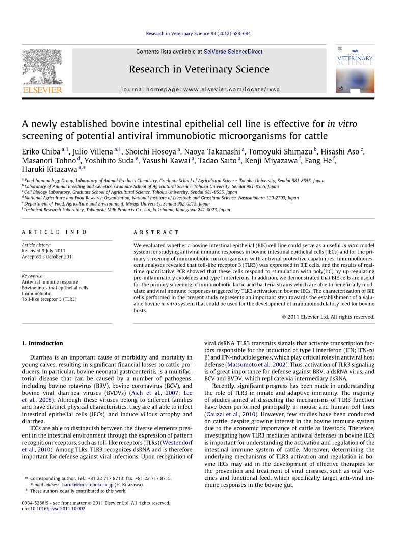
To evaluate the expression of TLR3 protein in BIE cells, we firstperformed immunohistochemical analysis. As shown in Fig. 1A, BIEcells strongly expressed TLR3 in the cytoplasm, whereas no expres-sion of TLR3 was detected at the cell surface (data not shown).
As delineating cytokine and chemokine responses to TLR3 stim-ulation is important to understand TLR3-mediated immune
Fig. 1. Functional expression of toll-like receptor 3 (TLR3) in bovine intestinal epithelial (Bindicates bovine TLR3-positive BIE cells, while nuclei were stained orange (red) with SYimages represent typical results of four independent experiments. (B) Expression of IFN-concentrations (12.5 or 60 ng/ml) of poly(I:C). Cytokine levels were evaluated at the ideviation (error bars). The results represent four independent experiments. ⁄⁄P < 0.01 areferences to colour in this figure legend, the reader is referred to the web version of th
responses and pathogenicity in IECs, we next evaluated the re-sponse of BIE cells to stimulation with poly(I:C). For these experi-ments, we treated BIE cells with two doses of TLR3 agonist toestablish the most appropriate dose to study TLR3-mediated im-mune responses. Challenge of BIE cells with both low (12.5 ng/ml) and high (60 ng/ml) concentrations of poly(I:C) significantlyincreased the expression of type I IFN, IFN-c, and pro-inflamma-tory cytokines and chemokines (Fig. 1B). The most significantchanges were observed in the expression of IFN-b and the pro-inflammatory cytokines IL-6, IL-8, and MCP-1. We also observedthat treatment of cells at the higher dose of poly(I:C) induced ear-lier up-regulation of IFN-b, as well as IL-6, IL-8, and MCP-1, whencompared with the lower dose. Thus, poly(I:C) at a concentrationof 60 ng/ml was selected for the following experiments.
3.2. Effect of LAB on cytokine expression by BIE cells
We evaluated changes in the expression of IFN-b, IL-6, IL-8, andMCP-1 in BIE cells after treatment with different LAB strains or theTLR2 agonist Pam3CSK4 for 24 and 48 h (Fig. 2). We observed that
IE) cells. (A) Immunofluorescent localization of TLR3 in BIE cells. Green fluorescenceTOX. Control experiments were performed by omitting the primary antibody. Thea, IFN-b, IFN-c, IL-6, IL-8, and MCP-1 mRNAs in BIE cells after stimulation with twondicated times post-stimulation. All values are presented as the mean ± standardnd ⁄P < 0.05 vs. cells cultured in the absence of poly(I:C). (For interpretation of theis article.)
E. Chiba et al. / Research in Veterinary Science 93 (2012) 688–694 691
L. rhamnosus GG was able to significantly increase IFN-b levels after48 h, whereas the other tested strains did not induce marked dif-ferences in IFN-b levels when compared with untreated BIE cells.In addition, L. rhamnosus LA-2 was the only strain able to increaseMCP-1 levels in BIE cells after 48 h of treatment (Fig. 2). Levels ofIL-6 mRNA were up-regulated within 24 and 48 h of treatmentwith Pam3CSK4, while the only LAB strain able to increase IL-6expression was S. thermophilus TMC1543 after 24 h of stimulation(Fig. 2). Our analysis also revealed that Pam3CSK4, L. rhamnosusLA-2, L. casei TMC0409, and both B. bifidum strains were able toup-regulate IL-8 after 24 h.
3.3. Effect of LAB on the response of BIE cells to poly(I:C) stimulation
We next evaluated the ability of BIE cells to serve as an in vitrosystem for the selection of LAB strains with the capacity to modulatethe response of bovine IECs to poly(I:C) challenge. As IFN-b is animportant cytokine for protection against viral infections, we aimedto identify LAB strains with the ability to augment the production ofIFN-b by BIE cells stimulated with TLR3 agonist. BIE cells were stim-ulated with the different LAB strains for 48 h and then challengedwith poly(I:C) (Fig. 3). The determination of IFN-b mRNA expressionlevels at 3 h (Fig. 3A) and 12 h (Fig. 3B) post-stimulation withpoly(I:C) revealed that only L. rhamnosus LA-2 and S. thermophilusTMC1543 were able to increase IFN-b levels after 12 h when
Fig. 2. Effect of lactic acid bacteria (LAB) on cytokine expression in bovine intestinal epistimulation with the bacteria Lactobacillus gasseri TMC0356, Lactobacillus rhamnosusTMC1543, Bifidobacterium bifidum 2-2, and B. bifidum 3-9, or the toll-like receptor 2 agovalues are presented as the mean ± standard deviation (error bars). The results represent fstimulation (broken lines).
compared to controls. Notably, the effect of L. rhamnosus LA-2 expo-sure was superior to that of S. thermophilus TMC1543. In addition, weobserved that L. rhamnosus LA-2 induced the down-regulation of IL-6 after 3 h (Fig. 3A), while S. thermophilus TMC1543, L. caseiTMC0409, and both B. bifidum strains had reduced the expressionof IL-6 by 12 h (Fig. 3B). In addition, L. gasseri TMC0356 down-regu-lated the expression of IL-8, while L. casei TMC0409 reduced IL-8 andMCP-1 mRNAs levels after 12 h. These results were confirmed bycomparing the relative mRNA levels of cytokines in BIE cells stimu-lated with each LAB strain and challenged with poly(I:C) for 12 husing multi-comparison tests (Fig. 3C).
4. Discussion
Epithelial TLR expression is thought to play a key role in hostdefenses against pathogens. Epithelial cells, more than any othercell type, express TLR3 in numerous organs, including the gastroin-testinal tract (Cario and Podolsky, 2000). It was reported that hu-man IECs express low levels of TLR2 and TLR4, whereas TLR3appears to be abundantly expressed in the normal human smallintestine and colon (Cario and Podolsky, 2000). In addition, our lab-oratory has previously shown that TLR3 is also strongly expressedin swine IECs (Moue et al., 2008). Moreover, TLR3 transcription inbovine colonic epithelium was recently described for the first time(Bridger et al., 2010). In the present study, immunohistochemical
thelial (BIE) cells. Expression of IFN-b, IL-6, IL-8 and MCP-1 mRNA in BIE cells afterLGG, L. rhamnosus LA-2, Lactobacillus casei TMC0409, Streptococcus thermophilus
nist Pam3CSK4. Cytokines were studied at the indicated times post-stimulation. Allour independent experiments. ⁄⁄P < 0.05 vs. cells cultured in the absence of bacterial
Fig. 3. Effect of lactic acid bacteria (LAB) on the response of bovine intestinal epithelial (BIE) cells to poly(I:C) challenge. Expression of IFN-b, IL-6, IL-8, and MCP-1 mRNA inbovine intestinal epithelial (BIE) cells after stimulation with different bacterial strains and poly(I:C). BIE cells were stimulated with Lactobacillus gasseri TMC0356,Lactobacillus rhamnosus LGG, L. rhamnosus LA-2, Lactobacillus casei TMC0409, Streptococcus thermophilus TMC1543, Bifidobacterium bifidum 2-2, or B. bifidum 3-9 for 48 h. Cellswere then challenged with poly(I:C) (60 ng/ml) and cytokine levels were evaluated 3 h (A) and 12 h (B) post-challenge. All values are presented as the mean ± standarddeviation (error bars). The results represent four independent experiments. ⁄⁄P < 0.01 and ⁄P < 0.05 vs. cells cultured in the absence of bacterial stimulation and challengedwith poly(I:C). (C) Comparisons of relative mRNA levels of cytokines in BIE cells stimulated with the indicated LAB strains for 48 h and challenged with poly(I:C) for 12 h. One-way ANOVA and Fisher’s least significant difference multi-comparison test were used. P values of < 0.05 and < 0.01 are indicated by the two types of broken lines.
692 E. Chiba et al. / Research in Veterinary Science 93 (2012) 688–694
analyses revealed abundant expression of TLR3 in BIE cells. There-fore, BIE cells, in addition to displaying characteristics of epithelialcells, such as cobblestone morphology, microvilli-like structures,and strong expression of cell-to-cell junctional proteins and
cytokeratin (Miyazawa et al., 2010), also express TLR3 and thusresemble the IECs of other species.
We also evaluated the response of BIE cells to stimulation withthe TLR3 agonist poly(I:C) and found that the cells up-regulate the
E. Chiba et al. / Research in Veterinary Science 93 (2012) 688–694 693
expression of type I IFN, IFN-c, and pro-inflammatory cytokinesand chemokines. The observed changes in the expression of cyto-kines induced by poly(I:C) correlate with the changes reported invarious intestinal viral infections of cattle and other hosts. Forexample, increased gene expression of RANTES, IP-10, IL-8, andMCP-1 were observed in rotavirus-infected HT-29 cells (Rolloet al., 1999; Xu et al., 2009). In addition, in vitro studies with bovineintestinal tissues demonstrated that exposure to BRV activatedTLR3-induced up-regulation of NF-kB and IL-6 production (Aichet al., 2007). These findings, together with our present results, indi-cate that BIE cells are valuable tools for the in vitro study of im-mune responses triggered by TLR3 expressed on bovine IECs.
To date, a few studies have evaluated the antiviral effects ofprobiotic LAB strains in animals. In-vivo studies using gnotobioticpigs demonstrated that probiotic LAB administration has a signifi-cant influence on IFN-a, TGF-b, IL-4, and IFN-c serum levels in-duced by rotavirus infection (Wen et al., 2009). The antiviraleffects of immunobiotics have also been examined in a few animalcell lines. For example, Maragkoudakis et al. (2010) reported thatprobiotics are able to protect porcine and goat epithelial cellsagainst rotavirus and transmissible gastroenteritis virus chal-lenges; however, the immunological mechanisms involved in theprotective effect were not determined. Recent studies have alsodemonstrated that probiotics significantly decrease IL-6 produc-tion by porcine IPEC-J2 cells infected with porcine rotavirus, sug-gesting that probiotics have immunoregulatory effects (Liu et al.,2010). To our knowledge, no prior studies have investigated the ef-fects of probiotics in bovine IECs lines. Therefore, we evaluatedwhether our bovine in vitro system could be used for the selectionof LAB strains with antiviral immune-enhancing activities.
We first examined if the stimulation of BIE cells with differentLAB strains was able to induce changes in the expression of IFN-b, IL-6, IL-8, or MCP-1. We found that the different strains had dis-tinct effects on cytokine production by BIE cells. Notably, L.rhamnosus GG and S. thermophilus TMC1543 were the only strainsable to increase IFN-b and IL-6 mRNA, respectively, in BIE cellsafter 48 h of stimulation. In addition, only L. rhamnosus LA-2 in-creased MCP-1 mRNA, while this bacterium, L. casei TMC0409,and both B. bifidum strains up-regulated IL-8. It has been shownthat many of the immunomodulatory effects of probiotic microor-ganisms are mediated by their ability to activate TLR2 (Tohno et al.,2005, 2006, 2007; Kitazawa et al., 2008; Alvarez et al., 2009; Fujieet al., in press). In addition, TLR2 agonists are able to induce IFN-btranscription (Dietrich et al., 2010). Moreover, it was observed thatcertain lactobacilli trigger the expression of IFN-b genes in den-dritic cells in a TLR2-dependent manner (Weiss et al., 2010). Inour present analyses, we therefore expected to identify strainscapable of increasing IFN-b levels in BIE cells; however, we onlydetected a slight increase of this cytokine in BIE cells treated withL. rhamnosus GG. In addition, we found that the TLR2 agonistPam3CSK4 was not able to modify the expression of IFN-b in BIEcells. Therefore, the information obtained in these experimentsdid not allow us to draw any conclusions concerning the antiviraleffects of LAB strains in bovine IECs.
We next studied the effects of LAB strains on the response of BIEcells to TLR3 stimulation and found that only L. rhamnosus LA-2and S. thermophilus TMC1543 were able to increase IFN-b levelsby 12 h post-stimulation. The increased production of IFN-b byBIE cells in response to TLR3 activation induced by L. rhamnosusLA-2 may have significant in vivo effects in the protection againstenteric viruses. Global gene-expression analyses of bovine intesti-nal tissues following infection with BRV or BCV indicate that sev-eral IFN-regulatory and -stimulatory genes are down-regulated,supporting the conclusion that both viruses may have evolvedmechanism(s) to inhibit IFN-mediated immune responses (Aichet al., 2007). Moreover, it was shown that BVDV significantly inter-
feres with the induction of type I IFN, which impairs not only in-nate defenses, but also interferes with the establishment ofadaptive immune responses (Peterhans et al., 2003; Lee et al.,2008). Based on these findings, L. rhamnosus LA-2, which enhancesIFN-b production in BIE cells, may play an important role in theimprovement of innate and specific immune responses against bo-vine intestinal virus. Thus, our studies with the BIE cell line haveallowed us to identify a LAB strain that is a good candidate for fu-ture in vivo studies.
As the degree and duration of pro-inflammatory cytokine secre-tion after TLR3 recognition of dsRNA can become harmful to thehost (Vercammen et al., 2008), we also evaluated the productionof the inflammatory cytokines IL-6, IL-8, and MCP-1 in BIE cells.Our analyses demonstrated that BIE cells pretreated with L. caseiTMC0409 produce lower levels of the three pro-inflammatorycytokines when compared with control cells 12 h post-stimulationwith poly(I:C). It was reported that TLR3 mediates harmful inflam-matory responses in the intestine, thus contributing to the patho-genesis of viral infections (Zhou et al., 2007). Therefore, the lowerproduction of pro-inflammatory cytokines following exposure to L.casei TMC0409 may allow for the efficient regulation of inflamma-tory responses and avoidance of tissue injury, offering a differentprotection mechanism against bovine viral infection.
In conclusion, the in vitro system described in this study was use-ful for the primary screening of two types of immunomodulatoryLAB strains that would be able to protect against viral intestinal dis-eases in cattle: strains capable of increasing antiviral defenses andstrains with anti-inflammatory capacities. To define the characteris-tic immunomodulatory abilities of individual LAB strains, theirinfluence on cytokine production after challenge of BIE cells withTLR3 agonist can be studied using statistical multi-comparison tests,as shown in Fig. 3C. We also demonstrated that TLR3 is expressed inBIE cells and that these cells respond to stimulation with the TLR3agonist poly(I:C). Characterization of the inflammatory immune re-sponse triggered by TLR3 activation in BIE cells showed that thisin vitro system can be used for the study of TLR3-mediated immuneresponses in bovine IECs. In addition, our findings indicate that BIEcells are useful for the primary screening of immunobiotic LABstrains which are able to beneficially modulate the antiviral immuneresponse triggered by TLR3 activation in bovine IECs. Although it isdifficult at present to predict if the observed in vitro changes in geneexpression are biologically relevant in vivo, given the diverse factorscapable of influencing intestinal immune responses, we proposethat BIE cells can serve as a useful in vitro tool to identify a smallnumber of potentially immunobiotic strains which can then be sub-jected to appropriate in vivo trials. In fact, the use of immunobioticmicroorganisms in animal feeding is expected to be enhanced bypreliminary host-specific in vitro screening tests. The present char-acterization of BIE cells represents an important step towards theestablishment of a valuable bovine in vitro system that could be usedfor the development of immunomodulatory feed for bovine hosts.
5. Conflict of interest statement
The authors declare no financial or commercial conflicts ofinterest.
Acknowledgements
This study was supported by a Grant-in-Aid for Scientific Re-search (B)(2) (No. 21380164) from the Japan Society for the Promo-tion of Science (JSPS), the Kieikai Research Foundation, and theJapan Racing Association to Dr. H. Kitazawa. Dr. Julio Villena wassupported by JSPS (Postdoctoral Fellowship for Foreign Research-ers, Program No. 21-09335).
694 E. Chiba et al. / Research in Veterinary Science 93 (2012) 688–694
References
Aich, P., Wilson, H.L., Kaushik, R.S., Potter, A.A., Babiuk, L.A., Griebel, P., 2007.Comparative analysis of innate immune responses following infection ofnewborn calves with bovine rotavirus and bovine coronavirus. Journal ofGeneral Virology 88, 2749–2761.
Alvarez, S., Villena, J., Tohno, M., Salva, S., Kitazawa, H., 2009. Modulation of innateimmunity by lactic acid bacteria: impact on host response to infections. CurrentResearch in Immunology, Research Media (Ed). India, 3, 87–126.
Bridger, P.S., Mohr, M., Stamm, I., Fröhlich, J., Föllmann, W., Birkner, S., Metcalfe, H.,Werling, D., Baljer, G., Menge, C., 2010. Primary bovine colonic cells: a model tostudy strain-specific responses to Escherichia coli. Veterinary Immunology andImmunopathology 137, 54–63.
Cario, E., Podolsky, D.K., 2000. Differential alteration in intestinal epithelial cellexpression of Toll-like receptor 3 (TLR3) and TLR4 in inflammatory boweldisease. Infection and Immunity 68, 7010–7017.
Dietrich, N., Lienenklaus, S., Weiss, S., Gekara, N.O., 2010. Murine toll-like receptor 2activation induces type I interferon responses from endolysosomalcompartments. PLoS One 5, e10250.
Fujie, H., Villena, J., Tohno, M., Morie, K., Shimazu, T., Aso, H., Suda, Y., Shimosato, T.,Iwabuchi, N., Xiao, J.Z., Yaeshima, T., Iwatsuki, K., Saito, T., Numasaki, M.,Kitazawa, H., 2011. Toll-like receptor-2 activating bifidobacteria strainsdifferentially regulate inflammatory cytokines in porcine intestinal epithelialcell culture system: finding new anti-inflammatory immunobiotics. FEMSImmunology and Medical Microbiology 63, 129–139.
Gauzzi, M.C., Del Cornò, M., Gessani, S., 2010. Dissecting TLR3 signalling in dendriticcells. Immunobiology 215, 713–723.
Kitazawa, H., Tohno, M., Shimosato, T., Saito, T., 2008. Development of molecularimmunoassay system for probiotics via toll-like receptors based on foodimmunology. Animal Science Journal 79, 11–21.
Liu, F., Li, G., Wen, K., Bui, T., Cao, D., Zhang, Y., Yuan, L., 2010. Porcine smallintestinal epithelial cell line (IPEC-J2) of rotavirus infection as a new model forthe study of innate immune responses to rotaviruses and probiotics. ViralImmunology 23, 135–149.
Maragkoudakis, P.A., Chingwaru, W., Gradisnik, L., Tsakalidou, E., Cencic, A., 2010.Lactic acid bacteria efficiently protect human and animal intestinal epithelialand immune cells from enteric virus infection. International Journal of FoodMicrobiology 141, S91–S97.
Matsumoto, M., Kikkawa, S., Kohase, M., Miyake, K., Seya, T., 2002. Establishment ofa monoclonal antibody against human Toll-like receptor 3 that blocks double-stranded RNA-mediated signaling. Biochemical and Biophysical ResearchCommunications 239, 1364–1369.
Miyazawa, K., Hondo, T., Kanaya, T., Tanaka, S., Takakura, I., Itani, W., Rose, M.T.,Kitazawa, H., Yamaguchi, T., Aso, H., 2010. Characterization of newlyestablished bovine intestinal epithelial cell line. Histochemistry and CellBiology 133, 125–134.
Moue, M., Tohno, M., Shimazu, T., Kido, T., Aso, H., Saito, T., Kitazawa, H., 2008. Toll-like receptor 4 and cytokine expression involved in functional immuneresponse in an originally established porcine intestinal epitheliocyte cell line.Biochimica et Biophysica Acta 1780, 134–144.
Peterhans, E., Jungi, T.W., Schweizer, M., 2003. BVDV and innate immunity.Biologicals 31, 107–112.
Tohno, M., Shimosato, T., Kitazawa, H., Katoh, S., Iliev, I.D., Kimura, T., Kawai, Y.,Watanabe, K., Aso, H., Yamaguchi, T., Saito, T., 2005. Toll-like receptor 2 isexpressed on the intestinal M cells in swine. Biochemical and BiophysicalResearch Communications 330, 547–554.
Tohno, M., Shimosato, T., Moue, M., Aso, H., Watanabe, K., Kawai, Y., Yamaguchi, T.,Saito, T., Kitazawa, H., 2006. Toll-like receptor 2 and 9 are expressed andfunctional in gut-associated lymphoid tissues of presuckling newborn swine.Veterinary Research 37, 791–812.
Tohno, M., Shimosato, T., Kawai, Y., Aso, H., Ikegami, S., Takemoto, N., Saito, T.,Kitazawa, H., 2007. Advanced molecular immunoassay system forimmunobiotic lactic acid bacteria using a transfectant of Toll-like receptor 2.Animal Science Journal 78, 195–205.
Vercammen, E., Staal, J., Beyaert, R., 2008. Sensing of viral infection and activation ofinnate immunity by toll-like receptor 3. Clinical Microbiology Review 21, 13–25.
Wen, K., Azevedo, M.S., Gonzalez, A., Zhang, W., Saif, L.J., Li, G., Yousef, A., Yuan, L.,2009. Toll-like receptor and innate cytokine responses induced by lactobacillicolonization and human rotavirus infection in gnotobiotic pigs. VeterinaryImmunology and Immunophatology 127, 304–315.
Westendorf, A.M., Fleissner, D., Hansen, W., Buer, J., 2010. T cells, dendritic cells andepithelial cells in intestinal homeostasis. International Journal of MedicalMicrobiology 300, 11–18.
Xu, J., Yang, Y., Wang, C., Jiang, B., 2009. Rotavirus and coxsackievirus infectionactivated different profiles of toll-like receptors and chemokines in intestinalepithelial cells. Inflammation Research 58, 585–592.
Zhou, R., Wei, H., Sun, R., Tian, Z., 2007. Recognition of double-stranded RNA byTLR3 induces severe small intestinal injury in mice. Journal of Immunology 178,4548–4556.