A novel device for assessing darkadaptation in field settingsAlain B. Labrique*, Amanda C. Palmer, Katherine Healy, Sucheta Mehra, Theodor C. Sauer, Keith P. West Jrand Alfred Sommer
Abstract
Background: Aberrant dark adaptation is common to many ocular diseases and pathophysiological conditions,including vitamin A deficiency, cardiopulmonary diseases, and hypoxia. Scotopic vision and pupillary responsivenesshave typically been measured using subjective, time-consuming methods. Existing techniques are particularlychallenging for use in developing country settings, where vitamin A deficiency remains a major public health problem.Our aim was design a compact, low cost, and easily operated device to assess dark adaptation in the field.
Methods: The Portable Field Dark Adaptometer (PFDA) incorporates a digital camera, a retinal bleaching flash, and aGanzfeld light source inside a pair of light-obscuring goggles. After a ~10 min period of dark adaption, the infraredcamera digitally records afferent pupillary responses to graded light stimuli (−2.9 to 0.1 log cd/m2). We tested thisdevice in a variety of field settings to assess: a) ease of use and b) whether test data could clearly and accurately depictthe well-known dose-response relationship between light intensity and pupil contraction. A total of 822 videos werecollected. We used an open source video analysis software to measure pupil size in pixel units. Pupillary responsivenesswas expressed as the percent change in pupil size from pre- to post-light exposure. Box plots, t test, and multi-levelmixed effects linear regression modeling were used to characterize the relationship between light intensity andpupillary response.
Results: The PFDA was employed with only minor technical challenges in Bangladesh, Kenya, Zambia, and Peru. Ourdata show a clear linear increase in pupillary constriction with increasing log light intensity. Light intensity was a strongpredictor of pupillary response, regardless of baseline pupil size.
Conclusions: The consistent physiological response demonstrated here supports the use of the PFDA as a reliable toolto measure dark adaptation. As a next step, PFDA measurements will be validated against biochemical indicators ofvitamin A status and hypoxemia. Ultimately, this new technology may provide a novel approach for nutritionalassessment, with potential clinical applications.
Keywords: Dark adaptometry, Pupil dynamics, Pupillary threshold, Night blindness, Vitamin A
BackgroundDark adaptation is the visual adjustment that occurswhen transitioning from a high- to low-light settings. Itis characterized by pupil dilation and increased activityof the rod photoreceptors that line the retina. A widevariety of pathophysiological conditions can impair darkadaptation. The biochemical relationship between vita-min A and scotopic vision was first described in 1925when Holm observed that regeneration of visual purple
(rhodopsin) was slowed in vitamin A deficient rats [1].The eye’s adaptability in dim light is also compromisedin hypoxic individuals. This was first noted in fighter pi-lots during World War II, who described difficulties withtheir vision when flying at high altitudes; mountainclimbers are similarly affected [2]. There is now a well-documented association between oxygen deficiency andimpaired dark adaptation [3]. Night vision is extremelysensitive to even mild hypoxia, regardless of the under-lying cause. For example, dark adaptation is impaired inpatients with carotid artery disease due to their de-creased arterial oxygen saturation [4].
* Correspondence: [email protected] of International Health, Johns Hopkins Bloomberg School ofPublic Health, Baltimore, MD, USA
Given the functional consequences of impaired darkadaptation, it has been extensively studied in both aca-demic and military research as a sequelae of disease andmarker of individual capacity [5, 6]. Tests for dark adap-tation have typically focused on identification of a pa-tient’s pupillary threshold using a stepwise series of lightintensities [7]. The pupillary threshold is the lowest lightintensity to cause a significant pupillary contraction in adark-adapted eye [8]. Over a dozen dark adaptometerswere produced during the 1940s, ranging from Wald’s“portable dark adaptometer” for vitamin A research to theBritish Army Medical Service’s “Middle East adaptometer”designed to test night vision in soldiers [1, 9–11]. Thesedevices relied on subjective responses by an examiner and,in the end, did not correlate with biochemical measures ofvitamin A status [12]. Until recently, the Goldmann-Weekers dark adaptometer (Haag-Streit) served as thegold-standard for to assess dark adaption; however, its sizeand complexity rendered this device unsuitable for fielduse.To address the need for a portable and relatively inex-
pensive calibrated light source, Congdon et al developedand tested the Scotopic Sensitivity Tester-1 (SST-1; LKCTechnologies, Maryland, USA). They found that SST-1measures were significantly correlated with serum ret-inol concentration [8, 13]. In the hands of trainedpersonnel, the SST-1 showed potential as a non-invasive
measure of population vitamin A status [14, 15]; how-ever, it still relied on subjective measurements, as anexaminer had to observe “major” pupillary changes, andrequired a dark room. Nearly a century after the firstproposal that dark adaptation could serve as an indirectmeasure of vitamin A status, we remain without a reli-able, field-friendly dark adaptometer.We tested the ease of use and performance of a novel
Portable Field Dark Adaptometer (PFDA) under fieldconditions. Performance was graded on the device’s abil-ity to accurately and clearly depict the well-known dose-response relationship between pupil contraction andlight intensity.
MethodsDevice design and testingThe PFDA is comprised of a rubber shell housing, de-signed to fit snugly to the contours of the face, with softfoam padding around the eyes to ensure that no lightenters once the goggles are in place. It is secured with awide head strap (Fig. 1). This design creates a mobiledark room such that assessments can be done outdoorsor in ambient light settings. An infrared mini-camera ismounted inside the right eyepiece to record pupillary re-sponse and aberrant activities (e.g., blinking, lookingaway) that may impact data quality. The infrared tech-nology enables examiners and readers to easily
Fig. 1 Schematic diagram of the Portable Field Dark Adaptometer (PFDA) developed to assess impaired pupillary responses to a graded series ofGanzfeld light stimuli applied within a pair of “dark-room” goggles (a,b) with an embedded microcircuit design (c) and regulated by a laptop-poweredcontroller box (d)
Labrique et al. BMC Ophthalmology (2015) 15:74 Page 2 of 9
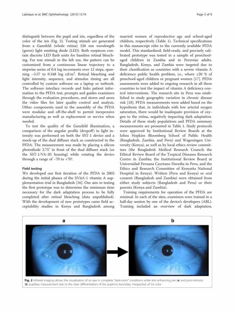
distinguish between the pupil and iris, regardless of thecolor of the iris (Fig. 2). Testing stimuli are generatedfrom a Ganzfeld (whole retina) 526 nm wavelength(green) light emitting diode (LED). Both eyepieces con-tain discrete LED flash units for baseline retinal bleach-ing. For test stimuli in the left eye, the pattern can becustomized from a continuous linear trajectory to astepwise series of 0.4 log increments over 12 steps, span-ning −3.57 to 0.348 log cd/m2. Retinal bleaching andlight intensity, sequence, and stimulus timing are allcontrolled by custom software on a laptop or netbook.The software interface records and links patient infor-mation to the PFDA test, prompts and guides examinersthrough the evaluation procedures, and stores and savesthe video files for later quality control and analysis.Other components used in the assembly of the PFDAwere modular and off-the-shelf to permit inexpensivemanufacturing as well as replacement or service whenneeded.To test the quality of the Ganzfeld illumination, a
comparison of the angular profile (dropoff ) in light in-tensity was performed on both the SST-1 device and amock-up of the dual diffuser stack as constructed in thePFDA. The measurement was made by placing a siliconphotodiode 2.75" in front of the dual diffuser stack (orthe SST-1/VA-20 housing) while rotating the devicethrough a range of −70 to +70°.
Field testingWe developed our first iteration of the PFDA in 2003during the initial phases of the JiVitA-1 vitamin A sup-plementation trial in Bangladesh [16]. Our aim in testingthe first prototype was to determine the minimum timenecessary for the dark adaptation process to be fullycompleted after retinal bleaching (data unpublished).With the development of new prototypes came field ac-ceptability studies in Kenya and Bangladesh among
married women of reproductive age and school-agedchildren, respectively (Table 1). Technical specificationsin this manuscript refer to the currently available PFDAmodel. This standardized, field-ready, and precisely cali-brated prototype was tested in a sample of preschool-aged children in Zambia and in Peruvian adults.Bangladesh, Kenya, and Zambia were targeted due totheir classification as countries with a severe vitamin Adeficiency public health problem, i.e., where ≥20 % ofpreschool-aged children or pregnant women [17]. PFDAassessments were added to ongoing research in all threecountries to test the impact of vitamin A deficiency con-trol interventions. The research site in Peru was estab-lished to study geographic variation in chronic diseaserisk [18]. PFDA measurements were added based on thehypothesis that, in individuals with low arterial oxygensaturation, there would be inadequate provision of oxy-gen to the retina, negatively impacting dark adaptation.Details of these study populations and PFDA summarymeasurements are presented in Table 1. Study protocolswere approved by Institutional Review Boards at theJohns Hopkins Bloomberg School of Public Health(Bangladesh, Zambia, and Peru) and Wageningen Uni-versity (Kenya), as well as by local ethics review commit-tees (the Bangladesh Medical Research Council; theEthical Review Board of the Tropical Diseases ResearchCentre in Zambia; the Institutional Review Board atUniversidad Peruana Cayetano Heredia in Peru, and theEthics and Research Committee of Kenyatta NationalHospital in Kenya). Written (Peru and Kenya) or oralconsent (Bangladesh and Zambia) were obtained fromeither study subjects (Bangladesh and Peru) or theirparents (Kenya and Zambia).Training requirements for operation of the PFDA are
minimal. In each of the sites, examiners were trained in ahalf-day session by one of the device’s developers (ABL).Training included an overview of dark adaptation,
Fig. 2 Infrared imaging allows the visualization of an eye in complete “dark-room” conditions while also enhancing pre (a) and post-stimulus(b) pupillary measurement due to the clear differentiation of the pupil-iris boundary, irrespective of iris color
Labrique et al. BMC Ophthalmology (2015) 15:74 Page 3 of 9
introduction to the device, use of the custom software,interactions with study subjects, testing procedures(described below), and trouble-shooting in the field. Alltrainees tested the PFDA procedures on volunteers. Thestrongest candidates were selected based on the devel-oper’s assessment of aptitude, with emphasis on thetrainee’s ability to interact and guide subjects through thetesting phase. Selected examiners continued with practice-testing on a daily basis for approximately one week, withoversight and regular feedback provided by a supervisor.
Assessment procedureExaminers first attach the PFDA googles and adjust themuntil the subject is comfortable. He or she checks the per-imeter of the goggles using a bright flashlight to identifyand correct errant light penetration through gaps. Afterinstructing the subject to open his or her eyes and lookforward, the examiner initiates the test using the customsoftware. This begins with the bleaching of both retinaswith a bright flash of light, exceeding 3400 cd-s/m2. A 10-min countdown then ensues, during which time the sub-ject’s vision is expected to fully dark-adapt. The 10-minadaptation period is based on previous studies of darkadaptation, as well as testing results from the first PFDAprototype [8, 15]. A set of toy plastic animals was usedduring the dark adaptation period of younger children, to
keep them from touching their goggles and disturbingtheir dark adaptation process. They were asked to feel andguess the animal as a game. At the end of the adaptationperiod, the software issues a warning to the operator thatthe test phase is about to begin. He or she then directs thesubject to open their eyes and look forward. The examprotocol consists of nine light stimuli ranging from −2.9to 0.1 log cd/m2, comparable to previous studies using theSST-1 device [8, 15]. Stimuli increase in increments of 0.4log cd/m2 and last for one second each. There is a 10-srest between stimuli to provide the pupil time to re-dilateto its pre-stimulus size. Prior to each of the nine stimuli,participants are reminded to open their eyes wide andlook straight forward. They are also asked to blink as littleas possible during the test. Aberrant activities like blinkingand looking away are also recorded and can be activelymonitored and discouraged during the testing window. Atthe end of test, the video is automatically saved on the lap-top or netbook. Overall, the duration of PFDA testing pro-cedures is approximately 15 min per subject.
Video analysisPFDA video recordings are assessed by a trained readerusing Tracker 4.85 (Douglas Brown, Davidson, NorthCarolina), an open source video analysis and modelingsoftware. The software’s “Tape Measure” tool enables
Table 1 Pupil response metrics in four study populations using the Portable Field Dark Adaptometer (2010–2013)
Country, Assessment Year Bangladesh, 2010 Kenya, 2010 Zambia, 2012 Peru, 2013
Study Population Pregnant women School-aged children Preschool-aged children Adults
Impaired: −0.5 to 0.1 cd/m2 8.7 33.7 24.0 11.01Pre- to post-stimulus change pupil diameter (in pixels), expressed as proportion of pre-stimulus diameter; figures are mean +/- SD; lower values reflect a greaterresponse/better dark adaptation2Absolute value of the difference in video frame number from pre- to post-stimulus, divided by 30 frames/s; higher values reflect a faster response/betterdark adaptation3Pupillary threshold defined as first stimulus at which pupil diameter decreased by 20 % or more; abnormal as defined by Congdon et al. [8], i.e., pupillarythreshold > = −0.5 log cd/m2
Labrique et al. BMC Ophthalmology (2015) 15:74 Page 4 of 9
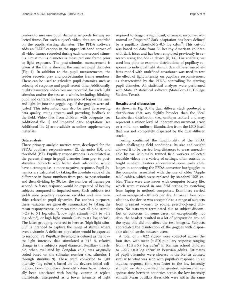
readers to measure pupil diameter in pixels for any se-lected frame. For each subject’s video, data are recordedon the pupil’s starting diameter. The PFDA softwareadds an “LED” caption in the upper left-hand corner ofall video frames recorded during each one-second stimu-lus. Pre-stimulus diameter is measured one frame priorto light exposure. The post-stimulus measurement istaken at the frame showing the smallest pupil diameter(Fig. 4). In addition to the pupil measurements, thereader records pre- and post-stimulus frame numbers.These can be used to calculate pupil dynamics such asvelocity of response and pupil resent time. Additionally,quality assurance indicators are recorded for each lightstimulus and/or the test as a whole, including: blinking;pupil not centered in image; presence of fog on the lens;and light let into the goggle, e.g., if the goggles were ad-justed. This information can also be used in assessingdata quality, rating testers, and providing feedback tothe field. Video files from children with adequate [seeAdditional file 1] and impaired dark adaptation [seeAdditional file 2] are available as online supplementarymaterials.
Data analysisThree primary analytic metrics were developed for thePFDA: pupillary responsiveness (R), dynamics (D), andthreshold (PT). Pupillary responsiveness is calculated asthe percent change in pupil diameter from pre- to post-stimulus. Subjects with better dark adaptation wouldhave a stronger, i.e., a more negative, response. Pupil dy-namics are calculated by taking the absolute value of thedifference in frame numbers from pre- to post-stimulusand then dividing by 30, i.e., the number of frames persecond. A faster response would be expected of healthysubjects compared to impaired ones. Each subject’s testyields nine pupillary response variables and nine vari-ables related to pupil dynamics. For analysis purposes,these variables are generally summarized by taking themean responsiveness or mean time over all nine stimuli(−2.9 to 0.1 log cd/m2), low light stimuli (−2.9 to −1.3log cd/m2), or high light stimuli (−0.9 to 0.1 log cd/m2).The latter grouping, referred to here as “high light stim-uli,” is intended to capture the range of stimuli whereeven a vitamin A deficient population would be expectedto respond [7]. Pupillary threshold is defined as the low-est light intensity that stimulated a ≥15 % relativechange in the subject’s pupil diameter. Pupillary thresh-old, when evaluated by Congdon et al, was originallycoded based on the stimulus number (i.e., stimulus 1through stimulus 9). These were converted to lightintensity (log cd/m2), based on the device’s initial cali-bration. Lower pupillary threshold values have historic-ally been associated with healthy, vitamin A repleteindividuals, interpreted as a lower intensity of light
required to trigger a significant, or major, response. Ab-normal or “impaired” dark adaptation has been definedby a pupillary threshold ≥ -0.5 log cd/m2. This cut-offwas based on data from 56 healthy American childrenwith dark irises and has been employed previously in re-search using the SST-1 device [8, 14]. For analysis, weused box plots to examine distributions of pupillary re-sponse to individual light stimuli. A multilevel mixed ef-fects model with undefined covariance was used to testthe effect of light intensity on pupillary responsiveness,as characterized by the PFDA, controlling for startingpupil diameter. All statistical analyses were performedwith Stata 12 statistical software (StataCorp LP, CollegeStation, Texas).
Results and discussionAs shown in Fig. 3, the dual diffuser stack produced adistribution that was slightly broader than the idealLambertian distribution (i.e., uniform scatter) and mayrepresent a minor level of inherent measurement erroror a mild, non-uniform illumination from the LED itselfthat was not completely dispersed by the dual diffuserstack.Testing confirmed the functionality of the PFDA
under challenging field conditions. Its size and weightallowed it to be carried long distances to areas unreach-able by car. Minimally trained field workers collectedreadable videos in a variety of settings, often outside inbright sunlight. Testers encountered some early chal-lenges in connecting the PFDA convertor box/goggles tothe computer associated with the use of older “Appletalk” cables, which were replaced by standard USB ca-bles. There were also issues with computer battery life,which were resolved in one field setting by switchingfrom laptop to netbook computers. Examiners carriedout an average of ~10 tests per day. Across all four pop-ulations, the device was acceptable to a range of subjectsfrom pregnant women to young, preschool-aged chil-dren. No tests were terminated due to subject discom-fort or concerns. In some cases, on exceptionally hotdays, the headset resulted in a lot of perspiration aroundthe eyes; this did not affect the test integrity. Subjectsappreciated the disinfection of the goggles with dispos-able alcohol swabs between users.A total of n = 822 videos were collected across the
four sites, with mean (± SD) pupillary response rangingfrom -15.5 ± 5.8 log cd/m2 in Kenyan school childrento −22.7 ± 8.0 log cd/m2 in Peruvian adults. Estimatesof pupil dynamics were slowest in the Kenya dataset,similar to what was seen with pupillary response. In allstudies, response time was faster to the low intensitystimuli; we also observed the greatest variance in re-sponse time between countries across the low intensitystimuli. Mean pupillary thresholds were within the same
Labrique et al. BMC Ophthalmology (2015) 15:74 Page 5 of 9
range as previously reported findings [7] and show a similarpattern as the other metrics; the highest threshold was inKenya, where 33.7 % of children were classified as havingimpaired dark adaptation. Results were roughly equivalentin the data from Bangladesh and Peru, where the preva-lence of impaired dark adaptation was ~10 %. Althoughbiochemical data are not yet available to test this hypo-thesis, the marked variation in pupil response metricsacross sites is likely due to differences in vitamin A status.Young children, in particular, are at an increased risk ofvitamin A deficiency [19], which we suspect as theunderlying cause of impaired dark adaptation in ourpreschool- and school-aged samples. Ethnic differencescould conceivably influence our finding as well. How-ever, the challenge of measuring pupil response indarkly pigmented eyes has largely been overcome withthe use of infrared technology.There was a clear trend of increased pupillary response
with increasing light intensity across all four sites (Fig. 4).Regression modeling confirmed a highly significant rela-tionship between pupillary response and light intensity,irrespective of starting pupil diameter, with values rangingfrom −2.6 log cd/m2 (95 % CI: −2.7, −2.5) amongreproductive-aged women in Bangladesh to −3.7 logcd/m2 (95 % CI: −3.9, −3.6) in Zambian preschool-agedchildren. While validation of the PFDA was beyond the
scope of this work, the design of the PFDA was guided bythe SST-1 device, which compares favorably to theGoldmann-Weekers Dark Adaptometer—previously con-sidered the gold standard for assessing dark adaptation[13]. Employment of an infrared camera and objectivevideo analysis may represent an advance over theseprevious psychophysical testing protocols. We expectthat the PFDA’s automated test procedure would alsoimprove the reliability of dark adaptation assessmentsby reducing variability associated with examiner tech-nique [20]. We did note some instances, particularly inthe Zambia dataset (2 % of measurements; 12 % ofchildren), of pupil dilation in response to light stimuli(Fig. 4). While not statistically significant, the mean ageof children with implausible values was lower than thatof their peers (difference = 0.37 ± 0.44 years; p = 0.1).These implausible values likely reflect the challenge ofworking with younger subjects, who may be less able tokeep their eyes fixed straight ahead for the entire testingperiod, thus complicating pupil diameter measure-ments. They also underscore the need for close super-vision of examiners in the field, who must continuouslymonitor eye position on the computer screen andencourage their subjects, particularly children, to lookstraight ahead with their eyes wide open during thetesting phase.
Fig. 3 Comparison of the angular profile in light intensity between the VA-20 Ganzfeld source in the commercial SST-1 device, the mock-up of thedual diffuser stack in the Portable Field Dark Adaptometer, and the ideal Lambertian reflectance across an angular illumination range of -70 to +70°
Labrique et al. BMC Ophthalmology (2015) 15:74 Page 6 of 9
a
-60
-40
-20
020
40
-2.9
-2.5
-2.1
-1.7
-1.3
-0.9
-0.5
-0.1 0.
1
b
-60
-40
-20
020
40
-2.9
-2.5
-2.1
-1.7
-1.3
-0.9
-0.5
-0.1 0.
1
c
-60
-40
-20
020
40
-2.9
-2.5
-2.1
-1.7
-1.3
-0.9
-0.5
-0.1 0.
1
d
-60
-40
-20
020
40
-2.9
-2.5
-2.1
-1.7
-1.3
-0.9
-0.5
-0.1 0.
1
Pre
to p
ost-
stim
ulus
cha
nge
in p
upil
diam
eter
(%
)
Light Stimulus Intensity (log cd/m 2)Fig. 4 Trend in pupillary response across nine log-incremental steps of light intensity (left to right) in Bangladesh (a), Kenya (b), Zambia (c), andPeru (d). Pupillary response is defined as the percent change in pupil diameter from pre- to post-stimulus
Labrique et al. BMC Ophthalmology (2015) 15:74 Page 7 of 9
ConclusionsField testing of the PFDA confirmed its ease of use formeasuring dark adaptation, even under challenging fieldconditions. Testing in four populations clearly replicatedthe well-recognized linear relationship between pupillaryresponsiveness over log-incremental increases in lightintensity. We also noted faster response times at thelower light intensity stimuli, indicative of the slightpupillary fatigue experienced between the beginning ofthe test, after 10 min of dark adaptation, and the nineconsecutive, log-incremental pupillary stimuli. The indi-ces of pupillary response and pupil dynamics generatedhere from PFDA measurements are not exhaustive.However, we do recommend that future PFDA studiesreport pupillary threshold to ensure comparability withprevious research [7], and in the case of vitamin Adeficiency-related research, consider a similar focus onthe high light intensity stimuli.The PFDA was initially designed for use in public
health nutrition research and practice. Based on the pre-vious findings showing a direct relationship betweenpupillary responsiveness and serum retinol concentra-tion [8, 14, 15] and modified dose response [8], thePFDA is currently undergoing validation as a tool toscreen populations for vitamin A deficiency. It also beingemployed in efficacy trials of provitamin A carotenoidbiofortified foods [21], where dark adaptation is a pri-mary functional outcome. There are potential applica-tions for the PFDA in research on non-communicablediseases and clinical practice as well. For example, itmight serve as a rapid and non-invasive status test forpatients with complex gastrointestinal and hepatobiliaryconditions linked to poor vitamin A absorption [22].Dark adaptometry can also be employed in the diagnosisof age-related macular degeneration, photoreceptor dys-trophies, and glaucoma [23–25]. Finally, there is a well-established, causal relationship between poor scotopicvision and hypoxemia [3], which suggests a potential ap-plication of this device in the search for early predictorsof cardiopulmonary disease.
Additional files
Additional file 1: Example PFDA video from a child with normaldark adaptation.
Additional file 2: Example PFDA video file from a child withabnormal dark adaptation.
AbbreviationsPFDA: Portable Field Dark Adaptometer; SST-1: Scotopic Sensitivity Tester-1.
Competing interestsThe authors declare that they have no competing interests.
Authors’ contributionsABL designed the device, planned and oversaw field testing, and guided thedata analysis and manuscript. ACP and KH conducted the data analysis anddrafted the manuscript. SM and TS provided feedback on device iterationsand assisted in redesign. KPW and AS helped design the device and assistedin planning and conduct of studies. All authors read and approved the finalmanuscript.
AcknowledgementsThe Instrument Development Group at the Johns Hopkins UniversityBloomberg Center for Physics and Astronomy designed and assembled thedevice. We acknowledge the following collaborators for field testing thedevice, providing feedback to the design team, and contributing data: inBangladesh, Saijuddin Shaikh and Hasmot Ali from Johns Hopkins University(JHU); in Kenya, Elise Talsma, Alida Melse-Boonstra, and Inge Brouwer fromWageningen University; in Zambia, Maxwell Barffour from JHU and WardSiamusantu from the National Food and Nutrition Commission; and in Peru,William Checkley from JHU and David Danz Cruz from the CRONICAS CohortStudy. This work was funded by Foreign Affairs, Trade and DevelopmentCanada. PFDA design was supported by grants from the Johns HopkinsUniversity (JHU) Faculty Innovation Fund and Center for Global Health. Fieldtesting was supported by the Control of Micronutrient Deficiency Grant #614from the Bill and Melinda Gates Foundation (Bangladesh); Challenge ProgramGrant #8251 from HarvestPlus (Zambia); grant agreement #211484 from theEuropean Union Framework Program [FP7/2007-2013] and HarvestPlus(Kenya); JHU Center for Global Health, contract # HHSN268200900033C withthe National Heart, Lung and Blood Institute, National Institutes of Health,and Fogarty International Center Grant #5R25TW009340 (Peru). None of thesponsors had any role in the design or evaluation of the instrument; thecollection, management, analysis, or interpretation of the data; or the preparation,review, or approval of the manuscript.
Received: 30 November 2014 Accepted: 23 June 2015
References1. Koch W. A new instrument for dark adaptation tests. Br J Ophthalmol.
1945;29(5):234–43.2. Working Group on Night Vision. Night Vision: Current Research and Future
Directions, Symposium Proceedings. Washington, D.C.: National AcademiesPress; 1987.
3. Havelius U, Hansen F, Hindfelt B, Krakau T. Human ocular vasodynamicchanges in light and darkness. Invest Ophthalmol Vis Sci. 1999;40(8):1850–5.
4. Havelius U, Bergqvist D, Falke P, Hindfelt B, Krakau T. I. Impaired darkadaptation in symptomatic carotid artery disease. Neurology.1997;49(5):1353–9.
5. Lamb TD, Pugh Jr EN. Phototransduction, dark adaptation, and rhodopsinregeneration the proctor lecture. Invest Ophthalmol Vis Sci.2006;47(12):5137–52.
6. Carr CJ, Fisher KD. A study of individual variability in dark adaptation andnight vision in man. Washington, D.C: Life Sciences Division, Army ResearchOffice; 1970.
7. Congdon NG, West Jr KP. Physiologic indicators of vitamin A status. J Nutr.2002;132(9 Suppl):2889S–94S.
8. Congdon N, Sommer A, Severns M, Humphrey J, Friedman D, Clement L,et al. Pupillary and visual thresholds in young children as an index ofpopulation vitamin A status. Am J Clin Nutr. 1995;61(5):1076–82.
9. Dowling JE, Wald G. Vitamin A Deficiency and Night Blindness. Proc NatlAcad Sci U S A. 1958;44(7):648–61.
10. Wald G. A portable visual adaptometer. J Opt Soc Am. 1941;31:235–8.11. Wilson WH. The Middle East Adaptometer. Br J Ophthalmol.
1946;30(11):645–57.12. Yarbrough ME, Dann WJ. Dark adaptometer and blood vitamin A
measurements in a North Carolina Nutrition Survey. J Nutr. 1941;22:597–607.13. Peters AY, Locke KG, Birch DG. Comparison of the Goldmann-Weekers dark
adaptometer and LKC Technologies Scotopic Sensitivity tester-1. DocOphthalmol. 2000;101(1):1–9.
14. Congdon NG, Dreyfuss ML, Christian P, Navitsky RC, Sanchez AM, Wu LS,et al. Responsiveness of dark-adaptation threshold to vitamin A and beta-carotene supplementation in pregnant and lactating women in Nepal.Am J Clin Nutr. 2000;72(4):1004–9.
Labrique et al. BMC Ophthalmology (2015) 15:74 Page 8 of 9
15. Sanchez AM, Congdon NG, Sommer A, Rahmathullah L, Venkataswamy PG,Chandravathi PS, et al. Pupillary threshold as an index of population vitaminA status among children in India. Am J Clin Nutr. 1997;65(1):61–6.
16. West Jr KP, Christian P, Labrique AB, Rashid M, Shamim AA, Klemm RD, et al.Effects of vitamin A or beta carotene supplementation on pregnancy-related mortality and infant mortality in rural Bangladesh: a cluster randomizedtrial. JAMA. 2011;305(19):1986–95.
17. World Health Organization. Global prevalence of vitamin A deficiency inpopulations at risk 1995–2005. WHO Global Database on Vitamin ADeficiency. Geneva, Switzerland: World Health Organization; 2009. p. 116.
18. Miranda JJ, Bernabe-Ortiz A, Smeeth L, Gilman RH, Checkley W, CronicasCohort Study Group. Addressing geographical variation in the progressionof non-communicable diseases in Peru: the CRONICAS cohort studyprotocol. BMJ Open. 2012;2(1), e000610.
19. Sommer A, West Jr KP. Vitamin A and childhood morbidity. Lancet.1992;339(8804):1302.
20. Kiser AK, Mladenovich D, Eshraghi F, Bourdeau D, Dagnelie G. Reliability andconsistency of dark-adapted psychophysical measures in advanced eyedisease. Invest Ophthalmol Vis Sci. 2006;47(1):444–52.
21. De Moura FF, Palmer AC, Finkelstein JL, Haas JD, Murray-Kolb LE, WengerMJ, et al. Are biofortified staple food crops improving vitamin a and ironstatus in women and children? New evidence from efficacy trials. Adv Nutr.2014;5(5):568–70.
22. Abbott-Johnson WJ, Kerlin P, Abiad G, Clague AE, Cuneo RC. Darkadaptation in vitamin A-deficient adults awaiting liver transplantation:improvement with intramuscular vitamin A treatment. Br J Ophthalmol.2011;95(4):544–8.
23. Jackson GR, Edwards JG. A short-duration dark adaptation protocol forassessment of age-related maculopathy. J Ocul Biol Dis Inform. 2008;1(1):7–11.
24. Kalaboukhova L, Fridhammar V, Lindblom B. Relative afferent pupillarydefect in glaucoma: a pupillometric study. Acta Ophthalmol Scand.2007;85(5):519–25.
25. Owsley C, McGwin Jr G, Jackson GR, Kallies K, Clark M. Cone- and rod-mediateddark adaptation impairment in age-related maculopathy. Ophthalmology.2007;114(9):1728–35.
Submit your next manuscript to BioMed Centraland take full advantage of:
• Convenient online submission
• Thorough peer review
• No space constraints or color figure charges
• Immediate publication on acceptance
• Inclusion in PubMed, CAS, Scopus and Google Scholar
• Research which is freely available for redistribution
Submit your manuscript at www.biomedcentral.com/submit
Labrique et al. BMC Ophthalmology (2015) 15:74 Page 9 of 9