of these materials is prohibited without the express written authorization of RN.com
Acknowledgements RN.com acknowledges the valuable contributions of… Suzan Miller-Hoover DNP, RN, CCNS CCRN-K Conflict of Interest and Commercial Support RN.com strives to present content in a fair and unbiased manner at all times, and has a full and fair disclosure policy that requires course faculty to declare any real or apparent commercial affiliation related to the content of this presentation. Note: Conflict of Interest is defined by ANCC as a situation in which an individual has an opportunity to affect educational content about products or services of a commercial interest with which he/she has a financial relationship. The planners of the educational activity have no conflicts of interest to disclose. There is no commercial support being used for this course. Purpose The purpose of this continuing nursing education course is to provide healthcare professionals with an overview of breast cancer. Advances in the diagnosis and management of the disease today have improved outcomes for breast cancer victims. Education about risk factors and treatment options is an important part of the healthcare professional’s role. Learning Objectives After successful completion of this course, you will be able to:
1. Discuss the incidence and types of breast cancer in the US today 2. Identify modifiable and non-modifiable risk factors for developing breast cancer 3. Discuss appropriate breast cancer screening 4. Discuss predictive and prognostic markers 5. Discuss the various treatment options available 6. Identify components of a survivorship plan
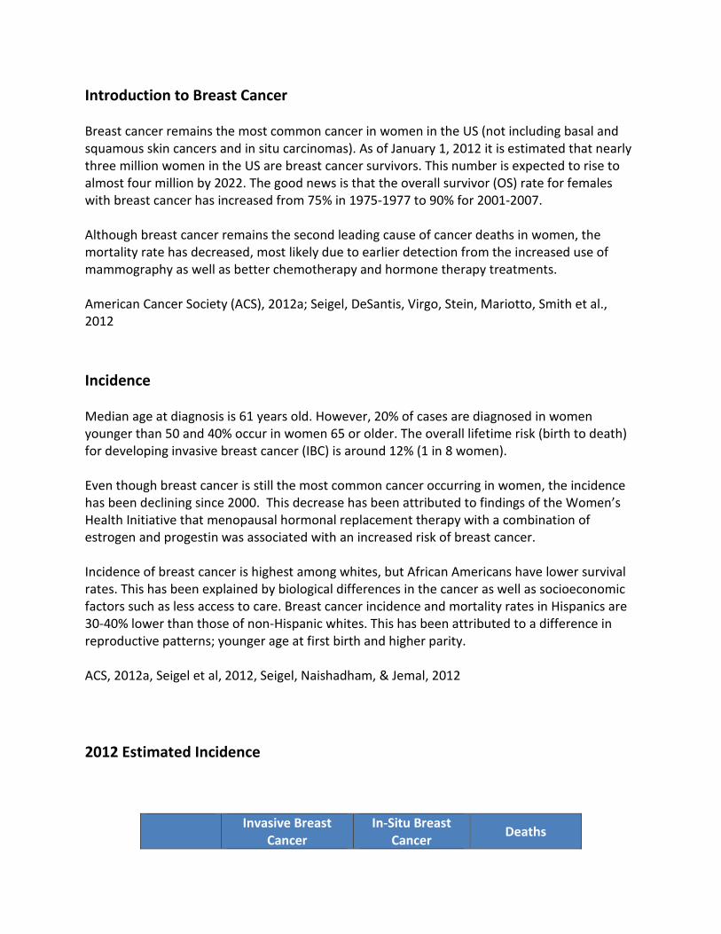
Introduction to Breast Cancer Breast cancer remains the most common cancer in women in the US (not including basal and squamous skin cancers and in situ carcinomas). As of January 1, 2012 it is estimated that nearly three million women in the US are breast cancer survivors. This number is expected to rise to almost four million by 2022. The good news is that the overall survivor (OS) rate for females with breast cancer has increased from 75% in 1975-1977 to 90% for 2001-2007. Although breast cancer remains the second leading cause of cancer deaths in women, the mortality rate has decreased, most likely due to earlier detection from the increased use of mammography as well as better chemotherapy and hormone therapy treatments. American Cancer Society (ACS), 2012a; Seigel, DeSantis, Virgo, Stein, Mariotto, Smith et al., 2012 Incidence Median age at diagnosis is 61 years old. However, 20% of cases are diagnosed in women younger than 50 and 40% occur in women 65 or older. The overall lifetime risk (birth to death) for developing invasive breast cancer (IBC) is around 12% (1 in 8 women). Even though breast cancer is still the most common cancer occurring in women, the incidence has been declining since 2000. This decrease has been attributed to findings of the Women’s Health Initiative that menopausal hormonal replacement therapy with a combination of estrogen and progestin was associated with an increased risk of breast cancer. Incidence of breast cancer is highest among whites, but African Americans have lower survival rates. This has been explained by biological differences in the cancer as well as socioeconomic factors such as less access to care. Breast cancer incidence and mortality rates in Hispanics are 30-40% lower than those of non-Hispanic whites. This has been attributed to a difference in reproductive patterns; younger age at first birth and higher parity. ACS, 2012a, Seigel et al, 2012, Seigel, Naishadham, & Jemal, 2012 2012 Estimated Incidence
Invasive Breast Cancer
In-Situ Breast Cancer Deaths
Females 226,870 63,000 39,510 Males 2,190 N/A 410
ACS, 2012, Seigel et al., 2012 Test Yourself The five-year survival rate in breast cancer has ___________ since the 1970’s.
a) Increased b) Decreased c) Stayed the same
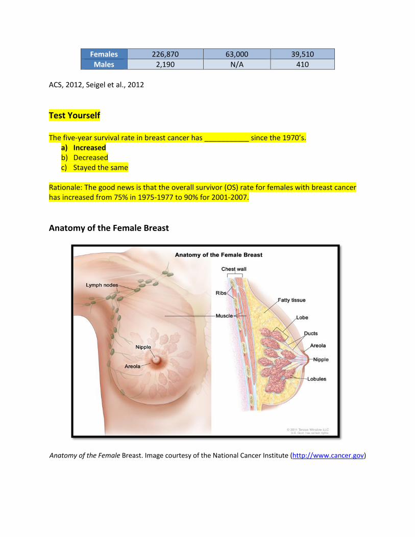
Rationale: The good news is that the overall survivor (OS) rate for females with breast cancer has increased from 75% in 1975-1977 to 90% for 2001-2007. Anatomy of the Female Breast
Anatomy of the Female Breast. Image courtesy of the National Cancer Institute (http://www.cancer.gov)
Risk Factors Many factors have been identified that increase one’s risk for breast cancer. They do not all instill the same degree of risk. Also some are under one’s control and can be modified. Others cannot. For example, the two greatest risk factors for breast cancer are being female and increasing age. Neither of these factors can be modified. Other identified risk factors are listed in the following table.
Strong Increase Moderate Increase Weak Increase
BCRA1 or BCRA2 gene mutation Not having children or having first child after age 35 Age at first period <12
More than 1 immediate family member One immediate family member Age at menopause >55
High breast density High levels of blood estrogens or androgens post-menopause Alcohol consumption
Atypical hyperplasia or LCIS Typical hyperplasia Ashkenazi Jewish heritage
Lobular carcinoma in situ (LCIS) Weight gain after menopause High socioeconomic status
Personal history of cancer Radiation exposure from x-rays
Radiation treatment during youth
Post-menopausal estrogen and progestin Post-menopausal estrogen
High bone density (probable) Current or recent use of birth control pills
Post-menopausal type 2 diabetes Not breastfeeding
Being tall Komen, 2012, Jancin, 2012 Risk Factors Defined Non-modifiable Risk Factors Age: Rates increase with age. At age 20, one’s risk is 1 in 1,681. Rates start increasing around age 40 (1 in 69) and are highest in women and men 70 or older (1 in 27). The median age in men is higher than in women; 68 v. 61 years.
Sex: Breast cancer is about 100 times more common in women. Inherited Gene Mutations: In addition to BRCA1 and BRCA 2, p53, PTEN/MMA1, CHEK2, and ATM mutations have also been linked to breast cancer. Mutations only account for 5-10% of all breast cancers. BRCA genes normally prevent cancer by stopping cells from mutating. Those who inherit a mutation from either parent lose this protection. Women who have a BRCA of 1/2 mutations have a lifetime risk of 40-65% for developing breast cancer. If they are diagnosed with breast cancer, they then face of 40% risk of a contralateral breast cancer within ten years. Hereditary cancers are often diagnosed at an earlier age and may be bilateral. Family History: One first degree family member (parent, sibling, or child) with breast cancer doubles one’s risk. Having more than one first degree family member increases the risk 3-4 times. The greatest risk is when the family member was diagnosed before age 50. Race & Ethnicity: Being white increases one’s risk, while being of Asian American or Pacific Islander decreases one’s risk. These differences have been associated with differences in various risk factors including:
• Age at menarche • Age at menopause • Age at first birth • Number of births • Body weight • Breastfeeding patterns
Ashkenazi Jewish Heritage: These women have a higher prevalence of BRCA 1/2 mutations. Breast Density on Mammogram: Women with dense breasts have more glandular tissue and less fatty tissue. This increases one’s risk 4-5 times that of a person with low breast density. It also may be harder to see abnormalities on a mammogram. Benign Proliferative Breast Conditions: This refers to an overgrowth of cells that line either the breast lobules or milk ducts. If the lobules are involved it is called lobular hyperplasia. If the ducts are involved, it is called ductal hyperplasia. Hyperplasia may be usual or typical or it may be atypical. Usual hyperplasia increases risk two times that of a person without hyperplasia, while atypical hyperplasia increases the risk 4-5 times. Risk Factors Defined Lobular Carcinoma in Situ: In this condition, abnormal cells grow inside the lobules only. Presence of LCIS increases one’s risk for breast cancer 7-12 times, and is considered a risk factor for the development of breast cancer in either breast. The breast cancer that develops may be either ductal or lobular.
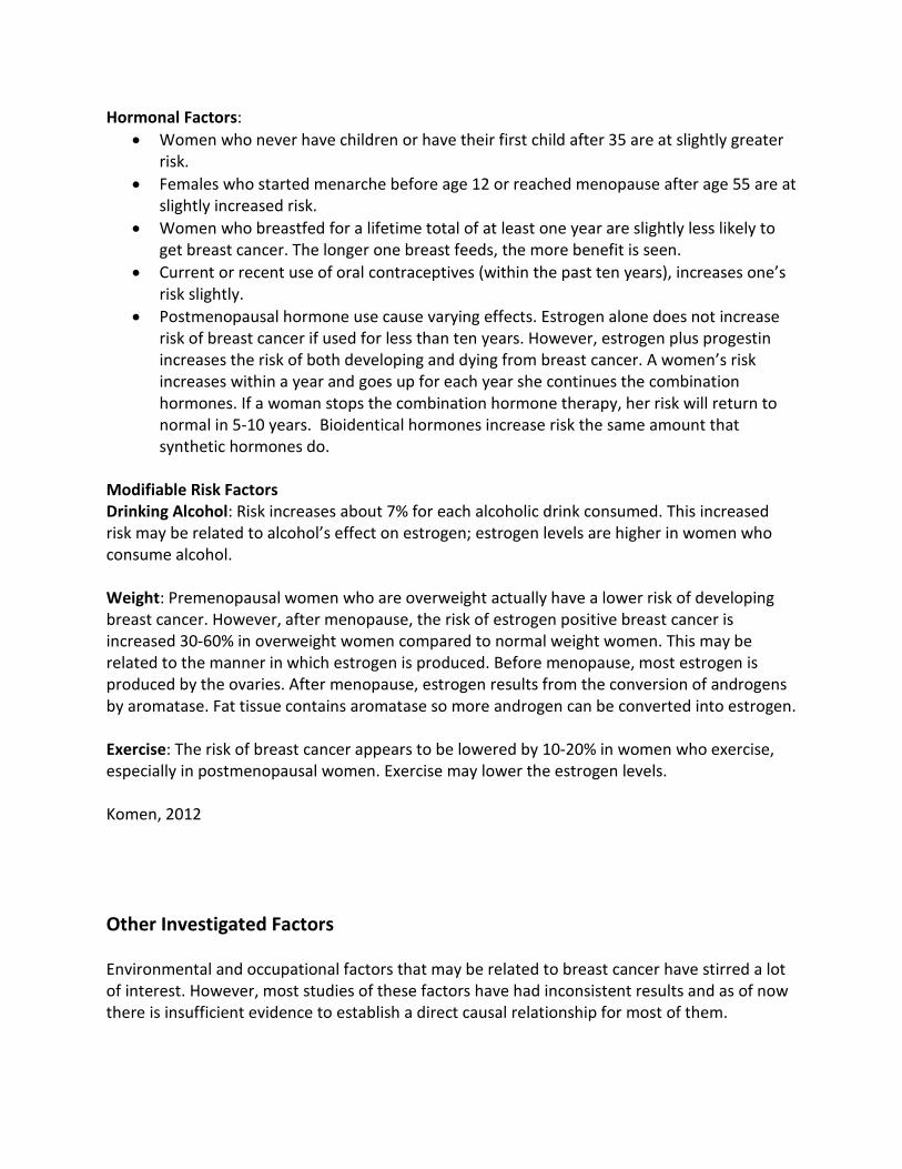
Hormonal Factors: • Women who never have children or have their first child after 35 are at slightly greater
risk. • Females who started menarche before age 12 or reached menopause after age 55 are at
slightly increased risk. • Women who breastfed for a lifetime total of at least one year are slightly less likely to
get breast cancer. The longer one breast feeds, the more benefit is seen. • Current or recent use of oral contraceptives (within the past ten years), increases one’s
risk slightly. • Postmenopausal hormone use cause varying effects. Estrogen alone does not increase
risk of breast cancer if used for less than ten years. However, estrogen plus progestin increases the risk of both developing and dying from breast cancer. A women’s risk increases within a year and goes up for each year she continues the combination hormones. If a woman stops the combination hormone therapy, her risk will return to normal in 5-10 years. Bioidentical hormones increase risk the same amount that synthetic hormones do.
Modifiable Risk Factors Drinking Alcohol: Risk increases about 7% for each alcoholic drink consumed. This increased risk may be related to alcohol’s effect on estrogen; estrogen levels are higher in women who consume alcohol. Weight: Premenopausal women who are overweight actually have a lower risk of developing breast cancer. However, after menopause, the risk of estrogen positive breast cancer is increased 30-60% in overweight women compared to normal weight women. This may be related to the manner in which estrogen is produced. Before menopause, most estrogen is produced by the ovaries. After menopause, estrogen results from the conversion of androgens by aromatase. Fat tissue contains aromatase so more androgen can be converted into estrogen. Exercise: The risk of breast cancer appears to be lowered by 10-20% in women who exercise, especially in postmenopausal women. Exercise may lower the estrogen levels. Komen, 2012 Other Investigated Factors Environmental and occupational factors that may be related to breast cancer have stirred a lot of interest. However, most studies of these factors have had inconsistent results and as of now there is insufficient evidence to establish a direct causal relationship for most of them.
Weak Increase No Increase or Decreased Risk Insufficient Evidence
Light at night and shift work Blood organochlorine levels (from pesticides, industrial chemicals, etc.)
Plastics or cosmetics containing parabens
Breast implants Smoking and secondhand smoke
Electromagnetic fields Trauma to breast Deodorant/antiperspirant use Caffeine Soy intake, dietary fat, fruits and vegetables, and carbohydrate consumption
Cell phones
Abortion or fertility drug use Hair relaxers Hair dyes Bras and underwire bras
Komen, 2012 Test Yourself Which breast cancer risk factors can be modified?
a) Time of menarche, number of children, and age b) Alcohol consumption, breast density, and diagnosis of LCIS c) Postmenopausal weight gain, inactivity, and alcohol consumption
Rationale: Modifiable Risk Factors Drinking Alcohol: Risk increases about 7% for each alcoholic drink consumed. This increased risk may be related to alcohol’s effect on estrogen; estrogen levels are higher in women who consume alcohol. Weight: Premenopausal women who are overweight actually have a lower risk of developing breast cancer. However, after menopause, the risk of estrogen positive breast cancer is increased 30-60% in overweight women compared to normal weight women. This may be related to the manner in which estrogen is produced. Before menopause, most estrogen is produced by the ovaries. After menopause, estrogen results from the conversion of androgens by aromatase. Fat tissue contains aromatase so more androgen can be converted into estrogen. Exercise: The risk of breast cancer appears to be lowered by 10-20% in women who exercise, especially in postmenopausal women. Exercise may lower the estrogen levels.
How Can Risk be Reduced? Women can reduce the risk for developing breast cancer by:
• Maintaining a healthy lifestyle to reduce modifiable risk factors. • Undergoing consistent and timely routine screening. • Utilize risk reduction agents if necessary. • Considering a risk reducing mastectomy and /or Bilateral Salpingo-Oophorectomy (BSO)
if necessary. Healthy Lifestyle Modifications Alcohol: Three drinks a week gives you a 15% greater chance of getting breast cancer than those who do not drink. The risk increases about 10% for each additional drink a day. Body Weight: Overweight and obese women (BMI >25) after menopause have greater risk of getting breast cancer than women of healthy weight. Being overweight also increases one’s chance of recurrence. Diet: There is no evidence that a correlation exists between diet with breast cancer risk, but a healthy diet does promote overall health. Exercise: Exercising regularly at a moderate or intense level for 4-7 hours per week is associated with a lower risk of breast cancer. HRT: Estrogen alone does not increase risk of breast cancer if used for less than ten years. However, estrogen plus progestin increases the risk of both developing and dying from breast cancer. A women’s risk increases within a year and goes up for each year she continues the combination hormones. Smoking: Studies linking smoking to breast cancer have had mixed results. It is possible that smoking before first child birth may increase risk. (Komen, 2012) Screening Screening does not reduce the risk of getting breast cancer but it does decrease the risk of dying from it. Cancers can be identified at an earlier and more treatable stage. Note! 20-30% of all breast cancers will not be detected on a mammogram so this alone is not sufficient for screening.
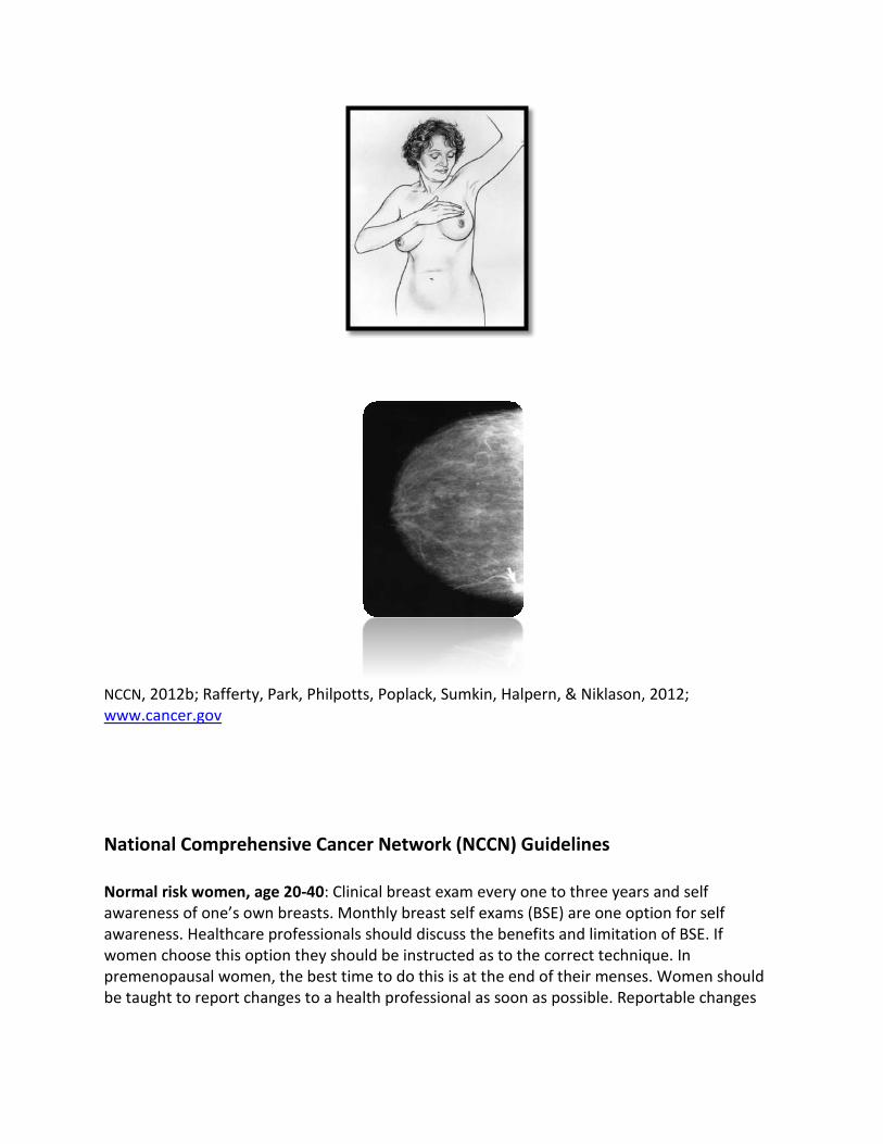
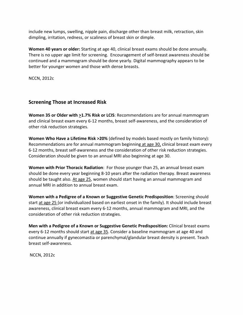
NCCN, 2012b; Rafferty, Park, Philpotts, Poplack, Sumkin, Halpern, & Niklason, 2012; www.cancer.gov National Comprehensive Cancer Network (NCCN) Guidelines Normal risk women, age 20-40: Clinical breast exam every one to three years and self awareness of one’s own breasts. Monthly breast self exams (BSE) are one option for self awareness. Healthcare professionals should discuss the benefits and limitation of BSE. If women choose this option they should be instructed as to the correct technique. In premenopausal women, the best time to do this is at the end of their menses. Women should be taught to report changes to a health professional as soon as possible. Reportable changes
include new lumps, swelling, nipple pain, discharge other than breast milk, retraction, skin dimpling, irritation, redness, or scaliness of breast skin or dimple. Women 40 years or older: Starting at age 40, clinical breast exams should be done annually. There is no upper age limit for screening. Encouragement of self-breast awareness should be continued and a mammogram should be done yearly. Digital mammography appears to be better for younger women and those with dense breasts. NCCN, 2012c Screening Those at Increased Risk Women 35 or Older with >1.7% Risk or LCIS: Recommendations are for annual mammogram and clinical breast exam every 6-12 months, breast self-awareness, and the consideration of other risk reduction strategies. Women Who Have a Lifetime Risk >20% (defined by models based mostly on family history): Recommendations are for annual mammogram beginning at age 30, clinical breast exam every 6-12 months, breast self-awareness and the consideration of other risk reduction strategies. Consideration should be given to an annual MRI also beginning at age 30. Women with Prior Thoracic Radiation: For those younger than 25, an annual breast exam should be done every year beginning 8-10 years after the radiation therapy. Breast awareness should be taught also. At age 25, women should start having an annual mammogram and annual MRI in addition to annual breast exam. Women with a Pedigree of a Known or Suggestive Genetic Predisposition: Screening should start at age 25 (or individualized based on earliest onset in the family). It should include breast awareness, clinical breast exam every 6-12 months, annual mammogram and MRI, and the consideration of other risk reduction strategies. Men with a Pedigree of a Known or Suggestive Genetic Predisposition: Clinical breast exams every 6-12 months should start at age 35. Consider a baseline mammogram at age 40 and continue annually if gynecomastia or parenchymal/glandular breast density is present. Teach breast self-awareness. NCCN, 2012c
Other Screening Modalities Ultrasound: Several studies support the use of ultrasound as an addition to mammography in high risk women with dense breasts. Dense breasts limit the sensitivity of mammograms. Tomosynthesis or 3D Mammography: This technology was approved by the FDA in 2011. Early studies show promise for this adjunct but there is insufficient evidence at this time by NCCN to recommend it for routine use. However, a large (1,192 women from five different sites), recently published study has demonstrated that adding this three dimensional breast imaging technique to standard digital mammography not only significantly increased radiologists’ diagnostic accuracy but also decreased the number of false positive readings. Interestingly, the amount of radiation from the combined procedure is less than three milligray which is the FDA limit for a mammography alone. MRI: Expert opinion states that MRI screening should not be utilized for women with less than a 15% lifetime risk for breast cancer. NCCN, National Comprehensive Cancer Network (NCCN), 2012b; Rafferty et al., 2012 Risk Reduction Agents Risk reduction agents include tamoxifen, raloxifene, and exemestane. Tamoxifen, taken daily for five years, reduces the risk of breast cancer by 49%. Data on raloxifene is limited to postmenopausal women. Initially the risk reduction is similar to tamoxifen, but it appears to lessen in the long term. However, the better side effect profile may make this a good choice for some women. A single large randomized study of exemestane in postmenopausal women with an increased risk for breast cancer showed a decrease of 65% in the risk that carried over for three years. These agents will be discussed further in the treatment section. While a good choice for many, not all women can tolerate these agents. Also, the effectiveness of these agents is not proven in women with BRCA 1/2 mutations or those with prior thoracic radiation. NCCN,2012b
Risk Reducing Surgery Bilateral prophylactic mastectomy reduces the risk of developing breast cancer by at least 90% in moderate to high risk women. This surgery should be considered only in women with BRCA 1/2 mutations or other strongly predisposing gene mutations, those with compelling family histories, and possibly in women with LCIS or prior thoracic radiation before age 30. Women should receive consultation from an interprofessional team, which includes a counselor, before deciding to undergo this surgery. Bilateral Salpingo-Oophorectomy (BSO) is also associated with a significant decrease in breast cancer in women specifically with BRCA 1/2 mutations without a prior history of breast cancer. The risk is reduced by about 50%. The greatest reductions are seen in women who had the surgery before age 40. The combination of risk reduction mastectomy and BSO in mutation carriers confers a risk reduction of around 95%. NCCN, 2012b Diagnosis If a suspicious finding is found on any of these modalities, a biopsy of the tissue must be done before a diagnosis of cancer can be made. The gold standard for tissue acquisition in the minimally invasive breast biopsy (MIBB), also known as a percutaneous needle biopsy. MIBBs are just as accurate as open surgical biopsies and offer numerous advantages. First, since most suspicious findings are benign, many patients will be spared surgery and the associated morbidity. MIBBs are less expensive and less disfiguring. Interprofessional treatment planning can be done which should mean a greater chance of an adequate resection being done with only one surgery. Open surgical biopsies should be reserved for the 5-10% of patients who are not amenable to MIBBs such as those with lesions in difficult places to access such as near the chest wall or an implant.
BIOPSY IS THE ONLY SURE WAY TO DIAGNOSE BREAST CANCER! (Gutwein, Darwin, Liu, Marshall, Hochwald, Copeland, & Grobmeyer, 2011) Types of Breast Biopsies Fine Needle Aspiration (FNA): A very thin needle attached to a syringe is inserted into the suspicious area and a small amount of tissue is withdrawn. If the area cannot be felt, ultrasound
can be used to guide the placement of the needle. Sometimes, not enough tissue can be obtained to make a diagnosis using this method.
Fine Needle Biopsy. Image provided courtesy of National Institutes of Health (2012). Core Needle Biopsy (CNB): A core needle is a larger, hollow needle that can withdraw cylinders of tissue from the area. Usually 3-6 tissue samples are obtained, thus providing more tissue than a FNA so a more accurate diagnosis can be made. Ultrasound guidance is usually used to guide the needle.
Magnum Hand Core Biopsy Needle, courtesy of C. R. Bard, Inc. Types of Breast Biopsies Stereotactic Core Needle Biopsy: Computers use mammograms to map the location of the area. This is done in conjunction with a CNB. Often done to biopsy macrocalcifications that can be seen on x-ray but not palpated.
Vacuum-Assisted Core Biopsy: These biopsies require the purchase of special systems. Computer mapping of mammograms, ultrasound, or MRI can be used for guidance. The skin is numbed, and a small cut is made in order to insert a hollow probe. A cylinder of tissue can then be removed. Multiple tissue samples are obtained. Suturing is not needed.
EnCor Enspire Vacuum Assisted Device, courtesy of C. R. Bard, Inc.
Surgical (Open) Biopsy: All (an excisional biopsy) or part (an incisional biopsy) of a lump is removed. If the lump is hard to find or see, a thin hollow needle may be inserted using mammographic guidance. A thin wire is then inserted through the needle and left in place to identify the correct area to biopsy in the operating room. Suturing is required to close a surgical biopsy site and scarring may result.
Open Surgical Biopsy. Image provided courtesy of Wikipedia (2013).
Types of Breast Cancer Breast cancers are very heterogeneous. In addition to cellular classifications, histological and molecular profiles are important particularly because of their influence on treatment and prognosis. Breast cancer can be invasive or non-invasive. Noninvasive cancer is referred to as in situ. The cancer remains “in place” or confined within the ducts or lobules.
Carcinoma in Situ. Image courtesy of the website of the National Cancer Institute
(http://www.cancer.gov). Ductal carcinoma in situ (DCIS) is classified as Stage 0. It is rarely palpable and is usually diagnosed by mammography. DCIS may progress to an invasive cancer. Lobular carcinoma in situ (LCIS) is not a cancer, but rather a risk factor for the development of cancer in either breast. National Cancer Institute (NCI), 2012 Cellular Classifications of Invasive Breast Cancer (IBC) Breast cancer that has spread outside the membrane of the duct or lobule is referred to as invasive. This type of cancer can invade the surrounding tissue and is able to spread to other parts of the body.
Invasive (or infiltrating) ductal carcinoma (IDC) accounts for 70-80% of all IBC. Ductal means that the cancer originated in the milk ducts of the breast. There are many subtypes of ductal carcinoma.
Most breast cancers are invasive! In Situ versus Invasive Breast Cancer
Breast Cancer Ductal Carcinoma in Situ and Invasive Breast Cancer. Images courtesy of the website of the National Cancer Institute (http://www.cancer.gov)
Sub-Types of IDC
Type Characteristic Origin Percentage
Cribiform Cells invade the stroma in nest-like formations resulting in holes between cancer cells 5-6%
Medullary Soft, fleshy mass resembling the medulla of the brain 3-5%
Mucinous (colloid) Cells float in pools of mucin which becomes part of the tumor 2-3%
Papillary Consists of small finger-like projections 1-2%
Tubular Usually small tumors made up of tube-like structures 1-4%
www.breastcancer.org, 2012b Other Types of IBC Invasive lobular carcinoma (ILC) is the second most common type and accounts for 10% of IBCs. This cancer originates in the milk producing lobules of the breast. It is harder to see on mammogram because the cells tend to form more of a thick line than a lump. Inflammatory breast cancer is probably a distinct biologic entity. It accounts for 1-5% of IBCs. It is a type of cancer that is very aggressive and always locally advanced, if not metastatic, on diagnosis. The diagnosis is primarily a clinical one; diffuse erythema and edema, often without a mass, on the majority of the breast. Paget’s disease of the nipple accounts for <5% of IBCs. Cancer cells start in the ducts of the nipple and then spread to the areola causing the nipple and areola to become red, scaly and itchy. Many patients diagnosed with Paget’s will also have cancer elsewhere in the breast. Phyllodes tumors account for <1% of IBCs. These tumors grow in a leaf like pattern. These tumors rarely metastasize. ACS, 2012b, Duskin & Cristofanilli, 2011 Very Rare Types of IBC Adenoid cystic accounts for <0.1% of IBCs. It looks like an adenoid cystic carcinoma that would be found in the salivary glands. It has a good prognosis. Apocrine accounts for about 0.4% of IBCs. Some argue that it is a subtype of IDC and not a distinct entity. It commonly is estrogen and progesterone receptor negative and androgen receptor positive. Metaplastic (MeBC) accounts for 0.2-0.6% of IBCs. It is a mix of adenocarcinoma with spindle cell, squamous and mesenchymal cells features. Tumors are sub classified by the predominant type of cell differentiation. There are no reported cases of men having this tumor. This type is associated with a poor prognosis.
Secretory accounts for 0.15% of IBCs and 0.2% of male breast cancers. It occurs at younger ages; 25-40 in females and 17 in males, making it the most common breast cancer seen in childhood. The tumors may be large; up to 16 cm has been reported. They seem resistant to chemotherapy but have an indolent clinical course. Cadoo, McArdel, O’Shea, Power, & Hennessy, 2012 Prognosis Prognosis and selection of therapy is based on many factors. These include:
• Age and menopausal status • Stage of disease • Nuclear grade of the tumor (how different does the cell look from the normal parent
cell) • Hormone receptor status of the tumor (estrogen and progesterone) • Human epidermal growth factor type 2 receptor expression • Proliferative capacity of the tumor • Presence of lymphovascular invasion • Gene profiles • Molecular subtypes
Predictive Markers Biomarkers are characteristics that can be measured to indicate normal biological and pathogenic processes or therapeutic responses, and are an important prognostic and predictive marker of breast cancer. Predictive biomarkers provide information on the effect of a therapeutic intervention, and prognostic biomarkers provide information about a patient’s overall cancer outcome, independent of therapy. It is recommended by NCCN and American Society of Clinical Oncology (ASCO) that the following biomarkers be measured routinely on diagnosis of breast cancer and at the time of any recurrence as the status of the markers can change over time:
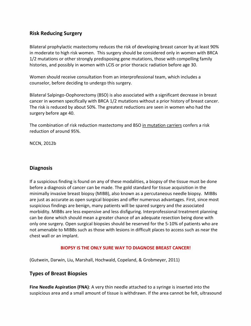
ER status has a significant predictive value for whether or not the tumor will respond to hormone therapy in both the adjuvant and metastatic settings. It is less clear what the prognostic implications Progesterone Receptors status has, but there is evidence that tumors with both positive ER and PR receptors respond best to hormonal therapy. ER/PR negative tumors tend to have a worse prognosis and do not respond to hormonal therapy. HER2 is a tyrosine kinase growth factor receptor. When stimulated it initiates signal transduction pathways which stimulates tumor growth. It is over-expressed in 15-30% of breast cancers and indicates a poorer prognosis unless it is blocked by one of the targeted agents like trastuzumab. Bishop, 2011 Estrogen Receptors
Estrogen Receptors and Estrogen Receptors: Trigger Gene Activation. Images courtesy of the website of
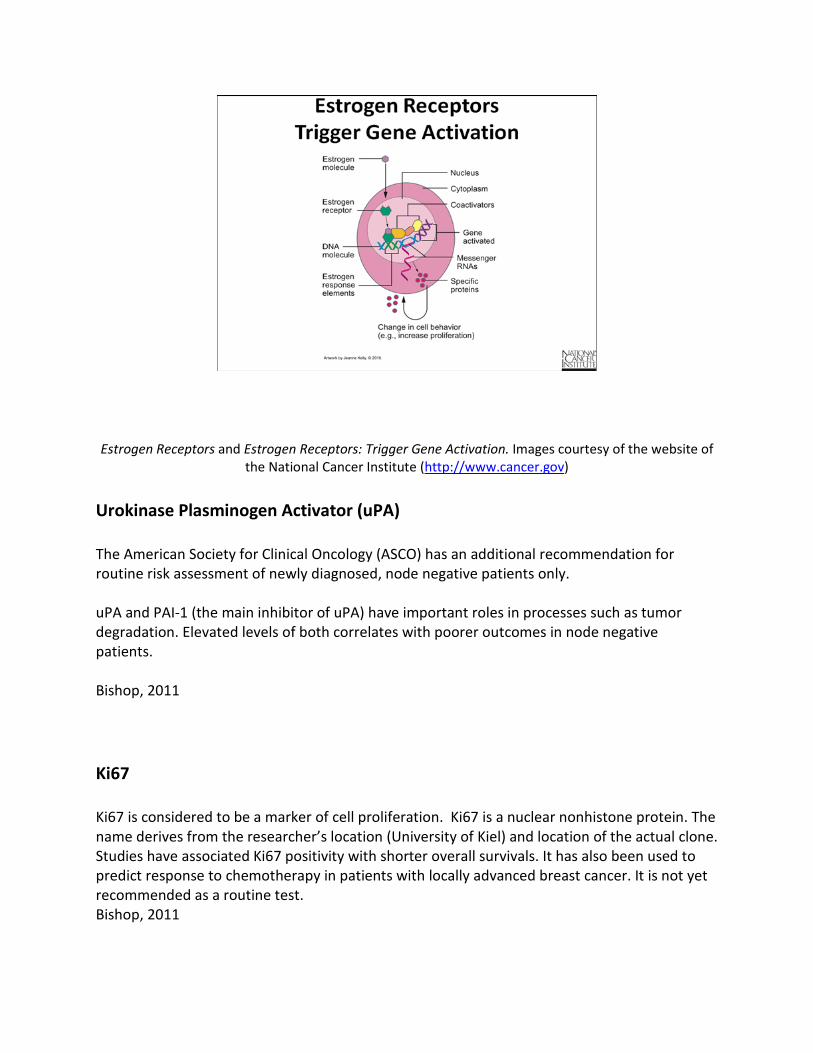
the National Cancer Institute (http://www.cancer.gov) Urokinase Plasminogen Activator (uPA) The American Society for Clinical Oncology (ASCO) has an additional recommendation for routine risk assessment of newly diagnosed, node negative patients only. uPA and PAI-1 (the main inhibitor of uPA) have important roles in processes such as tumor degradation. Elevated levels of both correlates with poorer outcomes in node negative patients. Bishop, 2011 Ki67 Ki67 is considered to be a marker of cell proliferation. Ki67 is a nuclear nonhistone protein. The name derives from the researcher’s location (University of Kiel) and location of the actual clone. Studies have associated Ki67 positivity with shorter overall survivals. It has also been used to predict response to chemotherapy in patients with locally advanced breast cancer. It is not yet recommended as a routine test. Bishop, 2011
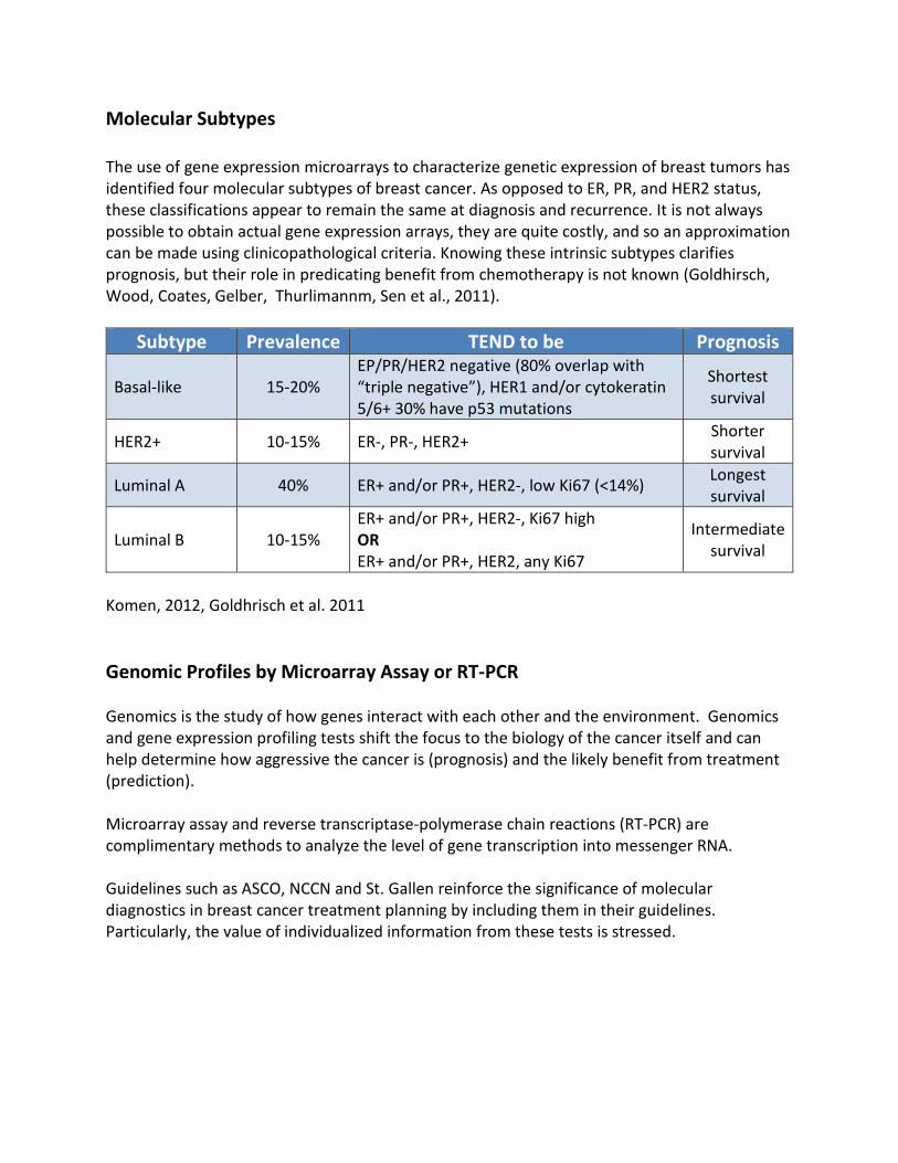
Molecular Subtypes The use of gene expression microarrays to characterize genetic expression of breast tumors has identified four molecular subtypes of breast cancer. As opposed to ER, PR, and HER2 status, these classifications appear to remain the same at diagnosis and recurrence. It is not always possible to obtain actual gene expression arrays, they are quite costly, and so an approximation can be made using clinicopathological criteria. Knowing these intrinsic subtypes clarifies prognosis, but their role in predicating benefit from chemotherapy is not known (Goldhirsch, Wood, Coates, Gelber, Thurlimannm, Sen et al., 2011).
Subtype Prevalence TEND to be Prognosis
Basal-like 15-20% EP/PR/HER2 negative (80% overlap with “triple negative”), HER1 and/or cytokeratin 5/6+ 30% have p53 mutations
Luminal B 10-15% ER+ and/or PR+, HER2-, Ki67 high OR ER+ and/or PR+, HER2, any Ki67
Intermediate survival
Komen, 2012, Goldhrisch et al. 2011 Genomic Profiles by Microarray Assay or RT-PCR Genomics is the study of how genes interact with each other and the environment. Genomics and gene expression profiling tests shift the focus to the biology of the cancer itself and can help determine how aggressive the cancer is (prognosis) and the likely benefit from treatment (prediction). Microarray assay and reverse transcriptase-polymerase chain reactions (RT-PCR) are complimentary methods to analyze the level of gene transcription into messenger RNA. Guidelines such as ASCO, NCCN and St. Gallen reinforce the significance of molecular diagnostics in breast cancer treatment planning by including them in their guidelines. Particularly, the value of individualized information from these tests is stressed.
Gene Profiles There are three commercial tests genetic profiling tests in the United States: OncotypeDXTM, MammaPrint®, and Mammastrat®. These tests look at genes in the tumor to see if they are overexpressed, under-expressed, or expressed normally. Then the results are programmed into a mathematical algorithm to calculate the results. All three tests can be used to help determine prognosis. The resulting scores carry more significance than any one clinicopathologic factor. However, they should not be used alone to determine prognosis, rather in conjunction with clinicopathologic factors. If one of these tests indicates a good prognosis, the healthcare provider and patient may decide to forego chemotherapy. Of note, while all of these tests predict prognosis, OncotypeDX is the only one of the tests shown to actually predict chemotherapy benefit in hormone receptor positive disease (Goldhirsch et al., 2011). Genome-Wide Profiling: Gene Expression Profiles
Genome-Wide Profiling: Gene Expression Profiles. Image courtesy of the website of the National Cancer
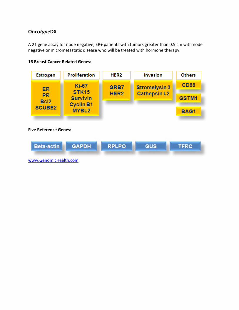
OncotypeDX A 21 gene assay for node negative, ER+ patients with tumors greater than 0.5 cm with node negative or micrometastatic disease who will be treated with hormone therapy. 16 Breast Cancer Related Genes:
OncotypeDX OncotypeDX Recurrence Score® (RS) Provides an Individual Risk Score for Distant Recurrence at 10 Years
Predicting Chemotherapy Response with OncotypeDx OncotypeDX is a strong and significantly statistical predictor of both recurrence free and overall survival. It also predicts the amount of benefit women with estrogen positive breast cancer can expect from chemotherapy. Patients with a low RS (0-<18) will get no benefit from chemotherapy, but see a very good response from the use of endocrine therapy. Patients with an intermediate RS (18-30) will get a very small benefit from chemotherapy. Patients with a high RS, >30, will see about a 28% increase in disease free survival at ten years. The TAILORx trial was conducted to determine which ER+, node negative women with intermediate RS do not benefit from chemotherapy. Data was collected on over 10,000 women. The results are being collated at the present. OncotypeDX has been shown to be prognostic and predictive for women with ER+ node positive breast cancer also. Chemotherapy has demonstrated benefit for all of these women, even those with four or more positive nodes. However, women with 1-3 positive nodes and a low RS seem to do as well as node negative women with a low RS. Therefore, a study has been devised to
randomize women with 1-3 positive nodes and a RS of 25 or less to hormone therapy +/- chemotherapy. The study, called RxPONDER is presently accruing. www.genomichealth.com MammaPrint ® MammaPrint® is a test that utilizes microarray techniques to analyze 70 genes whose activity provides information about breast cancer recurrence at five and ten years. The test is approved to predict distant metastasis in women with Stage I or II breast cancers regardless of estrogen receptor status. Approval in the US extends only to lymph node negative patients. Outside of the US, the test has been approved for women with up to three positive nodes. Based on MammaPrint®, women will be classified as either low risk or high risk. A high risk score means a patient has a 29% risk her cancer will recur within ten years if she has no additional treatment. A low risk patient has a 10% risk. There is not an individual score and the test has not been validated as to its ability to identify which patients will have a better outcome if a treatment decision is made based on the results of this assay. Agendia, the company that makes MammaPrint®, is now also marketing an 80 gene profile test that can classify breast cancer into basal, luminal and HER2 molecular subtypes. The test is called BluePrint®. www.agendia.com, 2012 Mammostrat® This test does not look at RNA but rather uses immunohistochemistry antibody testing to looks at the expression of five genes in the breast cancer cells. Patients are then being stratified into risk groups which can inform decision making. The test was validated on ER+, node negative patients but recent studies have shown it to be valid as a prognostic tool in ER-, node positive disease also. The scores are an independent predictor of recurrence free survival (RFS), distant recurrence free survival (DRFS), and overall survival (OS). Five year survival rates in patients with any nodal status and no chemotherapy:
Bartlett, Bloom, Piper, Lawton, van de Velde, Ross et al., 2012 Test Yourself Which is the only genomic test validated to predict chemotherapy benefit?
a) OncotypeDX b) MammaPrint® c) Mammostrat®
Rationale: OncotypeDX is a strong and significantly statistical predictor of both recurrence free and overall survival. It also predicts the amount of benefit women with estrogen positive breast cancer can expect from chemotherapy. Patients with a low RS (0-<18) will get no benefit from chemotherapy, but see a very good response from the use of endocrine therapy. Patients with an intermediate RS (18-30) will get a very small benefit from chemotherapy. Patients with a high RS, >30, will see about a 28% increase in disease free survival at ten years Staging of Breast Cancer The term staging is used to classify the extent of disease in the body. It is an important factor in determining prognosis and treatment options. It summarizes information to aid in communication among members of the healthcare team. The TNM (tumor, node, and metastasis) system is the most commonly used staging system. At times there may be a prefix before the stage notation. This indicates the timing of the staging:
• C = clinical staging done before treatment and utilized as the guide for primary treatment.
• P = pathologic staging which is done after surgical exploration and is utilized as a guide for adjuvant treatment and estimation of prognosis.
• R = retreatment staging which is done after a disease free interval and is utilized as a guide before further treatment.
Clinical Staging System: Tumor The American Joint Committee on Cancer (AJCC) recommends the use of the following clinical staging system measuring tumor, nodes, and metastasis (TNM):
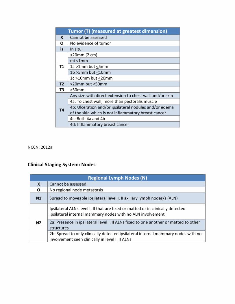
Tumor (T) (measured at greatest dimension) X Cannot be assessed O No evidence of tumor is In situ
T1
<20mm (2 cm) mi <1mm 1a >1mm but <5mm 1b >5mm but <10mm 1c >10mm but <20mm
T2 >20mm but <50mm T3 >50mm
T4
Any size with direct extension to chest wall and/or skin 4a: To chest wall, more than pectoralis muscle 4b: Ulceration and/or ipsilateral nodules and/or edema of the skin which is not inflammatory breast cancer 4c: Both 4a and 4b 4d: Inflammatory breast cancer
NCCN, 2012a Clinical Staging System: Nodes
Regional Lymph Nodes (N) X Cannot be assessed O No regional node metastasis
N1 Spread to moveable ipsilateral level I, II axillary lymph nodes/s (ALN)
N2
Ipsilateral ALNs level I, II that are fixed or matted or in clinically detected ipsilateral internal mammary nodes with no ALN involvement
2a: Presence in ipsilateral level I, II ALNs fixed to one another or matted to other structures 2b: Spread to only clinically detected ipsilateral internal mammary nodes with no involvement seen clinically in level I, II ALNs
N3
Cancer in ipsilateral infraclavicular (axillary level III) lymph node with or without level I, II ALN involvement; or in clinically detected ipsilateral internal mammary nodes with clinically evident level I, II ALN involvement; or cancer in ipsilateral supraclavicular lymph node/s with or without axillary or internal mammary lymph node involvement
3a: In ipsilateral infraclavicular node/s 3b: In ipsilateral internal mammary and axillary lymph nodes 3c: In ipsilateral supraclavicular nodes
NCCN, 2012a Clinical Staging System: Metastasis
Distant Metastasis (M) M0 No evidence
cM0 (1+) No clinical or radiographic evidence of distant metastasis, but presence of molecularly or microscopic tumor cells (no larger than 0.2mm) in circulating blood, bone marrow, or other non-regional nodal tissue in a patient with no other signs or symptoms of metastasis
M1 Distant metastasis larger than 0.2mm as determined by clinical or radiographic means
NCCN, 2012a Stage/Prognostic Groups
Stage TNM % Year Survival Rates for Women Dx in 2001-2002
Stage 0 Tis N0 M0 93% Stage 1A T1 N0 M0 Stage 1 A and B: 88%
Stage 1B T0 N1miM0 T1 N1miM0
Stage 11A T0 N1 M0 T1 N1 M0 T2 N0 M0
81%
Stage 11B T2 N1 M0 74%
T3 N0 M0
Stage 111A
T0 N2 M0 T1 N2 M0 T2 N2 M0 T3 N1 M0 T3 N2 M0
67%
Stage 111B T4 N0 M0 T4 N1 M0 T4 N2 M0
41%
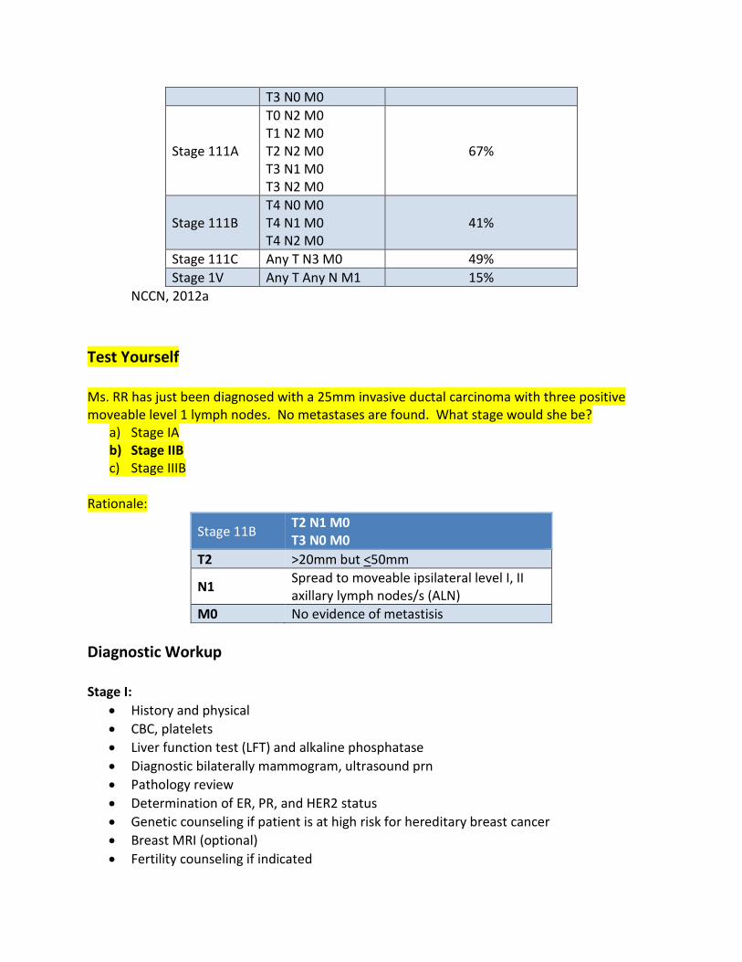
Stage 111C Any T N3 M0 49% Stage 1V Any T Any N M1 15%
NCCN, 2012a Test Yourself Ms. RR has just been diagnosed with a 25mm invasive ductal carcinoma with three positive moveable level 1 lymph nodes. No metastases are found. What stage would she be?
a) Stage IA b) Stage IIB c) Stage IIIB
Rationale:
Stage 11B T2 N1 M0 T3 N0 M0
T2 >20mm but <50mm
N1 Spread to moveable ipsilateral level I, II axillary lymph nodes/s (ALN)
M0 No evidence of metastisis Diagnostic Workup Stage I:
• History and physical • CBC, platelets • Liver function test (LFT) and alkaline phosphatase • Diagnostic bilaterally mammogram, ultrasound prn • Pathology review • Determination of ER, PR, and HER2 status • Genetic counseling if patient is at high risk for hereditary breast cancer • Breast MRI (optional) • Fertility counseling if indicated
Stage IIA or IIB:
• Consider additional studies only if indicated by signs and symptoms • Bone scan for localized bone pain or elevated alkaline phosphatase • Abdominal pelvic CT and/or MRI if elevated alkaline phosphatase, abnormal LFTs, or
abnormal physical exam of area • CT for pulmonary symptoms
Stage IIIA
• Chest CT • Abdominal pelvic and/or MRI • Bone scan or fluoride PET/CT • FDG PET/CT
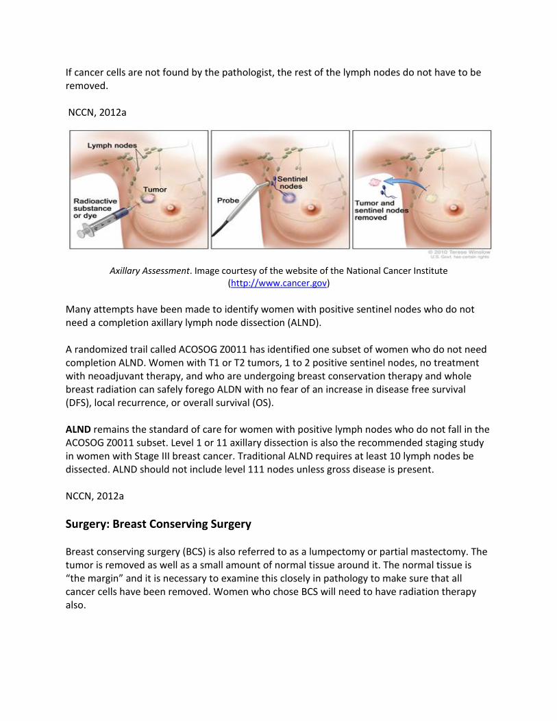
NCCN, 2012a Treatment Treatment options for breast cancer involve multiple modalities. For the best outcomes, patients should meet with an interprofessional oncology healthcare team. This allows multiple disciplines to have input into the plan of care and allows for more personalized care. Treatment needs to be individualized with consideration to the type of breast cancer, stage, individual comorbidities, menopausal status, and personal preferences. Treatment consists of both local, as well as systemic treatment. Local treatment can be surgery and/or radiation. Systemic treatment can be chemotherapy, endocrine therapy, and/or biologic therapy. A patient may receive one or all of these as they proceed along the treatment continuum. Axillary Assessment The standard of care for axillary node pathological assessment in women with Stage I or II breast cancer is a sentinel node biopsy. The sentinel node is the first node/s to receive lymphatic drainage form the tumor. A blue dye and/or radioactive substance is injected near the tumor. The surgeon can trace the path of this substance/s to identify the sentinel node/s. This procedure needs to be done by an experienced sentinel node team.
If cancer cells are not found by the pathologist, the rest of the lymph nodes do not have to be removed. NCCN, 2012a
Axillary Assessment. Image courtesy of the website of the National Cancer Institute
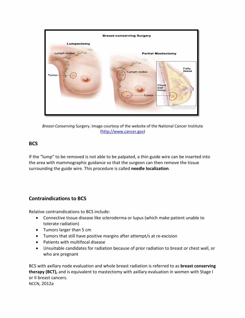
(http://www.cancer.gov) Many attempts have been made to identify women with positive sentinel nodes who do not need a completion axillary lymph node dissection (ALND). A randomized trail called ACOSOG Z0011 has identified one subset of women who do not need completion ALND. Women with T1 or T2 tumors, 1 to 2 positive sentinel nodes, no treatment with neoadjuvant therapy, and who are undergoing breast conservation therapy and whole breast radiation can safely forego ALDN with no fear of an increase in disease free survival (DFS), local recurrence, or overall survival (OS). ALND remains the standard of care for women with positive lymph nodes who do not fall in the ACOSOG Z0011 subset. Level 1 or 11 axillary dissection is also the recommended staging study in women with Stage III breast cancer. Traditional ALND requires at least 10 lymph nodes be dissected. ALND should not include level 111 nodes unless gross disease is present. NCCN, 2012a Surgery: Breast Conserving Surgery Breast conserving surgery (BCS) is also referred to as a lumpectomy or partial mastectomy. The tumor is removed as well as a small amount of normal tissue around it. The normal tissue is “the margin” and it is necessary to examine this closely in pathology to make sure that all cancer cells have been removed. Women who chose BCS will need to have radiation therapy also.
Breast-Conserving Surgery. Image courtesy of the website of the National Cancer Institute (http://www.cancer.gov)
BCS If the “lump” to be removed is not able to be palpated, a thin guide wire can be inserted into the area with mammographic guidance so that the surgeon can then remove the tissue surrounding the guide wire. This procedure is called needle localization. Contraindications to BCS Relative contraindications to BCS include:
• Connective tissue disease like scleroderma or lupus (which make patient unable to tolerate radiation)
• Tumors larger than 5 cm • Tumors that still have positive margins after attempt/s at re-excision • Patients with multifocal disease • Unsuitable candidates for radiation because of prior radiation to breast or chest wall, or
who are pregnant BCS with axillary node evaluation and whole breast radiation is referred to as breast conserving therapy (BCT), and is equivalent to mastectomy with axillary evaluation in women with Stage I or II breast cancers. NCCN, 2012a
Surgery: Mastectomy Mastectomy is surgery to remove the whole breast. A total or simple mastectomy only removes the breast tissue.
Total (Simple Mastectomy). Image courtesy of the website of the National Cancer Institute (http://www.cancer.gov)
A subcutaneous mastectomy is one in which the nipple and areolar are left. It is also referred to as a nipple-areola complex sparing mastectomy. There is more breast tissue left after this procedure. Current data does not support the use of subcutaneous mastectomy when the surgery is done for cancer treatment. Women who are having, or considering having, breast reconstruction will have a better cosmetic result if a skin sparing mastectomy is performed. In this procedure, the nipple areolar complex along with any biopsy sites is removed and the rest of the breast skin is left intact. The evidence suggests that this surgery is equivalent to standard mastectomy as far as the risk of local and regional cancer recurrence. NCCN, 2012a
Surgery: Mastectomy Modified radical mastectomy removes the entire breast along with level I and II axillary lymph nodes. No muscle is removed. Radical mastectomy is the most extensive type. The entire breast, level I, II, and III axillary nodes and the pectoralis muscle are all removed. Radical mastectomies are rarely done today and should only be done if the cancer has spread to the pectoralis muscle. www.breastcancer.org, 2012a
Modified Radical Mastectomy. Image courtesy of the website of the National Cancer Institute (http://www.cancer.gov)
Test Yourself A total mastectomy includes:
a) The breast and levels I, II, and III axillary lymph nodes b) The breast and levels I and II lymph nodes c) The breast only
Rationale: Mastectomy is surgery to remove the whole breast. A total or simple mastectomy only removes the breast tissue.
Postoperative Nursing Care Lumpectomy and/or sentinel node biopsy (SNB): These are outpatient surgeries with small incisions and patients often go home with just steri-strips. Wound care consists of washing with soap and water. Teach patient to report any signs or symptoms of infections or presence of a seroma. Patients also need to be informed of the small risk of lymphedema (around 5% ten years after treatment) after a SNB and risk reduction behaviors. Mastectomy and/or axillary node dissection: These patients usually have a drain and spend 1-2 nights in the hospital. In addition to wound care, they need to be taught how to manage their drain. The drain usually stays in place until the drainage has decreased to 25-30 ml per day. This usually takes a week to ten days. Patients need to be taught arm stretching exercises to prevent loss of range of motion in the shoulder/s of the operated side/s. The risk of lymphedema after ALND rises to around 35% in ten years. Therefore, patients need to learn behaviors to reduce the risk of lymphedema. The National Lymphedema Network is a wonderful resource for both patients and nurses. Fortenbaugh, Rummel, & Dell, 2012 Reconstructive Surgery Breast reconstruction can be immediate, done at the time of the mastectomy, or delayed, any time following the completion of cancer treatment. Options for breast reconstruction include the use of tissue expanders and implants, the use of autologous tissue, or a combination of the two. The goal of reconstruction is to produce symmetry by matching the remaining breast in terms of contour, dimension, and position. However, it is necessary to know that even the best reconstruction is not able to replace the natural breast. The reconstructed breast will feel different, will not have erogenous sensation, and will not function to breast feed. However, reconstruction can somewhat compensate for the loss of body image and cosmesis and for resulting psychosocial issues. Patients who opt not to have reconstruction should be provided with information on obtaining a breast prosthesis after mastectomy. NCCN, 2012a
Relative contraindications include smoking and obesity (BMI >30-35). The NCCN guidelines state that:
“Smoking increases the risk of complications for all types of reconstruction … patients should be made aware of the increased rates of wound healing complications and partial or complete flap failure among smokers.”
NCCN, 2012a Tissue Expansion/Implant Reconstruction The simplest method of reconstruction is the use of a tissue expander which consists of an inflatable silicone balloon. The expander is placed into a submuscular pocket deep to the pectoralis major muscle. It is only partially inflated at insertion to allow safe closure of overlying muscle and skin. The expander is then stretched by a series of postoperative saline injections that start 1-4 weeks after surgery and continue every 1-2 weeks until the desired size is obtained. Expansions may continue for as long as six months. There are three different techniques in use by plastic surgeons. For a woman with a small breast, the expander may be skipped and a fixed volume implant may be inserted at the initial surgery. Some surgeons utilize a combination variable volume expander-implant. The majority of reconstructive plastic surgeons seem to prefer a temporary tissue expander replaced at a later time by a permanent implant. The implant may be saline or silicone. Autologous Reconstruction Autologous reconstruction refers to tissue transferred from one part of one’s own body to another part. Musculocutaneous flaps, which consists of a segment of vascularized muscle with the overlying skin and fat, are one way to accomplish this. There are two types of musculocutaneous flaps utilized for breast reconstruction; pedicle flaps, and free flaps.
With pedicle flaps, the vascular origin remains intact on the dissected skin, fat and muscle. There are two common pedicle flaps utilized in breast reconstruction.
1. Latissimus Dorsi (LD) flaps can be done alone or combined with the insertion of an implant if more size is needed. The thoracodorsal artery and vein are used and the flap is tunneled under the axilla to the mastectomy site.
2. Transverse Rectus Abdominis Myocutaneous (TRAM) flaps utilize the superior epigastric artery (SEA) and superior epigastric vein (SEV). The flap is tunneled under skin and subcutaneous tissue to the mastectomy site.
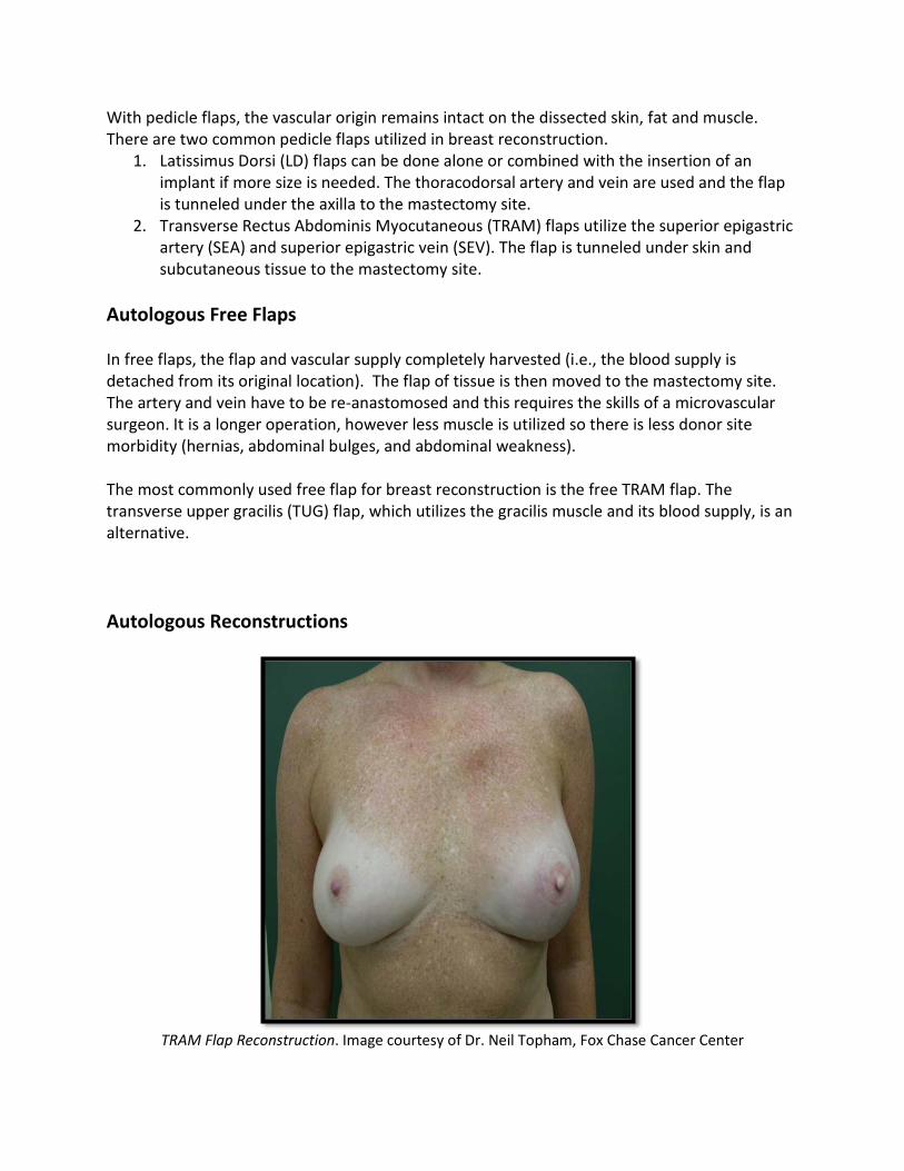
Autologous Free Flaps In free flaps, the flap and vascular supply completely harvested (i.e., the blood supply is detached from its original location). The flap of tissue is then moved to the mastectomy site. The artery and vein have to be re-anastomosed and this requires the skills of a microvascular surgeon. It is a longer operation, however less muscle is utilized so there is less donor site morbidity (hernias, abdominal bulges, and abdominal weakness). The most commonly used free flap for breast reconstruction is the free TRAM flap. The transverse upper gracilis (TUG) flap, which utilizes the gracilis muscle and its blood supply, is an alternative. Autologous Reconstructions
TRAM Flap Reconstruction. Image courtesy of Dr. Neil Topham, Fox Chase Cancer Center
Perforator Flaps
Perforator flaps are a type of free flap that harvest blood vessels that traverse muscle and perforate fascia (called perforators) to supply skin and fat above. No muscle is needed. These flaps are technically the most difficult to do. Flaps that can be utilized for breast reconstruction are the:
• Deep Inferior Epigastric Perforator (DIEP) flap • Superficial Inferior Epigastric Artery Perforator (SIEAP) flap • Superficial Gluteal Artery Perforator (SGAP) flap • Inferior Gluteal Artery Perforator (IGAP) flap • Stacked DIEP flap which uses bilateral flaps for the reconstruction of very large breast
Perforator Magnified
Perforator Magnified. Image Courtesy of Sameer Patel MD, Fox Chase Cancer Center Nipple-Areola Reconstruction Nipple-areola complex (NAC) reconstruction leads to increased patient satisfaction. Placement of the NAC is very important for a good aesthetic result so the surgery is not usually done until postoperative swelling is gone and the breast mound has had time to “settle”.
The nipple is created about 8-12 weeks after the reconstructive surgery by a small tissue flap that can done in an outpatient setting. The areola is tattooed on about six weeks later. Alternatively, there are some tattoo artists who are able to create a 3D illusion nipple tattoos. Polyurethane removable nipples are available for those who do not want more surgery. General Nursing Care after Reconstruction Patients require standard postoperative care for pulmonary toilet and prophylaxis of wound infection, deep vein thrombosis, stress ulcers, and pain management. They will be in the hospital 3-5 days depending on what procedure they have. The patient returns home with several Jackson-Pratt drains in place so they need to know how to manage these. Arm stretching exercises should begin about one week after surgery to prevent loss of shoulder range of motion. The flaps require careful and frequent monitoring after surgery to insure viability of the flap. Patients may have restrictions on body positioning to prevent wound dehiscence and/or expander movement. Additionally there are weight lifting restrictions; usually no more than five pounds for four weeks after an expander and 6-8 weeks after a flap. Radiation Therapy Radiation therapy is done to ensure local control of clinically undetectable cancer cells following surgery. Radiation will decrease the chance of breast cancer recurrence around 70%. Sites may include the breast and/or the axilla and/or chest wall. Radiation may be done after breast conserving surgery, after mastectomy in patients with either a large tumor (greater than 5 cm), when cancer is found in the lymph nodes (definitely recommended for four or more positive nodes and should be considered for those with one to three positive nodes), and/or for chest wall involvement. If adjuvant chemotherapy is necessary for the patient, radiation treatment should be given after the completion of chemotherapy. NCCN states the radiation therapy, as a component of BCT, may be omitted in selected women 70 years old or older with T1 breast cancer with negative margins by pathology, ER+, and negative nodes who will receive tamoxifen or an aromatase inhibitor. NCCN, 2012a, ACS, 2012a Treatment planning, called stimulation needs to be done with the aid of CT scans to insure even distribution of radiation to the tumor and minimal toxicity to the surrounding normal tissue. Traditional external radiation therapy targets the whole breast. This treatment only takes a few minutes but needs to be done five days a week for 5-6 weeks. Additionally, women at higher risk for local failure, such as those younger than 50, positive axillary nodes, positive
lymphovascular invasion, or close margins, should have a boost to the tumor bed which requires another week of therapy. Intensity Modulated Radiation Therapy (IMRT) is a method to deliver whole breast radiation in which the beams vary in length of radiation delivered so aim is more accurate. Three-dimensional CT is used for planning. It can be completed in four weeks. Hypofractionated radiotherapy increases each dose of radiation and delivers the total dose in less time. At the 2012 CTRC-AACR San Antonio Breast Symposium held in December, 2012, a long term study from the United Kingdom (UK) confirmed that a total low dose of radiation delivered in fewer, slightly larger doses, was as effective in controlling local early stage breast cancer as the standard five week course given in the UK. The study followed 4,451 women for ten years. This three week, 15 dose schedule is now the standard of care in the UK. American Association for Cancer Research, 2012; ACS, 2012a Accelerated Partial Breast Irradiation Most patients are able to continue to work and maintain their same activities of daily living during treatment. However, the time requirements can be prohibitive for some. It may be difficult to get off work every day or find (and afford) child care on a daily basis. For other women, the location of radiation centers can pose a travel burden. Therefore many efforts are underway to find alternative, shorter courses of treatment that are just as effective. Accelerated Partial Breast Irradiation (APBI) is one way to do this. Mammosite® system is a very popular way to accomplish this. A balloon catheter is placed in the lumpectomy site at the time of surgery or afterwards. A radioactive seed is inserted into the balloon two times a day for five days. No radiation remains in the body between treatments. The American Society for Radiation Oncology (ASTRO) states that the following patients may be considered for this type of therapy:
• Those 60 years old or older • Negative for BRCA mutations • Treated with primary surgery for a T1N0 ER+ tumor with a ductal histology and negative
margins
NCCN recommends patients be on a clinical trial if possible.
Note: Multi-lumen balloon catheters such as CONTURA® or SAVI® are now available that offer the promise of optimizing dose distribution. NCCN, 2012a
Intra-Operative Radiation Intra-operative radiation is given during lumpectomy surgery right after the cancer is removed. A single high dose is given directly to the breast tissue. Delivery methods vary, taking between 2-10 minutes, and then the surgery is completed as usual. More research needs to be done in this area to see if this is an effective procedure. ACS, 2012a Contraindications to Radiation Therapy A relative contraindication to radiation therapy is connective tissue disease involving the skin such as scleroderma or lupus. Contraindications are previous moderate or high dose radiation to breast or chest wall and/or pregnancy. If post-mastectomy radiation is required, it is preferable to delay autologous breast reconstruction until after the radiation is completed to prevent any loss of cosmesis. However, with implant reconstruction, expansion should take place before radiation as it is very hard to expand radiated skin. Some experienced teams are able to radiate reconstructed breasts with minimal complications. NCCN, 2012a Side Effects of Radiation The most common bothersome effect of radiation therapy is skin reactions like a sunburn. These reactions can vary from a mild redness to symptoms of a bad sunburn such as pain, itching, and peeling. Moist or dry desquamination may occur. This is the breakdown of the
squamous layer of the skin causing dryness and flaking, severe pruritus, redness and possible seeping of fluid. Nurses need to teach patients to keep skin dry and moisturized. Sometimes aloe vera or 1% hydrocortisone will help. At times a prescription strength steroid cream may be necessary. Prior to use, patients should okay any creams, ointments, or powders with their radiation nurse as some items have ingredients that could block the radiation. Sun exposure should be avoided. Many patients also find that radiation causes fatigue, especially toward the end of treatment. The best treatment preventive measure for this is exercise before, during, and after treatment. If radiation involves the axilla, lymphedema may occur. ACS, 2012a Systemic Treatment Systemic therapy in the form of chemotherapy, biologic therapy, and/or hormonal therapy is needed for patients with cancers that have features suggesting a high risk of recurrence. It is usually administered intravenously or orally, occasionally it may be given intramuscularly. Systemic treatment should be considered for all patients after surgery if less than 70 years old. However, the risk of disease recurrence with local treatment alone must be balanced against the toxicity and morbidities of systemic therapy. Adjuvant therapy is given in addition to surgery and/or radiation therapy to women with no clinical evidence of breast cancer metastasis, but with the potential for occult cells. Neoadjuvant therapy is given before surgery in cases of very aggressive cancer or for patients with a large tumor who hope to shrink it so they can have BCT. Systemic therapy is also used to treat metastatic or recurrent disease. Fertility Studies have proven that having a child after treatment for breast cancer does not increase the chance of recurrence or death nor does it cause birth defects in children. However, breast cancer treatments, especially chemotherapy, may impair fertility. Therefore, young women
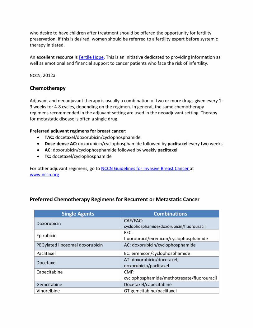
who desire to have children after treatment should be offered the opportunity for fertility preservation. If this is desired, women should be referred to a fertility expert before systemic therapy initiated. An excellent resource is Fertile Hope. This is an initiative dedicated to providing information as well as emotional and financial support to cancer patients who face the risk of infertility. NCCN, 2012a Chemotherapy Adjuvant and neoadjuvant therapy is usually a combination of two or more drugs given every 1-3 weeks for 4-8 cycles, depending on the regimen. In general, the same chemotherapy regimens recommended in the adjuvant setting are used in the neoadjuvant setting. Therapy for metastatic disease is often a single drug. Preferred adjuvant regimens for breast cancer:
• TAC: docetaxel/doxorubicin/cyclophosphamide • Dose-dense AC: doxorubicin/cyclophosphamide followed by paclitaxel every two weeks • AC: doxorubicin/cyclophosphamide followed by weekly paclitaxel • TC: docetaxel/cyclophosphamide
For other adjuvant regimens, go to NCCN Guidelines for Invasive Breast Cancer at www.nccn.org Preferred Chemotherapy Regimens for Recurrent or Metastatic Cancer
Embelin NCCN, 2012a Side Effects of Chemotherapy The adverse effects of chemotherapy depend on the type of drug, the dose, and the length of treatment. Common adverse effects of systemic chemotherapy occur because chemotherapy targets not only cancer cells all rapidly dividing cells in the body. General side effects include:
White blood cell growth factor (filtrate [Neurogenic®] or pegfilgrastim [Neulasta®]) support is necessary for regimes that pose a high risk (>20%) for febrile neutropenia. This helps ensure that patients are able to get their chemotherapy on time. Delivering doses on time increases the chance of survival. Medication Side Effects Doxorubicin (Adriamycin) is an anthracycline. Anthracyclines should always be included in a regime for HER2+ patients. The drug is a vesicant so must be given in a running IV with a good blood return. Urine may be colored pink to red for up to 48 hours. The drug is a potent myelosuppressive agent and also causes GI toxicities such as mucositis, esophagitis, and diarrhea. Cardiac toxicity limits the safe amount that can be given. Irreversible CHF may result so patients’ left ventricular ejection fractions (LVED) need to be assessed before therapy starts and patients need to be monitored for signs and symptoms of CHF or arrhythmias. Alopecia is a distressing side effect. Secondary acute myelocytic leukemia can result at a later time but this is a rare occurrence. Cyclophosphamide (Cytoxan) is an alkylating agent. This drug also causes myelosuppression, nausea vomiting, anorexia, stomatitis, diarrhea, and hepatic toxicity. Hemorrhagic cystitis may also occur so renal function must be monitored and patients must be taught to report hematuria, urinary frequency, or dysuria and to drink three liters of fluid a day and empty their bladder often. Alopecia also occurs in 30-50% of patients.
Docetaxel (Taxotere) is a mitotic spindle poison. This drug can cause severe hypersensitivity or anaphylactic reactions so patients need to be premedicated with dexamethasone. Neutropenia can be severe and is dose related. Febrile neutropenia requires IV antibiotics and/or hospitalization. Weakness, fatigue, and anemia are common. Fluid retention is often a problem. Edema begins in the periphery and may become generalized. It may be minimized by dexamethasone for three days prior to therapy. Peripheral neuropathy can affect up to 49% of patients and may indicate need to hold drug or dose reduce. Paclitaxel (Taxol) is also a mitotic spindle poison. Like docetaxel, this drug can cause severe hypersensitivity or anaphylactic reactions so patients need to be premedicated with dexamethasone. Neutropenia can be severe and is dose related. Fluid retention is not usually a problem with this drug, but neuropathy occurs in 60% and is severe in about 3%. Fatigue, arthralgias, and myalgias are frequent complaints. Wilkes & Barton-Burke, 2012 Test Yourself Systemic therapy given in addition to surgery and/or radiation therapy to women with no clinical evidence of breast cancer metastasis, but with the potential for occult cell is called what?
a) Adjuvant therapy b) Neoadjuvant therapy c) IV therapy
Rationale: Adjuvant therapy is given in addition to surgery and/or radiation therapy to women with no clinical evidence of breast cancer metastasis, but with the potential for occult cells. Targeted Therapies Targeted therapies target mutations unique to neoplastic cells, killing only the malignant cells and causing little or no damage to normal tissue, thus, less toxicity results. Targets are proteins and receptors vital for the proliferation, survival, or prevention of programmed cell death of cancer cells. Targeted agents used commonly in breast cancer are those that block the over-expression of HER2.
Targeted Therapies. Image courtesy of the website of the National Cancer Institute (http://www.cancer.gov)
Trastuzumab Trastuzumab (Herceptin®) is a monoclonal antibody that interacts with the HER2 receptors on the cell surface preventing them from activating signaling pathways that cause the cell to proliferate. It should be part of adjuvant therapy for all patients with HER2 positive tumors larger than 1 cm. Herceptin is given concurrently with paclitaxel as part of the AC followed by paclitaxel regime or after the completion of chemotherapy with the CEF (cyclophosphamide/epirubicin/fluorouracil) regimen. Treatment should continue for one year. This drug should not be given concurrently with an anthracycline because of cardiac toxicity. Cardiac monitoring of LVEF is necessary at baseline and at three, six, and nine months. This agent is a humanized antibody so rarely causes hypersensitivity or allergic reactions and no premedication is needed. NCCN, 2012a Targeted Therapy Patients with recurrent or metatasitc HER2+ disease who have already been treated with trastuzumab may be treated with pertuzamab (PerjetaTM) or lapatinib (Tykerb®) or in combination with trastuzumab, or selected chemotherapy agents. Pertuzamab: Like trastuzumab, it is also a humanized monoclonal antibody that binds to a different part of the HER2 receptor. It has a different yet complementary action to trastuzumab. Together they have a greater effect than one alone. The most common adverse reactions to this agent are diarrhea, rash, mucosal inflammation, and dry skin.
Lapatinib: Is a tyrosine kinase inhibitor. It is a small molecule that is taken orally and can cross the cell membrane and interfere with proteins located inside the cell. This agent inhibits the tyrosine kinase of HER1 and HER2 leading to the arrest of growth and death of tumor cells that depend on HER1 and HER2 cell signaling. Common side effects are diarrhea, rash, nausea vomiting, and fatigue. Lapatinib may overcome the resistance to trastuzumab and it is able to cross the blood brain barrier which trastuzumab isn’t. LVEF needs to be monitored. NCCN, 2012a; Wilkes & Barton-Burke, 2012 New and On the Horizon T-DM1: Is an investigational antibody-drug conjugate of trastuzumab and emtansine that has been found to have better results and less toxicity than the present standard treatment for advanced HER2+ breast cancer. This agent carries potent chemotherapy into the cancer cell, thus it is sometimes referred to as a “smart bomb.” It is indicated for unresectable locally advanced or metastatic disease. The FDA has granted a priority review designation to this drug. PARP (poly ADP-ribose polymerase) Inhibitors: Are agents that targets DNA repair enzymes that are over expressed. Many cancer treatments damage the DNA of the cancer cells. If the genes are repaired, tumor cells will survive. These agents prevent the repair genes from doing their job. They have shown promise in BRCA positive and triple negative breast cancers. They are presently being studied in clinical trials. WX-671 (upamostat): Is an inhibitor of uPA. In a phase 2 trial it has been shown to increase progression free survival when given with capecitabine first line in metatasitc HER2- breast cancer. Everolimus (Afinitor®): Has been approved for postmenopausal women with advanced hormone receptor+, HER2- breast cancer in combination with exemestane after failure of treatment with letrozole or anastrozole. This drug is classified as an mTOR (mammalian target of rapamycin) inhibitor. If not inhibited, mTOR promotes cell growth and proliferation, increases nutrient uptake, and promotes angiogenesis. Vorinostat (Zolinza®) and Panobinostat: Are HDAC (histone deacetylase) inhibitors. In combination with a PARP inhibitor or cisplatin they cause cell death in human triple negative breast cancer cells. They are available only on clinical trials. PD033299: Is a cyclin-dependent kinase inhibitor. This drug inhibits out of control cell cycling. It is a cytostatic agent which has been shown in a randomized phase 2 trials to significantly improve progression free survival in postmenopausal women with ER+ HER2- breast cancer when given in combination with letrozole.
National Cancer Institute, n.d.; ACS, 2012; Bhallal, Raol, Sharmal, Das Guptal, Chauhan, Stecklein, & Fiskus, 2012 Endocrine Therapy All patients with ER and/or PR positive cancers should be considered for adjuvant endocrine therapy. The only possible exceptions would be patients with tumors <0.5 cm or 0.6-1.0 cm with favorable prognostic indicators. Tamoxifen (Nolvadex®) is a selective estrogen receptor modulator or SERM. This means it blocks the action on some estrogen receptors in the body but not others. Adjuvant tamoxifen can be used in both premenopausal and postmenopausal women. Five years of adjuvant tamoxifen decreases the annual odds of recurrence by 39% and the odds of death by 31%. If the patient is to receive chemotherapy, that should be given before the tamoxifen is started. NCCN, 2012a Estrogen and Cancer
Estrogen and Cancer. Image courtesy of the website of the National Cancer Institute
(http://www.cancer.gov) Endocrine Therapy: Tamoxifen Traditionally, tamoxifen has been given for five years; however, a large randomized trial just published the results of continuing tamoxifen for ten years. It was found that a further reduction in recurrence and mortality can be seen after ten years in women who take the drug for ten years. In fact, it seems
that ten years of treatment will actually halve the breast cancer mortality during the second ten years after diagnosis. This study promises to be practice changing. Common side effects are hot flashes, vaginal dryness or discharge, irregular periods, nausea, and cataracts. Rare, but more serious, side effects are an increased risk for endometrial cancer and blood clots. Some serotonin uptake inhibitors (such as fluoxetine [Prozac], paroxetine [Paxil]) decrease the formation of the active metabolite of tamoxifen in women who have a suppression of the CYP206 enzyme, thus possible decreasing its effectiveness. Since many women with breast cancer are treated with antidepressants for anxiety, depression, and/or hot flashes, the prescriber should choose a drug that does not have this effect on tamoxifen metabolism. Davies, Pan, Godwin, Gray, Arriagada, Raina et al., 2012, Wilkes & Barton-Burke, 2012 Selective Estrogen Receptor Modulators (SERMS) Selectively inhibit or stimulate estrogen receptors on different targets.
SERMs. Image courtesy of the website of the National Cancer Institute (http://www.cancer.gov)
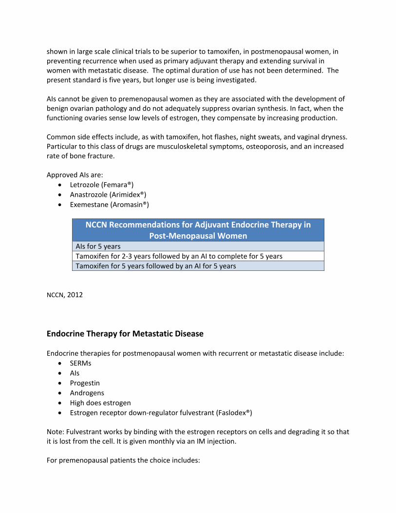
Endocrine Therapy: Aromatase Inhibitors (AIs) AIs take a different approach to hormone therapy; they prevent postmenopausal women from producing estrogen rather than blocking its activity. AIs inhibit the enzyme aromatase, which catalyzes the final step in the synthesis of estrogen from its steroid precursors. AIs have been
shown in large scale clinical trials to be superior to tamoxifen, in postmenopausal women, in preventing recurrence when used as primary adjuvant therapy and extending survival in women with metastatic disease. The optimal duration of use has not been determined. The present standard is five years, but longer use is being investigated. AIs cannot be given to premenopausal women as they are associated with the development of benign ovarian pathology and do not adequately suppress ovarian synthesis. In fact, when the functioning ovaries sense low levels of estrogen, they compensate by increasing production. Common side effects include, as with tamoxifen, hot flashes, night sweats, and vaginal dryness. Particular to this class of drugs are musculoskeletal symptoms, osteoporosis, and an increased rate of bone fracture. Approved AIs are:
NCCN Recommendations for Adjuvant Endocrine Therapy in
Post-Menopausal Women AIs for 5 years Tamoxifen for 2-3 years followed by an AI to complete for 5 years Tamoxifen for 5 years followed by an AI for 5 years
NCCN, 2012 Endocrine Therapy for Metastatic Disease Endocrine therapies for postmenopausal women with recurrent or metastatic disease include:
• SERMs • AIs • Progestin • Androgens • High does estrogen • Estrogen receptor down-regulator fulvestrant (Faslodex®)
Note: Fulvestrant works by binding with the estrogen receptors on cells and degrading it so that it is lost from the cell. It is given monthly via an IM injection. For premenopausal patients the choice includes:
• SERMs • LHRH agonists (goserelin and leuprolide) • Oophorectomy • Progestin • Androgens • High dose estrogen
NCCN, 2012 Survivorship Having breast cancer is a life altering experience. Women look and feel different and they must adjust to a “new normal.” A survivorship care plan needs to be developed for each patient who has completed therapy with curative intent. It should include breast cancer post-treatment follow-up.
• History and physical exams every 3-6 months for the first three years, every 6-12 months for years four and five, and then annually.
• Mammography should be performed one year after the initial mammogram and at least six months after completion of radiation therapy for women undergoing BCT. Subsequently they should be obtained every 6-12 months. If findings are stable, they should be obtained yearly. Mammography is not needed on reconstructed breasts.
• Teach breast self examination. • MRI may be used for surveillance in women at high risk of recurrence or bilateral
disease such as those with BRCA ½ mutations. • The routine performance of alkaline phosphatase, liver function tests, tumor markers,
bone scans, CT, MRI, PET, or US is not recommended. • Educate patients about symptoms of recurrence that should be reported such as new
lumps, bone pain, chest pain, dyspnea, abdominal pain, or persistent headaches. • Women at high risk who have not been evaluated by a genetic counselor should be
referred to one. • Regular gynecologic exams are recommended for all women. Women on tamoxifen
should immediately report any vaginal bleeding as they are at increased risk for uterine cancer.
• The risk of breast cancer recurrence continues more than 15 years after the completion of primary treatment. Care for patients should be performed by a healthcare professional experienced in surveillance of patients with cancer and in doing breast exams. Follow-up by a primary care provider can have the same healthcare outcomes as follow up by a specialist. Transfer of care of patients with early stage breast cancer may be done after one year if the care plan is communicated to them.
• Patients on adjuvant endocrine therapy may need help with symptom management and their adherence to ongoing medication regimen needs to be assessed.
• Adoption of a healthy lifestyle should be encouraged. Patients should try to maintain an ideal body weight, eat healthy foods, and engage in physical activity.
• Hormonal birth control methods are not recommended. • Patients should be given a list of resources for both information and support. The fear of
recurrence can be overwhelming for some. If metastases are present, the patient may be facing the threat of death. The American Society of Clinical Oncology (ASCO) has Breast Cancer Treatment Plan and Summary templates as well as a Breast Cancer Survivorship Plan available online at www.asco.org/treatmentsummary.
NCCN, 2012; Khatcheressian, Hurley, Bantug, Esserman, Grunfeld, Halberg et al. (2012) Summary Nurses are an excellent source of support and information for patients and their families all through the breast cancer journey. Letting patients know what to expect and when and where to find the information and support they need, and actively listening to their concerns can make a tremendous difference in their experience. Nevertheless, no one can do this alone. An interprofessional healthcare team with a focus on breast cancer will ensure that the patient has the best outcome. Adjuvant! Online Another online decision making tool to help health professionals discuss the risk and benefits of getting additional therapy with patients at the time of initial diagnosis is Adjuvant! Online.
Adjuvant! Online is based on classic pathologic criteria and host factors. It predicts ten year disease free and overall survival. The predictions assume that the cancer is completely resected and that radiation will follow breast conserving therapy. It is optimized for describing distributions of populations and results are not individual, but rather population-based. A printed results sheet like the one that follows can be used for patient discussions.
www.adjuvantonline.com Resources American Cancer Society
American Association for Cancer Research. Dec. 6, 2012. Hypofractionated Radiotherapy Was Safe, Effective for Early Breast Cancer Treatment at 10-year Follow-up. Retrieved December 14, 2012 from http://www.aacr.org/home/public--media/aacr-press-releases.aspx?d=2995. American Cancer Society. (2012a) Cancer Facts & Figures 2012. Atlanta: American Cancer Society, 2012. American Cancer Society. (2012b). For Women Facing a Breast Biopsy. Retrieved November 30, 2012 from http://www.cancer.org/treatment/understandingyourdiagnosis/examsandtestdescriptions/forwomenfacingabreastbiopsy/breast-biopsy-biopsy-types. Bartlett, J., Bloom, K., Piper, T. Lawton, T., van de Velde, C., Ross, D. et a. (2012). Mammostrat as an Immunohistochemical Multigene Assay for Prediction of Early Relapse Risk in the Tamoxifen Versus Exemestane Adjuvant Multicenter Pathology Study. Journal of Clinical Oncology, published ahead of print on October 8, 2012 as 10.1200/JCO.2012.42.8896. Becze, E. (2011). An Update on Breast Cancer Biomarkers, Clinical Journal of Oncology Nursing, Feb 2011. Retrieved from: http://www.onsconnect.org/2011/02/5min/an-update-on-breast-cancer-biomarkers. Bhallal, K., Raol, R., Sharmal. P. Das Guptal, S. Chauhan, L, Stecklein, S & Fiskus, W. 2012. Treatment with Histone Deacetylase Inhibitors Creates BRCAness and Sensitizes Human Triple Negative Breast Cancer Cells to PARP Inhibitors and Cisplatin, abstract 3-7. 2012 CTRC-AACR San Antonio Breast Cancer Symposium. Bishop, C. (2011). Biomarkers in Breast Cancer. Journal of Advanced Practitioner Oncology, 2(2), 101-110. Breastcancer.org. (2012a). Treatment & Side Effects. Retrieved December 12, 2-012 from http://www.breastcancer.org/treatment. Breastcancer.org. (2012b). Types of Breast Cancer. Retrieved November 30, 2012 from http://www.breastcancer.org/symptoms/types. Cadoo, K., McArdel, O., O’Shea, A., Power, C., & Hennessy, B. (2012). Management of Unusual Histological Types of Breast Cancer. The Oncologist,17,1135-1145. References Davies, C., Pan, H., Godwin, J. Gray, R., Arriagada, R., Raina, V. et al. (2012. Long-term effects of continuing adjuvant tamoxifen to 10 years versus stopping at 5 years after diagnosis of oestrogen receptor-positive breast cancer: ATLAS, a randomized trial. www.thelancet.com, published online December5, 2012.
Dushkin, H. and Cristofanilli, M. (2011). Inflammatory Breast Cancer. Journal of the National Comprehensive cancer Network, 9(2), 233-241. Fortenbaugh, C., Rummel, M. & Dell, D. (2012). Navigating the Breast Cancer Journey, Part 2. Nursing Spectrum, October 22, 20-25. Goldhirsch, A., Wood, W., Coates, A. Gelber, R. Thurlimannm B. Senn, H. et al. (2011). Strategies for subtypes-dealing with the diversity of breast cancer: highlights of the St. Gallen International Expert Consensus on the Primary Therapy of Early Brest Cancer 2011. Annals of Oncology, 22, 1736-1747. doi: 10:10.1093/annonc/mdr304. Gutwein, L., Darwin, A. Liu, H., Marshall, J., Hochwald, M. Copeland, M. & Grobmeyer, S. (2011). Utilization of minimally invasive breast biopsy for the evaluation of suspicious breast lesions. The American Journal of Surgery, 202,127-132. doi: 10.1016/j.amsurg.2010.09.005. Jancin, B. (2012). Diabetes confers 27% increase in breast cancer risk.OncologyPractice.com. Retrieved December 26, 1012 from www.oncologypractice.com. Khatcheressian, J., Hurley, P., Bantug, E., Esserman, L., Grunfeld, E., Halberg, A. et al. (2012), Breast Cancer Follow-Up and Management After Primary Treatment: American Society of Clinical Oncology Practice Guideline Update. JCO, published ahead of print on November 5, 2012. National Cancer Institute. 2012. Breast Cancer Treatment (PDQ®). Retrieved from http://www.cancer.gov/cancertopics/pdq/treatment/breast/healthprofessional/page1/AllPages. References National Cancer Institute. n.d. Targeted Therapies for Breast Cancer Tutorial. Retrieved from http://www.cancer.gov/cancertopics/understandingcancer/targetedtherapies/breastcancer_htmlcourse. National Comprehensive Cancer Network (NCCN). 2012a. Breast Cancer, Version 3.2012. Retrieved December 3, 2012 from http://www.nccn.org/professionals/physician_gls/pdf/breast.pdf. National Comprehensive Cancer Network (NCCN). 2012b. Breast Cancer Risk Reduction, Version 1.2012. Retrieved November 28, 2012 from http://www.nccn.org/professionals/physician_gls/pdf/breast_risk.pdf.
National Comprehensive Cancer Network (NCCN). 2012c. Breast Cancer Screening and Diagnosis, Version 1.2012. Retrieved November 28, 2012 from http://www.nccn.org/professionals/physician_gls/pdf/breast-screening.pdf. Rafferty, E., Park, J., Philpotts, L., Poplack, S. Sumkin, J., Halpern, E. & Niklason. (2012). Assessing Radiologist Performance Using Combined Digital Mammography and Breast Tomosynthesis Compared with Digital Mammography Alone: Results of a Multicenter, Multireader Trial. Radiology. Published online before print November 20, 21012, doi: 10.1148/radiol.12120674. Seigel, R., DeSantis, C., Virgo, K., Stein, K., Mariotto, A, Smith, T. et al. 2012. Cancer Treatment and Survivorship Statistics, 2012. CA A Cancer Journal for Clinicians, 62(4), 220-242. Seigel, R., Naishadham, D. & Jemal, A. 2012. Cancer Statistics for Hispanics/Latinos, 2012. CA A Cancer Journal for Clinicians, 62(5), 283-298. Susan G. Komen for the Cure. 2012. Understanding Breast Cancer Guide. Retrieved from http://ww5.komen.org/understandingbreastcancerguide.html. Wilkes, G. & Barton-Burke, M. (2012). Oncology Nursing Handbook. Burlington, MA: Jones & Bartlett Learning. Disclaimer This publication is intended solely for the educational use of healthcare professionals taking this course, for credit, from RN.com, in accordance with RN.com terms of use. It is designed to assist healthcare professionals, including nurses, in addressing many issues associated with healthcare. The guidance provided in this publication is general in nature, and is not designed to address any specific situation. As always, in assessing and responding to specific patient care situations, healthcare professionals must use their judgment, as well as follow the policies of their organization and any applicable law. This publication in no way absolves facilities of their responsibility for the appropriate orientation of healthcare professionals. Healthcare organizations using this publication as a part of their own orientation processes should review the contents of this publication to ensure accuracy and compliance before using this publication. Healthcare providers, hospitals and facilities that use this publication agree to
defend and indemnify, and shall hold RN.com, including its parent(s), subsidiaries, affiliates, officers/directors, and employees from liability resulting from the use of this publication. The contents of this publication may not be reproduced without written permission from RN.com. Participants are advised that the accredited status of RN.com does not imply endorsement by the provider or ANCC of any products/therapeutics mentioned in this course. The information in the course is for educational purposes only. There is no “off label” usage of drugs or products discussed in this course. You may find that both generic and trade names are used in courses produced by RN.com. The use of trade names does not indicate any preference of one trade named agent or company over another. Trade names are provided to enhance recognition of agents described in the course. Note: All dosages given are for adults unless otherwise stated. The information on medications contained in this course is not meant to be prescriptive or all-encompassing. You are encouraged to consult with physicians and pharmacists about all medication issues for your patients. The author of this course, Deena Dell, is a member of the speakers' bureau for Genomic Health, who manufactures Oncotype DX. To avoid bias, discussions of three commercial genetic profiling tests are reviewed in this course. This activity has also been independently reviewed by an Advisory Board and Nurse Planner.