A Pain In The Neck: Sports Related Cervical Spine Conditions Edward R. Laskowski, M.D., F.A.C.S.M. Co-Director, Mayo Clinic Sports Medicine Professor, Department of Physical Medicine and Rehabilitation
Transcript
A Pain In The Neck: Sports Related Cervical Spine Conditions Edward R. Laskowski, M.D., F.A.C.S.M. Co-Director, Mayo Clinic Sports Medicine Professor, Department of Physical Medicine and Rehabilitation
Epidemiology • ~10,000 cases of cervical spinal injuries
documented in the U.S. annually
• ~10% occur during athletic event
• 2nd leading cause of SCI in first three decades of life
• Comprise 2-3% of all sports-related injuries
• Majority occur during “unsupervised” sports-related activities: diving, skiing, “sand-lot” games
Epidemiology • In absolute numbers, recreational diving is
leading cause SCI leading to quadriplegia- alcohol, diving height/water depth, pike diving (Maroon et al Spine 1996)
• More public attention given to those injuries that occur during organized and/or televised competition- American football, wrestling, rugby, hockey, gymnastics, equestrian...
High-Risk Sports • Football (USA)
• Gymnastics (USA)
• Wrestling (USA)
• Rugby (Europe)
• Ice Hockey (Canada)
Kim et al Current Opinion Ortho 2003
• Snow sports
• Cycling
• Equestrian
Cervical Spine Injury and Football • Most high-profile group of athletes
• 1.2 million high-school and 200,000 college and pro football players annually.
• Incidence of C-spine injury estimated as high as 10-15% (Meyer AJSM 1994)
• 684 deaths in amateur/pro football 1945-1994, 17% related to cervical spine injury
• Cervical SCI (fx & neuro), 1977-2012:
266 HS, 38 college, 15 professional (Mueller, 2015)
Ice Hockey and Cervical Spine Injury • Canada: 258 CSI between 1943 and 2005
• 65% involve contact with boards
• 35% check from behind
• Since 2001, cervical spine injuries from hockey have decreased 69% following rule change/penalty increase for checking from behind, boarding (Tator, CJSM, 2009)
Objective Findings • Reproducible pain with palpation in the
paraspinous soft tissues.
• No deformity or step-off which would suggest more urgent problem
• No neuro deficit on careful exam
• Pain with ROM and focal TTP over spinous process or interspinous ligament after acute injury should be immobilized in rigid C-collar until imaging can be obtained
Evaluation
• Initial Radiographs- A/P, Lateral, odontoid (remember to see T-1)
• Keep in C-collar until acute symptoms subside (7-10 days), then Flex-Ex to r/o instability (F/E acutely may miss due to spasm)
• If continued TTP or painful ROM, consider MRI or CT
• CT: sagittal plane >3.5mm or rotation > 20%=instability
Ligamentous Instability
• > 3.5 mm horizontal displacement
• >11 degree rotation
• Absolute Contraindication
Treatment • Hard cervical collar during acute period
• Activity modification, analgesics
• Rehabilitation: include initial midline isometrics, followed by concentric resistance, allowing gradually increasing pain-free arc of motion and progressing to cervico-thoracic stabilization
• Avoid stretching during acute phase (72 hrs)
III. Intervertebral Disk • Acute traumatic disc herniation can lead to
central cord compression and myelopathy or if lateral, can cause radiculopathy
• Symptoms range from upper extremity radiculopathy to myelopathy with weakness and loss of sensory in all extremities, reflex changes, bowel/bladder changes
Upper Motor Neuron Injury vs Lower Motor Neuron Injury
• Vertebral osteophytes and disk material: form “hard disk” Radharkrishan, Brain, 1994
Evaluation • Detailed History- mechanism of injury, pre-
existing symptoms, neurologic review of systems
• Document motor, sensory and reflexes.
• C-spine x-rays
• MRI or CT-myelogram to evaluate the amount of cord and/or nerve root compression, especially with severe or progressive neuro deficit or bilateral symptoms
• Often will have early spondylosis-narrowed disc space, uncovertebral joint and facet hypertrophy, asymptomatic disc bulges: 75% of freshman football players (Thomas JAAOS 1999)
Treatment • Usually non-surgical management
• Therapy: modalities, activity modification; oral steroids or epidural steroid injection if pain severe and refractory to rehabilitation interventions
• If progressive neuro deficit or uncontrolled pain, surgery is option, though one year outcome similar to non-op
• ACDF usually surgery of choice for one-two level disease
IV. Transient Quadriplegia
• Described by Torg and others
• Described clinical entity of “neurapraxia of the cervical cord”
• Symptoms include bilateral burning pain, tingling, and loss sensation in arms and/or legs
• Varies from mild weakness to complete paralysis
• Transient- 10 minutes to 48 hours
• During 1984 NCAA season reported in 1.3/10,000 players
Mechanism of Injury
• Axial load with hyperextension or hyperflexion
• Transient cord compression via Pincer Mechanism (Penning Neurology 1962)
Pavlov, Torg, and the Ratio...
• Pavlov et al devised an objective measurement to determine if patients had congential cervical stenosis.
• Spinal canal-vertebral body ratio- the distance from midpoint of posterior vertebral body to the nearest point on spinolaminar line (a) divided by A/P width of vertebral body (b)
Torg Ratio and Transient Quadriplegia
• Torg et al in JBJS 1996 described relationship of a Pavlov ratio <0.8 (congenital stenosis) and Transient Neurapraxia in college football players
• 93% sensitivity in players with at least one episode of TQ
• Low positive predictive value: <0.8 only 12-33% of those who will have low functional csf reserve on MRI
Torg Ratio and its Role • Very sensitive- the players with TQ will most likely have
Torg Ratio of <0.8
• Low specificity- many football players will have a ratio of <0.8 (41% asymptomatic college players)
• Football players likely to have larger vertebral bodies (Herzog Spine 1991)
• Low positive predictive value- value of <0.8 not more likely to suffer catastrophic neurologic injury
• Therefore, x-ray screening using Torg ratio is not recommended
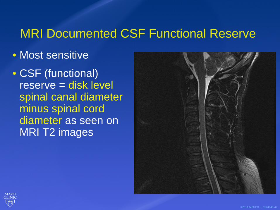
MRI Documented CSF Functional Reserve
• Most sensitive
• CSF (functional) reserve = disk level spinal canal diameter minus spinal cord diameter as seen on MRI T2 images
• Most athletes with permanent spinal cord injuries have unstable fractures/dislocations
• No direct relationship b/w canal geometry and risk of SCI (Torg 1986)
• Incidence of head injury decreased in the 1960-70’s while catastrophic SCI rose
• Incidence of fx/dislocations up 204% with perm. SCI up 116%- related to new/better helmets and dangerous tackling behavior. (Schneider et al 1964)
Axial load, Spear Tackling, and Mechanics of Fracture • Axial loading of the cervical spine is the prime
mechanism
• C-spine can absorb energy of collision by dissipating through paraspinal muscles, the disc, and the normal lordotic curve
• When neck flexed 30 degrees, as in spear tackling, forces transmitted along longitudinal axis of spine
• Compression of disc, angular deformation, failure in flexion with fracture, sublux/dislocation
Spear Tackler’s Spine
Torg’s “Caveat”- spear-tackler’s spine
• Torg described a subset of this population with a constellation of radiographic findings
• Ratio of <0.8 • loss of normal cervical lordosis-straight spine • post-traumatic degenerative disease
• Combined with propensity to spear tackle, creates high likelihood of neuro injury and CONTRAINDICATION to play
19 yo college linebacker with video documentation of spear-tackling, shows “spear-tackler’s spine”--taken 1 week prior to being rendered quadriplegic following tackle. (Med Sci Sport Exerc 1997)
The benefits of the research...
• As a result of detailed analysis of data on rates of C-spine injury, NCAA in 1976 banned spear-tackling
• Dramatic reduction in fracture/dislocation and SCI- overall approx. 70% in high-school and college football (Torg JBJS 2002)
Field Evaluation and Initial Treatment
• Initial treatment often begins on the field/rink
• Essential sideline equipment- spine board, stretcher, tools for removing mask from helmet (bolt-cutter, utility knife; newer helmets with release latch-Riddell, Schutt)
• If unconscious player, protect airway while protecting the c-spine- leave helmet on, jaw thrust and chin-lift, not head tilt
• Neck pain, focal midline tenderness (on spinous processes or interspinous ligaments), painful/limited neck ROM, or neurologic signs/symptoms needs to be immobilized and transported to ED.
• Immobilize on spine board, helmet and pads on.
• Remove only when in controlled setting, pull off in line with spine. Can change alignment if done incorrectly! (CJSM 2003)
• If needed, get films with gear in place
Helmet and Pads On… or Off? • Second Inter-Association Task Force on the
Prehospital Care of the Suspected Spine Injured Athlete, Jan 2015; 24 medical organizations
• “When deemed necessary by onsite medical personnel, protective equipment may be removed prior to transport.”
Helmet & pads, airway access, facilitate ER & hospital management
• Limitations: facemask removal often enough, less cervical motion, less personnel required
IV Transient Quadriplegia Absolute contraindications for return to play after an episode of TQ • Persistent neurological findings, cervical pain, or loss of ROM
• MRI evidence of spinal cord defect or edema
• Functional spinal stenosis on MRI
• Acute cervical fracture or ligamentous disruption
• Acute or chronic cervical disc herniation
• Cervical spine segmental instability
• Arnold-Chiari malformation
• Basilar invagination, os odontoideum
• Atlanto-occipital fusion or instability
• Klippel-Feil fusion greater than two levels
• Multi level surgical fusion Leah G. Concannon,; Mark A. Harrast,; and Stanley A. Herring, 2012
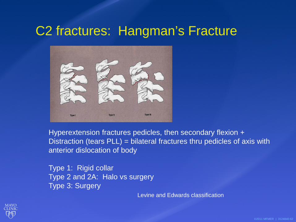
Hyperextension fractures pedicles, then secondary flexion + Distraction (tears PLL) = bilateral fractures thru pedicles of axis with anterior dislocation of body Type 1: Rigid collar Type 2 and 2A: Halo vs surgery Type 3: Surgery Levine and Edwards classification
abnormalities associated with Down syndrome. Int Orthop. 2006;30:284–289.
• Bailes JE. Experience with cervical stenosis and temporary paralysis in athletes. J Neurosurg Spine. 2005;2(1):11-16.
• Bransford R, Falicov A, Nguyen Q, Chapman J. Unilateral C-1 lateral mass sagittal split fracture: An unstable Jefferson fracture variant. J Neurosurg Spine. 2009;10:466–473.
• Brukner P, Khan K. Brukner & Khan’s Clinical Sports Medicine. Sydney, Australia: McGraw-Hill Book Company Australia; 2011. p. 313-341.
• Castro FP, Jr, Ricciardi J, Brunet ME, Busch MT, Whitecloud TS, 3rd. Stingers, the Torg ratio, and the cervical spine. Am J Sports Med. 1997;25:603–608.
• Gill SS, Boden BP. The epidemiology of catastrophic spine injuries in high school and college football. Sports Med Arthrosc. 2008;16:2–6.
• Herzog RJ,Wiens JJ, Dilingham MF, Sontag MJ. Normal cervical spine morphometry and cervical spine stenosis in asymptomatic professional football players: Plain film radiography, multiplanar computed tomography, and magnetic resonance imaging. Spine. 1991;16(Suppl):178–186.
• Pavlov H, Torg JS, Robie B, Jabre C. Cervical spinal stenosis: determination with vertebral body ratio method. Radiology. 1987;164:771–775.
• Plastaras CT, Pang S. Chapter 18: Cervical Spine Injuries and Conditions. In: Harrast MA, Finnoff JT. Sports Medicine: Study Guide and Review for the Boards. New York, NY: Demos Medical; 2011. p. 199-208.
Selected References
• Samartzis DD, Herman J, Lubicky JP, Shen FH. Classification of congenitally fused cervical patterns in Klippel-Feil patients: Epidemiology and role in the development of cervical spine-related symptoms. Spine. 2006;31(21):798–804.
• Schuenke M, Schulte E, Schumacher U. Thieme Atlas of Anatomy: General Anatomy and Musculoskeletal System. New York, NY: Thieme Medical Publishers; 2010. p. 84-85, 92-93, 96-103, 120-125, 138-143.
• Tassone JC, Duey-Holtz A. Spine concerns in the special Olympian with Down syndrome. Sports Med Arthrosc. 2008;16:55–60.
• Torg JS, Naranja RJ, Pavlov H, Talinat BJ,Warren R, Stine RA. The relationship of developmental narrowing of the cervical spinal canal to reversible and irreversible injury of the cervical spinal cord in football players. J Bone Joint Surg Am. 1996;78:1308–1314.