48
A Patient’s Handbook on Breast Health Management | weillcornell.org/breastcenter | nyp.org/breastcancer |

A Patient’s Handbook onBreast Health Management
| weillcornell.org/breastcenter | nyp.org/breastcancer |


On behalf of the entire Breast Surgery team at Weill Cornell Medicine and NewYork-Presbyterian, we thank you for entrusting us with any aspects of your breast care. It is an honor to serve you.
This handbook is intended to provide general information regarding management of various cancerous as well as benign breast problems. For the malignant (cancerous) problems, we focus on treatment of patients diagnosed with early-stage and operable disease. We have therefore included specific sections that discuss preoperative surgical planning and postoperative care. There are several areas where information is repeated, as we understand that many patients may utilize only segments of this guide, and there are areas where management issues for different breast problems will overlap.
Please note that we provide very little statistical data on breast cancer survival and recurrence rates. Happily, the majority of our breast cancer patients will have effective treatment and excellent outcomes. As treatment advances are made continuously, statistics on outcomes are constantly evolving.
We recognize that this guide is not a comprehensive textbook. We are therefore also happy to provide additional references, web-based resources, and support/advocacy programs, however, we strongly encourage patients to seek further information via the following sources:
National Cancer Institutewww.cancer.gov/types/breast
American Cancer Societywww.cancer.org/cancer/breastcancer/detailedguide
Susan G. Komenww5.komen.org
Please also feel free to contact us with any comments or recommendations that you have regarding this handbook.
Wishing you all the very best in health and happiness,
Greetings
Lisa A. Newman, MD, MPH, FACS, FASCOChief, Breast Surgical ServiceChief, Interdisciplinary Breast ProgramWeill Cornell Medicine and NewYork-Presbyterian
Vivian J. Bea, MDChief, Section of Breast Surgery NewYork-Presbyterian Brooklyn Methodist Hospital
Beth M. Siegel, MD, FACSChief, Section of Breast Surgery NewYork-Presbyterian Queens
Jennifer L. Marti, MD, FACSSite Director, Breast & Endocrine SurgeryNewYork-Presbyterian/Lower Manhattan Hospital
Manmeet K. Malik, DO, FACSDirector, Breast CenterNewYork-Presbyterian Queens


General Breast HealthImportance of Breast Cancer Screening and Diagnostic Testing.........................................................8Management Options for Women at “High-Risk” for Developing Breast Cancer..............................12Diagnosing Breast Cancer..................................................................................................................16Options for Reconstruction after Breast Surgery................................................................................23Decisions & Treatment Options Regarding Chemotherapy/Other Types of Systemic Therapy.........26Types of Breast Surgical Procedures..................................................................................................33Getting Prepared for Pre and Post-Operative Breast/Axillary Surgery...............................................35Contraception, Fertility Preservation and Breast Cancer in Premenopausal Women........................41
Unusual Breast Cancer ScenariosPregnancy-Associated Breast Cancer................................................................................................42Inflammatory Breast Cancer...............................................................................................................42Paget’s Disease of the Nipple.............................................................................................................43Phyllodes Tumors of the Breast..........................................................................................................43Breast Cancer Presenting as Axillary Metastases with Occult Primary.............................................43Male Breast Cancer............................................................................................................................43
Management of Common Breast ProblemsMastitis...............................................................................................................................................44Fibrocystic Breast Changes (including fibrocystic breast pain).........................................................44Nipple Discharge................................................................................................................................44Breast Abscess...................................................................................................................................45Chronic/Recurrent Perioareolar Breast Abscesses.............................................................................45Fibroadenoma.....................................................................................................................................45Lactating Adenoma.............................................................................................................................45Breast Cyst.........................................................................................................................................45Mondor’s Disease...............................................................................................................................45
Table Of Contents


7
General Breast Health

8
What is breast cancer screening and why is it important?
Importance of Breast CancerScreening & Diagnostic TestingBreast cancer is the most common malignancy diagnosed in adult American females. Early detection (catching and diagnosing a breast cancer when it is small) coupled with comprehensive treatment (which may involve combinations of surgery, medical therapy, and radiation), are the most effective strategies for reducing the life-threatening risks of breast cancer. Thanks to advances in breast cancer screening and treatment, the majority of patients will have an excellent outcome and long-term survival. It is therefore essential that all women understand basic information regarding breast health awareness, utilizing surveillance practices (such as screening mammography) appropriately and recognizing breast cancer danger signs requiring prompt medical attention.
Many women will experience breast problems that are benign (non-cancerous), such as fibrocystic changes and/or mastitis (benign breast inflammation/infection requiring antibiotics). Breast health awareness enables women to work in partnership with their health care providers so that both benign and malignant problems are managed properly.
Mammographically - Detected Breast Findings
Further Diagnostic Work-Up for Mammographically - Detected Breast Findings

9
What is breast cancer screening and why is it important? Any cancer can be potentially deadly and therefore must be taken seriously. Screening for breast cancer refers to strategies that detect breast cancers at early stages and/or small sizes, when they are less-likely to be life-threatening. Screening for breast cancer falls into three basic categories: (i) screening mammography, which is a specialized X-Ray evaluation of the breasts; (ii) clinical breast examination; and (iii) breast self-examination. Other imaging strategies are available such as magnetic resonance imaging (MRI) and whole-breast ultrasound, which are useful in special circumstances, such as screening of high-risk women (patients that are more likely to develop breast cancer, such as those with inherited predisposition). Breast cancer screening refers to routine evaluation of a woman’s breasts, in the absence of any symptoms or danger signs of breast cancer. Breast cancer symptoms require prompt attention (called diagnostic evaluation), which may include additional mammography, ultrasonography, and possible biopsy.
Screening MammographyThe Breast Oncology Program at Weill Cornell Medicine and NewYork-Presbyterian recommends that average-risk women undergo yearly mammograms beginning at the age of 40. Incorporating regular mammography into the routine health care plan reduces breast cancer mortality (death rates) by 20-30%. However, it is important for women to understand that mammograms are not perfect, and some cancers will be invisible on mammography. General awareness of new lumps or changes in the breast therefore remains important. Furthermore, since the likelihood of developing breast cancer increases with the normal aging process, it is more likely that a mammogram will identify a breast problem in an older woman.
Selected patients may therefore opt to defer their initial screening mammogram until they reach 40-45 years, and they may consider switching to alternate year mammography after reaching the age of 55. While recommendations regarding the optimal age for obtaining the first screening mammogram in average-risk, asymptomatic women is controversial, all guideline groups recommend that women have access to screening mammography beginning at the age of 40, and should discuss this decision with their health care team.
Types of MammogramsThe conventional screening mammogram consists of two basic views of each breast - a profile image, where the breast is being photographed from the side; and an image of the breast being photographed from top to bottom. Both images require some compression, so that cancerous abnormalities are not obscured/hidden by the normal surrounding breast tissue.
Digital mammography incorporates computerized assistance to obtain more detailed images of breast tissue. Three-dimensional mammography (also called digital breast tomosynthesis, DBT) is another computer-assisted variant of breast imaging which provides more detailed radiographic evaluation, and this technology can be especially useful in women that have breast tissue appearing dense (thick) on routine mammograms.
All forms of mammography involve some radiation exposure, but the benefits of breast cancer early detection outweigh the likelihood of any significant health damage from screening mammography.
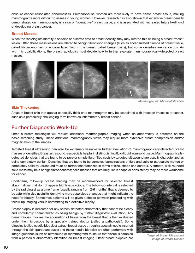
Examples of Mammographically - Detected Breast FindingsMicrocalcificationsMicrocalcifications are tiny spots that can appear on a mammogram, similar to grains of salt sprinkled on the mammographic images. Despite the name, microcalcifications are not related to calcium levels in a patient’s blood or calcium consumed in the diet. Some microcalcifications seen on mammogram are caused by completely benign fibrocystic changes; others are caused by breast cancer. Breast radiologists must use their expertise and skill to characterize the pattern of microcalcifications on an individual patient’s mammogram and assess the most appropriate follow-up.
Breast DensityThe radiologist will evaluate a mammogram for extent of generalized breast “thickness”, called breast density. Breast tissue that is very dense can be more challenging to evaluate mammographically, because the thick breast tissue can
Mammography
10
obscure cancer-associated abnormalities. Premenopausal women are more likely to have dense breast tissue, making mammograms more difficult to assess in young women. However, research has also shown that extensive breast density demonstrated on mammography is a sign of “overactive” breast tissue, and is associated with increased future likelihood of developing breast cancer.
Breast MassesWhen the radiologists identify a specific or discrete area of breast density, they may refer to this as being a breast “mass” lesion. Often these mass lesions are related to benign fibrocystic changes (such as encapsulated clumps of breast tissue, called fibroadenomas; or encapsulated fluid in the breast, called breast cysts), but some densities are cancerous. As with microcalcifications, the breast radiologist must decide how to further evaluate mammographically-detected breast masses.
Skin ThickeningAreas of breast skin that appear especially thick on a mammogram may be associated with infection (mastitis) or cancer, such as a particularly challenging form known as inflammatory breast cancer.
Further Diagnostic Work-UpOften a breast radiologist will request additional mammographic imaging when an abnormality is detected on the basic screening study. These additional mammography views may require more extensive breast compression and/or magnification of the images.
Targeted breast ultrasound can also be extremely valuable in further evaluation of mammographically-detected breast masses or densities. Breast ultrasound is especially helpful in distinguishing fluid/liquid from solid tissue. Mammographically-detected densities that are found to be pure or simple fluid-filled cysts by targeted ultrasound are usually characterized as being completely benign. Densities that are found to be complex (combinations of fluid and solid or particulate matter) or completely solid by ultrasound must be further characterized in terms of size, shape and contour. A smooth, well-rounded solid mass may be a benign fibroadenoma; solid masses that are irregular in shape or consistency may be more worrisome for cancer.
Short-term, follow-up breast imaging may be recommended for selected breast abnormalities that do not appear highly-suspicious. The follow-up interval is selected by the radiologist as a time frame (usually ranging from 2-6 months) that is deemed to be safe while also useful in identifying more suspicious changes that might indicate the need for biopsy. Sometimes patients will be given a choice between proceeding with follow-up imaging versus committing to a definitive biopsy.
Breast biopsy is indicated for any screen-detected abnormality that cannot be clearly and confidently characterized as being benign by further diagnostic evaluation. Any breast biopsy involves the acquisition of tissue from the breast that is then evaluated under the microscope by a specially trained doctor (called a pathologist). Some biopsies (called needle biopsies) extract breast tissue through a special needle inserted through the skin (percutaneously) and these needle biopsies are often performed with image-guidance (such as ultrasound or mammogram) to insure that tissue is sampled from a particular abnormality identified on breast imaging. Other breast biopsies are
Mammographic Microcalcification
Targeted Breast UltrasoundImage of Breast Cancer

11
performed as surgical procedures, where the patient is taken to the operating room and an incision is made on the breast skin so that the surgeon can remove a wedge of breast tissue. Some surgical biopsies are performed in conjunction with breast imaging, so that the surgeon can remove a wedge of abnormal breast tissue that was identified by mammogram and/or ultrasound. These are called image-guided localization surgical biopsies, and with these procedures the radiologist inserts a wire into the woman’s breast just prior to the surgery in order to point out the area of breast tissue that needs to be removed by the surgeon. An alternative image-guided localization procedure involves having the breast radiologist insert a special “seed” into the abnormal area of the breast (depending on the type of seed utilized, this insertion may occur from one to several days in advance of the surgical procedure) and the surgeon will then use a specially-designed probe (localizer) in the operating room to identify the area in the breast where the seed and abnormal breast tissue is located. The type of biopsy that is appropriate for an individual patient is determined by the type of abnormality and body/breast size, as well as patient preference.
Most image-guided needle biopsies will reveal a benign breast condition and the patient will then resume her usual breast health surveillance. If the needle biopsy reveals cancer, then the patient will be referred to the breast oncology team for treatment planning. Occasionally a woman will undergo an image-guided needle biopsy and will then require a follow-up surgical biopsy for more definitive evaluation to determine whether or not cancer is present. Examples of the latter scenario include cases where the needle biopsy was unsuccessful, inadequate, or showed some suspicious/high-risk pathology.
Possible Clip Placement at Biopsy SiteSince surgical biopsies require utilization of operating room services and can be more disfiguring, it is preferable to establish a breast cancer diagnosis via percutaneous core needle biopsy whenever possible. Core biopsy needles are special devices designed to extract tiny fragments of breast tissue from a lump or image-detected abnormality. Core needle biopsies can therefore be efficiently performed under local anesthesia in the clinic or breast imaging area, thereby avoiding utilization of surgical services and breast incisions. For palpable breast lumps, a breast specialist may be able to perform the percutaneous core needle biopsy freehand in the clinic. For non-palpable, image-detected breast abnormalities, the core needle biopsy is performed in the breast imaging suite by the radiologist. An image-detected core needle biopsy is more likely to yield a successful diagnostic specimen, since the breast imaging can confirm that the needle is extracting tissue from the correct area within the breast abnormality. Image-guided core needle biopsies may performed with mammography (also called stereotactic), ultrasound, or MRI assistance. The type of imaging used to guide these biopsies is determined by the radiologist based upon the appearance of the abnormality. When the radiologist performs an image-guided core needle biopsy, he/she will usually insert a tiny clip or marker to document the spot where the biopsy tissue was extracted. If the core needle biopsy is non-diagnostic, unsuccessful, or reveals some high-risk pathology (such as atypia or lobular carcinoma in situ), then the patient is referred to undergo the more definitive surgical biopsy.
Clinical Breast Examination (CBE)While it is reasonable for adult women to have a breast exam included in her usual overall physical exam, it is important for women to understand that many general health care providers are not skilled or experienced with performing a comprehensive breast exam. The benefits of routine CBE in terms of breast cancer early detection and reduced breast cancer mortality are therefore not well-documented.
Breast Self-Examination (BSE)As with CBE, the effectiveness of BSE is not well documented and the value of regular BSE has therefore been questioned. However, a general awareness of changes in a woman’s breast can be extremely important. This general awareness should be based upon visual inspection (looking for changes in the skin of the breast or nipple-areolar complex such as inflammatory changes/redness; dimpling; retraction; eczematous/flaky patches or bloody nipple discharge) and palpation of the breast checking for new lumps or densities.

12
Am I “High-Risk” for Developing Breast Cancer and What Does This Mean?
Management Options for Women at “High-Risk” for Developing Breast CancerAlthough breast cancer can occur in men, male breast cancer is very rare and being female is the strongest risk factor for developing breast cancer. Furthermore, while breast cancer can occur at any age, it increases in likelihood for all women with the normal aging process. A prior history of breast cancer also increases the likelihood of developing a second breast cancer. Beyond gender, age, and personal history, several risk factors have been characterized that identify women that are more likely to develop breast cancer at some point in their lifetime compared to other women. The majority of breast cancer patients however, do NOT have any identifiable risk factor(s).
Several of the known breast cancer risk factors are listed below. Some risk factors (such as hereditary predisposition) are quite strong and might influence recommendations for breast cancer screening or consideration of risk-reducing intervention such as chemoprevention (medical treatments to reduce likelihood of developing breast cancer) or even bilateral/double prevention/prophylactic mastectomy. Some risk factors are related to lifestyle and can be modified (such as obesity and alcohol intake). Other risk factors are weaker, cannot be modified, and do not influence screening recommendations (such as age at first menstrual cycle/menarche).
Management Options for Women at “High-Risk” for Breast Cancer
How do I know if I am at “High-Risk” for Breast Cancer?

13
Am I “High-Risk” for Developing Breast Cancer and What Does This Mean?Family HistorySome families carry genetic abnormalities (mutations) that are passed along generations and that are associated with inherited predisposition for breast cancer. These genetic mutations can be carried through the mother or the father and it is therefore important for patients to be familiar with the cancer history among both maternal and paternal relatives. Mutations in the BRCA1 and BRCA2 genes are among the most common genetic mutations causing hereditary breast cancer, but they account for fewer than 10% of the overall, general population of breast cancer cases. Families with male breast cancer; ovarian cancer; and/or multiple relatives diagnosed with breast cancer (especially if bilateral and/or diagnosed at young/premenopausal ages) are at particularly high risk for harboring BRCA mutations. BRCA mutations are also more common in Ashkenazi Jewish families. Women diagnosed with particular patterns of breast cancer (such as those known as triple negative breast cancer) are more likely to carry BRCA1 mutations.
Mutations in other genes aside from BRCA1 and BRCA2 can also increase risk of breast cancer. Families with these mutations may feature multiple relatives with colon cancer, melanoma, thyroid cancer, and/or pancreatic cancer.
Women belonging to families with possible hereditary predisposition for breast cancer should be referred for genetic counseling. When genetic testing is indicated, it may be performed through DNA extraction from a saliva specimen or from blood. The most definitive testing is performed on a family member that has been diagnosed with cancer, and these test results can streamline the testing that is performed on other, non-cancer-affected relatives. Patients should understand that genetic testing has not been perfected; some families with obvious hereditary cancer predisposition will have negative genetic testing. Genetic counseling is therefore useful in conjunction with genetic testing for appropriate interpretation of test results.
Therapeutic Chest Wall Radiation Exposure During Adolescence and Early Adult LifePatients that receive therapeutic doses of radiation to the chest wall at young ages (when the formative breast tissue is most susceptible to radiation damage) face an increased risk of breast cancer; these cancers are often bilateral and commonly occur during the premenopausal age range. Examples include patients receiving Mantle Irradiation for Hodgkins Lymphoma during the second and third decades of life.
Alcohol IntakeRegular consumption of significant quantities of alcoholic beverages is associated with increased breast cancer risk.
Postmenopausal ObesityAfter menopause, women have increased quantities of circulating estrogenic hormones related to metabolism in fatty tissues. Obesity in postmenopausal women therefore increases breast cancer risk.
Pathologic Indices/Markers of Increased Breast Cancer RiskCertain benign breast biopsy patterns are associated with increased future risk of breast cancer. Examples include atypical ductal hyperplasia, atypical lobular hyperplasia, and lobular carcinoma in situ (LCIS). Women that have had benign breast biopsy should therefore be aware of their detailed pathology report, as the presence of one or more of these high-risk features could indicate that more intensive breast cancer surveillance or risk-reducing intervention should be considered.
Mammographic DensityBreast tissue that appears particularly thick or dense on screening mammogram is associated with increased breast cancer risk. Extent of mammographic density is evaluated by the breast radiologist and may represent an indication for specialized forms of breast imaging.
Gynecologic and Reproductive PatternsBreast cancer is more common among populations of women that have more prolonged and uninterrupted breast tissue exposure to hormonal/estrogenic cycles. Young age at first menstrual cycle (age at menarche); late age at menopause; and late age at first live birth or nulliparity (no full-term pregnancies) are therefore all associated with increased breast cancer risk. In general, these risk factors are associated with an increased likelihood developing breast cancers that are hormone receptor-sensitive. Conversely, prolonged breast-feeding and early menopause via bilateral oophorectomy tends to reduce breast cancer risk.

14
Management Options for Women at “High-Risk” for Breast CancerAs discussed, women with possible hereditary predisposition for breast cancer based upon family history or personal history (breast cancer diagnosed at young age; bilateral breast cancer; triple negative breast cancer diagnosed younger than age sixty years; prior ovarian cancer), should be referred for genetic counseling and possible genetic testing.
Women found to be at high-risk for breast cancer (either by hereditary or non-hereditary factors) are candidates to consider a breast cancer risk
reducing intervention (this is called primary prevention) or to undertake more intensive surveillance for breast cancer early detection (also called secondary prevention). Primary prevention can be accomplished surgically through bilateral prophylactic mastectomy, or medically through chemoprevention. Secondary prevention can be conducted by annual breast MRI or whole breast ultrasound performed as a supplement to annual mammography. Furthermore, women with a family history of early-onset breast cancer should begin annual mammography 5 to 10 years younger than the youngest age of breast cancer diagnosis in the family.
Surgical Primary Prevention of Breast Cancer RiskBilateral prophylactic mastectomy surgery reduces breast cancer risk by 90-95%. Women considering this option must understand that the absolute benefit of prophylactic mastectomy surgery is therefore closely related to the woman’s individualized risk of developing breast cancer. A 25-year-old average-risk woman has a relatively low likelihood of developing breast cancer and is therefore not likely to benefit substantially from this operation. In contrast, a 25-year-old BRCA mutation carrier has a 40-85% lifetime risk of developing breast cancer and bilateral prophylactic mastectomy surgery can reduce this lifetime risk to less than 10%. Since most breast cancers can be treated effectively if detected at an early stage, the survival benefits of bilateral prophylactic mastectomy surgery are less well-defined. Women considering bilateral prophylactic mastectomy surgery must understand their individualized risk before committing to this irreversible procedure; they must understand that lifelong surveillance remains necessary because the surgery does not confer complete protection; and they should meet with one or more plastic surgeons to be fully informed regarding their breast reconstruction options.
Bilateral prophylactic mastectomy surgery can usually be performed with immediate/same-stage breast reconstruction; selected patients may opt for delayed reconstruction performed months or years later. Breast reconstruction does not compromise the effectiveness of the prophylactic mastectomy surgery, however the reconstructed breast will have insensate/numb skin. Some bilateral prophylactic mastectomy/immediate reconstructions are performed with the conventional, nipple/areolar-sacrificing approach, because of concerns that an increased quantity of microscopic breast tissue might be hidden within the nipple/areolar skin. The strategy of nipple-areolar skin preservation is becoming increasingly popular however, with favorable cosmetic and outcome results. Patients opting for nipple-sparing mastectomy and immediate reconstruction must understand that the possibility nonetheless exists that nipple-areolar preservation might negate some of the risk-reducing benefits of the surgery through possible residual breast tissue in the nipple areolar skin, and also because the very limited incision utilized for this operation can compromise the ability to surgically excise all of the breast tissue in peripheral/remote areas of the chest wall. The preserved nipple-areolar skin will also be insensate.
Bilateral prophylactic oophorectomy during the premenopausal age range can reduce breast cancer risk by approximately 50%. This surgical risk-reducing option is more common among women with BRCA mutations associated with increased risk of both breast and ovarian cancer.
Medical Primary Prevention of Breast Cancer (Chemoprevention)Women with increased risk of breast cancer can also choose to reduce their risk by taking a medication for up to five years. These pills lower breast cancer risk by 50-70%, depending on the selected category of chemoprevention. All are associated with risk of adverse effects, and so the decision to pursue chemoprevention must be made after carefully weighing the individual breast cancer risk versus the potential toxicity of medical risk reduction.
15
Premenopausal women are candidates for a medication called tamoxifen, which is a category of drugs called selective estrogen receptor modulators (SERMs). Raloxifene is an alternative SERM, however it is only approved for use in postmenopausal women. Similar to birth control pills and hormone replacement therapy, both SERMs can increase the potentially life-threatening risk of blood clots in the legs (deep vein thrombosis) which can then travel to the lungs (pulmonary embolism). These complications are called venous thromboembolism (VTE). Preexisting history of VTE is a contraindication to SERM therapy, and patients that develop VTE while taking SERMs must discontinue this form of chemoprevention. Treatment of VTE can involve long-term blood-thinning therapy (anticoagulation) and sometimes invasive interventional procedures aimed at filtering or blocking the transit of blood clots. Since surgery can increase the risk of VTE, patients should discontinue SERMs approximately two weeks prior to elective operative procedures. SERMs can also increase the risk of uterine cancer and cataracts. Approximately one-third of patients taking SERMs will experience new-onset or exacerbation of vasomotor symptoms such as hot flashes and night sweats. On the positive side, SERM therapy can reverse osteoporosis in postmenopausal women and can lower cholesterol levels.
Aromatase inhibitors (AIs) are an alternative category of medications that can reduce breast cancer risk, but they can only be used in postmenopausal women. AIs act by lowering estrogen production through fatty tissue metabolism of postmenopausal women. AIs are not associated with the VTE or uterine cancer risks that are seen with SERM therapy, however they can cause comparable vasomotor symptomatology. The bone effects of AIs are especially noteworthy, and AIs can substantially accelerate osteoporosis.
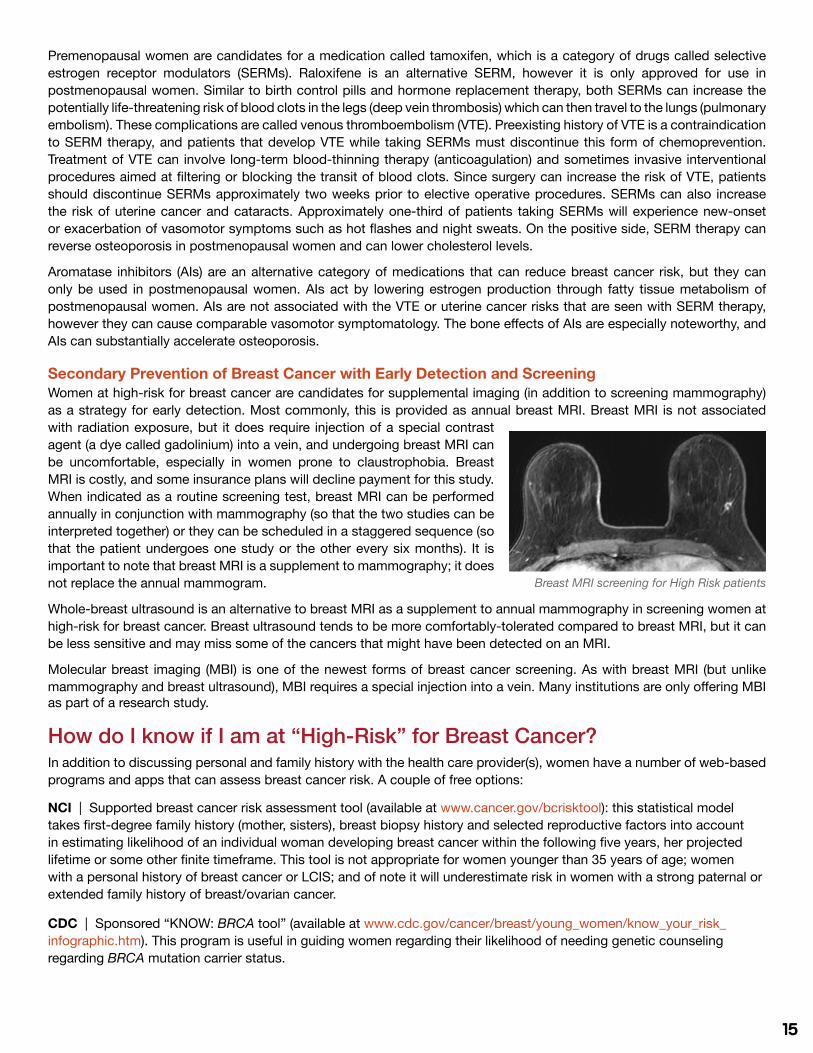
Secondary Prevention of Breast Cancer with Early Detection and ScreeningWomen at high-risk for breast cancer are candidates for supplemental imaging (in addition to screening mammography) as a strategy for early detection. Most commonly, this is provided as annual breast MRI. Breast MRI is not associated with radiation exposure, but it does require injection of a special contrast agent (a dye called gadolinium) into a vein, and undergoing breast MRI can be uncomfortable, especially in women prone to claustrophobia. Breast MRI is costly, and some insurance plans will decline payment for this study. When indicated as a routine screening test, breast MRI can be performed annually in conjunction with mammography (so that the two studies can be interpreted together) or they can be scheduled in a staggered sequence (so that the patient undergoes one study or the other every six months). It is important to note that breast MRI is a supplement to mammography; it does not replace the annual mammogram.
Whole-breast ultrasound is an alternative to breast MRI as a supplement to annual mammography in screening women at high-risk for breast cancer. Breast ultrasound tends to be more comfortably-tolerated compared to breast MRI, but it can be less sensitive and may miss some of the cancers that might have been detected on an MRI.
Molecular breast imaging (MBI) is one of the newest forms of breast cancer screening. As with breast MRI (but unlike mammography and breast ultrasound), MBI requires a special injection into a vein. Many institutions are only offering MBI as part of a research study.
How do I know if I am at “High-Risk” for Breast Cancer?In addition to discussing personal and family history with the health care provider(s), women have a number of web-based programs and apps that can assess breast cancer risk. A couple of free options:
NCI | Supported breast cancer risk assessment tool (available at www.cancer.gov/bcrisktool): this statistical model takes first-degree family history (mother, sisters), breast biopsy history and selected reproductive factors into account in estimating likelihood of an individual woman developing breast cancer within the following five years, her projected lifetime or some other finite timeframe. This tool is not appropriate for women younger than 35 years of age; women with a personal history of breast cancer or LCIS; and of note it will underestimate risk in women with a strong paternal or extended family history of breast/ovarian cancer.
CDC | Sponsored “KNOW: BRCA tool” (available at www.cdc.gov/cancer/breast/young_women/know_your_risk_infographic.htm). This program is useful in guiding women regarding their likelihood of needing genetic counseling regarding BRCA mutation carrier status.
Breast MRI screening for High Risk patients

How is Breast Cancer Diagnosed?
Diagnosing Breast CancerHealth care providers may suspect the presence of breast cancer based upon some clinical finding(s) on breast exam, and/or breast radiologists may suspect breast cancer based upon an abnormality seen on breast imaging. However, a definitive diagnosis of breast cancer can only be established with certainty when a breast biopsy has been performed, yielding tissue that can be evaluated microscopically by a pathologist. Several types of breast biopsies are available that can provide these diagnostic samples. The health care provider/breast oncology team (including the breast imager/radiologist) can guide patients regarding when a biopsy is necessary and the type of biopsy that is appropriate. Some patients will require several types of biopsies in order to have a comprehensive picture of the type and extent of breast cancer, as well as to determine the best treatment options. Pathology reports can usually be completed within 2-4 business days.
What Happens When a Breast Cancer is Diagnosed from Breast Biopsy?
How is Treatment Planned for a Newly-Diagnosed Breast Cancer Patient?

17
How is Breast Cancer Diagnosed?Surgical Diagnostic Breast Biopsy (with versus without wire localization or seed localization)Surgical removal of a wedge or lump of breast tissue is the most definitive strategy for determining whether a breast abnormality is cancerous, because this approach provides the pathologist with the largest sample of breast tissue for microscopic evaluation compared to other biopsy strategies. When a surgical biopsy is necessary for a breast abnormality forming a palpable lump, the surgeon decides on the location of the surgical incision and the site of breast tissue removal based upon what he/she can feel on breast examination.
For non-palpable breast abnormalities detected by breast imaging (mammogram, ultrasound, and/or breast MRI), surgical excision of the lesion requires image-guidance, also known as wire localization or seed localization surgical biopsy. With wire localization, the patient undergoes repeat breast imaging on the day of the surgical procedure and after injecting a local anesthetic into the breast skin, the radiologist inserts a thin metallic filament (wire) into the breast to point out the abnormality for the surgeon. The surgeon then plans the breast incision and removal by excising the tissue surrounding the localizing wire. A mammogram is then performed on the excised tissue to confirm that the appropriate specimen has been removed.
Seed localization involves insertion of a tiny device (about the size of a grain of rice) into the breast at the site of the abnormality by the radiologist - usually several days prior to the day of surgery. Some seeds contain a tiny quantity of radioactivity, other seeds are magnetic or radar-based. The surgeon then uses a special probe, or localizer in the operating room that allows her/him to find the location of the seed and the breast abnormality. The surgeon removes the area of breast tissue containing the seed and the abnormality. As with the wire localization, this excised breast tissue is radiographed in the operating room to confirm inclusion of the targeted area.
Percutaneous Core Needle Biopsy (with versus without image-guidance)Since surgical biopsies require utilization of operating room services and can be more disfiguring, it is preferable to establish a breast cancer diagnosis via percutaneous core needle biopsy whenever possible. Core biopsy needles are special devices designed to extract tiny fragments of breast tissue from a lump or image-detected abnormality. Core needle biopsies can therefore be efficiently performed under local anesthesia in the clinic or breast imaging area, thereby avoiding utilization of surgical services and breast incisions. For palpable breast lumps, a breast specialist may be able to perform the percutaneous core needle biopsy freehand in the clinic. For non-palpable, image-detected breast abnormalities, the core needle biopsy is performed in the breast imaging suite by the radiologist. An image-detected core needle biopsy is more likely to yield a successful diagnostic specimen, since the breast imaging can confirm that the needle is extracting tissue from the correct area within the breast abnormality. Image-guided core needle biopsies may performed with mammography (also called stereotactic), ultrasound, or MRI assistance. The type of imaging used to guide these biopsies is determined by the radiologist based upon the appearance of the abnormality. When the radiologist performs an image-guided core needle biopsy, he/she will usually insert a tiny clip or marker to document the spot where the biopsy tissue was extracted. If the core needle biopsy is non-diagnostic, unsuccessful, or reveals some high-risk pathology (such as atypia or lobular carcinoma in situ), then the patient is referred to undergo the more definitive surgical biopsy.
Percutaneous Fine Needle Aspiration Biopsy (with versus without image-guidance)Fine needle aspirations are commonly performed for cystic (fluid-filled) lesions of the breast, but core needle biopsies are preferred when tissue needs to be sampled in order to evaluate for the presence of a cancer. Core needle biopsies require skill and experience as well as availability of these specially-designed devices. If the core needle biopsy technology is unavailable, patients may occasionally undergo needle biopsy with a conventionally-available “skinny” needle, which is the same apparatus used for routine blood-drawing (phlebotomy). These procedures are called “fine needle aspiration (FNA)” biopsies. FNA biopsies yield clusters of individual breast tissue cells, rather than actual fragments of breast tissue. Interpretation of these specimens require the expertise of breast pathology specialists, cytopathologists. FNA biopsies require less costly resources, but they are associated with a higher risk of inadequacy because of the scanty amount of breast cellular tissue retrieved. Furthermore, if cancer cells are identified in an FNA biopsy specimen, the minimal tissue yield is often insufficient to comprehensively characterize the pattern of the breast cancer. Lastly, FNA biopsies are especially challenging in the breasts of pregnant women, because the hormonal-associated changes of pregnancy can make individual breast tissue cells appears cancerous on cytologic evaluation. As with surgical and core needle biopsies, FNAs can be performed either with or without image-guidance, and image-guidance can improve the success rate.

18
Punch Skin BiopsiesPunch biopsy devices are circular scalpels that are designed to extract small ellipses of skin. These procedures are routinely utilized by dermatologists to biopsy skin moles and other lesions. Breast specialists may utilize punch biopsy devices to evaluate breast problems associated with skin symptoms. Examples of these problems include specialized forms of breast cancer such as Paget’s disease of the nipple, inflammatory breast cancer, and locally advanced breast cancers that have grown through the breast skin.
What Happens When a Breast Cancer is Diagnosed from Breast Biopsy?The breast oncology team will need to evaluate several characteristics of breast cancer from the biopsy material to assist in treatment planning and determining the underlying aggressiveness of the breast cancer:
HistopathologyThe pathologist must characterize the breast cancer as being invasive versus ductal carcinoma in situ. Invasive or “full-blown” breast cancers are tumors with breast cancer cells that have extended beyond the boundaries of breast ductal and/or lobular unit walls. While cancerous breast lumps are more likely to be associated with invasive cancers, the actual distinction between invasive and non-invasive breast cancers is based upon the microscopic evaluation of breast tissue samples by the pathologist. For invasive breast cancers, the pathologist will characterize the lesion as having ductal histology (a pattern that accounts for at least 75% of breast cancers) versus lobular versus ductal-lobular combination versus medullary versus papillary or some other pattern. In general, all of these invasive histopathology patterns are offered similar surgical treatment options based upon size of lesion and extent of abnormalities seen on breast imaging. Some histopathology patterns however, are more likely to have particular clinical features. For example, invasive lobular cancers tend to be more insidious in their presentation compared to invasive ductal carcinomas - tumors with invasive lobular histology frequently present as relatively subtle densities in the breast rather than a well-defined/dominant lump. Medullary cancers of the breast tend to have a more favorable biology; they are less likely to metastasize even when detected at relatively bulky tumor sizes and despite the fact that they usually have unfavorable molecular marker characteristics (such as being negative for the estrogen receptor, progesterone receptor, and HER2/neu markers - also called triple negative).
Molecular MarkersInvasive breast cancers must be evaluated for the presence versus absence of specific molecular markers that have prognostic as well as therapeutic significance. Expression (positivity) of the hormone receptors (estrogen receptor and progesterone receptor) tends to correlate with biologically less-aggressive disease, and these cancers can be manipulated with hormonally-active cancer fighting medications (e.g. tamoxifen, a selective estrogen receptor modulator, or one of the aromatase inhibitors). HER2/neu is a third marker - overexpression of this marker in and of itself tends to correlate with a biologically-aggressive breast cancer, however the advent of powerful targeted cancer treatments that focus on killing cells that express this marker has dramatically improved the outcome for these tumors. Tumors that are negative for all three markers are called triple negative breast cancers (TNBC), which tend to be more challenging to treat however histology can modify this correlation. For example, medullary cancers and secretory cancers of the breast are more likely to be TNBC, yet they have a more favorable prognosis.

19
How is Treatment Planned for a Newly-Diagnosed Breast Cancer Patient?The majority of breast cancer patients will be successfully treated, and this is because of advances that have been made in the multidisciplinary fields that are involved in breast cancer management and outcome:
Breast Imaging | Facilitating early detection and disease evaluation
Breast Surgical Oncology | Facilitating better operative planning and featuring expanded surgical options
Breast Pathology | Improved characterization of breast tumors and their biology
Breast Medical Oncology | Featuring more effective treatments that can eliminate microscopic breast cancer cells hidden in distant organs (such as liver, lungs, bones)
Breast Radiation Oncology | With more effective and safer radiation treatments designed to eradicate microscopic breast cancer cells hiding in the breast or in the chest wall
Breast Reconstruction/Plastic Surgery | With expanded options to reconstruct the breast after mastectomy, or to restore symmetry after lumpectomy
Reproductive Oncology | With expanded options for fertility preservation in premenopausal breast cancer patients
Physical Therapy/Occupational Therapy | With improved and more aggressive management of breast surgery side effects such as lymphedema
It is therefore essential for each newly-diagnosed breast cancer patient to have representatives from several of these disciplines involved in planning the most appropriate treatment options for their particular cancer. At Weill Cornell Medicine and NewYork-Presbyterian, our Breast Oncology Program convenes several times each week for multidisciplinary conferences that insure comprehensive evaluation of every breast cancer patient managed in our system. The care of most patients will be reviewed multiple times in this setting. For some patients the treatment team may have consensus that there is a single best treatment plan for the patient. For most patients however, there will be options that have equal outcomes but that vary in terms of type/extent of surgery (for example breast-saving surgery versus mastectomy) or sequence of treatments (for example chemotherapy before versus after surgery). These are commonly described as “standard-of-care” treatment options, or treatment plans that have been well-studied and proven through clinical research to be effective for breast cancer based upon stage and pattern of disease. All patients will be offered the standard-of-care options. In addition to these conventional/standard-of-care options, some patients will also be offered the option of clinical trial participation, where they have the opportunity to receive some new therapy or procedure that appears promising for improved effectiveness or reduced side effects. Clinical trials are rigorously planned and closely-monitored for safety. If you are offered clinical trial participation, you can rest assured that your care is being overseen by many teams of hospital-based and as well as federal regulators.

20
Treatment Planning for Invasive Breast CancerWhile control of the cancerous breast is extremely important for newly-diagnosed breast cancer patients, it also important for patients to understand that the life-threatening aspect of breast cancer is generally determined by the risk of damage to other organs (such as the liver, lungs, bones, brain) through metastatic spread. For many patients this risk is present at the time of initial breast cancer diagnosis, commonly referred-to as distant organ “micrometastases”. Disease in the breast/chest wall is typically controlled by surgery (with or without radiation), and micrometastases are controlled by medical treatments (also referred-to as “systemic therapy”). Systemic therapies include medical treatments such as hormonally-active cancer fighting pills (called endocrine therapy) or intravenous infusions (such as chemotherapy); since these treatments are absorbed into the bloodstream they circulate throughout the body and are quite effective at eliminating the micrometastatic disease, thereby improving breast cancer survival rates. Unfortunately, all systemic therapies have potentially dangerous side effects and so the multidisciplinary treatment team will use them only when they are likely to be helpful. Since micrometastases are usually invisible on body imaging studies such as CAT scans or bone scans, the treatment team has to rely on clues related to the tumor biology and stage/extent of disease in order to determine when and what type of systemic therapy is necessary. The treatment team will therefore recommend that every treatment plan address the following three principles so that the disease in the breast/chest wall is controlled while also obtaining the necessary staging information:
(i) Control of the primary breast tumor and any obvious sites of disease within the breast | Surgery is generally necessary as at least one component of care in order to address this principle for the high majority of breast cancer patients. The surgically-removed breast tissue is carefully analyzed by the pathology team to assess adequacy of the resection, as well as to determine the microscopic pattern of the cancer, which provides important clues regarding biology of the cancer and systemic therapy needs.
(ii) Control of microscopic, hidden cancer cells in other parts of the breast (disease that is separate from the primary or obvious cancerous growth in the breast) | Because of the risk of these hidden cancer cells, most (but not all) breast cancer patients need to receive some treatment to the entire breast. This may involve surgery alone (such as mastectomy) or it may involve combinations of surgery and radiation (such as lumpectomy and breast radiation). At Weill Cornell Medicine and NewYork-Presbyterian, there is a priority placed on avoiding side effects associated with radiation treatment. In recent years, the Department of Radiation Oncology has incorporated newer technologies to avoid radiation to the heart and lungs during breast radiation treatment. With these advances, the side effects during radiation treatment has lessened significantly, improving patient survivorship and the treatment experience.
(iii) Axillary Staging | Since the glands or lymph nodes of the underarm represent one of the first routes of cancer spread outside of the breast, it is important to evaluate these lymph nodes in cases of invasive breast cancer. Evaluating these lymph nodes is important regardless of the surgical plan chosen for the breast. When cancer cells are found in the axillary lymph nodes of a breast cancer patient, this is a powerful clue that the cancer may have also spread through the bloodstream into other organs, and helps to identify patients that will need systemic therapy in order to control these micrometastases. Axillary staging may be performed as a needle biopsy; as a minimally-invasive surgical procedure called lymphatic mapping and sentinel lymph node biopsy; or as a more extensive, anatomically-defined operation to remove the fat pad of the axilla, called an axillary lymph node dissection. Decisions regarding the approach to axillary staging are complex, and the multidisciplinary treatment team will discuss the appropriate options for each patient based upon extent of the individual’s disease.
In addressing these three principles for patients with invasive breast cancer, we have two categories of surgical plans:
(i) Mastectomy Surgery | With complete removal of the breast we are addressing the first two treatment principles with a single operation. Whenever feasible, mastectomy patients will be offered immediate breast reconstruction, which means that the breast mound is re-created by the plastic surgeon in the same operation as when the mastectomy is performed. For some patients the reconstruction surgery is performed in a delayed fashion, after completion of all breast cancer treatments. Some breast reconstruction is performed with implants, other types of reconstruction involve using the patient’s own body tissues (such as fatty tissue and skin from the abdomen, or muscle from the back) to recreate the breast mound.When the mastectomy surgery is planned with immediate reconstruction, the surgical team (breast surgeon working in partnership with the plastic surgeon) will try to enhance the cosmetic result by using a skin-sparing or nipple-sparing

21
technique. Sometimes however, features related to the cancer (location and/or extent of the tumor) may make the patient ineligible for these skin-sparing approaches.
(ii) Breast-Conserving (breast-saving) Surgery | Patients receiving the breast-conserving surgical approach undergo lumpectomy surgery to remove the primary site(s) of disease in the breast, and they usually receive breast radiation to control the microscopic cancer cells hiding in the remaining breast tissue. The length and location of the lumpectomy incision, as well as the size of the lumpectomy specimen is decided by the surgeon and is based upon the suspected size of the cancer. Lumpectomy surgery is often performed with assistance from the breast radiologists to remove areas of disease/cancer in the breast that are seen on breast imaging (mammogram or ultrasound) but that the surgeon cannot feel; this is performed as image-guided/image-localized lumpectomy. As described in the section on image-guided surgical biopsy surgery, an image-guided/image-localized lumpectomy may be planned with wire localization or with seed localization. Wire localization requires repeat breast imaging on the day of surgery, and the radiologist inserts a thin metallic filament into the breast to point out the location of the cancer for the surgeon. With seed localization, the radiologist inserts a tiny localizing seed (about the size of a grain of rice) into the breast cancer, usually within a few days prior to the surgery date. Some seeds are radioactive, others are magnetic or radar-based; the type of seed determines the timeframe for seed insertion. The surgeon uses a special probe in the operating room that localizes the seed, thereby enabling her/him to find and remove the cancer. Regardless of whether the surgeon is removing a palpable breast cancer or using either wire or seed localization to perform the lumpectomy, the success of the surgery is determined by obtaining “negative margins”, which means that no cancer cells are seen at the surfaces of the lumpectomy specimen when analyzed by the pathologist. Often the surgeon will work with the pathologist to obtain preliminary information regarding the margins in the operating room, or he/she may remove additional samples of tissue surrounding the lumpectomy cavity to increase the likelihood of achieving negative margins. The final, or permanent section pathology report regarding the lumpectomy margins is usually complete in 3 to 5 business days, and this final report determines whether additional surgery is necessary because of inadequate margins. There is no guarantee at the time of lumpectomy that the surgery has been successful with regard to obtaining negative margins. Some patients will also require additional mammograms following the lumpectomy surgery to insure that there are no breast abnormalities remaining in the breast (such as residual microcalcifications) that may require surgical removal. When additional surgery is necessary because of either inadequate lumpectomy margins or residual disease detected on follow-up/postoperative mammogram, it may be in the form of another lumpectomy (called re-excision lumpectomy) or the patient may proceed onto mastectomy. Once the multidisciplinary treatment team has deemed the lumpectomy surgery to be adequate, the patient will then proceed on to receive radiation therapy for treatment of the remaining breast tissue. Radiation treatments to kill hidden disease in the conserved breast are usually delivered on a daily (Monday through Friday) basis for several weeks. The number of weeks necessary to deliver the radiation therapy is planned by the radiation oncologist and in some cases of low-risk breast cancer (such as older patients with small tumors that have favorable biology as determined by microscopic and/or genetic evaluation) patients may be able to avoid radiation completely. At Weill Cornell Medicine and NewYork-Presbyterian, there is a priority placed on avoiding side effects associated with radiation treatment. In recent years, the Department of Radiation Oncology has incorporated newer technologies to avoid radiation to the heart and lungs during breast radiation treatment. With these advances, the side effects during radiation treatment has lessened significantly, improving patient survivorship and the treatment experience. Some patients will be candidates to receive radiation therapy delivered directly to the lumpectomy bed at the time of surgery, and this is called “intraoperative radiation therapy” (IORT).
Breast SurgerySince survival from breast cancer tends to be determined by the risk of metastatic spread, survival from breast cancer is equal for patients regardless of whether they choose the mastectomy or the breast-conserving surgery approach. The axillary staging is necessary for patients regardless of whether they choose mastectomy or breast-conserving surgery (because this information helps to determine the need for systemic treatment such as chemotherapy). Similarly, decisions regarding the need systemic therapy (including chemotherapy) are unaffected by a patient’s choice for breast-conserving or mastectomy surgery.
Some patients are better candidates for breast-conserving surgery than others. The surgical breast oncologist will try to guide patients regarding the likelihood of successful outcome following breast-conserving surgery, and factors that may affect the final decision include the following:

22
Patient Personal PreferencesThe decision to completely remove or try to save a cancerous breast is a deeply personal one, affected by many complex priorities. Some patients feel strongly motivated to pursue breast-conserving surgery, even if they are going to be left with substantial breast asymmetry following resection of a large lumpectomy because they want to preserve any breast tissue at all, or because they prioritize nipple-areolar preservation. Other patients may feel compelled to pursue mastectomy surgery even if they have a tiny breast tumor that appears ideal for lumpectomy because they are unwilling or unable to commit to breast radiation or because they are unwilling to accept the relatively small risk of developing a recurrent or new breast tumor in the preserved breast. Patients that are ambivalent regarding the choice of breast surgery should strongly consider pursuing lumpectomy surgery first, because mastectomy surgery is obviously irreversible. In some cases the choice between mastectomy versus breast-conserving surgery may be influenced by the axillary surgery needs, because the possible need for an axillary lymph node dissection may be related to whether the patient is receiving radiation following lumpectomy surgery. The surgical breast oncology team will discuss these issues with each individual patient.
Extent of Disease Based Upon Mammogram and UltrasoundThe extent of the lumpectomy surgery is determined by the size of any palpable tumor that the surgical breast oncologist can feel, as well as by non-palpable disease that is apparent on the patient’s breast imaging. Microcalcifications are tiny spots that look like grains of salt sprinkled on the mammogram images and they cannot be seen or felt by the surgeon in the operating room; when present they can provide clues regarding the extent of disease in the breast and suspicious (cancerous-appearing) microcalcifications must be removed completely in order to confirm that a lumpectomy has been successful. Similarly, some patients will have additional densities or non-palpable tumors seen on mammogram or ultrasound (often called “satellite lesions” or “satellite tumors”) surrounding the biopsy-proven cancer that must be removed with the lumpectomy specimen. Sometimes a lumpectomy is performed with image guidance provided by the breast radiologist (wire/seed localization) in order to remove these microcalcifications or satellite lesions. Patients with diffuse, cancerous-appearing microcalcifications on mammogram, or widely-separated satellite lesions in the breast may be informed at the time of initial diagnosis that mastectomy surgery is the best surgical plan.
Inability to Receive Breast RadiationSome patients are unable to commit to radiation because of extensive distance from a radiation treatment facility or because of transportation difficulties.
Medical Contraindication to Breast RadiationRadiation cannot be delivered because of toxicity risks in the following circumstances:
- During pregnancy.
- In patients that have received prior radiation to the breast and/or chest wall, such as patients that have been previously treated with breast-conserving surgery for a cancer in the same breast or patients that received prior Mantle radiation for Hodgkin’s Lymphoma. While some research studies are evaluating strategies to deliver repeat radiation, this re-irradiation approach is not widely-available at this time.
- In patients that have certain medical diseases predisposing them to adverse radiation effects/toxicity, such as Sjogren’s syndrome.
Ratio of Breast and Primary Tumor SizePatients that have relatively small or modest-sized breasts compared to what is anticipated as being a relatively broad lumpectomy specimen may have a significant degree of breast asymmetry with breast-conserving surgery. Breast radiation can cause further asymmetry because of progressive/ongoing treatment scarring (fibrosis); the final appearance of the breast may not be clear until 6 to 12 months following the radiation therapy. Ultimately, the combination of volume loss from surgery and radiation fibrosis may result in a breast appearance (shrunken breast; nipple deviation; and/or retraction/dimpling at the lumpectomy incision) that is unacceptable to the patient. The surgical breast oncology and radiation oncology teams will try to prepare patients for the likely or expected cosmetic results following breast-conserving surgery and the patient must then decide whether this option feels reasonable.

23
Options For Reconstruction After Breast SurgeryBreast Reconstruction with Mastectomy Surgery Most mastectomy patients will be candidates for breast reconstruction in the same operation as the mastectomy surgery. This is called immediate breast reconstruction. For a variety of reasons related to either personal choice or medical issues, some patients may undergo breast reconstruction months or even years after the breast cancer treatment (including mastectomy) has been completed. This is called delayed reconstruction. Some of the factors that influence eligibility for immediate reconstruction and overall outcomes of breast reconstruction include the following:
Smoking History | Smoking increases the complication risks of any surgery, but it can be especially hazardous for the blood flow to and the healing of the breast skin following a mastectomy.
Radiation to the Chest Wall | Breast radiation is routinely delivered following lumpectomy as a component of breast-conserving surgery, but it is usually not necessary following mastectomy surgery. Some breast cancer patients however, face an increased likelihood of breast cancer regrowth (recurrence) on the chest wall despite having undergone mastectomy surgery. These patients are candidates to receive radiation to the chest wall after the mastectomy surgery in order to reduce the risk of local recurrence, and this treatment is called postmastectomy radiation (PMRT). For optimal reconstruction, these patients typically undergo placement of a tissue expander at the time of mastectomy, then 6 months after the completion of the radiation therapy the tissue expander is exchanged for a breast reconstructed from their own tissue or an implant. Patients that are likely to require PMRT include patients with inflammatory breast cancer, patients with bulky/locally advanced breast cancer, and patients with multiple axillary/underarm lymph nodes that have metastatic disease.

24
Co-Morbidities | In patients that have multiple or poorly-controlled medical problems (called “co-morbidities”), it may be deemed unsafe to perform two surgeries simultaneously. These patients need to prioritize focusing on completing the cancer-directed surgery alone, and deferring reconstruction until the overall medical picture improves.
Intraoperative Issues | Occasionally the surgical breast oncologist or plastic surgeon may identify unexpected problems during a planned mastectomy/immediate breast reconstruction that require cancellation of the reconstruction portion of the surgery. Examples include more extensive cancer, unhealthy skin flaps, or hemodynamic instability/poor tolerance of anesthesia. Fortunately, these scenarios are extremely rare.
Implant ReconstructionBreast reconstruction (delayed or immediate) can be performed as an implant-based procedure or by using the patient’s own body tissues (called autologous reconstruction). Implant-based reconstruction is usually performed as a staged procedure. At the time of the reconstruction, the plastic surgeon inserts a plastic, fluid-filled capsule (called a tissue expander) either under the breast skin (pre-pectoral placement) or under the muscle that drapes across the chest wall (retropectoral placement). This decision is based on blood flow to the skin at the time of the mastectomy. Over a period of a couple of weeks-to-months, the patient then undergoes outpatient, office-based inflation of the tissue expander. Once the patient reaches her desired reconstructed breast size, the plastic surgeon schedules her for a return to the operating room in order to undergo exchange of the tissue expander for the final implant. The final implant may be saline or silicone. Selected patients (based upon breast and body size) may be candidates for a single-stage mastectomy with direct-to-implant reconstruction, or an implant that utilizes air insufflation.
Autologous ReconstructionBreast reconstruction which uses your own tissue is called autologous reconstruction. There are several areas of the body which can be utilized to make a new breast, these include the abdomen, back, buttock, inner and outer thighs. The most common area used is the tissue of the lower abdomen where a tummy-tuck is performed and the tissue is reconnected on the chest using a surgical microscope to connect blood vessels and possibly nerves depending on your anatomy. Very thin patients are not usually good candidates for autologous reconstruction, however if they have had radiation therapy they may need a combination of their own tissue and a breast implant. Your plastic surgeon will tailor/make your care depending on your desires, anatomy and overall medical condition. The benefits to using autologous tissue include: A natural feel to the breast, natural aging of the tissue, less surgeries in the future and the benefit of removing tissue from the donor sites (abdomen, buttocks, thighs).
Nipple Reconstruction Versus Nipple PreservationThe conventional mastectomy involves sacrifice and resection of the nipple-areolar skin, because of concerns that microscopic deposits of breast tissue and/or cancer might be hidden in the nipple-areolar skin. With mastectomy and immediate reconstruction, the breast skin surrounding the nipple-areolar complex is preserved, and this is called a skin-sparing mastectomy. Selected mastectomy/immediate reconstruction patients are candidates to preserve the entire breast skin envelope, including the nipple-areolar complex, and this called a nipple-sparing mastectomy. The nipple-sparing mastectomy and immediate reconstruction may be considered in cases where the patient has a small tumor that is located far from the nipple-areolar skin. Patients contemplating the option of nipple preservation must understand that the preserved nipple-areolar skin will be insensate (numb); it is at risk for partial or even complete necrosis (the death of cells in living tissue caused by external factors such as infection, trauma, or toxins) because of poor blood supply; and if intraoperative tests reveal some pathologic abnormality then the surgical breast oncologist may decide to sacrifice this skin as an intraoperative decision. Some plastic surgeons perform a nipple “delay” procedure 1 to 2 weeks prior to the mastectomy/reconstruction. The nipple delay involves raising a nipple-areolar skin flap and then re-suturing it back to the breast, so that the healing process enhances the blood supply of the nipple-areolar skin; biopsies to check for any abnormalities of the subareolar breast tissue can be performed at this time as well. Lastly, patients should understand
Tissue Expander

25
that long-term studies to document the safety of nipple-areolar preservation are still underway. The nipple-areolar skin can harbor microscopic areas of breast tissue and/or cancer, and the small incisions used with these procedures can potentially compromise the completeness of the mastectomy surgery. While outcome data thus far indicate that nipple-areolar preservation is safe, results from this procedure require ongoing monitoring.
In patients undergoing conventional mastectomy with sacrifice of the nipple and either immediate or delayed reconstruction, nipple-areolar reconstruction is performed as a delayed procedure. This involves reconstructing the 3-dimentional nipple, and later the areola and nipple can be tattooed.
Of note, the concept of skin-sparing and nipple-sparing mastectomy is only relevant for patients undergoing immediate reconstruction. For mastectomy without immediate reconstruction, the skin flaps should be relatively flat against the chest wall. An excessive or redundant skin flap(s) can be unsightly and may serve as a source of unnecessary fluid accumulation (seroma) and/or infection.
Some mastectomy patients that have not had reconstruction are left with thick, floppy skin flaps in the underarm/axillary aspect of the mastectomy incision. These are commonly called “dog-ears” and they can be unsightly, as well as uncomfortable. Mastectomy incision dog-ears are more common in heavy-set, obese patients. Symptomatic dog-ears can be corrected by the plastic surgeons through liposuction and/or scar revision.
Reconstruction After LumpectomyThe breast volume loss from lumpectomy coupled with the scarring and fibrosis of breast radiation can leave some breast-conserving surgery patients with a breast appearance that is unsatisfactory. Asymmetry related to a shrunken treated breast, nipple deviation, and/or retraction/dimpling at the lumpectomy incision can be indications for pursuing lumpectomy reconstruction. Radiation and scarring patterns evolve over time, and so the final breast appearance may not be clear until 6 to 12 months after surgery and radiation. Patients with an unacceptable result can undergo plastic surgery involving scar revision, flap surgery, liposuction, and/or fat grafting in order to improve the breast appearance. Conversely, some patients will undergo a lift and/or reduction mammoplasty of the contralateral, untreated/non-cancerous breast in order to restore breast symmetry. Occasional patients will be candidates for lumpectomy reconstruction at the time of the initial cancer surgery, and this is called oncoplastic lumpectomy surgery. The oncoplastic lumpectomy may be performed by the surgical breast oncologist alone or in conjunction with a plastic/reconstruction surgeon, depending on the complexity of the procedure.

26
Decisions and Treatment Options Regarding Chemotherapy or Other Systemic Therapy
When/How Are Decisions Made Regarding Whether I Need Chemotherapy or Another Type of Systemic Therapy?
When is Radiation Necessary After a Mastectomy?
Contralateral Prophylactic Mastectomy
Management of Ductal Carcinoma in Situ

27
When/How Are Decisions Made Regarding Whether I Need Chemotherapy or Another Type of Systemic Therapy?Medical therapy for breast cancer is also called systemic therapy; these treatments are absorbed into the bloodstream and then circulate throughout all body organs, such as the liver, lungs and bones. The life-threatening risk of breast cancer is usually related to the possibility of the breast cancer growing in and damaging other organs of the body through distant metastatic spread. Patients with invasive breast cancer are at risk for having microscopic amounts of breast cancer hiding in other organs, and this is called micrometastatic disease. If left untreated, this micrometastatic disease can evolve into life-threatening and incurable metastatic breast cancer (called Stage 4 disease). The goal of medical/systemic treatments is to eradicate micrometastases; this therapy can therefore be life-saving and accounts for many of the improvements in breast cancer survival that have been achieved over the past several decades. Systemic therapy is most effective for cases of low-volume micrometastases, such as in patients with early-stage breast cancer. Early detection of breast cancer therefore remains important for improving breast cancer outcomes. All systemic therapies have potential adverse side effects, and micrometastases are invisible on body imaging such as X-rays, CAT scans or bone scans; the multidisciplinary breast oncology team therefore must identify as many clues as possible regarding when systemic therapy is worthwhile for achieving the best possible survival rates from breast cancer. Some of these clues are based upon the stage of the breast cancer (size of the primary breast tumor as well as the status of the draining glands/lymph nodes); other clues are related to the microscopic appearance of the cancer and its protein/molecular marker components; still other clues are related to sophisticated studies of the tumor’s genetic machinery. These various clues furthermore guide decisions regarding the type of systemic therapy that will be necessary.
Decisions regarding recommendations for systemic therapy are based upon the factors described below. Combinations of these factors help to decide whether a patient will benefit from a combination of different types of systemic therapies.
Stage of Disease | Size of the cancerous growth in the breast, and whether or not any cancer cells are identified in the lymph nodes that drain the breast provide important clues regarding the underlying aggressiveness of the cancer. The pathologist(s) will also provide descriptions of the microscopic appearance of the cancer cells (called “grade” of the cancer, or extent of tumor “differentiation”). Clinical breast exam and breast imaging (mammogram and/or ultrasound) provide valuable clues regarding the size of the cancerous tumor, but the definitive assessment of tumor size is determined by measurements made by the pathologist after the tumor has been surgically-removed. Evaluation of the lymph nodes is sometimes done by imaging such as axillary ultrasound and sometimes ultrasound-guided needle biopsy. Most patients will undergo definitive surgical staging of the lymph nodes by a procedure called lymphatic mapping and sentinel lymph node biopsy. The sentinel lymph nodes represent the glands (lymph nodes) that are most likely to harbor cancer cells when the disease has metastasized to the regional nodes. In the majority of cases, the sentinel nodes are located in the axilla (underam, armpit) on the same side as the cancerous breast. Some breast cancer patients require a more extensive operation to remove the bulk of the axillary fat pad; this is an anatomically-defined procedure called an axillary lymph node dissection. When cancer cells are detected in the lymph nodes through any of these procedures, it is a powerful clue that that patient is at risk for having distant organ micrometastases and indicates that systemic therapy is likely to be critical in curing the cancer.
Molecular Marker Profile | Whenever an invasive breast cancer is confirmed on biopsy material, the pathology team will apply special stains to the biopsy material to assess for activity of three proteins/molecular markers:
- Estrogen Receptor (ER)- Progesterone Receptor (PR)- HER2/neu Importantly, these three markers can only be evaluated by microscopic pathology studies of breast tumor tissue. They cannot be determined by clinical examination, breast imaging, blood tests or by the surgeon’s intraoperative evaluation of breast tissue. Patients with ER and/or PR-positive breast cancer are candidates for endocrine therapy; patients with HER2/neu- overexpressing tumors are candidates for targeted anti-HER2/neu therapy. Patients with tumors that are negative for all three markers are described as having triple negative breast cancer (TNBC), and when systemic therapy is necessary for TNBC it must be in the form of chemotherapy because targeted anti-ER/PR therapy and targeted anti-HER2/neu therapy will be ineffective.

28
Genetic Profiling | As cancers evolve, they accumulate genetic abnormalities that enhance the ability of the tumor to grow and metastasize. These intratumoral genetic abnormalities are different from the results of genetic testing that is done to determine whether a patient has some inherited predisposition for cancer. Medical research has resulted in technologies that can evaluate the expression of a variety of genes in the tumor, and then these genetic abnormalities are evaluated to provide a summary of the underlying tumor aggressiveness. The most common gene expression profile (also called a genomic profile) utilized in the United States is called the Oncotype Dx Recurrence Score. The Oncotype Dx Recurrence Score is typically used in patients that have hormone receptor (ER and/or PR)-positive, HER2/neu-negative, lymph node-negative breast cancer to determine whether a patient would benefit from chemotherapy in addition to endocrine therapy. This score is independent from primary breast tumor size and patient age. Therefore, some patients with large ER-positive, node-negative breast cancers may be able to avoid chemotherapy if they have a low recurrence score. Conversely a small node-negative, ER-positive breast cancer may be found to benefit from chemotherapy if it is associated with a high recurrence score. Studies are ongoing to determine whether the Oncotype Dx Recurrence Score may be used to identify patients with node-positive breast cancer that might be able to avoid chemotherapy. Alternative genetic/genomic profiles including Mammaprint may also be indicated in selected cases.
ChemotherapyChemotherapy is generic or generalized/non-specific treatment that kills any rapidly-dividing cells, a feature that characterizes cancer. Many other normal tissues of the body are also rapidly-dividing, which accounts for the adverse effects or toxicity of chemotherapy: destruction of hair follicles, causing temporary hair loss (alopecia); damage to the lining of the gastrointestinal tract, causing nausea and sometimes vomiting; and disruption of the red blood cells, immune cells and blood-clotting platelet cells produced in the bone marrow, causing risk of infection and bleeding tendencies. Fortunately, medical advances have resulted in strategies that can reduce the GI, infectious, and bleeding risks of chemotherapy, although the temporary alopecia (which is usually reversed after completion of the chemotherapy) is an expected toxicity of chemotherapy. Some chemotherapy agents can also have an adverse effect on cardiac heart function, and patients receiving chemotherapy will often undergo testing to monitor their cardiac health. Certain chemotherapy agents can increase the risk of a future blood cancer or leukemia.
Scalp-cooling programs have been developed as a strategy to protect the hair follicles from the effects of chemotherapy, thereby protecting against the risk of alopecia. Scalp-cooling devices are now approved by the FDA and have documented effectiveness in reducing the risk of alopecia. These scalp-cooling programs do not eliminate the possibility of alopecia. Unfortunately they are also costly, and are usually not covered by health care insurance.
Most chemotherapy is delivered as intravenous infusions given every 1 to 3 weeks over a 3 to 4 month time frame. It is usually administered as outpatient treatment. Some patients can receive these treatments through intravenous lines that are inserted with every cycle; other patients have long-term intravenous lines (ports) inserted either into the neck/chest wall or arm/upper extremity for repeated re-use.
Endocrine TherapyEndocrine treatments are special therapies, usually delivered in pill form, that counteract breast cancer cells characterized by expression of specific markers, the estrogen receptor (ER) and the progesterone receptor (PR). Endocrine therapy options include tamoxifen, a selective ER modulator (SERM) and one of the oldest forms of systemic therapy for breast cancer, versus a selection of pills called aromatase inhibitors. All of these agents can result in patients experiencing vasomotor side effects such as hot flashes and night sweats. Many patients also report weight gain while taking these medications but it is uncertain whether the weight changes are due to the medication itself or other issues related to the breast cancer treatment experience.
Tamoxifen is the only endocrine pill that is approved for use in premenopausal breast cancer patients with ER-positive disease, and it can also be used in postmenopausal patients. Tamoxifen is given for 5 to 10 years as a pill, once daily. Tamoxifen’s favorable activity on ER present in bones can protect against osteoporosis in postmenopausal women, and tamoxifen can also lower cholesterol levels. Unfortunately, tamoxifen’s activity on ER in the uterus can result in the development of uterine cancer (a very uncommon event). Other potential complications of tamoxifen therapy include deep vein thrombosis (blood clots in the legs), pulmonary embolism (blood clots that travel to the lungs), and cataracts. Because of the thrombotic (clotting disturbances) risks, tamoxifen is contraindicated in patients that have are predisposed to these problems, and tamoxifen should ideally be temporarily discontinued two weeks prior to any planned/elective surgical procedures.

29
Aromatase inhibitors are medications that counteract hormonally-driven breast cancers (those that are ER and/or PR positive) by disrupting the metabolism of circulating hormones generated by fatty tissues of a woman’s body. Prior to menopause, hormone production occurs primarily through the functioning ovaries. Aromatase inhibitors are therefore only effective in women that have experienced either natural menopause (through aging); interventional menopause (through surgical removal of the ovaries or radiation to permanently damage the ovaries); or through medical treatments that can temporarily cause the ovaries to cease functioning (also called ovarian suppression therapy). Selected premenopausal breast cancer patients with ER and/or PR-positive disease may be considered to either undergo removal of their ovaries or to take special medication that suppresses ovarian function in order to become eligible for aromatase inhibitor therapy. A major risk of aromatase inhibitors is that they can aggravate osteoporosis and place women at risk for bone fractures.
Targeted Anti-HER2/neu TherapyPatients with breast cancers that overexpress HER2/neu are candidates for powerful medications that specifically focus on killing the HER2/neu-positive cancer cells. Targeting anti-HER2 medications are given during chemotherapy and then continued as an every three week infusion to complete one year total of therapy. Patients receiving chemotherapy and anti-HER2/neu therapy will also require cardiac monitoring. The targeted anti-HER2/neu therapy does not cause hair loss (alopecia) and patient’s hair grows back while the HER2/neu infusions continue following completion of the chemotherapy.
Sequence of Systemic Therapy and Surgery - Adjuvant versus Neoadjuvant TherapyMost patients with early-stage breast cancer will undergo surgery first and then final decisions regarding the need for systemic therapy are made on the basis of the initial biopsy results, the surgical staging information, molecular marker studies and possibly genetic profiling studies as well. This treatment sequence is called adjuvant systemic therapy.
Patients with breast cancers that appear to be particularly aggressive at the time of diagnosis are candidates to receive chemotherapy before they have surgery, and this treatment sequence is called neoadjuvant chemotherapy. It may also be described as preoperative or induction chemotherapy. Patients with a special form of breast cancer called inflammatory breast cancer, and patients with especially bulky cancer in the breast associated with extensive breast skin, chest wall, and/or lymph node involvement are routinely recommended to receive neoadjuvant chemotherapy. In these cases the preoperative chemotherapy will improve resectability of the cancer and facilitates the safety of the breast surgery.
Extent of response in the breast that is observed with neoadjuvant chemotherapy provides valuable clues regarding the effectiveness of the prescribed chemotherapy regimen, and this is called clinical response. If the disease in the breast/

30
axilla does not appear to be clinically responding, then the patient may be recommended to switch to an alternative chemotherapy. One advantage of neoadjuvant chemotherapy is therefore the prospect of reducing exposure of the patient to an ineffective chemotherapy regimen. Furthermore, selected patients that have a strong clinical response may become candidates for breast conserving surgery even if they were initially diagnosed with extensive disease.
The benefits of neoadjuvant chemotherapy identified in women with advanced disease have led to application of the neoadjuvant treatment sequence to selected patients with early stage disease. If it is completely clear that a patient with early-stage invasive breast cancer is going to need chemotherapy (such as on the basis of tumor size, lymph node status and/or molecular profile information) then the patient may be offered the neoadjuvant chemotherapy sequence. In this setting a patient may improve her eligibility for breast-conserving surgery with a smaller lumpectomy; or she can utilize the chemotherapy delivery time frame in order to pursue genetic counseling/testing and to clarify her personal preferences regarding choice of breast surgical planning. It is important for neoadjuvant chemotherapy patients to understand that regardless of the extent of clinical response, breast surgery remains necessary in order to remove the originally-detected site of disease. Furthermore, some patients may appear to have a complete clinical response but if the pre- and/or post-treatment breast imaging reveals diffuse breast abnormalities (such as microcalcifications or satellite tumors), then a mastectomy remains the preferred surgical procedure.
Chemotherapy is the most commonly-utilized neoadjuvant systemic therapy regimen. For HER2/neu-overexpressing breast cancer, targeted anti-HER2/neu therapy infusions will be delivered as part of the preoperative regimen, and it will continue postoperatively for one full year. Some patients are candidates to receive neoadjuvant endocrine therapy. The neoadjuvant endocrine therapy approach (which is usually delivered over a period of several months prior to surgery) is less common, but may be particularly attractive in elderly or frail patients that have tumors strongly-positive for hormone receptor expression and that are poor candidates for surgery/anesthesia.
When is Radiation Necessary After a Mastectomy?Radiation is a standard component of breast-conserving surgery and is usually not necessary when the breast has been completely removed with mastectomy surgery. Some patients however, face an increased risk of regrowth of the breast cancer on the skin of the chest wall or in the lymph nodes/glands beyond the standard surgical field even when a mastectomy has been performed. Examples of such cases include women with inflammatory breast cancer, locally advanced breast cancer (where the patient has a bulky cancer in the breast and/or axillary lymph nodes/glands) and patients that have several lymph nodes found to contain cancer. These patients are referred to receive radiation treatments to the chest wall after the mastectomy surgery has been performed in order to lower the likelihood of chest wall cancer recurrence. This is called postmastectomy radiation (PMRT). When it appears at the time of diagnosis that PMRT is likely to be necessary, patients may be recommended to delay any breast reconstruction until after all of their breast cancer treatment has been completed. PMRT can irreversibly damage an immediate breast reconstruction, and delayed reconstruction (performed at least 6 to 12 months after PMRT has been delivered) may be more likely to be successful in terms of cosmesis and long-term results.
Contralateral Prophylactic Mastectomy Patients with unilateral (single-sided) breast cancer face an increased risk of developing a completely new breast cancer in the opposite breast. This risk however, is relatively low for most patients, but it is a cumulative/additive risk that increases over time (less than 0.5-1% per year for most patients). This risk may be 4-5 times higher in women with hereditary, BRCA mutation-associated breast cancer. Regardless of whether a breast cancer is associated with hereditary predisposition or not, survival from breast cancer is generally determined by the stage and effectiveness of treatment for the first cancer that was identified. A new cancer in the opposite breast would however, require diagnosis/biopsy and treatment because leaving an untreated breast cancer would indeed be hazardous. Prevention surgery in the form of contralateral prophylactic mastectomy (removal of the opposite, non-cancerous breast) will reduce the likelihood of a woman developing a completely new breast cancer, but it is not likely to influence survival rates. Furthermore, contralateral prophylactic mastectomy is not a guarantee against developing a new tumor in the opposite breast, because microscopic amounts of breast tissue can potentially be left behind in any mastectomy surgery, hidden within the skin flaps, in the chest wall musculature, and/or in the fatty tissues of the underarm/armpit (axilla). Mastectomy surgery is however, the most aggressive intervention that a woman can pursue in order to minimize the likelihood of repeating the experience of diagnosing and treating a completely new breast cancer. Some patients undergoing mastectomy for a unilateral breast cancer may choose to undergo an elective contralateral prophylactic mastectomy.

31
Reasons a patient may choose to undergo a contralateral prophylactic mastectomy would include: - “Peace of mind” – not having to worry about a diagnosis of breast cancer in the opposite breast.- Avoidance of future imaging in the opposite breast.- Cosmetic symmetry to the cancerous breast mastectomy with reconstruction.
As alluded to above, patients contemplating this option must clearly understand the following issues:- Contralateral prophylactic mastectomy will not affect the treatment needs for the known first cancer, such as
whether or not chemotherapy is recommended.- Contralateral prophylactic mastectomy is not likely to affect survival rates or effectiveness of treatment for the
known first cancer.- Contralateral prophylactic mastectomy does not provide a guarantee against developing a new breast cancer;
prophylactic mastectomy is risk-reducing but not risk-eliminating.- Contralateral prophylactic mastectomy surgery increases anesthesia time and surgical complication risks.
Despite the above limitations, some patients may nonetheless choose to undergo contralateral prophylactic mastectomy. Potential advantages of this strategy (aside from the obvious prevention benefits) may be related to symmetry and/or reconstruction issues. Patients undergoing DIEP flap reconstruction (utilizing tissue from the abdomen) can only have tissue harvested/donated by the abdomen once and so a subsequent/future new breast cancer would require an alternative reconstruction option. If the abdomen has enough tissue to accommodate a bilateral reconstruction however, then this tissue can be used for the cancerous mastectomy and the contralateral prophylactic mastectomy reconstruction performed simultaneously. Another scenario that might add justification to the contralateral prophylactic mastectomy is the setting of a very large-breasted woman undergoing a unilateral mastectomy for the known cancer, but who is not a candidate for any type of reconstruction surgery. These patients may have a legitimate concern regarding the discomfort and imbalance of having a single large/pendulous breast; they may also prefer to have a symmetrically flat chest wall rather than having to utilize a large mastectomy prosthesis. Undergoing a contralateral reduction mammoplasty for the non-cancerous breast may be helpful in these situations.
DIEP Flap

32
Management of Ductal Carcinoma in SituDuctal carcinoma in situ (DCIS) is a preinvasive/premalignant form of breast cancer. Rarely it may be detected as a palpable breast lump, but the very high majority (more than 95%) of DCIS cases are detected as non-palpable, asymptomatic abnormalities found on screening mammography, such as microcalcifications.
Pathologically, DCIS is distinguished from invasive breast cancer by the microscopic appearance of cancer cells that are limited to the confines of breast ductal walls. When the cancer cells have violated these ductal walls, they have access to blood vessels and lymphatic structures in the breast, which can permit metastatic spread beyond the breast. Pure DCIS is also known as Stage 0 breast cancer, and since these cancer cells do not have the biologic ability to spread or metastasize, it is associated with more than 95% long term survival rates. The diseased breast tissue does however require cancer-directed therapy, and the surgical treatment planning for DCIS is therefore similar to the treatment planning for invasive breast cancer, but a few treatment issues differ for DCIS compared to invasive breast cancer.
- DCIS can be managed with breast-conserving surgery (lumpectomy, usually followed by radiation therapy) or mastectomy (with or without breast reconstruction). As with invasive breast cancer, eligibility for breast-conserving surgery is based upon extent of clinical and/or mammographically-evident disease; ability to obtain negative lumpectomy margins; ability to clear the mammogram of residual abnormalities following lumpectomy; and ability to receive breast radiation after lumpectomy.
- Breast biopsy tissue revealing DCIS must be tested for expression of the estrogen receptor (ER). DCIS that is ER-positive may be treated with endocrine therapy as chemoprevention, in order to reduce the likelihood of developing a recurrence of breast cancer in the treated breast after lumpectomy surgery, or to reduce the chances of developing a completely new breast cancer in either breast.
- The breast tissue removed for treatment of DCIS must be carefully examined pathologically to look for any evidence of co-existing invasive cancer. If an invasive cancer is identified, then all three molecular markers must be evaluated (estrogen receptor, progesterone receptor, and HER2/neu) on the invasive component. Adjuvant systemic therapy recommendations are then made on the basis of the size and molecular marker expression of the invasive disease.
- DCIS cases managed by breast-conserving surgery do not require axillary staging surgery, but if the lumpectomy specimen reveals an invasive component then the patient should be recommended to return to the operating room to undergo lymphatic mapping and sentinel lymph node biopsy.
- DCIS cases managed by mastectomy should undergo routine sentinel lymph node biopsy so that axillary staging information is available in the event that an invasive component of disease is present in the mastectomy specimen. If the mastectomy specimen reveals invasive breast cancer and sentinel lymph node biopsy has not been performed, then the sentinel lymph node biopsy is not feasible because the breast is no longer available for the lymphatic mapping injection(s).
- Endocrine therapy (tamoxifen or an aromatase inhibitor) may be considered for cases of DCIS that are positive for the estrogen receptor and/or the progesterone receptor. Endocrine therapy in these cases can reduce the risk of developing a new contralateral breast cancer and it can reduce the risk of a local recurrence (as well as that of a new primary breast cancer) after lumpectomy treatment.

33
Types of Breast Surgical ProceduresTotal (Simple) MastectomyComplete removal of the breast tissue, including the nipple-areolar skin complex. Mastectomy operations are usually accompanied by the insertion of a drain (rubber catheter) to remove the normal inflammatory fluids that can accumulate under the mastectomy skin flaps. This drain is removed as an outpatient in the clinic when the drainage output has decreased below a certain volume (usually 30 cc/24 hours for two consecutive days). Patients will be given the option to have visiting nurse services arranged in order to assist with drain care and output monitoring following discharge from the hospital. Some mastectomies can be performed as outpatient/ambulatory procedures, but most patients will stay overnight or longer, depending on individual circumstances. Mastectomy patients should resume as much normal activity as possible after surgery (ambulation/walking, using their arms/hands) to facilitate recovery. The surgical team may however recommend slight and temporary limitation of arm activity on the side of the surgery, so that the arm is not raised above the shoulder level. This limitation is aimed at reducing the volume of fluid through the drain, so that the drainage catheter can be removed sooner. Once the drain has been removed, the patient is encouraged to pursue arm exercises (sometimes in conjunction with a formal physical therapy program) to regain full range of motion after the surgery. Patients undergoing immediate reconstruction may have additional exercise restrictions determined by the plastic/reconstruction surgery team. These reconstruction patients need to remain in the hospital after surgery and the duration of hospital stay is dictated by the plastic/reconstruction surgical team.

34
Skin-Sparing MastectomyTotal mastectomy performed with immediate reconstruction, with sacrifice (removal) of the nipple-areolar skin but with preservation of the surrounding breast skin envelope. Hospitalization stay, surgical drain(s) and activity restrictions are determined by the plastic/reconstruction surgical team.
Nipple-Sparing MastectomyTotal mastectomy performed with immediate reconstruction, including preservation of the nipple-areolar skin complex. Hospitalization stay, surgical drain(s) and activity restrictions are determined by the plastic/reconstruction surgical team.
Modified Radical MastectomyTotal mastectomy performed in conjunction with an axillary lymph node dissection. Modified radical mastectomy specimens are usually performed with two drains left in place; one for the mastectomy portion of the surgery and a second to drain fluid collection from the axillary/underarm lymph node portion of the surgery. These drains are removed as an outpatient in the clinic when the drainage output has decreased below a certain volume (usually 30 cc/24 hours for two consecutive days). Patients will be given the option to have visiting nurse services arranged in order to assist with drain care and output monitoring following discharge from the hospital. Many modified radical mastectomies can be performed as outpatient/ambulatory procedures, but some patients will stay overnight or longer, depending on individual circumstances. Modified radical mastectomy patients should resume as much normal activity as possible after surgery (ambulation/walking, using their arms/hands) to facilitate recovery. The surgical team may however, recommend slight and temporary limitation of arm activity on the side of the surgery, so that the arm is not raised above the shoulder level. This limitation is aimed at reducing the volume of fluid through the drain, so that the drainage catheter can be removed sooner. Once the drain has been removed, the patient is encouraged to pursue arm exercises (sometimes in conjunction with a formal physical therapy program) to regain full range of motion after the surgery. Patients undergoing immediate reconstruction may have additional exercise restrictions determined by the plastic/reconstruction surgery team. These reconstruction patients need to remain in the hospital after surgery and the duration of hospital stay is dictated by the plastic/reconstruction surgical team.
Lumpectomy (with or without wire localization and/or seed localization)Removal of wedge or portion of breast tissue without specifying extent/volume or size of breast tissue removed. The term lumpectomy is usually utilized when the removal of breast tissue is performed for a cancer diagnosis. Some lumpectomies are performed with guidance from the breast imaging radiologist to include a wire or seed that assists the surgeon in removing the most important area of breast tissue. Patients undergoing non-mastectomy breast surgery without axillary surgery are encouraged to resume full activity (ambulation/walking) and full range of arm motion immediately after surgery (as tolerated in accordance with pain control). These procedures are also sometimes referred to as Quadrantectomy, Partial Mastectomy, or Segmentectomy, depending on the approximate volume of breast tissue that needs to be resected in order to successfully remove the cancer. A central segmentectomy (or central lumpectomy) refers to removal of a lumpectomy specimen that includes the nipple areolar skin, and this procedure may be necessary in patients that have disease located close to or directly involving the nipple-areolar skin (such as a condition called Paget’s disease of the nipple).
Excisional BiopsyRemoval of a wedge or portion of breast tissue. The term excisional biopsy is usually utilized when the removal of breast tissue is performed to evaluate some breast problem, but a diagnosis of cancer has not yet been established. Patients undergoing non-mastectomy breast surgery without axillary surgery are encouraged to resume full activity (ambulation/walking) and full range of arm motion immediately after surgery (as tolerated in accordance with pain control).
Incisional BiopsyRemoval of a wedge or portion of breast tissue, and implying that only a portion of an abnormal lump is being removed. Patients undergoing non-mastectomy breast surgery without axillary surgery are encouraged to resume full activity (ambulation/walking) and full range of arm motion immediately after surgery (as tolerated in accordance with pain control).

35
Getting Prepared for Pre and Post-Operative Breast/Axillary Surgery
What Are The Side Effects/Risks of Breast and/or Axillary Surgical Procedures?
What Are The Options for Axillary Staging and Surgical Procedures?
How Are Breast and Axillary Surgical Incisions Handled?
When Is A Drain Necessary With Breast Cancer Surgery and How Are These Drains Handled?
Useful Arm Exercises After Axillary Surgery

36
Preparing for Breast/Axillary SurgeryAny patients receiving anesthesia and undergoing surgery are at risk for complications. Most breast surgical procedures are relatively short in comparison to general thoracic, cardiac and abdominal operations, and so the corresponding risks are lower. Nonetheless, patients can and should minimize these risks further with the following precautions:
- Patients with any major medical problems such as cardiac disease, diabetes, hypertension, asthma or any condition requiring steroids should discuss any planned breast/axillary surgery with their primary medical team so that any necessary adjustments can be made to their medications for optimizing their health prior to and after surgery.
- Medications such as aspirin and nonsteroidal anti-inflammatory drugs (NSAIDs, such as motrin or advil) can increase the risk of postoperative bleeding and bruising; when feasible these medications should be discontinued at least 5 days prior to surgery and should not be resumed until at least 2 days postoperatively. Patients taking daily aspirin therapy for cardiac conditions should discuss this issue with their cardiology team.
- Patients that are taking anticoagulation or other blood-thinning/anticlotting medication (such as Coumadin or Plavix) should discuss management of these medications with their medical team as well as their surgery team. Sometimes these medications can be discontinued in advance of surgery but sometimes patients require temporary perioperative management with special injections.
What Are The Side Effects and Risks of Breast and/or Axillary Surgical Procedures?Breast and axillary surgery is generally associated with very low complication rates and the surgical incisions heal uneventfully, leaving few long-term side effects except for the surgical scar/incision. Some surgical side effects are expected, routine, and temporary; others are more serious and require intervention or even repeat surgical procedures. In general, patients undergoing breast and/or axillary surgery will have the most successful recovery and long-term outcomes when they are proactive and aggressive about range of motion and overall activity before as well as after surgery.
Pain (incisional; “spasmodic”; chronic)Any surgical procedure will cause pain at and surrounding the site of the surgical incision (scar). Most postoperative pain will be well-controlled by the medication prescribed by the surgical team, and usually this prescription contains a combination of Tylenol and Codeine or some other narcotic. The acute (early postoperative) pain usually resolves completely within 1 to 2 weeks. If it becomes suddenly worse several days after surgery, this may be a sign of a surgical wound infection, and the patient should contact her surgical team. While uncommon, some patients will experience chronic (long-standing) pains that may have an achy, tingling or spasmodic character. These symptoms sometimes spread down toward the nipple or down the arm on the side of the surgery. It is extremely rare for these chronic pain syndromes to be disabling, but when necessary, the patient may need to seek attention from a pain specialist.
SeromaIt is normal for fluid to fill in the surgical cavity, or the area of tissue that has been removed, and this collection of fluid is called a seroma. Often this seroma is helpful and maintains a normal-appearing contour of the breast even when large areas of breast tissue have been removed. Sometimes however, the seroma accumulation is excessive and causes an uncomfortable bulging; these patients may even have a sensation of fluid “sloshing around” under their incision. When a seroma collection is uncomfortable for the patient, it can usually be aspirated percutaneously with a skinny needle in the clinic, similar to the way that a cyst is aspirated.
Bleeding, Hematoma, and EcchymosisAny surgical procedure is at risk for some bleeding after the procedure and this bleeding usually occurs within the first 1 to 3 days after the operation. Minimal amounts of bleeding will be absorbed by the surrounding skin and leave a temporary bruised appearance. This bruising is called “ecchymosis” and when present it may cover only a small amount of skin or an extensive span of skin on the side of the surgery but it will generally resolve over a 1 to 3 week period along with the normal healing process. Ecchymotic skin is often achy and tender. When postoperative bleeding is excessive it may cause some leakage of bloody fluid through the incision, or it may cause a bulging fluid collection at the surgical site, often associated with an overlying skin ecchymosis; this is called a hematoma. Sometimes hematoma formation can be reversed (fixed) by applying a strong pressure dressing. The surgical team must decide whether a hematoma is severe enough to require pressure dressing versus some intervention such as aspiration or even return to the operating room to surgically evacuate (remove) the hematoma and re-close the surgical incision.

37
InfectionAny surgical incision is at risk for becoming infected, and surgical would infections usually occur approximately 5 to 10 days after the operation. Signs of a wound infection include increasing pain, redness and inflammatory changes of the skin surrounding the incision, and/or fever. Some wound infections are mild cases of incisional skin redness (called “erythema”) and are easily controlled by antibiotic pills. Other cases of incisional wound infection are more extensive and require hospitalization for intravenous antibiotics. Although rare, some patients will develop a wound infection that involves a collection of pus at the surgical site, and this is called an abscess. Most wound abscesses require re-opening of the surgical incision to evacuate (remove) the pus and clean out the wound; this is called incision and drainage (I&D). Often, the I&D must be performed in the operating room and when these procedures are necessary, the skin incision cannot be re-closed; the infected wound cavity must be left open to heal up and close on its own.
Insensate (numb) SkinPatients should expect the skin surrounding the surgical incision to be numb. This numbness is usually permanent, but some patients will experience return of skin sensation. The extent of numb skin may be limited to a small patch, or it may involve a broad surrounding area of skin. The extent and duration of numbness varies from one patient to the next and cannot be predicted by the surgeon. Mastectomy patients will usually have numb skin over the entire chest wall/skin flaps, and this will include the nipple-areolar skin complex in cases of nipple-sparing mastectomy. Patients undergoing axillary surgery (sentinel lymph node biopsy and/or axillary lymph node dissection) will have numb skin of the armpit that can extend down the arm and/or over the back of the shoulder. The numb skin does not cause any muscle dysfunction.
Arm and/or Shoulder StiffnessPatients are usually encouraged to resume normal use of their arm on the side of the breast surgery so that their range of motion is unchanged. Patients undergoing breast reconstruction and patients with drains in place may have special and/or temporary limitations on range of arm motion determined by the plastic surgeon and duration of needing the drainage catheter. Patients with drains in place will usually be asked to limit their range of arm motion so that the arm is not raised above shoulder, simply to reduce the amount of fluid drainage and duration of time that the drain is left in place. Many patients will reflexively limit their arm activity on the side of the surgery because of breast discomfort. Patients undergoing axillary surgery (sentinel lymph node biopsy and/or axillary lymph node dissection) may experience stiffness at the shoulder. Any limitation in arm range of motion can result in postoperative stiffness; some patients will need a formal physical therapy consult and exercise regimen to regain their full range of motion.
Incisional Retraction/DimplingThe location and configuration (shape) of an incision is determined by the surgical breast oncologist, based upon the extent and site of tissue being removed. Patterns of healing and scarring can vary between patients and are unpredictable. Some patients will have areas of the surgical incision that dimple or retract inward. These patterns can be aggravated by radiation treatments. Patients that develop an unsightly amount of retraction can be evaluated by the breast plastic/reconstruction surgeon for possible scar revision or special correctional procedures such as fat grafting or flap surgery.
Incisional KeloidMost incisions will leave a thin, “hairline” scar, but some incisions will be thick and broad. These are called “keloids”. Scars that form keloids are more common in African Americans but they can occur in patients with any racial-ethnic background. The surgical breast oncologist can try to orient an incision along the normal skin lines (when appropriate for the particular necessary procedure), but keloids can nonetheless occur unpredictably in patients that have previously formed non-keloid scars. Sometimes the plastic surgeons can perform scar revision procedures or steroid injections in order to reverse/correct steroid formation.
Incisional “Dog-Ears”Most incisions are flat and aligned with the patient’s normal breast skin contour. However sometimes the outer edges of a surgical incision will protrude or pucker outward, and this protrusion is called an incisional “dog-ear”. Incisional dog-ears are more common with mastectomy incisions performed on heavyset/obese patients, where they may be noticeable as flap or bulge of skin in the axillary (underarm, armpit) region. When the dog-ear is uncomfortable, it can be corrected by the plastic surgeon with scar revision and/or liposuction.

38
Scarring/Fat NecrosisSome patients will develop nodules or lumps that are related to surgical scarring after breast and/or axillary procedures, and these nodules are called “fat necrosis”. Pockets or nodules of fat necrosis are more common after radiation and/or reconstruction surgery. When they occur, they may need to be evaluated by breast imaging (mammogram and/or ultrasound); occasionally some type of biopsy will be necessary in order to confirm that they are benign and not related to cancer. Benign pockets of fat necrosis will sometimes resolve with progressive massage therapy.
Cords and Fibrous BandsSome patients will develop patterns of scarring that are noticeable as “bands” or “cords” of tissue that may extend under the skin of the breast or down the inner aspect of the arm. These bands will often resolve with physical therapy, range of motion exercises, and/or massage therapy.
LymphedemaLymphedema is the accumulation of special fluids from body tissue (called “lymph”) that can collect in either the breast or arm after breast and/or axillary surgery. This side effect most commonly affects the arm after a patient has undergone an axillary lymph node dissection. Lymphedema is extremely uncommon following a sentinel lymph node biopsy, but when it develops it is usually mild and short-lived/temporary, occurring within the first year of surgery. Lymphedema after an axillary lymph node dissection is a potentially more severe side effect; reported rates of lymphedema after an axillary lymph node dissection vary but are in the range of 10-40% of cases, depending on individual risk factors and treatment issues. Most cases of lymphedema will be transient episodes of heaviness or swelling in the affected arm, but severe cases can leave the patient with a chronically swollen arm and hand. Factors that can increase the risk of lymphedema after axillary surgery include radiation, obesity, and infections/skin breaks in the skin of the arm on the side of the surgery. Management options for lymphedema include aggressive physical therapy programs involving manual decompressive therapy, specially-fitted compression sleeves, and in the most severe cases patients may undergo surgery by the plastic surgeons to recreate lymphatic drainage channels. Air travel can predispose to fluid retention, and some patients will opt to utilize compressive sleeves while flying, but it is unclear whether this maneuver is truly effective. One of the myths regarding lymphedema is that patients should avoid blood pressure cuffs on the side of axillary surgery - there is no reason to believe that blood pressure cuffs will aggravate lymphedema and in fact compression is one of the management strategies for lymphedema. Patients can reduce their chances of experiencing lymphedema by the following strategies:
- Regulated exercise programs (often designed by lymphedema physical therapy specialists); some of these specialized programs involved swimming/water exercises or graded weight-lifting.
- Avoiding breaks in the arm skin on the side of the surgery.
- Avoiding damaged or cracked-skin in the arm skin on the side of the surgery by using moisturizers, especially in winter, when dry skin may predispose to skin damage).
- Being careful with arm skin protection/coverage during risky activities such as gardening or arts and craftwork.
Anesthetic Risks and ComplicationsPatients receiving anesthesia for any surgical procedure face risks of complications related to either general or sedative-type anesthesia. Fortunately, these risks are very uncommon in patients undergoing breast and/or axillary surgery. Examples of such risks include nausea/vomiting, pulmonary congestion, and urinary retention. These problems are usually readily reversed with antiemetic/anti-nausea medication, coughing/deep breathing exercises, and temporary bladder catheterization, respectively. Even more rare complications include cardiac problems, pneumonia, and blood clotting problems in the legs or lungs (called venous thromboembolism).
What Are The Options for Axillary Staging and Axillary Surgical Procedures?As discussed above, patients with invasive breast cancer and DCIS patients undergoing mastectomy surgery will nearly always require some type of axillary staging procedure. Decisions regarding how the axilla is handled can be complex, depending on the type/extent of breast disease, other treatments delivered such as radiation, and whether the patient has obvious or extensive disease in the axilla (which will usually require combinations of axillary surgery and radiation). Individual breast cancer patients will have in-depth conversations with their multidisciplinary treatment teams in order to determine which intervention(s) are most appropriate for their particular pattern of disease.

39
Fine Needle Aspiration BiopsySome patients will have either palpable glands/lymph nodes in the underarm or abnormal lymph nodes that are identified on ultrasound imaging. These cases may be further evaluated by skinny needle biopsy (often with ultrasound guidance) to determine whether or not a breast cancer has spread (metastasized) to the lymph nodes.
Core Needle BiopsySome patients will undergo needle sampling of abnormal lymph nodes using a specially-designed device called a core needle biopsy. These cores extract tiny fragments of tissue from the lymph node. These biopsies are usually performed by the breast imaging radiologist with ultrasound guidance, and the radiologist may leave a tiny metallic clip in the biopsied node to document exact location of the sampled lymph node. Depending on the cancer stage and treatment need, some patients may also undergo insertion of the seed into a lymph node that has been biopsied and found to be cancerous, in order to facilitate subsequent surgical removal the diseased lymph node(s).
Lymphatic Mapping and Sentinel Lymph Node BiopsyThe majority of patients with invasive breast cancer detected at an early stage will undergo a procedure called lymphatic mapping and sentinel lymph node biopsy. Lymphatic mapping is technology that allows the surgeon to find the most important lymph nodes that are responsible for draining a cancerous breast (called the sentinel lymph nodes); in more than 95% of cases the sentinel nodes are located in the axilla (underarm/armpit) on the same side as the cancerous breast. When cancer is found in the sentinel lymph nodes, this may reflect the presence of cancer cells hidden in other parts of the body (such as the liver, lungs, bones) and can be a powerful clue that the patient will benefit from medical treatment for the breast cancer, such as chemotherapy. Lymphatic mapping involves injection of a small quantity of a radioactive dye (tracer) into the breast either the day before the surgery or on the morning of the surgery. In the operating room, an incision is made in the axilla and the surgeon uses a probe (similar to a geiger counter) that allows for identification and removal of the radioactive sentinel lymph nodes. Alternatively, the surgeon may choose to either replace or supplement the radiotracer mapping with injection of a blue dye into the breast in the operating room. Once the incision is made, the surgeon looks for and removes the blue-stained lymph nodes as well. Furthermore, if the surgeon should remove any suspicious-appearing lymph nodes that are palpable or noticed during surgery, regardless of whether they are radioactive or blue.
Lymphatic Mapping Sentinel Lymph Node Biopsy
Axillary Lymph Node DissectionSome breast cancer patients require a more extensive, anatomically-defined operation that involves removal of the entire fat pad from the underarm area, and this is called an axillary lymph node dissection. When necessary, the axillary lymph node dissection may be a critical aspect of cancer control and management. However, it is associated with a higher risk of adverse side effects (such as lymphedema and shoulder/arm dysfunction and/or stiffness) and surgeons will therefore try to identify management options that can reduce the likelihood of patients needing an axillary lymph node dissection. For example, some patients will be candidates to replace an axillary lymph node dissection with radiation therapy.

40
How Are Breast and Axillary Surgical Incisions Handled?Final decisions regarding how the breast and/or axillary skin incisions are closed and dressed will be made by the surgical breast oncologist. These decisions will be influenced by personal preferences; patient-reported history of skin sensitivities (such as allergic reactions to adhesives); and amount of tension (tightness) on the skin closure. In patients undergoing immediate reconstruction, these decisions are made by the plastic/reconstruction surgeon. The surgical team will review any special instructions with the patient prior to discharge from the surgical suite.
Skin/Incision AdhesiveMany surgical breast oncologists will close the breast and axillary incisions with a special formulation of an adhesive (glue) placed directly on the skin incision. This adhesive will eventually slide off of the incision, leaving the patient with a thin, hairline-type of incision. This skin adhesive can be exposed to shower water as early as 24 to 48 hours after the surgery as directed by your surgeon. Patients should sponge bathe until they are ready to undergo showering.
Subcutaneous SuturesIn addition to the skin incisional adhesive, most breast/axillary surgery patients will have special sutures (stitches) placed underneath the skin to buttress (strengthen) the skin closure. These sutures are absorbable and do not need to be removed.
Incisional/Wound Dressings and Dressing ChangesMost patients will have a simple gauze dressing with surgical tape across the incision. Additional dressings including pressure dressings may be applied as per the preference of your surgeon. Ask your surgeon for specific postoperative instructions regarding these dressings.
Binders and Support BrasMost breast surgery patients will be sent home with a special surgical brassiere or an elastasized binder (appearing like a “tube top”) that will fit snugly and provide an up-lifting effect. It should be worn day-and-night for 1 to 2 weeks. The patient can replace this special binder whenever they resume showering or alternatively they can switch to using a support bra of their own choosing. The selected support bra should be snugly-fitting and uplifting, and worn day-and-night (except when showering) for 1 to 2 weeks. Self-selected support bras can be athletic/jogging bras, plain support bras or even underwire bras, as long as they are comfortable enough for the patient to wear continuously.
When Patients Can ShowerAs noted above, patients can potentially shower 24 to 48 hours after surgery.
Use of Support Bras and BindersAs noted above, patients should use either their surgical bra/binder or a self-selected snugly-fitting support bra day-and-night (except when showering) for 1 to 2 weeks.
When Is A Drain Necessary With Breast Cancer Surgery and How Are These Drains Handled?Plastic catheters attached to a capped-bulb (surgical drains) are usually left in place for any patients undergoing mastectomy surgery and/or axillary lymph node dissection surgery. These drains are NOT utilized for lumpectomy and/or sentinel lymph node biopsy surgery procedures. These drains collect normal inflammatory fluid that accumulates after breast and/or axillary surgery. They are typically secured in place with a suture/stitch exit the skin in the underarm area. Usually they are ready for removal 1 to 3 week after the surgery. Timing of drain removal is based upon the volume of drainage output. Patients and their families/support team are instructed regarding drain care and output measurements/monitoring. The drain is ready for removal when the drainage output is lower than 30 cc per 24 hours for two consecutive days. Removal of the drain is performed in the clinic as an outpatient by the surgeon or the surgical nursing staff; it involves removing the stitch and a brief tug. Drain removal does not require any anesthetic.
Surgical Drain CareThe skin exit site of the surgical drain should remain clean and dry. Some patients will have this dressing changed daily, but if clean and dry the same dressing may be left in place for several days. The surgical breast oncology team will determine the appropriate plan. Patients will be instructed regarding care of the bulb (which must be emptied regularly, squeezed

41
and re-capped to insure adequate function and drainage) as well as the catheter, which should be gently stretched (this is called drain “stripping”) to insure adequate functioning. If the drain catheter is blocked or plugged, it will not drain adequately and this may result in leakage of fluid around the drain exit site in the skin, resulting in staining and drainage around the dressing.
Visiting Nurse ServicesBreast surgery patients surgical drains will often be referred for visiting nurse services at home following discharge. The visiting nurse can assist with monitoring appropriate drain care.
Useful Arm Exercises After Axillary SurgeryIt is useful to exercise the arm following axillary surgery in order to avoid stiffness. Arm extension activity while any drain is in place may need to be limited to shoulder level so that drainage volume is minimized. If there is no drain in place (or after the drain has been removed), patients should exercise their arm to regain full range of motion. You may need a referral to a physical therapist in order to assist with these arm exercises.
Contraception, Fertility Preservation, Breast Cancer in Premenopausal WomenIntimacy involving a healthy sex life is safe during breast cancer treatment, but premenopausal women should consider a few important issues as soon as possible following the diagnosis. Meeting with a gynecologist that is familiar with breast cancer and fertility preservation options can be a valuable consultation.
- Oral contraception is contraindicated during breast cancer treatment and should be discontinued.
- Pregnancy can complicate breast cancer treatment planning, and some form of effective but non-hormonally active contraception is therefore strongly encouraged.
- It is safe for women to become pregnant after successful completion of breast cancer treatment, however some breast cancer treatments (such as chemotherapy) can interfere with ovarian function or even cause early menopause, and other treatments (such as tamoxifen) are contraindicated during pregnancy or pregnancy-planning. It can therefore be useful for premenopausal breast cancer patients to obtain information as soon as possible following a breast cancer diagnosis regarding strategies to preserve ovarian function and/or expand their options for future pregnancy. This is called fertility preservation counseling. Fertility preservation may involve medications and/or harvesting eggs from the ovaries of a premenopausal breast cancer patient.

42
Unusual Breast Cancer ScenariosPregnancy-Associated Breast CancerBreast cancers diagnosed during pregnancy are relatively uncommon, but are well-described and studied. Outcome/survival rates from pregnancy-associated breast cancer is related to stage and underlying tumor biology (e.g. molecular marker status). Patients with pregnancy-associated breast cancer should review their individualized treatment options with the multidisciplinary breast oncology management team, but the following general principles usually apply and may influence decisions regarding breast conserving surgery, neoadjuvant/preoperative chemotherapy, and/or axillary staging surgery:
- Radiation is contraindicated during any trimester of pregnancy because of potential fetal exposure.- Chemotherapy is contraindicated during first trimester of pregnancy but can be safely delivered during second
and third trimesters of pregnancy. Patients receiving chemotherapy during their second and/or third trimester of pregnancy must be evaluated by the high-risk obstetricians. These patients will typically be scheduled to undergo induced, elective labor that is planned around the timing of chemotherapy delivery so that the patient is not delivering the baby when she is experiencing low blood counts (nadir) from chemotherapy.
- Blue dye lymphatic mapping for sentinel lymph node biopsy is contraindicated during pregnancy, because safety of the blue dye related to fetal organogenesis (organ development) has not been established. Radio-isotope injections for lymphatic mapping appear to be safe based upon preliminary data demonstrating very low levels of isotope exposure to the pelvis and fetus.
- General anesthesia can usually be safely-administered during pregnancy, and the risks are lowest during the second trimester. During the first trimester there may be a small risk of spontaneous abortion and during third trimester there may be a small risk of premature labor. Decisions regarding fetal monitoring should be made in conjunction with the anesthesia and obstetrics staff.
Inflammatory Breast CancerPrimary inflammatory breast cancer is an unusual but well-studied pattern of breast cancer that typically presents with rapid-onset (within a few weeks-to-months) of inflamed, swollen breast skin that may be diffuse or extensive. The swollen breast skin is notable for enlarged breast pores, frequently described as looking like an orange peel (“peau d’orange”). An underlying breast mass/lump may or may not be present. Sometimes inflammatory breast cancer will look very similar to

43
breast mastitis. Some patients have secondary inflammatory breast cancer changes resulting from progressive changes related to an untreated/neglected breast cancer. A breast biopsy, which may be performed as a needle biopsy, punch biopsy and/or incisional biopsy is necessary to confirm the diagnosis of breast cancer, but the inflammatory nature of the disease is a clinical diagnosis made by the breast oncology team. Inflammatory breast cancer tend be very aggressive forms of breast cancer and they are therefore managed as follows:
- Staging evaluation with CAT Scans of the chest, abdomen and pelvis as well as a bone scan to rule out bulky distant metastatic disease (which may influence recommendations regarding extent of other treatments delivered).
- Neoadjuvant/preoperative chemotherapy with or without targeted anti-HER2/neu therapy for HER2/neu-overexpressing disease.
- Modified radical mastectomy.
- Postmastectomy radiation.
- Endocrine therapy for estrogen receptor and/or progesterone receptor-positive breast cancer.
Paget’s Disease of the NipplePaget’s disease of the nipple is a special form of breast cancer involving ductal carcinoma in situ, where the preinvasive cancer cells are located in the skin of the nipple-areolar complex. As with other forms of DCIS, Paget’s disease can be managed by breast-conserving surgery (central segmentectomy followed by radiation therapy) or mastectomy based upon patient preference and radiographic extent of disease. Sentinel lymph node biopsy is performed for Paget’s disease patients undergoing mastectomy surgery, and in cases associated with co-existing invasive breast cancer. Endocrine therapy is considered as chemoprevention for patients with ER-positive Paget’s disease, and any other systemic adjuvant therapy recommendations are based upon size of any associated invasive cancer, its molecular marker status, and lymph node status.
Phyllodes Tumors of the BreastPhyllodes tumors of the breast are uncommon lesions that can be either benign or malignant. They can appear deceptively similar to benign fibroadenomas of the breast, but are usually larger. They can be difficult to definitively distinguish from benign fibroadenomas and sometimes complete surgical excision is necessary as a diagnostic maneuver. Complete surgical excision is the standard treatment, and this may include mastectomy for the larger and/or malignant phyllodes tumors. The roles of radiation and/or chemotherapy for malignant phyllodes tumors remains uncertain and requires individualized patient planning. Evaluation of the axillary lymph nodes is usually not necessary for malignant phyllodes tumors.
Breast Cancer Presenting as Axillary Metastases with Occult PrimaryOccasional breast cancers will present as bulky axillary adenopathy with biopsy confirming adenocarcinoma consistent with breast cancer, but the primary tumor in the breast cannot be identified by clinical exam, mammogram or ultrasound. These patients should routinely undergo breast MRI to look for the primary breast tumor. If the breast primary tumor is identified then the cancer is managed similar to any other lymph node-positive breast cancer, and these patients typically have axillary lymph node dissection (because of the clinical extent of axillary disease) and lumpectomy or mastectomy depending on the size of the identified breast tumor. Systemic therapy usually includes chemotherapy and targeted therapy based upon molecular marker status. Most of these patients will also require adjuvant breast/nodal radiation therapy following lumpectomy, or postmastectomy radiation therapy because of their high-risk bulky nodal disease at presentation. If no primary tumor is identified in the breast, the patient can choose between whole-breast radiation therapy as breast-conserving therapy and mastectomy, but all other aspects of care remain the same.
Male Breast CancerBreast cancer in men accounts for only 0.5% of all breast cancers diagnosed in the United States. Survival is comparable to the survival of breast cancer in women, driven by stage and molecular marker status. Because of the smaller breast size, most cases of male breast cancer are managed by mastectomy. Axillary management options are the same as with breast cancers diagnosed in women, and include needle biopsy, sentinel lymph node biopsy, and/or axillary lymph node dissection. Systemic therapy (including options for neoadjuvant therapy) are based upon extent of disease in the breast and lymph nodes. Male breast cancer is a routine indication to undergo genetic counseling and testing to look for evidence of hereditary predisposition for cancer.

44
Management of Common Breast ProblemsMastitisMastitis is a benign infection of the breast skin and tissues underneath the breast skin (also called breast cellulitis). Risk factors for mastitis include trauma, nipple-piercing and breast feeding. Some cases of mastitis are brief and resolve quickly with antibiotics taken by mouth. Others are more severe and require intravenous antibiotics. Cases of mastitis that do not resolve completely with appropriate antibiotic treatment must undergo further evaluation to rule out either an associated abscess (requiring incision and drainage) or cancer (requiring biopsy and cancer treatment).
Fibrocystic Breast Changes (including fibrocystic breast pain)Most women will experience intermittent or cyclic breast pain related to fibrocystic, hormonally-driven changes. Sometimes these symptoms are diffuse and bilateral; other times they are focal and limited to a specific area of the breast. Some patients have pain that occurs during particular times of the menstrual cycle, other patients have nearly constant discomfort. In general, pain in the absence of breast skin changes or a dominant/discrete lump does not represent a danger sign of cancer, however changes or progression of breast symptoms should be discussed with the patient’s health care provider(s). Furthermore, many patients with fibrocystic breast changes will have areas of density or “lumpy-bumpiness” related to the various textures of underlying breast fatty, fibrous, and ductal tissues. It is important for patients to be familiar with the basic textures and appearance of their own breasts; when significant or progressive changes occur they should be reported to the healthcare provider(s). Some patients will have fibrocystic breast changes that cannot be definitively distinguished from breast cancer unless some type of breast biopsy is performed. Patients that appear to have fibrocystic breast pain alone may attempt to control these symptoms with one or more of the following strategies:
- A comfortable, snugly-fitted and supportive brassiere.- Abstinence from any caffeine-containing products, including coffee, tea, caffeinated soft drinks and chocolates.- Use of primrose oil and/or Vitamin E in combination with primrose oil.
Nipple DischargeMany patients experience nipple discharge related to fibrocystic breast changes, and this is called a “physiologic” discharge. Physiologic nipple discharges may be greenish, yellow, watery, or milky and can often be elicited from multiple and/or bilateral nipple openings. If the breast exam and age-based screening studies are negative, then no further work-up is necessary. Patients with any blood in their nipple discharge or with a serous (pinkish) discharge that is elicited (draining) from a single nipple duct are characterized as having a “pathologic” discharge. Patients with a pathologic discharge

45
require further work-up to confirm whether or not a cancer is present as the cause of the discharge. The necessary work-up usually include mammography with diagnostic (compression and magnification) views of the subareolar region, subareolar breast ultrasound, and in some situations a breast MRI. Some surgeons will recommend that a ductogram be performed as well, which is a procedure involving injection of a dye into the nipple duct opening where the discharge is seen followed by mammogram to provide a map of the abnormal ductal tree. The diagnostic imaging work-up is aimed at identifying a particular target for biopsy. Ultimately patients with a pathologic nipple discharge will require some type of biopsy to evaluate the tissues under the nipple even if the preoperative work-up is negative. If a target has been identified then the patient may have an image-guided needle biopsy, but often a surgical procedure called a terminal duct excision is necessary, where the breast tissue under the nipple skin is surgically removed for microscopic analysis.
Breast AbscessA breast abscess is a collection of infected fluid or pus in the breast, sometimes called a “boil”. Breast abscesses are typically extremely painful, associated with overlying mastitis and frequently thinned-out (“fluctuant”) pointing skin as well as fevers and chills. Breast abscesses can sometimes be treated with aspiration of the infection (pus) by a radiologist under ultrasound guidance and oral or intravenous antibiotics. Some abscesses require incision and drainage in the office OR operating room, and these cases may also require biopsy of the involved skin or abscess cavity wall as well. Incision and drainage wounds for breast abscesses must be managed with daily wound packing and dressing changes; the skin incision cannot be sutured closed because the infected/contaminated skin edges will not heal properly.
Chronic/Recurrent Perioareolar Breast AbscessesSome patients develop repeated episodes of mastitis and abscess formation in the vicinity of the nipple areolar complex. These frustrating cases are similar to an acne condition of the nipple-areolar skin and is sometimes called “Zuska’s disease”. Patients with a smoking history are at increased risk for this condition, in which case smoking cessation is an essential element of disease control. Otherwise, management of Zuska’s disease is based upon extent of symptoms, with periodic antibiotics, incision and drainage, and/or breast biopsy being necessary depending on extent of the clinical scenario.
FibroadenomaA fibroadenoma is a benign area of encapsulated breast tissue. Some fibroadenomas form palpable, obvious lumps in the breast, and others are detected incidentally on screening imaging of the breast. Decisions regarding whether these lumps should be needle biopsied, surgically-removed, or monitored with sequential imaging depends on a variety of factors including patient preference; underlying breast cancer risk factors/level of concern regarding breast cancer risk; size of lump; and its appearance on breast imaging.
Lactating AdenomaA lactating adenoma is a “clump” of encapsulated breast tissue and milk that can accumulate in a patient during breast feeding. Sometimes they will resolve with manual decompression and aggressive nursing/lactation. When they do not resolve then decisions regarding monitoring versus some type of biopsy are made on the basis of breast imaging appearance and clinical level of suspicion regarding a possible cancer diagnosis.
Breast CystA simple cyst of the breast is an encapsulated fluid collection. Some cysts form palpable lumps and others are detected incidentally on breast imaging/screening. Breast ultrasound is ideally-suited for evaluation of breast cysts. A “pure” or “simple” cyst appears perfectly encapsulated on ultrasound with no evidence of debris or particulate matter within it; these cysts are always characterized as benign. Complex or irregular cysts (cysts that have an unusual shape or that have fluid mixed with debris within them) may be referred for either surgical removal or ultrasound-guided aspiration. Cysts that recur repeatedly after aspiration, that have blood in the aspirate, or that have a residual solid lump after aspiration are routinely referred for surgical removal.
Mondor’s DiseaseMondor’s disease is thrombophlebitis (inflammation of a venous structure under the skin) of the breast/chest wall. Typically it presents as a cord, or linear band of dense tissue that is palpable, running from the lower aspect of the breast down toward the abdomen. These clotted venous bands will usually resolve with warm compresses and anti-inflammatory agents, but patients developing these symptoms should seek medical attention to be sure that no other suspicious findings are present.

Iris Cantor Infusion and Breast CenterNewYork-Presbyterian David H Koch Center1283 York Avenue, 4th FloorNew York, NY 10065 Phone: (646) 962-2330
Breast CenterNewYork-Presbyterian Brooklyn Methodist Hospital506 6th StreetBrooklyn, NY 11215Phone: (718) 780-3022
Breast CenterNewYork-Presbyterian Lower Manhattan Hospital156 William Street, 12th FloorNew York, NY 10038Phone: (646) 962-5213 | Fax: (646) 962-0157

Our Breast Centers are among the most modern and technologically advanced facilities of their kind. The clinicians at the Centers are accomplished, caring specialists who have a long-standing commitmentto women’s health. Patients receive comprehensive breast care in a nurturing and friendly environment at one of four locations.
Breast CenterNewYork-Presbyterian Queens58-04 Main StreetFlushing, NY 11355Phone: (718) 670-1185

Breast Center | New York, NYNewYork-Presbyterian Lower Manhattan Hospital
Phone: (646) 962-5213 | Fax: (646) 962-0157
Iris Cantor Infusion & Breast Center | New York, NYNewYork-Presbyterian David H Koch Center
Phone: (646) 962-2330
Breast Center | Brooklyn, NYNewYork-Presbyterian Brooklyn Methodist Hospital
Phone: (718) 780-3022
Breast Center | Flushing, NYNewYork-Presbyterian Queens
Phone: (718) 670-1185


