ACTA ENTOMOLOGICA MUSEI NATIONALIS PRAGAE Published 15.xii.2010 Volume 50(2), pp. 619–628 ISSN 0374-1036 A peculiar male genitalia monstrosity in Anthomyza neglecta (Diptera: Anthomyzidae) Jindřich ROHÁČEK Slezské zemské muzeum, Tyršova 1, CZ-746 46 Opava, Czech Republic; e-mail: [email protected]Abstract. An unusual monstrosity of the male genitalia of Anthomyza neglecta Collin, 1944 (Anthomyzidae) is described (and illustrated) based on a specimen reared from a tuft of sedge (Carex acuta L.) from Přemyšov Nature Reserve near Polanka nad Odrou (Czech Republic) and compared with normal genitalia of specimens from the same locality. The striking modifications of the genitalia of this aberrant specimen include: (1) complete disappearance of the internal genital structures (hypandrium, aedeagal complex) with only (somewhat enlarged) ejaca- podeme remaining outside the epandrial capsule under 8th sternum; (2) epandrium asymmetrical, dorsoventrally compressed and anteroventrally closed, with only a small circular opening for the alimentary canal; (3) medandrium greatly deformed (shortened and asymmetrical); (4) gonostyli strongly asymmetrical (particularly right gonostylus extremely malformed and wholly dissimilar to normal form) and (5) abnormally asymmetrical cerci. Keywords. Diptera, Anthomyzidae, Anthomyza neglecta, male genitalia mon- strosity, morphology Introduction Malformations and/or various abnormalities of male external genitalia are frequently en- countered in some nematocerous families of Diptera, particularly in sand flies (Psychodidae: Phlebotominae), mosquitos (Culicidae) or black flies (Simuliidae), see e.g. ABONNENC et al. (1971), MARCONDES (1999), REINERT (1999), BARRETO et al. (2000), GUERNAOUI et al. (2010); for review of gynandromorphic and intersex cases see BRUST (1966), DAVIES (1988) and NARITA et al. (2010). In higher Diptera, anomalous formations of the male genitalia rarely occur in nature, and the known cases are also mostly due to gynandromorphism (caused by genetic sexual mosaic of tissues) or intersex (caused by defects in process of sex determination and differentiation), viz., Drosophilidae (PATTERSON & STONE 1938); Sarcophagidae (THOMAS 1950,
Transcript
ACTA ENTOMOLOGICA MUSEI NATIONALIS PRAGAE Published 15.xii.2010 Volume 50(2), pp. 619–628 ISSN 0374-1036
A peculiar male genitalia monstrosity in Anthomyza neglecta (Diptera: Anthomyzidae)
Abstract. An unusual monstrosity of the male genitalia of Anthomyza neglecta Collin, 1944 (Anthomyzidae) is described (and illustrated) based on a specimen reared from a tuft of sedge (Carex acuta L.) from Přemyšov Nature Reserve near Polanka nad Odrou (Czech Republic) and compared with normal genitalia of specimens from the same locality. The striking modifications of the genitalia of this aberrant specimen include: (1) complete disappearance of the internal genital structures (hypandrium, aedeagal complex) with only (somewhat enlarged) ejaca-podeme remaining outside the epandrial capsule under 8th sternum; (2) epandrium asymmetrical, dorsoventrally compressed and anteroventrally closed, with only a small circular opening for the alimentary canal; (3) medandrium greatly deformed (shortened and asymmetrical); (4) gonostyli strongly asymmetrical (particularly right gonostylus extremely malformed and wholly dissimilar to normal form) and (5) abnormally asymmetrical cerci.
Keywords. Diptera, Anthomyzidae, Anthomyza neglecta, male genitalia mon-strosity, morphology
Introduction
Malformations and/or various abnormalities of male external genitalia are frequently en-countered in some nematocerous families of Diptera, particularly in sand flies (Psychodidae: Phlebotominae), mosquitos (Culicidae) or black flies (Simuliidae), see e.g. ABONNENC et al. (1971), MARCONDES (1999), REINERT (1999), BARRETO et al. (2000), GUERNAOUI et al. (2010); for review of gynandromorphic and intersex cases see BRUST (1966), DAVIES (1988) and NARITA et al. (2010). In higher Diptera, anomalous formations of the male genitalia rarely occur in nature, and the known cases are also mostly due to gynandromorphism (caused by genetic sexual mosaic of tissues) or intersex (caused by defects in process of sex determination and differentiation), viz., Drosophilidae (PATTERSON & STONE 1938); Sarcophagidae (THOMAS 1950,
ROHÁČEK: A peculiar male genitalia monstrosity in Anthomyza neglecta (Anthomyzidae)620
KURAHASHI 1997); Sciomyzidae (STEYSKAL 1964, 1974); Muscidae (IWASA & SHINONAGA 1982, O’HARA 1983); Faniidae (ROZKOŠNÝ et al. 1997); Tachinidae (O’HARA 1983); Sphaeroceridae (WHEELER 1992); Phoridae (DISNEY & ŠEVČÍK 2009). Major deformations of insect genitalia and associated postabdominal sclerites can also result from other factors such as teratological damage during ontogenetic development of a specimen. Experimental studies on species of Drosophila Fallén, 1823 show that several Hox (particularly Abdominal-A, Abdominal-B, caudal, Distal-less) and sex determination genes (doublesex) specify the growth and differ-entiation of the male (and female) genitalia, see e.g. MORENO & MORATA (1999), GORFINKIEL et al. (1999), ESTRADA & SÁNCHEZ-HERRERO (2001), SÁNCHEZ & GUERRERO (2001), ESTRADA et al. (2003), FORONDA et al. (2006), YODER & CARROLL (2006), so damage to these genes could also result in genitalic monstrosities.
Within the acalyptrate Diptera, male genitalic abnormalities have been recorded in Dro-sophilidae (gynandromorphs and intersexes reviewed by PATTERSON & STONE (1938) and NARITA et al. (2010); for anomalies induced by genetic experiments, see above), Sciomyzidae (STEYSKAL 1964, 1974), Agromyzidae (NOWAKOWSKI 1973, without description or illustration) and Sphaeroceridae (WHEELER 1992). In wild populations of higher Diptera, individuals with malformed genitalia are unlikely to reproduce and would thus be strongly selected against. During my more than 35-years-long dipterological career, I have examined hundreds of thousands of specimens of (mainly acalyptrate) flies but noted only two specimens with genitalic monstrosities: one case of abnormal genitalia in a male of Leptocera nigra Olivier, 1813 (Sphaeroceridae) and one such case in a female Anthomyza gracilis Fallén, 1823 (An-thomyzidae), the latter being described by ROHÁČEK (2009).
Recently, when examining specimens of Anthomyzidae reared from tussocks of various graminoid plants, I surprisingly found a male specimen of Anthomyza neglecta Collin, 1944 with very abnormally formed genitalia. This peculiar case is described below and compared with normally shaped male genitalia of this species.
Material and methods
Material. The male specimen of Anthomyza neglecta Collin, 1944 with modified genitalia was reared together with two (1 1 ) normal specimens of this species from a tuft of Carex acuta L. (Czech Republic: N. Moravia: Polanka nad Odrou, Přemyšov Nature Reserve, 49º47′24″N, 18º11′23″E, 212 m a.s.l., sample B13, boggy meadow, plant collected 11.v.2009, adults emerged 11.v.–9.vi.2009). Abdomen of the specimen was detached and cleared for genitalia study (see below) and preserved in glycerine; rest of specimen was dried and mounted on pinned triangular card and deposited (together with dissected abdomen in plastic tube pinned below it) in the Silesian Museum (Opava, Czech Republic).
Other comparative material of Anthomyza neglecta examined: 1 1 with the same data; 2 2 from the same sample but emerged 9.vi.–9.vii.2009; same locality, sample B17, reared from Carex elongata, 2 3 emerged 11.v.–9.vi. and 2 1 9.vi.–9.vii.2009; same locality, sample B14, reared from Carex vesicaria, 5 5 emerged 11.v.–9.vi. and
Acta Entomologica Musei Nationalis Pragae, 50(2), 2010 621
8 11 9.vi.–9.vii.2009; same locality, sample B16, reared from Scirpus sylvaticus, 3 1 emerged 11.v.–9.vi. and 2 9.vi.–9.vii.2009; same locality, sample B15, reared
from Typha latifolia, 1 emerged 11.v.–9.vi. and 1 9.vi.–9.vii.2009. In addition, a long series (more than 600 specimens) of A. neglecta from various localities in Austria, Czech Republic, Slovakia and Romania (for data see ROHÁČEK 2009: 40) was examined, including specimens collected mostly in various sedges (Carex, Scirpus) or reared from them.
Methods. The method of rearing flies from tussocks of graminoids is described by ROHÁČEK (2009). A sample of selected plant species (a tussock including rootball with some soil about 25 x 25 cm) was placed in a plastic rearing box equipped with a collecting head containing a 0.5 l polyethylene bottle filled with 75% ethanol in which emerged flies were trapped and preserved.
Male genitalia were examined after detachment, treating in hot 10% KOH, washing in water and dissection of the whole abdomen in a drop of glycerine under a bionocular microscope. After examination, all parts were transferred to a small plastic tube in glycerine and pinned below the respective specimens. Detailed examinations were performed with a compound microscope (Jenaval) and genitalic structures drawn by means of Abbe’s drawing apparatus on this microscope at higher magnification (130–350×). For more detail see ROHÁČEK (2006).
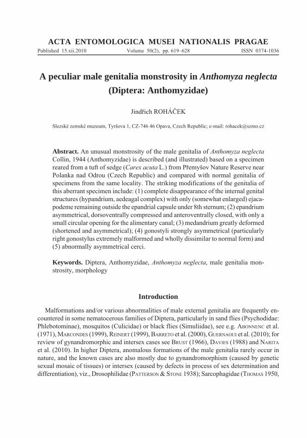
The morphological terminology used here follows that described in detail in the Morphology chapter in ROHÁČEK (2006). It is largely based on the “hinge” hypothesis of the origin of the eremoneuran hypopygium, re-discovered and documented by ZATWARNICKI (1996). Therefore, the following alterations of terms of the male genitalia need to be listed (synonymous terms used by other hypotheses in parentheses): ejacapodeme (ejaculatory apodeme), epandrium (periandrium), gonostylus (surstylus), medandrium (intraepandrial sclerite, intraperiandrial sclerite, bacilliform sclerites), pregonite (gonite), postgonite (paramere), phallapodeme (ae-deagal apodeme), phallophore (basiphallus), transandrium (posterior hypandrial bridge). For recognition of particular postabdominal and male genitalic structures in Anthomyzidae (includings several special terms only used in this family), see also Figs. 1–2, 11, 13 in this paper.
Abbreviations of morphological terms used in text and/or figures.afa – aedeagal part of folding apparatusce – cercuscp – caudal process of transandriumcs – connecting scleritedp – distiphallusea – ejacapodemeep – epandriumf – filum of distiphallusgs – gonostylushy – hypandriumlgs – left gonostylus
ma – medandriumpg – postgonitepha – phallapodemepp – phallophoreprg – pregonitergs – right gonostyluss – saccus of distiphallusS1–S8 – abdominal sternaT1–T6 – abdominal tergata – transandrium
ROHÁČEK: A peculiar male genitalia monstrosity in Anthomyza neglecta (Anthomyzidae)622
Results
Description of malformed male specimen of Anthomyza neglecta Collin, 1944(Figs. 3–5, 8–10)
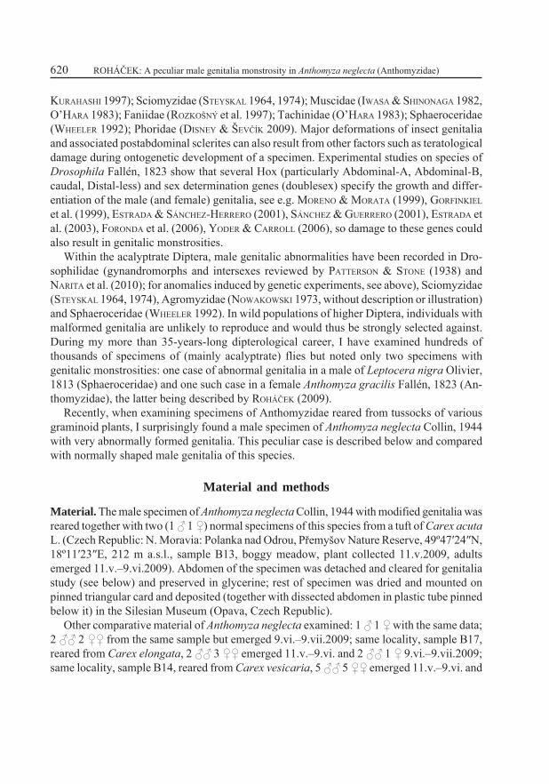
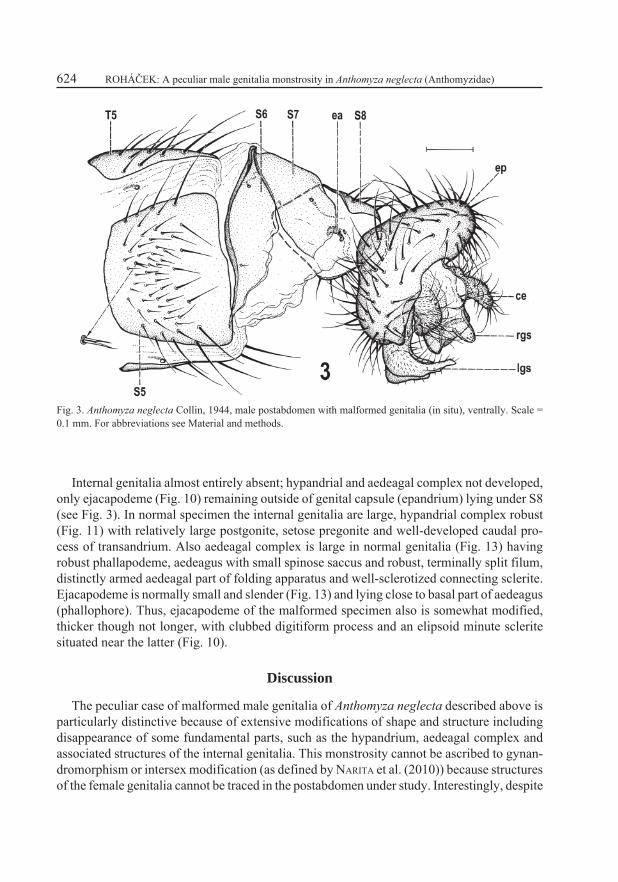
Most of body (including head and thorax with all extremities, and preabdomen) as in normal male of A. neglecta. An abnormally doubled seta (see Fig. 3) was found on pregenital (S5) sternum but this anomaly probably has nothing in common with the genitalic malformations described below. Postabdomen (Fig. 3) with majority of sclerites (T6, S6, S7) unmodified, except for S8 being distinctly narrower and with more bent sides (strongly convex, almost arch-shaped) than that of normal specimen.
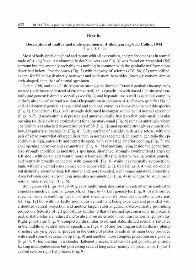
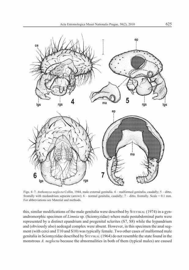
Genital (9th) and anal (11th) segments strongly malformed. External genitalia incompletely rotated (only inverted instead of circumverted), thus epandrium with dorsal side situated ven-trally and gonostyli directed dorsally (see Fig. 3) and hypandrium as well as aedeagal complex entirely absent – cf. normal position of hypandrium in abdomen of Anthomyza gracilis (Fig. 1) and of all internal genitalia (hypandrial and aedeagal complex) in postabdomen of this species (Fig. 2). Epandrium (Figs. 3–5) strongly deformed in comparison to that of normal specimen (Figs. 6–7), dorsoventrally depressed and anteroventrally fused so that only small circular opening (with heavily sclerotized rim) for alimentary canal (Fig. 5) remains anteriorly where epandrium was attached to posterior part of S8 (Fig. 3); anal opening strongly asymmetrical, low, irregularly subtriangular (Fig. 4). Outer surface of epandrium densely setose, with one pair of setae somewhat enlarged (less than in normal specimen). In normal genitalia the ep-andrium is high, anteriorly and ventrally open, with very large anterior opening (Fig. 7) and anal opening narrower and symmetrical (Fig. 6). Medandrium, lying inside the epandrium, also strongly modified in monstrous specimen, shortened, strongly asymmetrical (larger on left side), with dorsal and ventral more sclerotized ribs (the latter with subcircular branch), and ventrally broadly connected with gonostyli (Fig. 5) while it is normally symmetrical, high, with only ventral arms connected to gonostyli (Fig. 7). Cerci (Figs. 3, 4) well developed but distinctly asymmetrical, left shorter and more rounded, right longer and more projecting. Area between cerci surrounding anus also asymmetrical (Fig. 4) in contrast to situation in normal male specimen (Fig. 6).
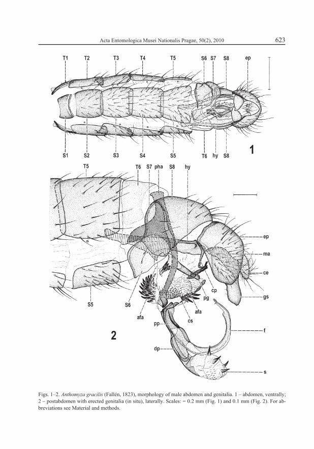
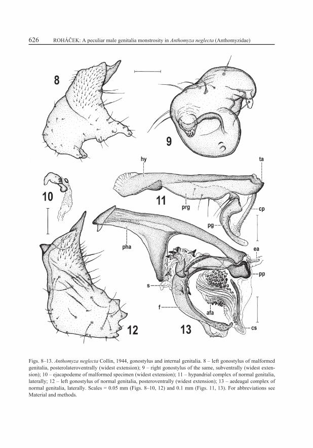
Both gonostyli (Figs. 4–5, 8–9) greatly malformed, dissimilar to each other (in contrast to almost symmetrical normal gonostyli, cf. Figs. 6–7). Left gonostylus (Fig. 8) of malformed specimen only resembling that of normal specimen in its proximal microtomentose part (cf. Fig. 12) but with markedly anomalous ventral half, being expanded and provided with a doubled ventral projection and another larger, subtriangular, posteroventrally protruding projection. Setosity of left gonostylus similar to that of normal specimen only in proximal part; distally setae are reduced and/or absent (on inner side) in contrast to normal gonostylus. Right gonostylus (Fig. 9) completely dissimilar to normal state, shifted medially (situated in the middle of ventral side of epandrium, Figs. 4, 5) and forming an extraordinary plump structure carrying peculiar process in the centre of posterior side of its main body provided with small spine-like setae on tip (Fig. 9) and another, more complex projection on right side (Figs. 4, 9) terminating in a slender flattened process. Surface of right gonostylus entirely lacking micropubescence but possessing several long setae (mainly on proximal part) plus 1 curved seta on right flat process (Fig. 9).
Acta Entomologica Musei Nationalis Pragae, 50(2), 2010 623
Figs. 1–2. Anthomyza gracilis (Fallén, 1823), morphology of male abdomen and genitalia. 1 – abdomen, ventrally; 2 – postabdomen with erected genitalia (in situ), laterally. Scales: = 0.2 mm (Fig. 1) and 0.1 mm (Fig. 2). For ab-breviations see Material and methods.
ROHÁČEK: A peculiar male genitalia monstrosity in Anthomyza neglecta (Anthomyzidae)624
Fig. 3. Anthomyza neglecta Collin, 1944, male postabdomen with malformed genitalia (in situ), ventrally. Scale = 0.1 mm. For abbreviations see Material and methods.
Internal genitalia almost entirely absent; hypandrial and aedeagal complex not developed, only ejacapodeme (Fig. 10) remaining outside of genital capsule (epandrium) lying under S8 (see Fig. 3). In normal specimen the internal genitalia are large, hypandrial complex robust (Fig. 11) with relatively large postgonite, setose pregonite and well-developed caudal pro-cess of transandrium. Also aedeagal complex is large in normal genitalia (Fig. 13) having robust phallapodeme, aedeagus with small spinose saccus and robust, terminally split filum, distinctly armed aedeagal part of folding apparatus and well-sclerotized connecting sclerite. Ejacapodeme is normally small and slender (Fig. 13) and lying close to basal part of aedeagus (phallophore). Thus, ejacapodeme of the malformed specimen also is somewhat modified, thicker though not longer, with clubbed digitiform process and an elipsoid minute sclerite situated near the latter (Fig. 10).
Discussion
The peculiar case of malformed male genitalia of Anthomyza neglecta described above is particularly distinctive because of extensive modifications of shape and structure including disappearance of some fundamental parts, such as the hypandrium, aedeagal complex and associated structures of the internal genitalia. This monstrosity cannot be ascribed to gynan-dromorphism or intersex modification (as defined by NARITA et al. (2010)) because structures of the female genitalia cannot be traced in the postabdomen under study. Interestingly, despite
Acta Entomologica Musei Nationalis Pragae, 50(2), 2010 625
Figs. 4–7. Anthomyza neglecta Collin, 1944, male external genitalia. 4 – malformed genitalia, caudally; 5 – ditto, frontally with medandrium separate (arrow); 6 – normal genitalia, caudally; 7 – ditto, frontally. Scale = 0.1 mm. For abbreviations see Material and methods.
this, similar modifications of the male genitalia were described by STEYSKAL (1974) in a gyn-andromorphic specimen of Limnia sp. (Sciomyzidae) where male postabdominal parts were represented by a distinct epandrium and pregenital sclerites (S7, S8) while the hypandrium and (obviously also) aedeagal complex were absent. However, in this specimen the anal seg-ment (with cerci and T10 and S10) was typically female. Two other cases of malformed male genitalia in Sciomyzidae described by STEYSKAL (1964) do not resemble the state found in the monstrous A. neglecta because the abnormalities in both of them (typical males) are caused
ROHÁČEK: A peculiar male genitalia monstrosity in Anthomyza neglecta (Anthomyzidae)626
Figs. 8–13. Anthomyza neglecta Collin, 1944, gonostylus and internal genitalia. 8 – left gonostylus of malformed genitalia, posterolateroventrally (widest extension); 9 – right gonostylus of the same, subventrally (widest exten-sion); 10 – ejacapodeme of malformed specimen (widest extension); 11 – hypandrial complex of normal genitalia, laterally; 12 – left gonostylus of normal genitalia, posteroventrally (widest extension); 13 – aedeagal complex of normal genitalia, laterally. Scales = 0.05 mm (Figs. 8–10, 12) and 0.1 mm (Figs. 11, 13). For abbreviations see Material and methods.
Acta Entomologica Musei Nationalis Pragae, 50(2), 2010 627
only by failure of the genitalia to circumvert during development, resulting in displacement and some modification of pregenital sclerites (no elimination or deformation of the genitalia proper recorded). The male internal genitalia are also lacking in the gynandromorph of Ra-chispoda subpiligera (Malloch, 1914) (Sphaeroceridae) discovered by WHEELER (1992) but in this specimen the female parts predominate in the postabdomen with only a modified male synsternite (S6–S8) retained, although the pregenital (5th) segment is typically male.
The only other case of abnormally formed (asymmetrical) terminalia in Anthomyzidae, described in female Anthomyza gracilis Fallén, 1823 by ROHÁČEK (2009: 11–12), was con-sidered a product of teratological damage inasmuch as these modifications (asymmetry and fusion of some postabdominal sclerites) were not so extensive and no part of female genitalia was missing.
Because of the huge changes in the male genitalia of Anthomyza neglecta under study (not only the absence of internal genitalia but also the completely dissimilar epandrium, medandrium and right gonostylus), this monstrosity is not likely to be of teratological origin. Most probably it is a consequence of genetic mutation or damage affecting some (or more) of the genes governing the development of male genital and anal primordia as recognised in Drosophila (cf. MORENO & MORATA 1999, ESTRADA & SÁNCHEZ-HERRERO 2001, SÁNCHEZ & GUERRERO 2001, ESTRADA et al. 2003, FORONDA et al. 2006).
Acknowledgements
It is an agreeable duty to express my sincere gratitude to Miroslav Barták (Praha, Czech Republic), Miloš Černý (Halenkovice, Czech Republic), Peter Chandler (Melksham, England), Jan Máca (České Budějovice, Czech Republic), Stephen A. Marshall (Guelph, Canada) and Rudolf Rozkošný (Brno, Czech Republic) for their information on genital malformations in higher Diptera and provision of relevant literature. Kevin N. Barber (Sault Ste. Marie, Canada), Peter Chandler, Stephen A. Marshall and both reviewers of the manuscript are also thanked for helpful comments and other improvement of the paper. The study was supported by the grant No. P506/10/1666 of the Czech Science Foundation.
ReferencesABONNENC E., POINSOT S. & RIOUX J.-A. 1971: Tératologie des Phlébotomes (Diptera: Psychodidae). Révision
et nouvelles observations. Cahiers de l’ORSTOM, Série Entomologie Médicale et Parasitologie 9: 307–316.BARRETO M., BURBANO M. E. & BARRETO P. 2000: Lutzomyia sand flies (Diptera: Psychodidae) from
Middle and Lower Putumayo Department, Colombia, with new records to the country. Memórias do Instituto Oswaldo Cruz 95: 633–639.
BRUST R. A. 1966: Gynandromorphs and intersexes in mosquitoes (Diptera: Culicidae). Canadian Journal of Zoology 44: 911–921.
DAVIES D. M. 1988: Gynandromorphs in Canadian Simuliidae (Diptera). Proceedings of the Entomological Society of Ontario 119: 87–89.
DISNEY R. H. L. & ŠEVČÍK J. 2009: A new species of fungus breeding Triphleba Rondani (Dipt., Phoridae) from Malaysia, including an intriguing mutant. Entomologist’s Monthly Magazine 145: 203–210.
ESTRADA B., CASARES F. & SÁNCHEZ-HERRERO E. 2003: Development of the genitalia in Drosophila melanogaster. Differentiation 71: 299–310.
ROHÁČEK: A peculiar male genitalia monstrosity in Anthomyza neglecta (Anthomyzidae)628
ESTRADA B. & SÁNCHEZ-HERRERO E. 2001: The Hox gene Abdominal-B antagonizes appendage development in the genital disc of Drosophila. Development 128: 331–339.
FORONDA D., ESTRADA B., DE NAVAS L. & SÁNCHEZ-HERRERO E. 2006: Requirement of Abdominal-A and Abdominal-B in the developing genitalia of Drosophila breaks the posterior downregulation rule. Develop-ment 133: 117–127.
GORFINKIEL N., SÁNCHEZ L. & GUERRERO I. 1999: Drosophila terminalia as an appendage-like structure. Mechanisms of Development 86: 113–123.
GUERNAOUI S., RAMAOUI K., RAHOLA N., BARNABE C., SERENO D. & BOUMEZZOUGH A. 2010: Malformations of the genitalia in male Phlebotomus papatasi (Scopoli) (Diptera: Psychodidae). Journal of Vector Ecology 35: 13–19.
IWASA M. & SHINONAGA S. 1982: A wild-caught gynandromorph in a muscid fly, Hydrotaea zao Shinonaga et Kano, 1973 (Diptera, Muscidae). Kontyû 50: 649–651.
KURAHASHI H. 1977: A gynandromorph in Parasarcophaga harpax (Diptera, Sarcophagidae), with special reference to “hypopygium circumversum” of the calyptrate postabdomen. Japanese Journal of Entomology 45: 372–376.
MARCONDES C. B. 1999: Anomalies of Lutzomyia intermedia (Lutz & Neiva, 1912) (Diptera, Psychodidae, Phlebotominae). Memórias do Instituto Oswaldo Cruz 94: 365–366.
MORENO E. & MORATA G. 1999: Caudal is the Hox gene that specifies the most posterior Drosophile segment. Nature (London) 400: 873–877.
NARITA S., PEREIRA R. A. S., KJELLBERG F. & KAGEYAMA D. 2010: Gynandromorphs and intersexes: poten-tial to understand the mechanism of sex determination in arthropods. Terrestrial Arthropod Reviews 3: 63–96.
NOWAKOWSKI J. T. 1973: Monographie der europäischen Arten der Gattung Cerodontha Rond. (Diptera, Agro-myzidae). Annales Zoologici (Warszawa) 31: 1–327.
O’HARA J. E. 1983: Two bilateral gynandromorphs in the Calyptratae (Diptera): Hydrotaea meteorica (Muscidae) and Siphona hokkaidensis (Tachinidae). Canadian Entomologist 115: 379–386.
PATTERSON J. T. & STONE W. 1938: Gynandromorphs in Drosophila melanogaster. University of Texas Pub-lication 3825: 1–67.
REINERT J. F. 1999: Morphological abnormalities in species of the quadrimaculatus complex of Anopheles (Diptera: Culicidae). Journal of the American Mosquito Control Association 15: 8–14.
ROHÁČEK J. 2006: A monograph of Palaearctic Anthomyzidae (Diptera). Part 1. Časopis Slezského Zemského Muzea, Opava (A) 55(supplement 1): 1–328.
ROHÁČEK J. 2009: A monograph of Palaearctic Anthomyzidae (Diptera). Part 2. Časopis Slezského Zemského Muzea, Opava (A) 58(supplement 1): 1–180.
ROZKOŠNÝ R., GREGOR F. & PONT A. C. 1997: The European Faniidae (Diptera). Acta Scientiarum Naturalium Academiae Scientiarum Bohemicae Brno, Nova Series 31(2): 1–80.
SÁNCHEZ L. & GUERRERO I. 2001: The development of the Drosophila genital disc. BioEssays 23: 698–707.STEYSKAL G. C. 1964: Two cases of abnormal development in male postabdomen of flies of the family Sciomyzidae
(Diptera). Papers of the Michigan Academy of Science, Arts and Letters 49: 195–198.STEYSKAL G. C. 1974: A gynandromorphic specimen of the genus Limnia (Diptera: Sciomyzidae). Journal of
the Washington Academy of Sciences 64: 11.THOMAS H. T. 1950: A gynandromorph Sarcophaga Meigen (Diptera: Calliphoridae), with notes on the sexual
dimorphism of that genus. Proceedings of the Zoological Society of London 120: 155–163.WHEELER T. A. 1992: A gynandromorph of Rachispoda subpiligera (Malloch) (Diptera, Sphaeroceridae), with
notes on asymmetry, circumversion, and the structure of the male postabdomen. Canadian Entomologist 124: 729–735.
YODER J. H. & CARROLL S. B. 2006: The evolution of abdominal reduction and the recent origin of distinct Abdominal-B transcript classes in Diptera. Evolution & Development 8: 241–251.
ZATWARNICKI T. 1996: A new reconstruction of the origin of eremoneuran hypopygium and its implications for classification (Insecta: Diptera). Genus 7(1): 103–175.