Vol. 188, No. 2, 1992 BIOCHEMICAL AND BIOPHYSICAL RESEARCH COMMUNICATIONS October 30, 1992 Pages 671-677 A PEPTIDE SEQUENCE ON CARCINOEMBRYONIC ANTIGEN BINDS TO A 80kD PROTEIN ON KUPFFER CELLS Peter Thomas, Anthony T. Petrick, Carol A. Toth, Eben S. Fox, James J. Elting and Glenn Steele Jr. Laboratory of Cancer Biology, Department of Surgery, The New England Deaconess Hospital, HarvardMedical School, BostonMA Molecular Diagnostics Inc., West Haven, CT 06516 Received September 2, 1992 Clearance of carcinoembryonic antigen (CEA) from the circulation is by binding to Kupffer cells in the liver. We have shown that CEA binding to Kupffer cells occurs via a peptide sequence YPELPK representing amino acids 107-112 of the CEA sequence. This peptide sequence is located in the region between the N-terminal and the first immunoglobulin like loop domain. Using native CEA and peptides containing this sequence complexed with a heterobifunctional crosslinking agent and ligand blotting with biotinylated CEA and NCA we have shown binding to an 80kD protein on the Kupffer cell surface. This binding protein may be important in the development of hepatic metastases. Q 1992 Academic Press, Inc. Carcinoembryonic antigen (CEA) is a glycoproteinwhose measurement inblood is used to monitor patients with colorectal carcinoma and other solid cancers (1). CEA is single chain of 668 amino acids with an N-terminal domain and three repeating immunoglobulin like disulfide loop domains of the C2 type (2,3). The deduced sequence from the cloned gene also predicts a short hydrophobic tail at the C-terminus but this is lost prior to secretion (4,5). Furthermore there are 28 potential sites for N-linked glycosylation (2). CEA is a member of a family of molecules that includes the non specific cross reacting antigen (NCA), the biliary glycoproteins (BGPs) and the pregnancy specific glycoproteins (PSGs). These molecules belong to the larger immunoglobulin supergene family (6). Recently a functional role of these proteins as adhesion molecules has emerged. Rodent cell lines transfected with cDNAs specific for CEA gene family members showed that both CEA and NCA can function as Ca++ independent intercellular adhesion molecules (7,8,9). Rojas et al (10) showed that BGP also acts as an intercellular adhesion molecule, however, binding was Ca++dependent. It has been suggested that CEA can function as a mediator of intercellular adhesion during embryonic development and in tumorigenesis (7). A more comprehensive discussion of the possible functions of the CEA family canbe found in a review by Thomas et al (11). 0006-291X/92 $4.00 671 Copyright 0 1992 by Academic Press, Inc. All rights of reproduction in any form reserved.
Transcript
Vol. 188, No. 2, 1992 BIOCHEMICAL AND BIOPHYSICAL RESEARCH COMMUNICATIONS October 30, 1992 Pages 671-677
A PEPTIDE SEQUENCE ON CARCINOEMBRYONIC ANTIGEN BINDS TO A 80kD PROTEIN ON KUPFFER CELLS
Peter Thomas, Anthony T. Petrick, Carol A. Toth, Eben S. Fox, James J. Elting and Glenn Steele Jr.
Laboratory of Cancer Biology, Department of Surgery, The New England Deaconess Hospital, HarvardMedical School, BostonMA
Molecular Diagnostics Inc., West Haven, CT 06516
Received September 2, 1992
Clearance of carcinoembryonic antigen (CEA) from the circulation is by binding to Kupffer cells in the liver. We have shown that CEA binding to Kupffer cells occurs via a peptide sequence YPELPK representing amino acids 107-112 of the CEA sequence. This peptide sequence is located in the region between the N-terminal and the first immunoglobulin like loop domain. Using native CEA and peptides containing this sequence complexed with a heterobifunctional crosslinking agent and ligand blotting with biotinylated CEA and NCA we have shown binding to an 80kD protein on the Kupffer cell surface. This binding protein may be important in the development of hepatic metastases. Q 1992 Academic Press, Inc.
Carcinoembryonic antigen (CEA) is a glycoproteinwhose measurement inblood
is used to monitor patients with colorectal carcinoma and other solid cancers
(1). CEA is single chain of 668 amino acids with an N-terminal domain and three
repeating immunoglobulin like disulfide loop domains of the C2 type (2,3). The
deduced sequence from the cloned gene also predicts a short hydrophobic tail at
the C-terminus but this is lost prior to secretion (4,5). Furthermore there are
28 potential sites for N-linked glycosylation (2). CEA is a member of a family
of molecules that includes the non specific cross reacting antigen (NCA), the
biliary glycoproteins (BGPs) and the pregnancy specific glycoproteins (PSGs).
These molecules belong to the larger immunoglobulin supergene family (6).
Recently a functional role of these proteins as adhesion molecules has
emerged. Rodent cell lines transfected with cDNAs specific for CEA gene family
members showed that both CEA and NCA can function as Ca++ independent
intercellular adhesion molecules (7,8,9). Rojas et al (10) showed that BGP also
acts as an intercellular adhesion molecule, however, binding was Ca++dependent.
It has been suggested that CEA can function as a mediator of intercellular
adhesion during embryonic development and in tumorigenesis (7). A more
comprehensive discussion of the possible functions of the CEA family canbe found
in a review by Thomas et al (11).
0006-291X/92 $4.00
671 Copyright 0 1992 by Academic Press, Inc.
All rights of reproduction in any form reserved.
Vol. 188, No. 2, 1992 BIOCHEMICAL AND BIOPHYSICAL RESEARCH COMMUNICATIONS
Our interest is in determining the mechanism of clearance of CEA from the
circulation and we have shown in both experimental animals and humans that this
occurs via receptor mediated endocytosis by the Kupffer cell (12,13). A
deglycosylatedpeptide (5.5 kD) isolated from a pepsin digest of CEA and covering
the junction of the N-terminal and first loop domains from amino acid 106 to
approximately 150. This peptide binds to Kupffer cells and binding is inhibited
by intact CEA (17). Glycopeptides covering sequences in other regions of the
molecule did not bind to Kupffer cells (14).
In the present study we have attempted to map this binding site and
identify the minimum sequences required for binding. Furthermore we have used
synthetic peptides to confirm the identity of the CEA binding protein on the rat
Kupffer cell as an 80kD surface protein.
MATERIALS and METHODS
Glvcooroteins. CEA was purified from a single colorectal carcinoma hepatic metastasis, as
previously described (13). The preparation was characterized by sodium dodecyl sulfate-polyacrylamide gel electrophoresis (SDS-PAGE), high-pressure liquid chromatography analysis, and activity in commercial CEA assay systems.
Normal Cross Reacting Antigen (NCA) was purified from colorectal cancer hepatic metastases as described previously (13), and subjected to the same criteria of purity. The MW of the purified NCA was approximately 55,000 by SDS- PAGE and HPLC.
Protein Modification. CXA (100 ug) was radiolabeled with 1 mCi Na lz51, (17 Ci/mg) (New England
Nuclear, Boston, MA), using the chloramine T procedure (15). The labeled CEAhad a specific radioactivity of -6 mCi/mg. CEA (lmg) was also conjugated to fluorescein isothiocyanate (20 ug) overnight at 4°C at neutral pH. The conjugate was separated from unreacted fluorescein by chromatography on Sephadex G-25.
CEA was conjugated to (sulfosuccinimidyl 2-(p-azidosalicylamido)ethyl- 1,3'dithiopropionate (SASD) (Pierce, Rockford, IL U.S.A.) using a modification of the manufacturers instructions. SASD (1.5pg) was radioiodinated with Na'=I (1mCi) by the Chloramine T procedure. After stopping the iodination reaction CEA (1OOpg) was added and the pH adjusted to 8.4 with a O.lM borate buffer. The reaction was carried out for 30 min. at ambient temperature. The conjugate was purified by chromatography on Sephadex G-25. All procedures were carried out in the dark.
Both CEAandNCAwere conjugatedwithbiotinusing 3(N-maleimidopropionyl)- biocytin. Previous studies had shown that intact disulfide bridges were not needed for CEA binding to Kupffer cells (13).
Svnthetic Peotides. Peptides were synthesizedonanAppliedBiosystems 430Apeptide synthesizer
usingt-BOG chemistry and cleaved from the resin using anhydrous HF. Purification was performed by reverse phase HPLC using two solvent systems. Solvent system 1 was A:50 mM ammonium acetate in water and B:50 mM ammonium acetate in 75% acetonitrile/water. Solvent system 2 was A:O.l% TFA in water and B:O.l% TFA in 90% acetonitrile/water. A linear gradient of 1% B/minute was used for both solvent systems. All peptides were analyzed for amino acid composition.
Isolation of Rat Kuoffer Cells. Kupffer cells were isolated from fasting male Sprague Dawley rats (-300
gm.) by collagenase perfusion of the liver followed by differential
672
Vol. 188, No. 2, 1992 BIOCHEMICAL AND BIOPHYSICAL RESEARCH COMMUNICATIONS
centrifugation and purification on a metrizamide gradient, as described previously (13). Further purification of the cells was achieved by allowing them to attach overnight to plastic tissue culture dishes. The cells were >95% viable by trypan blue exclusion. The preparation was greater than 85% Kupffer cells based on staining for endogenous peroxidase and their ability to phagocytose 1.1~ latex beads.
LiPand Binding Assays. '?I-Peptides (1Opg) or lZ51-CEA were incubated up to 45 min. with rat
Kupffer cells in triplicate using a modification of the method of Stahl et al (16). Free ligandwas separated from cell bound ligand by centrifuging at 11,000 rpmthrough an oil phase consisting of dibutylphthalate:dioctylphthalate 3:l (17) as described previously (13).
Affinitv chromatograohv of Kuoffer cell surface oroteins. Isolated rat Kupffer cells were surface labeled with '%I using the
lactoperoxidase procedure. The labeled cells were dissolved in 0.5% TritonXlOO in O.lM Tris buffered saline pH 7.4 (TBS) containing 5mM PMSF and lpg/ml leupeptin, and centrifuged in an Eppendorf microfuge for 10 mins to remove insoluble material. The supernatant containing the labelled cell surface proteins was chromatographed on a column of immobilized CEA (2 mg CEA/ml of wet CNBr activated Sepharose) in 0.1% Triton X-100, TBS, with 10 mM CaC12 at 4OC. The CFA/Sepharose was washed well with the above buffer. Bound material was eluted from the CEA/Sepharose by 0.1% Triton X100, in TBS containing 10 mM EDTA. The eluted radioactive fractions were pooled concentrated and examined on 10% SDS-PAGE. Three major proteins were present (18). Because it was likely that at least one of these proteins was a galactose recognizing lectin the eluted radiolabeled proteins were rechromatographed on a asialo fetuin Sepharose 4B column. The eluted proteins were dialyzed against the 0.1% Triton X100, TBS, 1OmM CaCl, buffer overnight at 4°C and subjected to chromatography on immobilized asialofetuin Sepharose. The unbound protein fraction was examined by SDS-PAGE as above. The SDS gels were examined by coomassie blue staining and exposure to Kodak Xomat X-ray film at -7O'C.
Crosslinking of CEA and Peotides to Isolated Kuoffer Cells. Kupffer cells (1 X 10⁷) in O.lM phosphate buffered saline (PBS) pH 7.4
were reacted with 5pg of labelled conjugate (approximately 2pCi) for 30 min. at 37"C, and crosslinked with short wavelength UV light for 10 min. The cells were washed 3X in PBS and extracted and reduced with 0.5% SDS, 5% 2-mercaptoethanol in O.lM tris buffered saline pH 7.4 (TBS) with 1mM PMSF and lpg/ml leupeptin. The extracts were dialyzed overnight against 0.5% SDS in TBS and concentrated using a Centricon- microconcentrator. The samples were examined by SDS-PAGE on 10% gels and crosslinked proteins were visualized by autoradiography.
Lieand BlottinP Assavs. Isolated rat Kupffer cells were extracted with O.lM TBS pH 7.4 containing
1% Triton X-100 and inhibitors of proteolysis (1mM PMSF, lpg/ml leupeptin and aprotinin). Extracts were separated on 10% SDS-PAGE with biotinylated MW standards and the proteins transferred to nitrocellulose. The membrane was blocked with 10% BSA for 1 hour at 37°C and washed in 20mM TBS with 1OmM Ca++ and Mg++. The membrane was incubated with the biotinylated CEA or NCA (2pg/ml) for 1 hour at 37'C and washed 3 times (10 min.) in the buffer at 25V. The membrane was incubated with avidin/alkaline phosphatase in TBS for 10 min. and developed with a solution of 5-bromo-4-chloro-3-indolyl phosphate and tetranitro blue tetrazolium.
RESULTS and DISCUSSION
To identify the site on CEA recognized by the Kupffer cell we synthesized
four pentadeca-peptides with 5 amino acid overlaps based on the sequence of the
673
Vol. 188, No. 2, 1992 BIOCHEMICAL AND BIOPHYSICAL RESEARCH COMMUNICATIONS
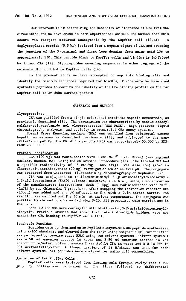
105 115 125 135 100 110 120 130 140
1 I I 1 I 1 1 I 1 5.5kD+- RVYPELPKPSISSNNSKPVEDAVAFTCEPETQDATYLW
c-75 - TGQFRVYPELPKPSI
C-76' - PKPSISSNNSKPVED
c-77' - KPVEDKDAVAFTCEP
C-78 - FTCEPETQDATYLWW
C-85 - YPELPK
Fig.1. Structure of the 5.5kD pepsin peptide and the 5 synthetic peptides. l Data from Thomas and Toth (14). * Peptides C-76 and C-77 were synthesized with an extra tyrosine residue at the C-terminus to allow them to be radioiodinated. All peptides were labeled with "'1 using the Chloramine T procedure. Each peptide (1OOpg) was labeled to a specific radioactivity of approximately 4mCi/mg.
previously identified 5.5kD binding peptide (14). The four 15 amino acid peptides
and a hexameric peptide are shown in Fig. 1. Fig. 2A shows that only one of
these 15 amino acid peptides (C-75) was bound and endocytosed by isolated rat
Kupffer cells. The binding of lz51 labeled C-75 could be inhibited by unlabeled
C-75 and CF,A but not by the overlapping pentadeca-peptide C-76 (Fig. 2B). These
results were further confirmed using immunofluorescence of isolated rat Kupffer
cells with FITC labelled peptides. This localizes the binding site to amino
acids 101-115 in the CEA sequence. This sequence has the expected homology with
the CEA related glycoproteins NCA, and BPGs, however the sequence (RVYPELPKPSI)
also showed a high degree of homology with the human proteolytic enzyme,
prostromelysin (PDLPK, amino acids 123-127) (19) with human complement Cls
precursor (PELPK) (20) and with human collagenase 1 (PDLPR) (19). These
substitutions represent only conservative changes.
Because of the appearance of the sequence PELPK in complement subcomponent
Cls, we synthesized the hexapeptide YPELPK (C-85) to determine if this was also
the binding site for CEA. This peptide was rapidly endocytosed by Kupffer cells
and could be inhibited by both the larger peptide C-75 and by CEA itself (Fig.
2C).
Because of the possible implications of CEAbinding to Kupffer cells in the
development of hepatic metastases from colorectal tumors (21,22,23) we were
interested in identifying the binding site for these peptides on isolatedKupffer
cells. Previous studies using affinity chromatography with CEA-Sepharose showed
the presence of three major CEA binding proteins on the Kupffer cell surface all
of which require Ca++ and, or Mg++ for binding (18). Chromatography of this mixture on asialo fetuin Sepharose removed both the higher (170kD) and lower
molecular weight (35kD) proteins. This resulted in an almost homogeneous
674
Vol. 188, No. 2, 1992 BIOCHEMICAL AND BIOPHYSICAL RESEARCH COMMUNICATIONS
15 45 TIME (minutes)
B 16
C-76
6
Control CEA c-75 CCdd
Fip;. 2. A. Uptake of the 15 amino acid 's51-Peptides (1Opg) by isolated rat Kupffer cells. - l , C-75, -v C-76, - v C-77, - 0 C-78. B. Inhibition of Kupffer cell uptake of peptide C-75. "sI C-J5 uptake was measured in the presence of an equimolar concentration of CEA (100 fold excess by weight) and a 100 fold molar excess of peptides C-75 or C-76. C. Inhibition of Kupffer cell uptake of peptide C-85. '=I C-85 uptake was measured in the presence of an equimolar concentration of CEA and a 100 fold molar excess of unlabeled peptide C-75. All determinations were carried out in triplicate, the error bars represent one standard deviation.
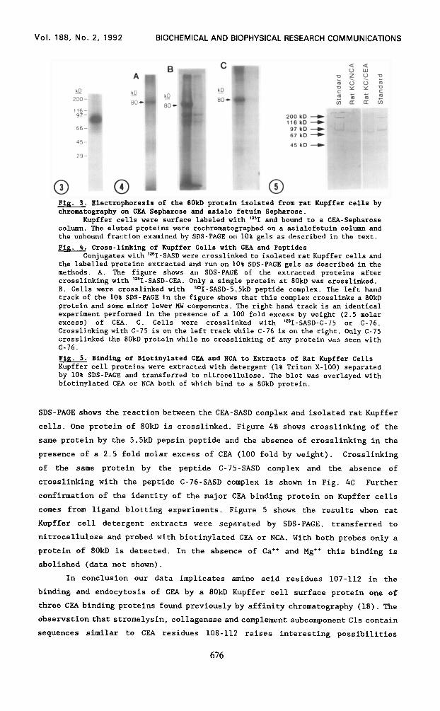
radiolabelled protein of 80kD (Fig. 3). To confirm that the 80kD protein was
responsible for the specific binding of these peptides we used a photoactivatable
dithiopropionate (SASD) which specifically labels the binding protein following
reduction. The crosslinker was labeled with '*sI and coupled to CEA, to the 5.5kD
peptide produced by pepsin digestion of CEA (17) and to the reactive C-75 peptide
as well as the unreactive peptide C-76. In Fig. 4A an autoradiogram of a 10%
Vol. 188, No. 2, 1992 BIOCHEMICAL AND BIOPHYSICAL RESEARCH COMMUNICATIONS
200 kD 4 116 kD +
97 kD + 67 kD +
45 kD +
Fig. 3. Electrophoresis of the BOkD protein isolated from rat Kupffer cells by chromatography on CKA Sepharose and asialo fetuin Sepharose.
Kupffer cells were surface labeled with "'1 and bound to a CBA-Sepharose column. The eluted proteins were rechromatographed on a asialofetuin column and the unbound fraction examined by SDS-PAGE on 10% gels as described in the text.
Fig. 4. Cross-linking of Kupffer Cells with CKA and Peptides Conjugates with 'e51-SASDwere crosslinked to isolated rat Kupffer cells and
the labelled proteins extracted and run on 10% SDS-PAGE gels as described in the methods. A. The figure shows an SDS-PAGE of the extracted proteins after crosslinking with 'e51-SASD-CKA. Only a single protein at 80kD was crosslinked. B. Cells were crosslinked with "'1-SASD-5.5kD peptide complex. The left hand track of the 10% SDS-PAGE in the figure shows that this complex crosslinks a 80kD protein and some minor lower MW components. The right hand track is an identical experiment performed in the presence of a 100 fold excess by weight (2.5 molar excess) of CPA. c. Cells were crosslinked with '=I-SASD-C-75 or C-76. Crosslinkingwith C-75 is on the left track while C-76 is on the right. Only C-75 crosslinked the 80kD protein while no crosslinking of any protein was seen with C-76. Fig. 5. Binding of Biotinylated CBA and NCA to Extracts of Rat Kupffer Cells Kupffer cell proteins were extracted with detergent (1% Triton X-100) separated by 10% SDS-PAGE and transferred to nitrocellulose. The blot was overlayed with biotinylated CPA or NCA both of which bind to a 80kD protein.
SDS-PAGE shows the reactionbetween the CEA-SASD complex and isolated rat Kupffer
cells. One protein of 80kD is crosslinked. Figure 4B shows crosslinking of the
same protein by the 5.5kD pepsin peptide and the absence of crosslinking in the
presence of a 2.5 fold molar excess of CEA (100 fold by weight). Crosslinking
of the same protein by the peptide C-75-SASD complex and the absence of
crosslinking with the peptide C-76-SASD complex is shown in Fig. 4C Further
confirmation of the identity of the major CEA binding protein on Kupffer cells
comes from ligand blotting experiments. Figure 5 shows the results when rat
Kupffer cell detergent extracts were separated by SDS-PAGE, transferred to
nitrocellulose and probed with biotinylated CEA or NCA. With both probes only a
protein of 80kD is detected. In the absence of Cat+ and Mg++ this binding is
abolished (data not shown).
In conclusion our data implicates amino acid residues 107-112 in the binding and endocytosis of CEA by a 80kD Kupffer cell surface protein one of
three CEA binding proteins found previously by affinity chromatography (18). The
observation that stromelysin, collagenase andcomplement subcomponent Cls contain
sequences similar to CEA residues 108-112 raises interesting possibilities
676
Vol. 188, No. 2, 1992 BIOCHEMICAL AND BIOPHYSICAL RESEARCH COMMUNICATIONS
regarding their interaction with Kupffer cells. We will now be able to use
peptides that bind to this 80kD protein as inhibitors of CEA uptake in vivo, to
determine, if, like CEA, theywillenhance metastases fromcolorectalcancer cell
lines.
ACKNOWLEDGMENTS
This investigation was supported by grants numbers CA44585 and CA44704
from the National Cancer Institute, and DK44305 from the National Institute of
Diabetes and Digestive and Kidney Diseases, United States Public Health Service.
1.
2.
3.
4.
5.
6. 7.
a.
9. 10.
11.
12.
13.
14.
15.
16.
17. 18.
19.
20.
21. 22.
23.
REFERENCES
Zamcheck, N., Steele, G.D., Thomas, P. and Mayer, R.J. (1986) In: The Manual of Clinical Immunology, (Rose, Freidman and Fahey, eds.) Am. Sot. Microbial. pp. 802-809. Oikawa, S., Nakazato, H. and Kosaki, G. (1987) Biochem. Biophys. Res. Commun. 142, 511-518. Kamarck, M., Elting, J., Hart, J., Gobel, S., Rae, P.M.M., Nortdurft, M.A., Nedwin, J. and Barnett, T. (1987) Proc. Natl. Acad. Sci. U.S.A. 84, 5350-5354. Hefta, S.A., Hefta, L.J.F., Lee, T., Paxton, R. and Shively, J.E. (1988) Proc. Natl. Acad. Sci. U.S.A. 85, 4648-4652. Sack, T.L., Gum, J.R., Low, M.G. and Kim, Y.S. (1988) J. Clin. Invest. 82, 586-593. Williams, A.F. and Barclay, A.N. (1988) Annu. Rev. Immunol. 6, 381-405. Benchimol, S., Fuks, A., Jothy, S., Beauchemin N., Shirota, K. and Stanners, C.P. (1989) Cell 57, 327-334. Oikawa, S., Inuzuka, C., Kuroki, M., Matsuoka, Y., Kosaki, G. and Nakazato, H. (1989) Biochem. Biophys. Res. Commun. 164, 39-45. Zhou, H., Fuks, A. and Stanners, C.P. (1990) Cell GrowthDiffer. 1, 209-215. Rojas, M., Fuks, A. and Stanners, C.P. (1990) Cell growth Differ. 1, 527- 533. Thomas, P., Toth, C.A., Saini, K.S., Jessup, J.M. and Steele, G. (1990) Biochim Biophys Acta 1032, 177-189. Toth, C.A., Thomas, P., Broitman, S.A. and Zamcheck, N. (1982) Biochem. J. 204, 377-381. Toth, C.A., Thomas, P., Broitman, S.A. and Zamcheck, N. (1985) Cancer Res. 45, 392-397. Thomas, P. and Toth, C.A. Biochem. Biophys. Res. Commun. (1990) 170, 391- 396. Greenwood, F.C., Hunter, W.M. andGlover, J.S. (1963) Biochem. J. 89: 114- 123. Stahl, P., Rodman, J., Miller, M. and Schlesinger P. (1978) Proc. Natl. Acad. Sci. U.S.A. 75, 1399-1403. Maynard, Y. and Baenziger, J. (1981) J. Biol. Chem. 15, 8063-8068. Thomas, P., Toth, C.A., Fox, E.S. and Steele, G. (1991) Carcinoembryonic antigen binding proteins from Kupffer cells. In: Cells of the Hepatic Sinusoid Vol 3, The Kupffer Cell Foundation, Rijswijk, The Netherlands, 512-514. Wilhelm. S.M., Collier, I.E., Kronberger, A., Eisen, A.Z., Marmer, B.L. Grant, G.A., Bauer, E.A. and Goldberg, G.I. (1987) Proc. Natl. Acad. Sci. U.S.A. 84, 6725-6729. Tosi, M., Duponchel, C., Meo, T. and Julier, C. (1987) Biochemistry 26; 8516-8524. Jessup,J.M. and Thomas, P.(1989)Cancer and Metastasis Reviews. 8,263-280. Hostetter, R.B., Augustus, L.B., Mankarious, R., Chi, K., Fan, D., Toth, C.A.,Thomas,P.andJessup, J.M. (1990) J. Natl. Cancer Inst. 82, 380-385. Wagner, H.E., Thomas, P., Wolf, B.C., Zamcheck, N., Jessup, J.M. and Steele, G.D. (1990) Invasion and Metastasis 10, 253-266.