Version/Version Date: 4.1 09/14/18 - i - IRB #: 441-13 A Phase II Study of Neoadjuvant Chemotherapy with and without Immunotherapy to CA125 (Oregovomab) followed by Hypofractionated Stereotactic Radiotherapy and Concurrent HIV Protease Inhibitor Nelfinavir in Patients with Locally Advanced Pancreatic Cancer *Principal Investigator: Chi Lin, MD, PhD 987521 Nebraska Medical Center, Omaha, NE 68198-7521 Phone: 402-552-3844 Fax: 402-552-3926 Email: [email protected]Secondary Investigators: Jean L. Grem, MD Michael A. Hollingsworth, PhD Quan P. Ly, MD Aaron R. Sasson, MD James K. Schwarz, MD Sarah Thayer, MD, PhD Lyudmyla Berim, MD Statistician: Jane Meza, PhD 984375 Nebraska Medical Center, Omaha, NE 68198-4375 Phone: 402-559-8407 Fax: 402-559-7259 Email: [email protected]Study Product: Nelfinavir (Viracept ® ) Oregovomab (Quest Pharma Tech Inc, B43.13 to CA125 IND#7112
Transcript
Version/Version Date: 4.1 09/14/18 - i -
IRB #: 441-13
A Phase II Study of Neoadjuvant Chemotherapy with and without Immunotherapy to CA125 (Oregovomab) followed by Hypofractionated
Stereotactic Radiotherapy and Concurrent HIV Protease Inhibitor Nelfinavir in Patients with Locally Advanced Pancreatic Cancer
*Principal Investigator: Chi Lin, MD, PhD
987521 Nebraska Medical Center, Omaha, NE 68198-7521 Phone: 402-552-3844 Fax: 402-552-3926 Email: [email protected]
Secondary Investigators: Jean L. Grem, MD Michael A. Hollingsworth, PhD Quan P. Ly, MD Aaron R. Sasson, MD James K. Schwarz, MD Sarah Thayer, MD, PhD Lyudmyla Berim, MD Statistician: Jane Meza, PhD 984375 Nebraska Medical Center, Omaha, NE 68198-4375 Phone: 402-559-8407 Fax: 402-559-7259 Email: [email protected] Study Product: Nelfinavir (Viracept®) Oregovomab (Quest Pharma Tech Inc, B43.13 to CA125 IND#7112
Subject with C1D1 C1D8 C1D15 C2D1 C2D8 C2D15 C3D1 C3D8 C3D15 C4D1 C4D8 C4D15 CO CA125 < 10 C C C C C C NFV NFV NFV NFV NFV C C VE CT
MRI SRT Pre-op
restage OR RY
Neo-Adjuvant Therapy C = Chemotherapy Regimen (SOC); Gemcitabine 750 (females) or 900 mg/m2 (males) IV by fixed dose rate infusion, leucovorin 50 mg/m² IV over 30 min, and 5-FU 2700 mg/m2 IV over 24 hrs REPEAT weekly for 2 of 3 weeks (day 1 and 8) x 4 Cycles (RESUME C after SRT for Cycle 4) I = Immunotherapy and Immunologic Assessment (Res): Oregovomab 2 mg IV over 20 min will be given every 3 weeks on day 15 x 4 Cycles (RESUME I after SRT for Cycle 4) BD= Blood Draw (Res) for immunologic assessment will be done Pre-chemotherapy, Prior to second infusion for the first 6 obtainable patients, Pre-SRT, Pre-surgery ET=Excess tumor tissue sample (Res) postsurgical resection for CA125 > 10 arm only NFV = Nelfinavir (Res) 1250 mg P.O. BID to start daily the Monday of week three of the third cycle of chemotherapy (2 weeks prior to the initiation of SRT) CONTINUE x 5 weeks ending the Friday 2 weeks after the end of SRT SRT = Stereotactic Radiotherapy (SOC) daily Monday-Friday x 1 week to start week 11 (SOC) OR= Surgery (SOC) to be performed sometime during week 17-18
CA125 andBlood Draw (Res) for immunological assessment Based on CA125 results:
Version/Version Date: 4.1 09/14/18 - iii -
Subject with C5D1 wk1
C5D8 wk2
C5D15 wk3
C6D1 wk4
C6D8 wk5
C6D15 wk6
C7D1 wk7
C7D8 wk8
C7D15 wk9
wk10
wk11
End of Study wk12
CA125 > 10 C C I C C I C C I BD BD
OR
Subject with C5D1 C5D8 C5D15 C6D1 C6D8 C6D15 C7D1 C7D8 C7D15 CA125 < 10 C C C C C C
Adjuvant Therapy C = Chemotherapy Regimen (SOC); Gemcitabine 750 (females) or 900 mg/m2 (males) IV by fixed dose rate infusion, leucovorin 50 mg/m² IV over 30 min, and 5-FU 2700mg/m2 IV over 24 hrs REPEAT weekly for 2 of 3 weeks (day 1 and 8) x 3 Cycles (Cycles 5-7) CA125> 10 Subjects Resume: I = Immunotherapy and Assessment (Res): Oregovomab 2 mg IV over 20min will be given every 3 weeks on day 15 x 3 Cycles (Cycles 5-7) BD= Blood Draw (Res) for immunological assessment will be done prior to restarting chemotherapy after surgery/post restaging if not resectable and 3 weeks post (week 12) or end of study.
NOTE: If the patient has CA 125 >= 10 who is not eligible for receiving oregovomab (e.g. allergic to the drug) but eligible for the rest of treatment, this patient should be accrued to the part of protocol without oregovomab. In another word, patients who have CA 125 >=10 can also be accrued and treated on the part of protocol without oregovomab if they cannot receive oregovomab but no contraindications to the rest of treatment.
**Post-operative RECOVERY / ** Post-restaging unresectable RESUME Adjuvant Chemotherapy: Three cycles of chemotherapy to start when the clinician’s determine that the patient has recovered from surgery
OR if the patient was unresectable and the clinician’s determine that chemotherapy should be resumed.
3. Eligibility Criteria ..................................................................................................................11 4. REGISTRATION PROCEDURES………………………………………………………..13 5. TREATMENT PLAN ............................................................................................................15 6. MEASUREMENT OF EFFECT ..........................................................................................28 7. STUDY PARAMETERS .......................................................................................................30 8. DRUG FORMULATION AND PROCUREMENT............................................................33 9. TOXICITY REPORTING GUIDELINES ..........................................................................47
10. STATISTICAL CONSIDERATIONS .................................................................................51 11. RECORDS TO BE KEPT .....................................................................................................53 12. PATIENT CONSENT FORM STATEMENT…………………………………………….54
13. REFERENCES .......................................................................................................................57 14. DATA FORMS Attached APPENDICES
APPENDIX A Criteria defining resectability status ..................................................................................62
APPENDIX B Performance Status Criteria ..............................................................................................63
APPENDIX C Eligibility Criteria CRF......................................................................................................64 APPENDIX D
Specimen Requirement and Measurement of Immunological Parameters………………68 APPENDIX E NCI Common Toxicity Criteria Version 4.0 .....................................................................71 APPENDIX F Pain Assessment Scale .......................................................................................................72
APPENDIX G MEDWATCH ……………………………………………………………………………73 APPENDIX H
Version/Version Date: 4.1 09/14/18 - ii -
Medication Information Sheet ……………………………………………………………...74
Page 1 of 81 Version/Version Date: 4.1 09/14/18
1.0 OBJECTIVES 1.1. Primary Objectives
1.1.1 To evaluate the efficacy of neoadjuvant chemotherapy, (gemcitabine, leucovorin, 5-FU) with or without Oregovomab, followed by hypofractionated stereotactic radiotherapy (SRT) concurrently with nelfinavir in patients with locally advanced pancreatic cancer that is CA125 positive (>10) or CA125 negative ( <10).
1.2 Secondary Objectives 1.2.1 To assess the safety of neoadjuvant chemotherapy, (gemcitabine, leucovorin, 5-FU) with or without Oregovomab, followed by SRT concurrently with nelfinavir in patients with locally advanced pancreatic cancer that is CA125 positive (>10) or CA125 negative (<10). 1.2.2 To assess the cellular and humoral immune responses to active immunotherapy with Oregovomab/ monoclonal antibody in patients with pancreas cancer with CA125 level greater than 10 undergoing chemotherapy and radiation treatments.
1.3 Correlative Studies 1.3.1 To evaluate tumor and organ motion with 4D CT and respiratory gating system and to evaluate the effect of tumor/organ motion on the dosimetry, local control and survival. 1.3.2 To evaluate inter- and intra-fractional target motion with Calypso system.
2.0 BACKGROUND 2.1 Current therapy for locally advanced pancreatic cancers
Approximately 37,000 individual in the US develop pancreatic cancers each year and almost an equal number of patients will die from the disease (1). Prognosis is directly related to the extent of tumor. The patients are usually classified into those with localized, locally advanced, or metastatic disease. The median survivals for patients in these groups range from 11-18 months, 10-12 months, and 5-7 months, respectively (2). The overall 5-year survival is less than 5%. Surgical resection offers the best chance for long-term survival. However, even in patients who undergo potentially curative surgery, 5-year survival has in general not exceeded 20%.
Because surgery alone rarely provides long-term cures, an alternative strategy is to treat pancreatic cancer before or after surgery with either systemic chemotherapy or combined chemo-radiation therapy. In the majority of studies, adjuvant therapy is administered following pancreatic resection. A US Intergroup study compared gemcitabine vs. infusional 5-FU chemotherapy for one month prior to and three months after chemoradiation (CRT) consisting of continuous infusional 5-FU as adjuvant therapy after pancreatic cancer resection; outcome in those with tumor located in the pancreatic head was the primary study endpoint (3). The gemcitabine plus CRT arm was superior to the 5-FU plus CRT arm with a median survival of 20.6 months vs 16.9 months and survival at 3-yr 32% vs 21%. This survival advantage came at a cost of appreciable toxicity, with grade 3-4 hematologic and non-hematologic toxicities occurring in 58% and 58% of subjects, respectively. Oettle et al compared gemcitabine given at 1000 mg/m² weekly for 3 of 4 weeks x 6 cycles to no additional therapy in 368 patients with resected pancreatic cancer (4). Adjuvant gemcitabine was associated with a significant improvement in disease-free survival (13.4 vs 6.9 months), and a trend towards improvement in overall survival (median 22.1 vs 20.2 months); 34% of those receiving gemcitabine were alive at 3 yr vs 20.5% with surgery alone. Grade 3-4 hematologic and non-hematologic toxicities occurred in fewer than 5% of subjects receiving gemcitabine.
Page 2 of 81 Version/Version Date: 4.1 09/14/18
While these studies indicate improvement with adjuvant therapy, there is still a need to improve upon these results. A disadvantage of adjuvant therapy is that as many as 25% of patients never receive adjuvant therapy or have their treatment delayed due to post-operative complications (5-7). In an effort to increase the number of patients receiving adjuvant therapy, chemotherapy and radiation therapy can be administered pre-operatively to potential surgical candidates. Additional potential benefits of pre-operative therapy include the delivery of therapy to well-oxygenated tissues, the potential to downstage tumors (particularly when the lesion is borderline resectable or unresectable because of regional factors such as large tumor size or involvement of the mesenteric or portal vein), and the opportunity to observe patients for the development of metastatic disease during therapy. After maximal tumor shrinkage and no interval development of metastatic disease, surgery can be considered. Preoperative chemoradiation therapy is a fairly recent approach with a theoretical advantage of improving the resectability. Single center studies from MD Anderson Cancer Center and Fox Chase Cancer Center have demonstrated favorable histopathologic features following neoadjuvant therapy (6, 8). In both studies the incidence of positive resection margins and positive lymph nodes were significantly less than in a cohort of patients that served as controls. Multiple studies have identified the presence of positive margins and positive lymph nodes as significant predictors of poor outcome (9-13). Because pancreatic cancer is characterized by early dissemination of disease, use of systemic chemotherapy is reasonable. A recent meta-analysis suggests that gemcitabine plus fluoropyrimdine combinations are superior to gemcitabine alone in the treatment of patients with metastatic pancreatic cancer: hazard ratio 0.90, p = 0.03 (14). We have experience with a combination of gemcitabine given with leucovorin-modulated 5-FU given as a 24-hour infusion weekly for two of three weeks, which is well tolerated The rationale for this regimen is based on preclinical studies conducted in Dr. Grem’s laboratory that showed the sequential administration of gemcitabine followed by either 5-fluorodoexyuridine (FdUrd) or 5-FU resulted in more than additive cytotoxicity in both cell growth and clonogenic assays [15]. The combination of gemcitabine followed by FdUrd produced greater damage to nascent DNA in an alkaline elution assay compared to either agent alone, and the locus of interaction was DNA-directed (15). This led to the development of a Phase I clinical trial of gemcitabine given with a 24-hour infusion of FdUrd weekly for three of four weeks. FdUrd was selected because it displays more selective TS inhibition than 5-FU. Thirty-eight patients were accrued into this trial before a nation-wide shortage of pharmaceutical grade FdUrd led to the trial’s premature closure (16). Analysis of the hematologic toxicity indicated that many patients could not receive the week 3 doses due to neutropenia or thrombocytopenia. Therefore, a new trial was implemented that evaluated escalating doses of gemcitabine given as a 30 minute infusion weekly for two of three weeks followed by a 24-hour infusion of 5-FU modulated by low-dose calcium leucovorin. This trial was conducted by Dr. Grem in three parts. In part 1, the initial dose level employed gemcitabine at 75% of its recommended dose as a single agent (750 mg/m²), 5-FU at about 50% of its recommended dose as a weekly 24 hr infusion (1150 mg/m²), and low-dose leucovorin for 2 weeks out of 3. In all stages of the protocol, to ensure tolerability, the patient received an initial cycle of gemcitabine alone. A conservative dose modification scheme for gemcitabine was used in the interests of patient safety to avoid untoward toxicity when 5-FU was added. If tolerated, 5-FU was added cycle 2. If the gemcitabine dose
Page 3 of 81 Version/Version Date: 4.1 09/14/18
was not tolerated, the dose of gemcitabine was decreased by one or two dose levels depending on the severity of toxicity, and a second cycle of gemcitabine alone was given. 5-FU was added the subsequent cycle. The dose of 5-FU were escalated in 25% increments in cohorts of three patients up to a planned dose of 2250 mg/m². Since dose-limiting toxicity was not observed at 2250 mg/m², the dose of gemcitabine was then escalated in 20% increments. Once the maximally tolerated dose of gemcitabine was determined (female patients, 900 mg/m², male patients, 1080 mg/m²), additional cohorts of patients have been treated with 5-FU increased in 20% increments while the gemcitabine dose has been held constant. The dose of fluorouracil has been escalated from 2250 mg/m² up to a maximum dose of 3888 mg/m². At the highest dose level, two patients experienced dose-limiting hematologic toxicity the cycle in which 5-FU was added and a third patient experienced ataxia.
Therefore, the recommended dose of 5-FU was 3240 mg/m². Thirteen patients received one or more cycles with gemcitabine combined with 3240 mg/m² 5-FU, including 7 female and 6 male patients. For the females, the gemcitabine dose was 900 mg/m² in 5 patients, 750 mg/m² in 1, and 600 mg/m² in 1. For the males, the gemcitabine dose was 1080 mg/m² in 4 patients, and 900 mg/m² in 2. The hematologic toxicity with gemcitabine combined with 5-FU 3240 mg/m² is summarized below according to gender.
gemcitabine mg/m²
WBC x 1000/μL median (range)
Hemoglobin g/dL median (range)
Platelets x 1000/μL median (range)
Granulocytes /μL median (range)
females 600-900
4.2 (1.7 - 4.8)
9.8 (6.9 - 10.5)
130 (82 - 268)
2491 (800 - 2632)
males 900-1080
2.85 (2.1 - 4.3)
9.7 (7.2 - 11.9)
81 (50 - 152)
1771 (832 - 2100)
females 900
4.2 (1.7 - 4.8)
9.9 (8.4 - 10.5)
131 (82 - 268)
2491 (800 - 2632)
males 1080
3.35 (2.3 - 4.3)
10.15 (7.2 - 11.9)
81 (50 - 152)
1771 (832 - 1935)
Non-hematologic toxicities were mild-moderate in severity. One patient each experienced grade 1 diarrhea and mucositis; grade 1 and 2 fatigue was seen in 4 and 1 patients, respectively. Two patients had grade 2 nausea/vomiting. Overall, this regimen is well-tolerated.
Analysis of the toxicity in the first nine patients enrolled in a clinical trial evaluating gemcitabine/5-FU/leucovorin (IRB protocol # 035-04) as a component of neoadjuvant chemoradiation therapy revealed that the major dose-limiting toxicities during the initial two cycles of chemotherapy with gemcitabine/5-FU/leucovorin at the recommended doses were myelosuppression (grade 4 neutropenia, n = 3) and grade 3 mucositis (n=1). To reduce the toxicity, the dose of gemcitabine was decreased to 750 mg/m² in females and to 900 mg/m² in males, and the dose of 5-FU was decreased from 3240 to 2700 mg/m². Only one of 11 subsequent patients experienced dose-limiting toxicity during the initial two cycles of chemotherapy administered prior to initiation of chemoradiation. In this clinical trial, to improve patient convenience, a single dose of leucovorin, 50 mg/m², will be given prior to the start of the 5-FU infusion rather than giving two doses of leucovorin on the day prior to, and
Page 4 of 81 Version/Version Date: 4.1 09/14/18
the day of, IV chemotherapy.
2.2. Rational for SRT and Dose The recently completed phase II trial on neoadjuvant regimen in our institution includes several months of chemotherapy followed by 5 – 6 weeks of radiation therapy concurrent with radiation sensitizing chemotherapy, followed by a 4 - 6 weeks of post chemoradiation therapy break prior to surgery. Preliminary data from our institution indicates patients may develop disseminated disease during this lengthy period and thus become ineligible for surgery. Further, the chemoradiation is fairly debilitating. ECOG (17) conducted a phase II trial of preoperative conventional (50.4 Gy, 1.8 Gy/fraction) chemoradiation. The study showed that 51% of patients had hospital admission because of toxicities. The treatment-related toxicities are proportional to the irradiated volume and radiation dose. In M.D. Anderson, an accelerated radiotherapy schedule using 30 Gy in 10 fractions appeared to be more tolerable and equally effective (18, 19). A recent randomized trial [20] has compared preoperative short-course radiotherapy with preoperative conventionally fractionated chemoradiation for rectal cancer. The results showed no difference in actuarial 4-year overall survival (67.2% in the short-course group vs. 66.2% in the chemoradiation group, P = 0.960), disease-free survival (58.4% vs. 55.6%, P = 0.820), and crude incidence of local recurrence (9.0% vs. 14.2%, P = 0.170). The study also reported similar late toxicity (10.1% vs.7.1%, P = 0.360) and higher early radiation toxicity in the chemoradiation group (18.2% vs. 3.2%, P < 0.001). These data suggest the equivalence in efficacy between short course and long course neoadjuvant therapy Koong et al. [21] has conducted a phase I study of stereotactic radiosurgery (SRS) in patients with unresectable pancreatic cancer. Fifteen patients were treated at 3 dose levels (3 patients received 15 Gy, 5 patients received 20 Gy, and 7 patients received 25 Gy). No Grade 3 or higher acute GI toxicity was observed. In the 6 evaluable patients who received 25 Gy, the median survival was 8 months. All of patients had local control until death or progressed systemically as the site of first progression. This study suggests the feasibility of SRS in pancreatic cancer. Following the methodology of Koong et al, one can apply the linear-quadratic formalism for radiation cell killing to “equate” schemes that vary the dose/fraction and number of fractions. This concept of biologically equivalent dose (BED) says that the total effect is given by:
+βαdnd 1)(
where n is the # of fractions and d is the dose/fraction. The “alpha-beta ratio” characterizes the radiation response of a particular tissue; a higher value is indicative of a tissue that responds acutely to the effects of radiation. Due to their highly proliferative nature, most tumors fall into this category. Because prolonging the treatment time introduces a sparing (repair) effect in acutely responding tissues, there is significant motivation to deliver radiation in larger fractions over a shorter time. Most recently, studies have shown that SBRT with sequential gemcitabine resulted in excellent local control of locally advanced pancreatic cancer with acceptable side effects (20, 21). The duodenum is in closest proximity to the majority of the pancreatic head tumors, it is impossible to avoid treating this structure to a relatively high dose. Koong et al.’s data suggest that it is possible to irradiate a small volume of duodenum to a dose of 22.5 Gy in one fraction with acceptable toxicity.
Page 5 of 81 Version/Version Date: 4.1 09/14/18
While the dose-fractionation scheme employed by Koong et al resulted in no significant morbidity, we proposed a phase I trial to test hypofractionated stereotactic radiotherapy (SRT) and concurrent HIV protease inhibitor Nelfinavir (radiation sensitizer) as part of a neoadjuvant regimen in patients with locally advanced pancreatic cancer. We used more conservative starting dose in this study (5 Gy x 5) since a radiosensitizer (nelfinavir) was used to enhance the anti-tumor effect. Dose escalation of SRT/Nelfinavir was as follows: 1) 5 Gy x 5/625 mg BID x 3 wk; 2) 5 Gy x 5/1250 mg BID x 3 wk; 3)6 Gy x 5/1250 mg BID x 3 wk; 4) 7 Gy x 5/1250 mg BID x 3 wk; 5) 7 Gy x 5/1250 mg BID x 5wk; and 6) 8 Gy x 5/1250 mg BID x 5 wk. Toxicity was assessed with CTCAE v3. Forty-six patients have been enrolled since October, 2008 and tolerated up to the dose level 6. Median follow up is 13 months (95% CI: 3-36 months). During RT and 1.5 month post RT, ≥ grade 3 GI, hematologic and other toxicities were 2.6%, 2.6% and 13% respectively. Some of the side effects during this period were carried over from the period of induction chemotherapy. Twelve patients had resection. The resection rate is 27% (12/44.) Two patients are going to be evaluated for resection in the near future. During postoperative period, ≥ grade 3 GI, hematologic and other toxicities were 8%, 8% and 24%, respectively. The rate of ≥ grade 3 toxicity for patients at dose level 6 is (4/20) 20% which is acceptable per protocol. The protocol defined unacceptable toxicity is 2/3, 66% or 2/6, 33%. The median pathologic response scores for resected tumors were 4 (range: 0-9) with 1 complete response. The overall survival for patients with a resected tumor is significantly longer than the patients with an unresectable tumor (Log-Rank p=0.03) (see figure 1 below). Among patients with unresectable tumor, the overall survival of patients who received ≥ 35 Gy in 5 fractions is significant longer than those who received < 35 Gy in 5 fractions (Log-Rank p=0.002) (see figure 2 below). We concluded that SRT dose of 40 Gy in 5 fractions concurrent with Nelfinavir 1250 mg BID as part of neoadjuvant regimen is safe and has survival advantage. It is recommended to be the dose for the phase II trial. Figure 1
2.3 Rational for using Nelfinavir (NFV) as a radiation sensitizer
2.3.1 Molecular Markers in pancreatic cancer and Radiosensitization Overexpression of EGFR and oncogenic/mutant K-Ras, and constitutive activation of the phosphatidylinositol 3-kinase (PI3K)-Akt signaling pathway is a frequent molecular alteration in pancreatic cancer. Over the past decade EGFR and Ras have been shown to modulate tumor radiosensitivity (22-24). EGFR has a number of downstream effectors that include Ras and PI3K. EGFR- and Ras-associated radioresistance is mediated, at least in part, by PI3K; phosphorylated Akt (P-Akt) is a good marker for this effect (25). Data from University of Pennsylvania have shown that blocking PI3K-Akt pathway enhances radiation response in vitro and in vivo (25-29). Radiosensitization occurs in cells in which this pathway is constitutively activated, but does not occur in cells (such as normal tissues) in which this pathway is not activated (25, 26, 28). Inhibition of this pathway, therefore, is an attractive approach for radiation sensitization.
Studies have shown that the HIV protease inhibitors (HPIs) interfere with PI3K-Akt signaling. These drugs given in combination with reverse transcriptase inhibitors are the mainstay of the current therapeutic regimens for HIV infected patients. The HPIs are peptidomimetics that inhibit the HIV aspartyl protease, a retroviral enzyme that cleaves the viral gag-pol polyprotein and is necessary for the production of infectious viral particles (30). It was found that Nelfinavir inhibited Akt at concentrations that are routinely achieved in patients. It also sensitized tumor cells both in vitro and in vivo to radiation. HPIs have been used continuously in patients with well-characterized pharmacokinetics. There are reports of HIV patients on protease inhibitors who have received radiation therapy; no increase in side effects from the radiation have been reported and clinical outcome may be improved (31). In summary, there is clearly strong rationale to proceed with a clinical trial of nelfinavir and radiation in pancreatic cancer: (a) Preclinical work demonstrates NFV results in down regulation of Akt signaling in cancer cells and results in radiation sensitization. (b) There is no sensitization of normal tissues to radiation. (c) There is a high frequency of Akt activation in pancreatic cancer. (d) NFV has been safely administered to HIV+ patients over the last decade with minimal side effects.
2.3.3 Nelfinavir Dose Rationale and Risks
The most common side effect with NFV is diarrhea which occurs in 40-50% of the people taking it. It is generally mild (WHO grade 1-2) and can be managed with over the counter anti-diarrheal agents. There are no reported differences in side effects in HIV patients vs. non-HIV patients (30, 32). Given that the risk of diarrhea is lower with the 625 mg formulation compared to the 250 mg capsules, we have chosen to treat patients on this protocol with the 625 mg capsules (33). The standard dose of NFV in HIV patients is 1250 mg BID given orally (30, 32) Other less frequently reported side effects include nausea, abdominal pain, rash, headache, fever, or discomfort. Patients who have been on long-term NFV (1-2 years) can have hyperlipidemia, insulin resistance, and fat redistribution. NFV interferes with the cytochrome p450 enzymes resulting in serum concentrations to be high of drugs metabolized by this pathway. These drugs include: phenytoin, diazepam, sildenafil, St. John’s Wort, etc. The use of these drugs will be monitored carefully.
2.4 Rationale for study of immune responses in patients with pancreas cancer
2.4.1 CA-125 Tumor-Associated Antigen CA-125 is a surface glycoprotein antigen that is expressed on more than 80% of all non-mucinous epithelial ovarian carcinomas and occurs at elevated levels in the serum of patients with ovarian cancer (34). Increased CA-125 serum levels have also been observed in patients with carcinomas of the pancreas, lung, colon, and other gastrointestinal tumors as well as with benign tumors. Its genetic structure has recently been elucidated (35) but little is known about its function. Oregovomab is an investigational drug previously in clinical trials as an immunotherapeutic treatment of ovarian cancer patients whose tumor cells express the tumor associated antigen, CA125. The active component of Oregovomab is the activated murine monoclonal antibody B43.13, an IgG1κ subclass immunoglobulin that binds with high affinity (1.16 x 10¹⁰/M) to CA125.
2.4.2 Mechanism of Action of Oregovomab
The immune system is carefully regulated to protect the body from foreign invaders, including bacteria, viruses, fungi, toxins, parasites, and tumors, while avoiding autoimmune diseases and destruction of the unborn fetus. That the immune system confers protection against tumors is well documented, where it
Page 8 of 81 Version/Version Date: 4.1 09/14/18
functions primarily to prevent the development of tumors through a process called immune surveillance whereby it destroys, or rejects, tumors that have developed and express mutated or altered proteins, glycoproteins or glycolipids that are recognized as being foreign to the host. The fact that a patient has a tumor implies that the immune system has failed in this important function.
A cancer continues to grow because the immune system has developed a passive relationship with the tumor that is referred to as “tolerance”. This state of tolerance is complex and may have one or more different mechanisms, including immunosuppression by the tumor, failure to recognize certain altered in the products produced by the tumors, or other factors. There is good scientific evidence that this state of tolerance can be overcome and that the immune system can be stimulated to destroy the cancer. Oregovomab is designed to stimulate the immune system to destroy pancreatic cancers by a unique mechanism. To understand how Oregovomab works, an understanding is needed of how the immune system is regulated and of the mechanism of tolerance.
Immune responses fall into two categories, namely humoral (or antibody) responses, and cellular responses. Antibodies are made by B cells and are important in the protection from bacteria, viruses, and toxins, while cellular responses are mediated by T cells and macrophages, and are important in the protection from fungi, some viruses, parasites, and tumors. Both humoral and cellular immune responses are regulated by T cells.
To initiate an immune response, T helper cells must be activated. This happens when the antigen is taken up and processed by what are referred to as antigen presenting cells. Several types of cells can function as antigen presenting cells, the most important of which are macrophages and dendritic cells. These cells take the antigen into specialized compartments within the cytoplasm; partially digest it with enzymes, and present small fragments of the antigen on the surface of the cell. These fragments are associated with specialized cell surface structures called MHC (Major Histocompatibility Complex) antigens. T cells have specialized receptors (T cell receptors) on them that recognize the combination of antigen fragment and MHC antigen.
The activation of the T cell requires two types of signals. The first type of signal occurs when cell-surface molecules on the Antigen Processing Cell (APC) and the T cell interact physically. These interactions include the T cell receptor interacting with the antigen fragment-MHC antigen complex on the antigen presenting cell, CD8 or CD4 on the T cell interacting with the MHC Class I or Class II molecules on the APC, and co-stimulatory molecules (“danger signals’) such as CD28 on the T cell interacting with B7 on the APC, among others. The interaction of the antigen presenting cells and T cells induces the secretion of the second set of signals in the form of small protein molecules called a cytokines, or lymphokines, including IL-1, IL-12, IL-4, Tumor necrosis factor-alpha, and others. Once this happens, the T cell is activated to produce a variety of additional cytokines and to undergo a number of cell divisions. Over a few days, the T cells mature into T helper cells which cause B cells to make IgG antibodies or cause macrophages and killer T cells to become activated and capable of killing cells infected with viruses or cancer cells.
If, on the other hand, the T cell receptor interacts with antigen fragment-MHC antigen complex on an antigen presenting cell, but no co-stimulatory “danger signals” molecules are present, or none of the activating cytokines are made at the same time, then the T cell is not activated. Rather, the T cell becomes
Page 9 of 81 Version/Version Date: 4.1 09/14/18
paralyzed and may actually die. In either case, no immune response is observed because no antibody or T killer cells can be measured. The absence of a measurable immune response is called “tolerance”. This state of tolerance can occur for several reasons, including having too little or too much antigen in the system. Ovarian, pancreatic and other adenocarcinomas frequently make a large amount of a specialized antigen, CA-125, which is thought to decrease the activation of T helper cells because either there is too much of it in the system, it cannot be processed by antigen-presenting cells, or it does not induce co-stimulatory danger signals.
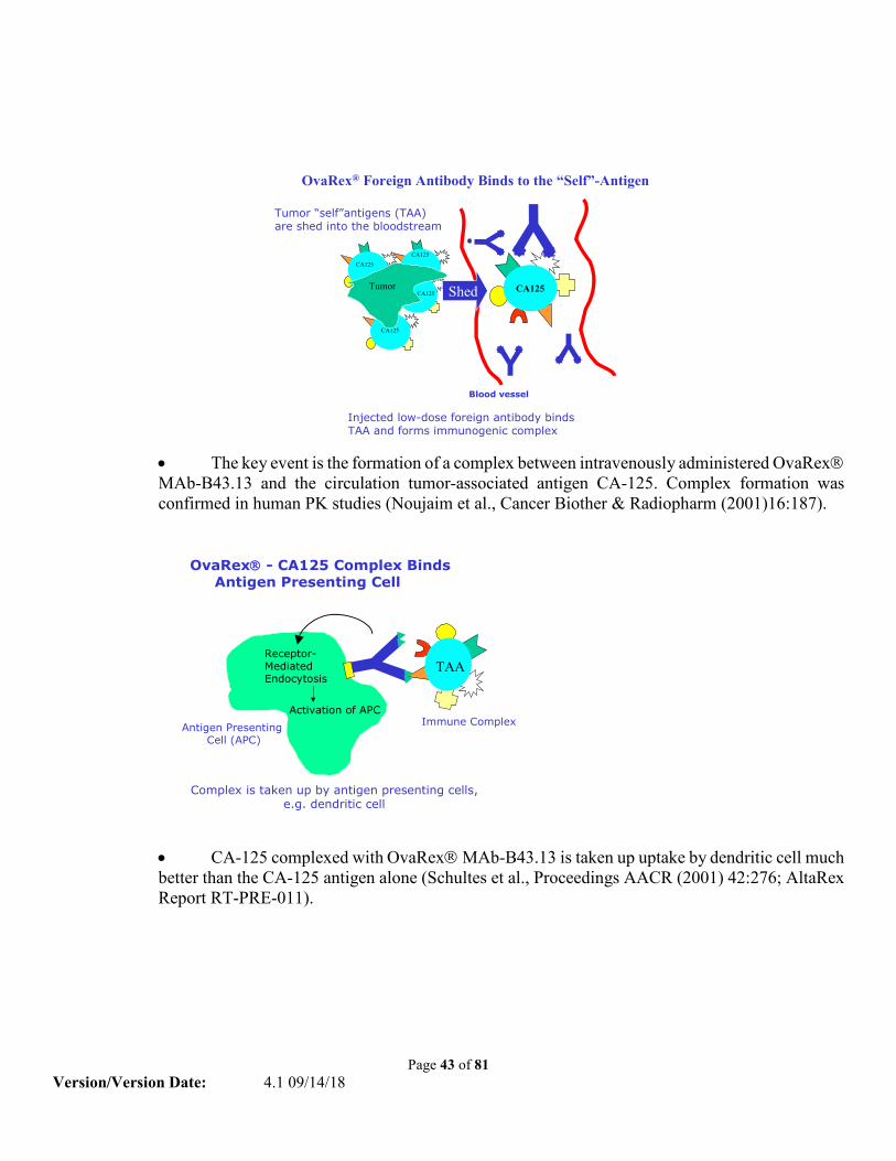
Oregovomab is a mouse monoclonal antibody that is specific for CA-125. When Oregovomab is injected intravenously at low doses into a patient, the antibody binds to circulating CA-125. Complexes are formed between the antibody and the CA-125 antigen (antigen-antibody complexes). These complexes are taken up by antigen presenting cells and at the same time, decrease the amount of CA-125 in the circulation. It is thought that the antigen-antibody complexes enhance the ability of the antigen presenting cells to present fragments of the CA-125 antigen on the surface of the cell and activate the antigen presenting cells to make co-stimulatory cell-surface molecules and cytokines. The combination of decreased amounts of CA-125 in the circulation and the enhanced presentation of CA-125 on activated antigen presenting cells results in a more efficient stimulation and activation of T helper cells. This would then result in a measurable immune response to CA-125 and the apparent reversal of “tolerance”.
2.4.3 Rationale for giving Oregovomab concurrently with chemotherapy
Most clinical trials with the agent have been conducted in maintenance settings when chemotherapy was not being administered (36, 37) and the magnitude of response in this clinical setting has proven inadequate to produce clinical benefit. Several reports, however, have suggested that administration of Oregovomab in association with chemotherapy may result in enhanced cellular immunity relative to the monotherapy settings (36, 38). In 2009, Braly published a randomized phase II study in which simultaneously administered Oregovomab with standard chemotherapy in a first group of patients and a week after chemotherapy in the second one. The study showed that the arm subjected to simultaneous immuno-chemotherapy developed a better immune response (contrary to what was previously thought considering the immunosuppressive effects of chemotherapy) (39). Further studies, however, are needed to completely assess the magnitude of the immune response. The measure of effectiveness of an immunotherapy in the treatment of cancer has been fraught with the inability to successfully measure direct effect on tumor burden similar to cytotoxic therapies. We therefore believe in the importance of assessing the rate of positivity obtained by ELISPOT method against Oregovomab, disease-free survival, overall survival (up to the date fixed as the last visit to complete the entire population evaluation).
2.4.4 Rationale for giving Oregovomab with the neoadjuvant regimen proposed in this study
Since the safety profile of our neoadjuvant regimen (chemotherapy with gemcitabine and 5FU followed by hypofractionated stereotactic radiotherapy concurrent with Nelfinavir) has been established in our phase I study, it provides an ideal clinical setting to study the potential benefit of addition of Oregovomab into this neoadjuvant regimen. It is possible that administration of Oregovomab with chemotherapy in patients with gross tumor may result in better immune responses due to more antigens being released by chemotherapy. Moreover, initiation of an immune response during the neoadjuvant phase may eliminate distant micrometastases that are believed to exist but have not been yet detected.
Page 10 of 81 Version/Version Date: 4.1 09/14/18
2.5 Special Studies Background
2.5.1. Rational for using real-time motion tracking during SRT The issue of respiratory motion has long been recognized as a major limitation in the management of radiotherapy patients. For patients receiving stereotactic body radiation therapy (SBRT), the dose per fraction is large. Therefore, the high-dose tumor volume has to be compact. We elect not to irradiate the lymphatic drainage area prophylactically since studies have shown that regional nodal failure is only 7-10% while local and distant failure has reached 50% (40, 41). With such a compact volume, tumor position must be accurately assessed throughout the radiation treatment, especially for the pancreatic tumor that moves with respiration. Minimizing the impact of respiratory motion is essential in order to achieve further gains in the treatment of pancreatic disease. In this study, we will apply advanced imaging and delivery technology to provide added confidence in imaging and targeting. All patients will undergo a planning 4D CT. CT images will be reconstructed as a function of respiratory phases. End exhale images will be used for planning purposes, as this has been shown to be the most reproducible phase of respiration with the longest duration (42-48). The linear accelerators with SRT capabilitywill be used to deliver SRT. For patients consented prior to diagnostic laparoscopy, Calypso Beacon markers will be placed during the laparoscopy. The Calypso System allows for real-time tracking of tumors during cancer radiation treatment. Calypso continuously tracks the position of the tumor using an innovative technology known as “GPS for the Body®.” This tracking technology is guided by three tiny Beacon transponders — wireless devices that each are about the size of a grain of rice. The transponders transmit safe radiofrequency waves that provide sub-millimeter, real-time information about the position and movement of the tumor. For patients who consent after diagnostic laparoscopy, Calypso Beacon markers will be placed by Interventional Radiology. For those patients who can not use Calypso system, radio-opaque gold markers will be placed. The Exac Trac incorporates stereotactic x-ray capabilities for verifying target position. For soft tissue targets the system is designed to be used with radio-opaque gold markers implanted near the target. These markers are implanted prior to CT imaging and treatment planning, and should be placed close enough to the target anatomy so that they can be observed within the field of view of the x-ray localization system at the time of treatment. The use of implanted makers for radiotherapy localization has been described for a number of tumor sites, including prostate (49, 50), liver (51), lung (52, 53) and pancreas (54). When the Artiste and Truebeam linear accelerators are used to deliver radiation therapy, the maximum dimension of the tumor can be up to 10 cm. CT on rail (Artiste) or KV cone beam CT (Truebeam) together with Calypso system will be used for image-guided radiation therapy.
3.0 ELIGIBILITY CRITERIA
3.1 Inclusion Criteria
Page 11 of 81 Version/Version Date: 4.1 09/14/18
3.1.1 Pathologically confirmed adenocarcinoma of the pancreas. Patients have resectable, borderline resectable disease, or unresectable disease with no evidence of distant metastases or peritoneal disease (Resectable or borderline resectable disease is defined in Appendix A). The maximum dimension of the tumor must be ≤ 10 cm.
3.1.2 Age: Patients must be 19 years of age or older. (This is the age of consent in Nebraska. Pancreatic cancer does not occur in the pediatric age group.)
3.1.3 Karnofsky Performance Status of 60% or better. (Appendix B) 3.1.4 Patients who received chemotherapy > 5 years ago for malignancies other than
pancreatic cancer are eligible, provided that chemotherapy was completed > 5 years ago and that there is no evidence of the second malignancy at the time of study entry.
3.1.5 Patients who received radiation therapy > 5 years ago for malignancies other than pancreatic cancer and whose radiation therapy field is not overlapping with the 20% isodose line of current radiation field are eligible, provided that radiation therapy was completed > 5 years ago and that there is no evidence of the second malignancy at the time of study entry.
3.1.6 All malignant disease must be able to be encompassed within a single irradiation field.
3.1.7 All patients must have radiographically assessable disease 3.1.8 Patients must have a ANC greater than or equal to 1500/μL and platelet count
greater than or equal to 100,000/μL 3.1.9 Patients must have a serum creatinine less than or equal to 2.0 mg/dL and total
bilirubin less than or equal to 2.0 mg/dL in the absence of biliary obstruction. If the patient has biliary obstruction, biliary decompression will be required. Either endoscopic placement of biliary stent (7 French or greater) or percutaneous transhepatic drainage are acceptable. Once biliary drainage has been established, institution of gemcitabine therapy may proceed when the total bilirubin falls to <= 4.0 mg/dL. Patients with biliary or gastroduodenal obstruction must have drainage or surgical bypass prior to starting chemoradiation.
3.1.10 The patient must be aware of the neoplastic nature of his/her disease and willingly provide written, informed consent after being informed of the procedure to be followed, the experimental nature of the therapy, alternatives, potential benefits, side-effects, risks, and discomforts.
3.1.11 No prior therapy with the exception of 1 cycle of chemotherapy based on current diagnosis and clinical condition.
3.1.12 Patients must have CA125 level ≥10 to participate in the immunotherapy aspect of the trial and receive oregovomab. If the patient has CA125 > 10 who is not eligible to receive oregovomab (e.g. allergic to the drug) but is eligible for the rest of treatment, this patient should be accrued to the part of protocol without oregovomab.
Page 12 of 81 Version/Version Date: 4.1 09/14/18
3.2 Exclusion criteria 3.2.1 Patients who cannot undergo staging laparoscopy. For example, this may include
patients with a prior history of multiple abdominal operations in which laparoscopy may not be technically feasible or potentially harmful. The patient is eligible if they have a common bile duct stent adjacent to the tumor that may be used as an internal marker, or if the patient has already had a staging laparoscopy without marker implantation and the markers can be implanted (by interventional radiology) prior to the beginning of radiation therapy.
3.2.2 Patients with a known allergy to murine proteins or have had a documented anaphylactic reaction or allergy to any of chemotherapy agents used in this protocol, oregovomab, or to antiemetics appropriate for administration in conjunction with protocol-directed therapy.
3.2.3 Uncontrolled inter-current illness including, but not limited to ongoing or active infection requiring intravenous antibiotics, symptomatic congestive heart failure, unstable angina pectoris, or serious, uncontrolled cardiac arrhythmia, that might jeopardize the ability of the patient to receive the therapy program outlined in this protocol with reasonable safety.
3.2.4 Pregnant and nursing women are excluded from this study because the chemotherapy agents, Oregovomab, Nelfinavir and abdominal radiation therapy all have the potential for teratogenic or abortifacient effects.
3.2.5 Patients with prior malignancy will be excluded except for adequately treated basal cell or squamous cell skin cancer, adequately treated noninvasive carcinomas, or other cancers from which the patient has been disease-free for at least 5 years.
3.2.6 Patients with active duodenal ulcer or bleeding or history of a gastrointestinal fistula or perforation or other significant bowel problems (severe nausea, vomiting, inflammatory bowel disease and significant bowel resection).
3.2.7 Patients with known HIV infection, or hepatic insufficiency. 3.2.8 Patients who cannot take oral medications. 3.2.9 Patients may not be receiving or have received any other investigational agents
during/or within 1 month prior to treatment with Oregovomab or Nelfinavir. 3.2.10 Patients with an active autoimmune disease (e.g., rheumatoid arthritis, systemic
3.2.11 Patients with a recognized acquired, hereditary, or congenital immunodeficiency disease including cellulaimmunodeficiency’s, hypogammaglobulinemiaor dysgammaglobulinemia.
3.2.12 Patients receiving the following drugs that are contraindicated with NFV (VIRACEPT) will be excluded if they cannot be change or discontinued.
Page 13 of 81 Version/Version Date: 4.1 09/14/18
3.2.13 Patients receiving the following drugs will be clinically evaluated as to whether
dosage/medication can be changed to permit patient on study:
3.3 Inclusion of Women and Minorities
Both men and women and members of all races and ethnic groups are eligible for this trial.
4.0 REGISTRATION PROCEDURES All patients with pancreatic cancer or suspicious pancreatic masses referred to the Nebraska Medical Center (NMC) / UNMC are evaluated in a multidisciplinary team conference. On initial presentation, a history and physical examination are performed, laboratory data obtained, and performance status is assessed. Imaging studies obtained include a high-resolution multi-detector computed tomography (CT) of the abdomen and pelvis as well as a chest radiograph (two views). Further imaging studies will be obtained as clinically indicated. Any pathologic specimens obtained at referring institutions are reviewed for accuracy. Patients with suspicious pancreatic masses will require pancreatic biopsy for confirmation of malignancy. Biopsy techniques available include percutaneous, endoscopic ultrasound guidance, and
Page 14 of 81 Version/Version Date: 4.1 09/14/18
laparoscopic. Patients without evidence of metastatic disease on imaging studies will be evaluated for potential resectability (NCCN Guidelines- Appendix A). Imaging studies available for use in defining resectability include CT scan (MRI scan will be done if CT scan is not sufficient for evaluating the resectability), endoscopic ultrasound, and laparoscopic ultrasound. The CT scan obtained is an imaging protocol specifically designed to evaluate the pancreas using a triple phase contrast scan with thin (3 mm) cross-sectional images with 3-dimensional reconstruction availability. CT mesenteric angiography will be obtained when clinically indicated. Endoscopic ultrasound and laparoscopic ultrasound will be utilized when additional information regarding tumor relationship with surrounding vascular structures, or when diagnostic tissue samples are required. Patients with resectable or borderline resectable pancreatic adenocarcinoma who meet the eligibility criteria will be offered participation in the treatment portion of this trial. More subjects will participate in this pre-therapy evaluation phase than will ultimately be found to be suitable candidates for the chemotherapy/radiation therapy. Patients without evidence of metastatic disease on imaging studies will then undergo laparoscopy to exclude the presence of occult metastatic disease. Routine inspection of the liver, peritoneal surfaces and serosa of abdominal viscera will be performed. Biopsy will be performed of suspicious lesions for histologic examination. Patients without peritoneal disease will have Calypso Beacons placed around the pancreatic cancer during laparoscopy. Patients who undergo a laparoscopy at an outside facility or have laparoscopy done prior to consent will have Beacons placed by Interventional Radiology prior to Radiation Therapy. When Beacons are not available, gold markers will be placed. The standard of care outside clinical trials setting for patients with localized or locally advanced pancreatic cancer with resectable disease is surgery followed by adjuvant therapy. In patients with locally advanced pancreatic cancer that is potentially resectable (provided an antitumor response is obtained) or unresectable, palliative chemotherapy alone or in combination with palliative radiation therapy is considered the standard of care. NOTE: Before patients are enrolled into the treatment portion of the study, an eligibility checklist (Appendix C) must be completed to verify the subject meets the eligibility and may be used as source documentation if it has been reviewed, signed, and dated prior to registration by the treating physician. Some insurance carrier’s may decline to cover the costs of usual medical care if the patient is participating in a clinical trial. The patient will be provided assistance by the research nurse coordinator in determining if the insurance carrier will decline coverage. Insurance carriers may or may not pay for study related expenses. The patient can then decide if they wish to participate.
4.1 Eligibility Verification/Registration Before patients are enrolled into the study, an eligibility checklist (Appendix C) must be completed to verify the subject meets the eligibility criteria. Date of enrollment is defined as the date of the start of study treatment / first protocol related intervention. The eligibility check list will be maintained in the study file. Study personnel will provide the UNMC Fred & Pamela Buffett Cancer Center PRMS office (ZIP 6805)
Page 15 of 81 Version/Version Date: 4.1 09/14/18
a copy of the signed and dated consent form for each UNMC subject registered to the protocol within 7 days that includes the following information:
• Protocol Number • Patient Identification: Patient’s name NMC medical record number • Patient demographics: gender, birth date (mm/dd/yyyy), race, ethnicity
5.0 TREATMENT PLAN
5.1 Draw Baseline CA125 level for research purposes Research(Res) Patients with a CA125 level ≥10 will participate in the immunotherapy aspect of the trial (see Section 5.5) and receive oregovomab. If the patient has CA125 > 10 who is not eligible to receive oregovomab (e.g. allergic to the drug) but is eligible for the rest of treatment, this patient should be accrued to the part of protocol without oregovomab.
5.2 Biospecimen Samples for research purposes only (Res) Patients who have CA125 level ≥10 will participate in the immunotherapy and immunologic assessment aspect of the trial and receive Oregovomab. A Blood Draw for immunological assessment of Oregovomab will be done Pre-chemotherapy, Prior to second infusion for the first 6 obtainable patients, Pre-SRT, Pre-surgery, and then prior to restarting chemotherapy after surgery/post restaging if not resectable and 3 weeks post cycle 7 chemotherapy (week 12) or end of study. An excess tumor tissue sample for immunological assessment of Oregovomab will be obtained s after the surgical resection. SEE APPENDIX D for instructions.
5.3 Staging laparoscopy and marker implantation Standard of Care (SOC) (day -3 to day -30) As stated above in Section 3.0, patients without evidence of metastatic disease on imaging studies will undergo laparoscopy to exclude the presence of occult metastatic disease. Routine inspection of the liver, peritoneal surfaces and serosa of abdominal viscera will be performed. Biopsy will be performed of suspicious lesions for histologic examination. Patients without peritoneal disease will have placement of Beacons around the pancreatic cancer during the procedure if they have already consented to participate in the study. The staging laparoscopy must be done within 30 days of treatment start. All patients CA125 positive (>10) or CA125 negative (<10) who have no peritoneal metastasis will undergo 4 cycles of neo-adjuvant therapy. The CA125 must be drawn within 10 days of treatment start. Patients who undergo a laparoscopy at an outside facility or have a laparoscopy done prior to consent, will have Beacons placed by Interventional Radiology prior to Radiation Therapy. The Beacons will be placed by Interventional Radiology during chemotherapy cycles. Prophylactic antibiotics will be given as necessary to prevent infection. Beacon position and geometry will be documented on a post-chemotherapy restaging CT scan. When Beacons are not available, gold markers will be placed.
5.4 Chemotherapy (SOC)
ASSESSMENT REMINDERS: Patients will be assessed and asked to complete a pain scale assessment tool (Appendix F) prior to the start of each chemotherapy cycle, 3-4 weeks post radiation, and 2-4 weeks
Page 16 of 81 Version/Version Date: 4.1 09/14/18
post-surgery. Week 1-9, Cycles 1-3 / Week 14-16, Cycle 4 / then resume Post-op/ post restaging, Cycles 5-7: Gemcitabine 750 mg/m² (females) or 900 mg/m² (males) IV as fixed dose rate infusion on days 1, 8 Calcium Leucovorin 50 mg/m² IV over 30 minutes on days 1, 8 5-Fluorouracil 2700 mg/m² IV over 24 hours starting on days 1, 8 Post-operative RECOVERY / Post-restaging unresectable: An additional three cycles (Cycles 5-7) of adjuvant chemotherapy to start when the clinician’s determine that the patient has recovered from surgery OR if the patient was unresectable and the clinician’s determine that chemotherapy should be resumed. A window of -2 up to +7 days will be allowed to start planned cycles of therapy provided all other criteria to restart the new cycle have been met. Appropriate dose modifications are described in Section 5.10. Week 8: Cycle 3 day 8 prior to NFV: CT scan of the chest will be performed for restaging; an MRI scan of the abdomen will be done for restaging and SRT planning. Patients with no metastasis will proceed with SRT.
5.5 Administration of Immunotherapy (Oregovomab) and Immunologic Assessment (Res) ASSESSMENT REMINDERS: If the patient has CA125 > 10 who is not eligible to receive oregovomab (e.g. allergic to the drug) but is eligible for the rest of treatment, this patient should be accrued to the part of protocol without oregovomab. Immunologic Assessment, patients who have CA125 level ≥10 will participate in the immunotherapy and immunologic assessment aspect of the trial and receive Oregovomab. A Blood Draw (appendix D) for immunological assessment of Oregovomab will be done Pre-chemotherapy, Prior to second infusion for the first 6 obtainable patients, Pre-SRT, Pre-surgery, and then prior to restarting chemotherapy after surgery/post restaging if not resectable and 3 weeks post cycle 7 chemotherapy (week 12) or end of study
Week 1-9, Cycles 1-3 / Week 14-16, Cycle 4 / then resume Post-op/ post restaging, Cycles 5-7: Oregovomab 2 mg IV over 20 min (in any case no less than 15 minutes and no greater than 30 minutes) on day 15 x 4 Cycles. Repeat at 3 week intervals x 3 Cycles (weeks 3, 6, 9) and then post SRT Cycle 4, week 16. Post-operative RECOVERY / Post-restaging unresectable: An additional three cycles (Cycles 5-7) of adjuvant chemotherapy to start when the clinician’s determine that the patient has recovered from surgery OR if the patient was unresectable and the clinician’s determine that chemotherapy should be resumed. We will try to minimize the use of corticosteroids. Appropriate dose modifications are described in Section 5.10. No investigational or commercial agents or therapies other than those described in this protocol may be administered with the intent to treat the patient's malignancy.
5.6 Administration of Nelfinavir (NFV) (Res)
Page 17 of 81 Version/Version Date: 4.1 09/14/18
Week 9-13: Start Nelfivavir (NFV) 1250 mg orally twice daily starting the Monday of Cycle 3 day 15(week 9), (2 weeks prior to the initiation of SRT) CONTINUE for 5 weeks to discontinue the Friday 2 weeks after SRT has completed (day 57-91). Treatment will be administered on an outpatient basis. Reported adverse events and potential risks are described in Section 8.Appropriate dose modifications are described in Section 5.10. No investigational or commercial agents or therapies other than those described in this protocol may be administered with the intent to treat the patient's malignancy. The patient will be given a “medication information sheet” at the beginning of their Nelfinavir treatment. The patient will also be asked to maintain documentation of medication adherence by filling out a calendar of when and how many tablets of Nelfinavir were taken. The patient will be asked to bring the calendar back along with any leftover medication at their convenience at the end of treatment. Week 9-10: Treatment planning for Stereotactic Radiotherapy (SRT).
5.7 Stereotactic Radiotherapy (SRT) (SOC) Week 11: Start Stereotactic Radiotherapy (SRT) the total dose of 40 Gy will be delivered in 5 Fractions over 5 consecutive days for 1 week (details of SRT are provided in Section 5.9). Continue Nelfinavir. Week 12: Stop SRT and continue Nelfinavir for 14 more days (week 12-13)
5.8 Surgery (SOC) Week 16-17: Restaging, a CT scan of the chest and abdomen will be performed for restaging 5-6weeks after completing SRT (week 16-17) to assess disease response. An MRI scan may be performed if CT scan is not sufficient for assessing the resectability. If surgical resection is not possible and the clinician determines that chemotherapy should be resumed, patients will receive an additional three cycles of chemotherapy to start when the clinician determine that chemotherapy should be resumed. Week 17-18: Surgery (if resectable or potentially resectable) Patients without metastasis and with resectable or borderline resectable disease will undergo definitive surgery. If no contraindication for surgical resection is identified, resection will be performed 6-8 weeks after completing SRT. At the time of surgical resection, an extensive examination of the abdomen will be performed to exclude the presence of metastatic disease. All operations will be performed with curative intent with resection of all gross tumor (ie R0 [negative margins]. Resection of adjacent involved organs or vascular structures will be performed as clinically indicated. Standardized histopathologic analysis of resected specimens will be performed. Margins to be evaluated include: common bile duct, pancreatic, retroperitoneal (tissue between superior mesenteric artery and duodenum), as well as tissue along the superior mesenteric vein and artery. Examination of regional lymphatics will be performed according to standard pathology techniques.
Page 18 of 81 Version/Version Date: 4.1 09/14/18
Upon recovery from surgery, the patients will receive three additional cycles (Cycles 5-7) of adjuvant chemotherapy with gemcitabine/leucovorin/5-FU. Patients who have CA125 level ≥10 will continue to participate in the immunotherapy and immunologic assessment aspect of the trial and receive Oregovomab on day 15 of these additional 3 cycles and a Blood Draw for immunological assessment will be done prior to restarting chemotherapy and at the end of study (week 12 of adjuvant therapy). Patients will then be followed at 3 month intervals with a history and physical exam, CT scan of the chest/abdomen/pelvis, and lab work.
5.9 Details of SRT
The SRT treatment will consist of image-guided radiotherapy.
5.9.1 Patient’s positioning The treatment position of the patient is supine, with the arms placed above the head. The immobilization device (thermoplastic mask) will include total body to make sure that the patients’ position is the same during planning, simulation and treatment. 5.9.2 Patient data acquisition. Treatment planning 4D CT scans are required to define tumor, clinical, and planning target volumes. The treatment planning 4D CT scan with IV contrast will be acquired with the patient in the same position and immobilization device as for treatment. All tissues to be irradiated will be included in the CT scan. CT scan thickness should be ≤ 3 mm through the region that contains the primary target volumes. Conventional MRI scans (T1 and T2) may be included to assist in definition of target volumes. FDG PET-CT information may be included in the treatment planning; no extra scans will be performed for study purposes. The GTV, CTV and PTV, and normal tissues (OAR) must be outlined on all CT slices in which the structures exist. 5.9.3 Volumes Definition of volumes: The definition of volumes will be in accordance with the 1993 ICRU Report #50:
5.9.4 Prescribing, Recording and Reporting Photon Beam Therapy. The Gross Tumor Volume (GTV) is defined as all known gross disease determined from CT, clinical information, endoscopic findings, FDG PET-CT and/or conventional MRI. The Integrated Tumor Volume based on CT/MRI/PET (GTVfusion) is defined as gross disease on the free breathing CT scan, MRI scan and FDG-PET scan. These scans will be correlated by imaging fusion technique. The volume will be delineated by the treating physician on the above scans separately. The GTVCT, GTVMRI and GTVPET (if done) will be eventually fused together to generate GTVfusion. Patients who have the maximal dimension of the GTVfusion > 8 cm will not be eligible for the study.
Page 19 of 81 Version/Version Date: 4.1 09/14/18
The Clinical Target Volume (CTV) is defined as the GTVs plus areas considered to contain potential microscopic disease. In this study, we have no intension to treat the potential microscopic disease with SRT, therefore, the CTV is defined as GTVs (i.e. both the primary tumor and the lymph nodes containing clinical or radiographic evidence of metastases) and areas between GTVs. The integrated CTV is created with 4D CT information to compensate internal organ motion. The Planning Target Volume (PTV) will provide a margin around integrated CTV to compensate for the variability of treatment set-up and internal organ motion. 5.9.5 Organs at Risk (OAR) The normal tissue volumes to be contoured include:
o the skin surface (the tissue within the skin surface and outside all other critical normal structures and PTVs is designated as unspecified tissue)
o spinal cord (spinal cord contours will be defined at least 5 mm larger in the radial dimension than the spinal cord itself, i.e. the cord diameter on any given slice will be 10 mm larger than the cord itself)
o duodenum o stomach o liver o right kidney o left kidney o small bowels exclude duodenum o spleen
5.9.6 The treatment technique. The linear accelerators that have SRT capability will be used to deliver SRT. The ExacTrac incorporates stereotactic x-ray capabilities for verifying target position. This consists of two floor mounted x-ray tubes and two opposing amorphous silicon (aSi) flat panel detectors mounted to the ceiling. Each x-ray tube/detector pair is configured to image through the linac isocenter with a coronal field of view of approximately 18cm in both the superior-inferior (S-I) and left-right (L-R) directions at isocenter. For soft tissue targets the system is designed to be used with radio-opaque gold markers or Beacon markers implanted near the target. These markers are implanted prior to CT imaging and treatment planning, and should be placed close enough to the target anatomy so that they can be observed within the field of view of the x-ray localization system at the time of treatment. Specific patient breathing characteristics is determined during 4D CT. If the breathing pattern is adequate, respiratory-gated delivery, which is, turning the beam on only at a specified phase of respiration will be determined. This “freezes” target motion and allows reduction of beam margins, thereby reducing the amount of irradiated normal tissue (in this case, normal liver). The Novalis system is well suited to gated delivery and has been evaluated extensively by Tenn et al [48]. The following is a brief procedural summary from that work which will be incorporated into this study: The patient is set up in the treatment room and IR reflective markers with adhesive bases are attached to their anterior surface so that breathing motion can be monitored. A second
Page 20 of 81 Version/Version Date: 4.1 09/14/18
set of IR reflective markers is rigidly attached to the treatment couch and used as a reference against which the movement of patient markers is measured. These rigidly mounted reflectors are also used to track couch location during the patient positioning process. The 3D movement of the patient’s anterior surface is tracked via the IR markers and the anterior-posterior (A-P) component of this trajectory is used to monitor breathing motion. The system plots breathing motion versus time and a reference level is specified on this breathing trace. This designates the point in the breathing trace at which the verification x-ray images will be triggered. The two images are obtained sequentially at the instant the breathing trace crosses this level during exhale phase. Because the patient is localized based on these images, the gating level is set at the same phase in the breathing cycle at which the planning CT data was obtained. Within each image the user locates the positions of the implanted markers. From these positions the system reconstructs the 3D geometry of the implanted markers and determines the shifts necessary to bring them into alignment with the planning CT. The patient is subsequently positioned according to the calculated shifts. Finally, a gating window (beam-on region) during which the linac beam will be delivered is selected about the reference level. The system can gate the beam in both inhale and exhale phases of the breathing cycle. Subsequent x-ray images verifying the location of the implanted markers locations are obtained at the gating level continuously during treatment. If marker positions remain within tolerance limits the target position may also be assumed to be correctly positioned. If they are outside the limit, the newly obtained images can be used to reposition the patient and maintain treatment accuracy. When the Artiste and Truebeam linear accelerators are used to deliver radiation therapy, the maximum dimension of the tumor can be up to 10 cm. CT on rail (Artiste) or KV cone beam CT (Truebeam) together with Calypso system will be used for image-guided radiation therapy. When Beacons are not available, gold markers will be used. 5.9.7 Dose computation. The treatment plan used for each patient will be based on an analysis of the volumetric dose, including DVH analyses of the PTV and critical normal structures. Treatment planning should be accomplished with multiple coplanar/noncoplanar conformal beams or arcs to allow for a high degree of dose conformality. The uniformity requirement will be +10% -5% of the total dose at the prescription point within the tumor volume. The IMRT may be used if there is a benefit of decreasing tissue complications. 5.9.8 Equipment and tools. Beam’s Eye View techniques will be used to select the beam isocenter and direction to fully encompass the target volume but minimizing the inclusion of the critical organs in order to select the plan that minimizes the dose to normal tissues. 5.9.9 Dose specification.
5.9.9.1 Dose prescription. The prescription dose is the isodose which encompasses at least 95% of the planning target volume (PTV). Prescription dose to the PTVs shall be according to the following:
Page 21 of 81 Version/Version Date: 4.1 09/14/18
The gross tumor and gross lymph node metastasis will receive a total 40 Gy. DVHs must be generated for all critical normal structures (OAR): The dose to the kidney will require careful monitoring and kidney volumes must be defined on simulation fields. The percent of total kidney volume (defined as the sum of the left and right kidney volume) receiving 15 Gy (3 Gy per fraction) should be required to be less than 35% of the total kidney volume. The maximum dose to any point within the spinal cord should not exceed 15 Gy (3 Gy per fraction). At least 700 ml or 35% of normal liver (entire liver minus cumulative GTV) should receive at total dose less than 15 Gy (3 Gy per fraction). The maximum point dose to the stomach or small bowel except duodenum should not exceed 80% of prescription dose. An isodose distribution of the treatment at the central axis indicating the position of kidneys, liver and spinal cord is required.
5.9.9.2 Dose recording.
The reported doses for each PTV shall include the prescription dose as well as the maximum point dose, % target volume receiving > 110% and >115% of its prescribed dose and the % target volume receiving < 93% of the prescribed dose, and the mean dose to the PTV. Doses to the organs at risk will also be recorded.
5.9.9.3 Dose homogeneity. No more than 20% of any planning target volume (PTV) will receive >110% of its prescribed dose. No more than 1% of any planning target volume (PTV) will receive <93% of its prescribed dose. No more than 1% or 1 cc of the tissue outside the PTVs will receive >110% of the dose prescribed to the primary PTV. 5.9.10 Fractionation schedule.
The total dose of 40 Gy will be delivered in 5 Fractions over 5 consecutive days. 5.9.11 Treatment Verification. The location of the implanted markers will be verified on daily x-rays. 5.9.12 Quality Assurance Documentation A copy of the daily treatment record will be maintained in the radiation oncology department. Isodose distribution at the central axis will be recorded. Exact track IGRT data will be collected. Breath gating will be used and data will be collected. 5.9.13 Radiation modification for hematologic toxicity Blood counts will be measured once weekly. Radiation will not be started if the AGC is < 1000/μl and platelets are < 50,000/μl, and the start of radiation will be delayed until the blood counts are above this level.
5.10 Dose Modifications (The NCI Common Toxicity Criteria version 4.0, APPENDIX E, will be used, available at http://ctep.cancer.gov/reporting/ctc_v40.html).
For all cycles, the planned treatment dosing may be either interrupted or omitted for significant treatment-related toxicities occurring on the day of or in the prior 24 hr of planned therapy: platelet count < 25,000/μL, AGC < 500/μl, grade 2 or worse non-hematologic toxicity (excluding nausea & vomiting on sub-optimal anti-emetic therapy, alopecia, elevated transaminases).
At the start of each new cycle, dose adjustments may also be made based upon significant toxicities occurring at any time in the previous cycle.
5.10.2 Initiation of Next Cycle of Gemcitabine/5-FU/LV The next treatment cycle will commence 22 days after the start of the prior cycle provided that the absolute granulocyte count is greater than or equal to 1500/μl, the platelet count is greater than or equal to 75,000/μl, and all clinically significant treatment-related non-hematologic toxicities have resolved to no more than grade 1 in severity. If multiple toxicities have been seen in the preceding cycle, the dose administered should be based on the most severe toxicity experienced.
5.10.3 Dose Reductions for Hematologic Toxicity Occurring During the Prior Cycle The dose-limiting toxicity of gemcitabine is myelosuppression, while grade 4 myelosuppression with a weekly 24-hr infusion of 5-FU/LV is uncommon. Therefore, the dose of gemcitabine will be preferentially reduced for myelosuppression. A 1-week delay will be allowed to permit recovery of counts, then the patient should be treated according to the Dose Modification table below. The dose of LV will not be adjusted for myelosuppression.
Therapy (Tx) delay
Day
AGC on Tx day (/μL)
Plt on Tx day (x10/μL)
AGC nadir (/μL)
Plt nadir (x10/μL)
Gemcitabine Dose
5-FU Dose
No
22
>/= 1500&
>/= 75
>/= 500 &
>/= 25
Same
Same
No
22
>/=1500 &
>/=75
< 500 or
< 25
Decrease 20%
Same
1 week 29 >/=1500 & >/=75 >/= 500 &
>/=25
Same
Same
1 week 29 >/=1500 & >/=75 < 500 or
< 25
Decrease 20%
Same
1 week
29
1000-1499 or
50-74.9
>/= 500 &
>/=25
Decrease 20%
Same
1 week
29
1000-1499 or
50-74.9
< 500 or
< 25
Decrease 40%
Same
1 week
29
< 500 or
< 50
Any
Any
Hold*
Hold*
During the neoadjuvant period, if chemotherapy cannot be resumed after a one-week delay, then the patient should proceed to NFV and SRT once the AGC is 1000/μL or higher and the platelets are 50,000/μL or higher.
Page 23 of 81 Version/Version Date: 4.1 09/14/18
5.10.4 Dose Reductions for Post-Operative Cycles of Gemcitabine/5-FU/LV Hematologic Toxicity
Day
AGC on Tx day (/μL)
Plt on Tx day (x10/μL)
AGC nadir (/μL)
Plt nadir (x10/μL)
Gemcitabine Dose
5-FU Dose
22
>/= 1500 &
75
>/= 500 &
>/= 25
Same
Same
22
>/= 1500 &
>/= 75
< 500 or
< 25
Decrease 20%
Same
Therapy delay Day
AGC (/μL) Platelets
(x10/μL) Gemcitabine Dose
5-FU Dose
29
<1500 or
<75
Delay 1 week
Delay 1 week
36
1000-1499 or
50-74.9
Decrease 20%
Same
36
< 1000
< 50
Hold
Hold
43
1000-14 or
50-74.9
Decrease 40%
Same
43
< 1000 or
< 50
Discontinue
Decrease 20%
5.10.5 Dose Modifications for Treatment-Related Non-Hematologic Toxicity Occurring the Prior Cycle of Gemcitabine/5-FU/LV The dose of 5-FU will be preferentially decreased for treatment-related mucositis or diarrhea. It will also be decreased for subjects who have recovered from treatment-related initial cardiotoxicity with preventive anti-angina therapy (calcium channel agonists, beta-blockers and nitrates). If mucositis or diarrhea recurs despite the 5-FU dose reduction, then the dose of Gemcitabine will also be decreased the subsequent cycle. The dose of Gemcitabine will be preferentially reduced for side effects that are primarily associated with Gemcitabine and uncommon with 5-FU (e.g., pulmonary, renal). If the patient develops signs of microangiopathic hemolytic anemia, Gemcitabine should be discontinued. Dose reduction for grade 3 nausea/vomiting will only be made if adequate anti-emetics were used. If anti-emetic therapy was suboptimal, adjust the antiemetic regimen and try the same chemotherapy doses.
Non-Hematologic Toxicity Toxicity Type
Toxicity Grade
Week 2 Tx held for toxicity?
Gemcitabine Dose
5-FU Dose
Diarrhea or
2
No
Same
Same
Page 24 of 81 Version/Version Date: 4.1 09/14/18
Mucositis 2
Yes
Same unless toxicity recurs despite 5-FU decrease, then decrease 20%
Decrease 20%
3
Either
Same unless toxicity recurs despite 5-FU decrease, then decrease 20%
Decrease 20%
Other
2
No
Same
Same
2
Yes
Same unless toxicity recurs despite 5-FU decrease, then decrease 20%
Decrease 20%
3
Either
Same unless toxicity recurs despite 5-FU decrease, then decrease 1 level
Decrease 20%
5.10.6 Dose Modifications for NFV Because the duration of nelfinavir therapy is limited, there are no planned dose modifications for grade 1-2 toxicities attributed to nelfinavir. In the event of grade 3 or worse toxicity attributed to nelfinavir, the drug will be held until the toxicity is grade 2 or less in severity, then the drug will be resumed at 650mg PO BID.
5.10.7 Dose Modification for SRT Because only 5 daily doses of SRT are planned, the radiation will be interrupted only if the patient experiences grade 3 or worse non-hematologic toxicity or grade 4 hematologic toxicity during the period in which SRT is administered. CBC and differential will be measured pre- and post-SRT.
5.10.8 Treatment of Allergic Adverse Events/ Dose Modifications for Oregovomab
The dose of Oregovomab will be administered by qualified, experienced nursing personnel under the supervision of a physician via a slow intravenous infusion to the patient in an appropriate treatment area.
An emergency crash cart will be present in the treatment area for immediate use in case of a life-threatening allergic reaction such as an anaphylactic reaction. The crash cart will contain instruments and medications in working order and within expiration dates suitable for the management of medical emergencies with particular reference to anaphylactic reactions, which may be associated with the administration of Oregovomab.
A physician will be immediately available for emergency treatment of the patient in the event of such a life-threatening reaction. The patient will then be continuously monitored until they are stabilized.
Page 25 of 81 Version/Version Date: 4.1 09/14/18
A physician will be present during the infusion period for patients who have previously experience NCI Common Toxicity Criteria Grade 2 allergic events, specifically bronchospasm, and subsequently receive additional doses of study medication.
Patients who experience NCI Common Toxicity Criteria Grade 3 or 4 allergic reactions will receive no further infusions of the study medications but will be followed for outcome.
5.11 Supportive Care Guidelines 5.11.1 Prophylactic Anti-Emetic Premedication Patients must be pre-medicated for nausea & vomiting with the following antiemetic regimens as outlined below. These recommendations follow the ASCO guidelines for the use of anti-emetic therapy (55): Gemcitabine/LV/5-FU: Dexamethasone 8 mg PO (or equivalent) 30 min prior to gemcitabine Radiation to upper abdomen: ondansetron 8 mg or granisetron 2 mg PO daily (or equivalent) < 1 hr prior to radiation, and Prilosec 20 mg PO daily(or equivalent) < 1 hr prior to radiation. Alternative anti-emetic agents:
• palonsetron 0.25 mg IV pre-therapy (or equivalent) • prochlorperazine 5-10 mg PO or 5-10 mg IV q 6-8 hr • promethazine 12.5-25 mg PO/PR/IM/IV q 4-6 hr • lorazepam 1-2.5 mg PO or IV given the night before and just after chemo • ondansetron 8 mg PO or IV
5.11.2 Diarrhea Patients will be instructed to begin taking loperamide after the first poorly formed or loose stool or first episode of 2 or more bowel movements in one day. Loperamide should be taken in the following manner:
*4 mg at the first onset of diarrhea *then, 2 mg every 2 hours around-the-clock until diarrhea-free for at least 12 hours
*Patients may take loperamide 4 mg every 4 hours during the night *Loperamide should not be taken prophylactically
Patients must notify the research team as to when they initiated loperamide therapy. If diarrhea persists despite loperamide therapy, then the patient should be evaluated for the need for IV fluid & electrolyte replacement.
Alternative anti-diarrhea agents: • Somatostatin analog (Octreotide7) 100 - 500 mcg SC/IV tid; maximum daily dose
= 1500 mcg/day; alternatively, somatostatin analog may be given at 25-50 mcg/hour as a continuous IV infusion.
Page 26 of 81 Version/Version Date: 4.1 09/14/18
• Atropine/diphenoxylate which is available as either a 0.025/2.5 tab, or 0.025/2.5 per 5 mL liquid. Patients should take 1-2 tabs PO tid or qid or 5-10 mL PO tid/qid.
• Atropine/difenoxin (Motofen7) 0.025/1 tab; 2 tabs PO x 1, then 1 tab PO q 2-4 hr (max 8 tabs per day)
• Paregoric: (an antidiarrheal opiate): 5 - 10 mL ORALLY 1-4 times daily: maximum 40 mL/day
• OTC meds: bismuth subsalicylate 262 mg tabs: 2 tabs PO q 1 hr prn; maximum 4200 mg/24 hr
5.11.3 Stomatitis Mild symptoms will be treated with topical antiseptic & analgesic agents. Various versions of "magic mouthwash" exist, and represent a combination product for the topical treatment of mild oropharyngeal pain. Such mouthwashes usually consist of viscous lidocaine, diphenhydramine, aluminum and magnesium hydroxide, 70% sorbitol, and orange flavoring. Both viscous lidocaine and diphenhydramine have local anesthetic properties, while the aluminum hydroxide component of Maalox has a beneficial drying property. Patients with severe pain require systemic narcotic analgesics. Topical anti-fungal agents will be added as clinically indicated. More severe symptoms will require all of the above as well as stronger analgesic agents & admission to hospital for IV fluid & electrolyte replacement if dehydrated. 5.11.4 Requirement for Venous Access Central venous access is required for protocol participation. A previously placed central venous access device that is functioning properly (free infusion of saline, unimpeded blood return, good condition of external appliance, no recent history of device infection or thrombosis) can be used. Should the patient require a central venous access, an implanted port central venous access device will be placed (e.g. a Medi-Port, Port-a-Cath, Power Port) after appropriate informed consent has been obtained. Other aspects of catheter/port management will be in accordance with standard nursing clinic central venous port procedures. 5.11.5 Treatment of Fever & Neutropenia Subjects developing a fever of 100.5º C or higher will have a CBC with WBC differential obtained along with a history & physical examination to look for signs of infection.If the ANC is < 500/μL, the patient will be treated with empiric antibiotic therapy as an inpatient & undergo appropriate radiographic & laboratory investigation for sources of infection, & development of a specific treatment plan. Fever & neutropenia occurring during a treatment cycle will require interruption of chemotherapy. The patient may resume chemotherapy at the start of the next scheduled cycle if therapy for infection has been completed & the patient meets other criteria for starting a new cycle If the ANC is between 500-1000/μL, antibiotic therapy will be instituted if there is clinical suspicion of an infection. Daily CBCs with differentials will be obtained if the patient remains febrile.
Page 27 of 81 Version/Version Date: 4.1 09/14/18
If the ANC is > 1000/μL, & there is no clinical evidence of an infection, then therapy may resume. 5.11.6 Use of Leukocyte Colony-Stimulating Factors and Erythropoetin Colony stimulating factors (CSFs) will be used as clinically indicated according to ASCO guidelines (56). Use of any CSFs must be discontinued at least 24 hours prior to initiation of the next cycle of chemotherapy & must be documented in the patient record. Epoetin will be used as clinically indicated according to ASCO guidelines (57). 5.11.7 Pancreatic Enzyme Replacement Patients with symptoms of pancreatic insufficiency should receive pancreatic enzyme supplement. 5.11.8 Nutritional Supplementation Patients will be evaluated by a nutritionist to review their caloric needs and daily caloric intake. Patients will be encouraged to supplement their nutrition with a high-protein, low fat nutritional drink during the neoadjuvant therapy phase of the trial. Strong consideration should be given for early, elective placement of an enteral feeding tube if the patient has lost 10% or more of their body weight.
5.12 Duration of Study We estimate that there will be about 66 patients enrolled into of this protocol treated over a 5 year period
5.13 Duration of Follow up If the patient undergoes surgical resection, after completing the three planned cycles of adjuvant chemotherapy, the patient will then be seen every 3 months for the 1st year and every 4 months for 2nd year, then every six months thereafter. The patients will be assessed for long term toxicity from combination of NFV, chemotherapy, immunotherapy and radiation. Restaging radiographic scans, CBC and chemistry panel will be performed at each of these visits as part of standard of care.
Post-trial Assessments Patients who go off study treatment at any time during the trial will be followed for 30 days after the last day of treatment or until other disease-related treatment begins. For all patients, drug-related SAEs and AEs will be followed until baseline or ≤ grade 1 levels. Patients may refuse to participate in the post-trial assessments.
5.14 Criteria for Removal from Study
5.14.1 Progression of Disease
Page 28 of 81 Version/Version Date: 4.1 09/14/18
5.14.2 If at any time the constraints of this protocol are detrimental to the patient's wellbeing, or if the patient is unable to comply with the requirements of the protocol, the patient will be removed from protocol therapy. In this event, the reason(s) for withdrawal will be documented. 5.14.3 If the patient experiences an adverse reaction that, in the opinion of the investigator, necessitates the removal of the patient from the study, including any unresolved serious adverse event. 5.14.4 Any patient who suffers a serious systemic allergic response or severe degree of intolerance to the study medication will be withdrawn from further study treatment but will be followed up for a period of three years following treatment. 5.14.6 Development of intercurrent medical problems that would make continued protocol therapy detrimental to the patient’s safety. 5.14.7 There is concurrent illness or other reasons that would, in the opinion of the investigator, affect assessment of clinical status or conduct of the study to a significant degree. 5.14.8 The patient completes study treatment as per study schedule. 5.14.9 The patient chooses to discontinue treatment or follow-up. In this event, the reason(s) for withdrawal will be documented.
The reason(s) for withdrawing the patient from the treatment portion of the study will be documented in the case report form. Where possible and feasible, patients who received at least one dose of Oregovomab should be subjected to the procedures scheduled at the end of the study and should be submitted to the follow up to assess disease progression. If available, the following information will be recorded in the case report form: date of disease relapse, date of death, cause of death, and autopsy report.
6.0 Measurement of Effect 6.1 Tumor Response Measurable Disease Response: CTEP’s. Criteria for Radiographic Response Evaluation (RECIST) guidelines will be followed: A quick reference to the RECIST guidelines can be downloaded at the following URL: http://ctep.cancer.gov/guidelines/recist.html Patients enrolled in this study must have a measurable pancreatic cancer which is defined as lesions that can be accurately measured in at least one dimension: [longest diameter to be recorded] on the CT scan or MRI scan. The same method of assessment & the same technique should be used to characterize each identified & reported lesion at baseline & during follow-up.
6.2 Response criteria Taking into account the measurement of the longest diameter only for those lesions with size response, response criteria are defined as:
• Complete Response (CR): the disappearance of a lesion. • Partial Response (PR): at least a 30% decrease in the longest diameter of a lesion,
taking as reference the longest diameter recorded since the treatment started. • Progressive Disease (PD): at least a 25% increase in the longest diameter of a
lesion, taking as reference the longest diameter recorded since the treatment started. • Stable Disease (SD): neither sufficient shrinkage to qualify for partial response nor
sufficient increase to qualify for progressive disease, taking as reference the longest diameter since the treatment started.
6.3 Time to Treatment Failure The time to treatment failure will be defined as from the first date of therapy until the date the patient is removed from study for any reason.
6.4 Survival Survival will be defined as from the first date of therapy until the date the patient dies. 6.5 Toxicity criteria The NCI Common Toxicity Criteria Adverse Events version 4.0 will be used to grade toxicity; it is available at the following internet site: http://ctep.cancer.gov/forms/CTCAEv4.pdf.
6.6 Monitoring of peri-and post-marker implantation morbidity The marker position and geometry will be documented on simulation CT images. Information regarding the development of infectious complications, bleeding, and any implantation associated adverse events will be recorded.
Page 30 of 81 Version/Version Date: 4.1 09/14/18
7.0 Study Parameters Neo-Adjuvant
Screening Staging Marker BASELINE
Cycle1
Cycle 2
Cycle 3
Cycle 4
R E C O
Week 1 2 3 4 5 6 7 8 9 10 11 12 13 14 15 16 17-18
VE
Day -3 to-30 1 8 15 1 8 15 1 8 15 1 8 15 R Eligibility X Y Serum Pregnancy Test (If Female)
X
Staging Laparoscopy (within 30 days of treatment start)
X X
CA-125 X Pain assessment Appendix F prior to the start of each chemotherapy cycle, 3-4 weeks post radiation, and 2-4 weeks post-surgery
X X X X X X 2-4 wks postOR
Chemotherapy Regimen X X X X X X X X Subject with CA 125>10 Immunotherapy/Oregovomab
X X X X
Subject with CA125>10 Blood Draw for Immunologic Assessment (Res) [HAMA/CA125, ELISPOT]
X
X X X
Nelfinavir X X X X X Medication info. sheet and med adherence form given
X
Stereotactic Radiotherapy X Surgery X
Page 31 of 81 Version/Version Date: 4.1 09/14/18
History X Every 3 weeks Physical examination X Every 3 weeks Weight X Every 3 weeks Performance status (Karnofsky)
X Every 3 weeks
CBC, diff, Plt X Every week and pre and post SRT
Comprehensive metabolic panel
X Every 3 weeks
CA19-9 X Every 3 weeks
CT Chest Prior to SRT week 8 MRI abdomen optional Prior to SRT week 8
Prior to Surgery if needed, PRN X
opt
CT Chest/abdomen/pelvis with oral/IV contrast
X Prior to Surgery X
NOTE: If the patient has CA 125 >= 10 who is not eligible for receiving oregovomab (e.g. allergic to the drug) but eligible for the rest of treatment, this patient should be accrued to the part of protocol without oregovomab. In another word, patients who have CA 125 >=10 can also be accrued and treated on the part of protocol without oregovomab if they cannot receive oregovomab but no contraindications to the rest of treatment. C = Chemotherapy Regimen (SOC); Gemcitabine 750 (females) or 900 mg/m2 (males) IV by fixed dose rate infusion, leucovorin 50 mg/m² IV over 30 min, and 5-FU 2700 mg/m2 IV over 24 hrs REPEAT weekly for 2 of 3 weeks (day 1 and 8) x 4 cycles(RESUME C after SRT for Cycle 4) I = Immunotherapy and Immunologic Assessment (Res): Oregovomab 2 mg IV over 20 min will be given every 3 weeks on day 15 x 4 Cycles (RESUME I after SRT for Cycle 4) BD= Blood Draw (Res) for immunologic assessment will be done Pre-chemotherapy, Prior to second infusion for the first 6 obtainable patients, Pre-SRT, Pre-surgery NFV = Nelfinavir (Res) 1250 mg P.O. BID to start daily the Monday of week three of the third cycle of chemotherapy (2 weeks prior to the initiation of SRT) CONTINUE x 5 weeks ending the Friday 2 weeks after the end of SRT SRT = Stereotactic Radiotherapy (SOC) daily Monday-Friday x 1 week to start week 11 (SOC) OR= Surgery (SOC) to be performed sometime during week 17-18
Page 32 of 81 Version/Version Date: 4.1 09/14/18
**Post-operative RECOVERY / ** Post-restaging unresectable RESUME Adjuvant Chemotherapy: Three cycles of adjuvant chemotherapy to start when the clinician’s determine that the patient has recovered from surgery OR if the patient was unresectable and the clinician’s determine that chemotherapy should be resumed+(Approx. 1 mo post-op).
Pre Therapy
Cycle5 Cycle6 Cycle7 End of Study (week 12) or Early Termination
Follow-Up
Day 1 8 15 1 8 15 1 8 15 Pain assessment Appendix F prior to the start of each chemotherapy cycle and end of study
X
X
X
X
Chemotherapy Regimen X X X X X X Subject with CA 125>10 Immunotherapy/Oregovomab
X X X
Subject with CA125>10 Blood Draw for Immunologic Assessment (Res)
X
X
Physical examination X Every 3 weeks X X Weight X Every 3 weeks X X Performance status (Karnofsky)
X Every 3 weeks X X
CBC, diff, Plt X Every week X X Comprehensive metabolic panel
X Every 3 weeks X X
CA19-9 X Every 3 weeks X X MRI (optional, prn) X prn PRN X prn X prn CT Chest/abdomen/pelvis with oral/IV contrast
X X Every 3 months
C = Chemotherapy Regimen (SOC); Gemcitabine 750 (females) or 900 mg/m2 (males) IV by fixed dose rate infusion, leucovorin 50 mg/m² IV over 30 min, and 5-FU 2700 mg/m2 IV over 24 hrs REPEAT weekly for 2 of 3 weeks (day 1 and 8) x 3 Cycles (Cycles 5-7) CA125> 10 Subjects Resume: I = Immunotherapyand Assessment (Res): Oregovomab 2 mg IV over 20 min will be given every 3 weeks on day 15 x 3 Cycles (Cycles 5-7)
Page 33 of 81 Version/Version Date: 4.1 09/14/18
BD= Blood Draw (Res) for immunological assessment will be done prior to restarting chemotherapy and at end of study.
Page 34 of 81 Version/Version Date: 4.1 09/14/18
8.0 Drug Formulation and Procurement
8.1 Nelfinavir (study drug) 8.1.1 Description VIRACEPT® (mesylate) is an inhibitor of the human immunodeficiency virus (HIV) protease. VIRACEPT Tablets are available for oral administration as a light blue, capsule-shaped tablet with a clear film coating in 250 mg strength (as NFV free base) and as a white oval tablet with a clear film coating in 625 mg strength (as NFV free base). Each tablet contains the following common inactive ingredients: calcium silicate, crospovidone, magnesium stearate, hypromellose, and triacetin. VIRACEPT Oral Powder is available for oral administration in a 50 mg/g strength (as NFV free base) in bottles. The oral powder also contains the following inactive ingredients: microcrystalline cellulose, maltodextrin, dibasic potassium phosphate, crospovidone, hypromellose, aspartame, sucrose palmitate, and natural and artificial flavor. The chemical name for nelfinavir mesylate is [3S-[2(2S*, 3S*), 3a,4ab,8ab]]-N-(1,1-dimethylethyl)decahydro-2-[2-hydroxy-3-[(3-hydroxy-2-methylbenzoyl)amino]-4-(phenylthio)butyl]-3-isoquinoline carboxamide mono-methanesulfonate (salt) and the molecular weight is 663.90 (567.79 as the free base). Nelfinavir mesylate has the following structural formula:
Nelfinavir mesylate is a white to off-white amorphous powder, slightly soluble in water at pH 4 and freely soluble in methanol, ethanol, 2-propanol and propylene glycol.
Pharmacokinetics The pharmacokinetic properties of NFV were evaluated in healthy volunteers and HIV-infected patients; no substantial differences were observed between the two groups. Absorption: Pharmacokinetic parameters of NFV (area under the plasma concentration-time curve during a 24-hour period at steady-state [AUC24], peak plasma concentrations [Cmax], morning and evening trough concentrations [Ctrough]) from a pharmacokinetic study in HIV-positive patients after multiple dosing with 1250 mg (five 250 mg tablets) twice daily (BID) for 28 days (10 patients) and 750 mg (three 250 mg tablets) three times daily (TID) for 28 days (11 patients) are summarized in Table 1.
Page 35 of 81 Version/Version Date: 4.1 09/14/18
The difference between morning and afternoon or evening trough concentrations for the TID and BID regimens was also observed in healthy volunteers who were dosed at precisely 8- or 12-hour intervals. In healthy volunteers receiving a single 1250 mg dose, the 625 mg tablet was not bioequivalent to the 250 mg tablet formulation. Under fasted conditions (n=27), the AUC and Cmax were 34% and 24% higher, respectively, for the 625 mg tablets. In a relative bioavailability assessment under fed conditions (n=28), the AUC was 24% higher for the 625 mg tablet; the Cmax was comparable for both formulations. In healthy volunteers receiving a single 750 mg dose under fed conditions, NFV concentrations were similar following administration of the 250 mg tablet and oral powder. Effect of Food on Oral Absorption: Food increases NFV exposure and decreases NFV pharmacokinetic variability relative to the fasted state. In one study, healthy volunteers received a single dose of 1250 mg of VIRACEPT 250 mg tablets (5 tablets) under fasted or fed conditions (three different meals). In a second study, healthy volunteers received single doses of 1250 mg VIRACEPT (5 x 250 mg tablets) under fasted or fed conditions (two different fat content meals). The results from the two studies are summarized in Table 2 and Table 3, respectively.
NFV exposure can be increased by increasing the calorie or fat content in meals taken with VIRACEPT. A food effect study has not been conducted with the 625 mg tablet. However, based on a cross-study comparison (n=26 fed vs. n=26 fasted) following single dose administration of NFV 1250 mg, the magnitude of the food effect for the 625 mg NFV tablet appears comparable to that of the 250 mg tablets. VIRACEPT should be taken with a meal. Distribution: The apparent volume of distribution following oral administration of NFV was 2-7 L/kg. NFV in serum is extensively protein-bound (>98%).
Page 36 of 81 Version/Version Date: 4.1 09/14/18
Metabolism: Unchanged NFV comprised 82-86% of the total plasma radioactivity after a single oral 750 mg dose of 14C-NFV. In vitro, multiple cytochrome P-450 enzymes including CYP3A and CYP2C19 are responsible for metabolism of NFV. One major and several minor oxidative metabolites were found in plasma. The major oxidative metabolite has in vitro antiviral activity comparable to the parent drug. Elimination: The terminal half-life in plasma was typically 3.5 to 5 hours. The majority (87%) of an oral 750 mg dose containing 14C-NFV was recovered in the feces; fecal radioactivity consisted of numerous oxidative metabolites (78%) and unchanged NFV (22%). Only 1-2% of the dose was recovered in urine, of which unchanged NFV was the major component.
Special Populations Hepatic Insufficiency: The multi-dose pharmacokinetics of NFV have not been studied in HIV-positive patients with hepatic insufficiency. Renal Insufficiency: The pharmacokinetics of NFV have not been studied in patients with renal insufficiency; however, less than 2% of NFV is excreted in the urine, so the impact of renal impairment on NFV elimination should be minimal. Gender and Race: No significant pharmacokinetic differences have been detected between males and females. Pharmacokinetic differences due to race have not been evaluated. 8.1.2 Treatment Regiment Since our phase I trial of chemotherapy followed by SRT with NFV has shown no SRT-NFV related DLT, 1 dose level of NFV (1250 mg BID) with SRT dose of 40Gy x 5 will be evaluated with the immune-chemotherapy. NFV is taken orally with food preferably high in fat because the bioavailability of the tablets increases under the influence of a high fat meal. NFV will begin 2 weeks prior to starting radiation and will continue 14 days after the completion of SRT. 8.1.3 Method for accruing patients The patients will be identified for this study in conjunction with the Department of GI Surgical Oncology and Medical Oncology based on their inclusion/exclusion criteria. All patients are treated as per protocol.
8.1.4 Preparation and Administration of Study Drug NFV is readily available in the inpatient pharmacy of the University of Nebraska Medical Center. When a patient is enrolled, the drug will be dispensed in the Department of Radiation Oncology. It is stored at room temperature.
8.1.6 Prior and Concomitant Therapy Any prior therapy is acceptable except radiation to the abdomen. The time between other investigational agents and enrollment on this study is at least 30 days.
8.1.7 Packaging NFV is available from the pharmacy in bottles containing 40 caplets of 625 mg drug. The drug will be dispensed to the patient in these marked bottles.
Page 37 of 81 Version/Version Date: 4.1 09/14/18
8.1.8 Blinding of Study Drug There is no blinding of NFV
8.1.9 Receiving, Storage, Dispensing and Return
8.1.9.1 Receipt of Drug Supplies: NFV will be bought as needed from the inpatient pharmacy. No inventory is needed. 8.1.9.2 Storage: NFV is bought as needed. No storage is required. 8.1.9.3 Dispensing of Study Drug: NFV will be dispensed by a research nurse in the Radiation Oncology Department. 8.1.9.4 Return or Destruction of Study Drug: Once a patient is finished with the NFV treatment, the un-used capsules will be brought back to the Radiation Oncology Department and destroyed.
8.2 Gemcitabine hydrochloride 8.2.1 Chemistry Other Names: Gemzar Chemical name: 2'-2'-difluorodeoxycytidine monohydrochloride. Molecular formula is C9H12ClF2N3O4 Molecular weight is 299.7 g/mol.
8.2.2 Mechanism of Action Gemcitabine (dFdC) is classified as an antimetabolite. This prodrug requires intracellular metabolic activation to the 5'-diphosphate and -triphosphate derivatives to exert cytotoxicity. The diphosphate metabolite inhibits ribonucleotide reductase, thereby depleting deoxyribonucleotide pools. The triphosphate derivative incorporates into DNA, thereby interfering with DNA chain elongation and cause nucleotide mis-incorporation.
8.2.3 Clinical Formulation Gemcitabine is commercially available as a lyophilized product containing the equivalent of 200 mg or 1000 mg gemcitabine as the hydrochloride salt, mannitol, USP (200 mg and 1000 mg, respectively), and sodium acetate, USP and ACS (12.5 mg and 62.5 mg, respectively).
Solution preparation: Normal Saline is the only diluent approved. To make a solution containing 10 mg/ml final concentration, add 20 ml normal saline to the 200 mg vial or 100 ml normal saline to the 1000 mg vial. To make a solution containing 40 mg/ml final concentration, add 5 ml normal saline to the 200 mg vial or 25 ml normal saline to the 1000 mg vial. The solution may be diluted with up to 100 ml normal saline.
Storage: The lyophilized compound should be stored at controlled room temperature (151 to 301 C)
Stability: No degradation of the drug product in the dry state has been observed after 6 months at 401 C with 75% relative humidity or after 3 years at controlled room temperature (301 C). The
Page 38 of 81 Version/Version Date: 4.1 09/14/18
reconstituted solution should be stored at controlled room temperature or at 41 C. Since the solution does not contain antibacterial preservatives, it should be used within 24 hr.
8.2.4 Clinical Pharmacology Gemcitabine is primarily cleared by metabolic conversion to the inactive metabolite, dFdU, which is mediated by the enzyme cytidine deaminase. Both parent drug and the metabolite are excreted through the kidneys. Pharmacokinetic parameters reported by Eli Lilly were derived using data from patients treated for varying durations of therapy given weekly with periodic rest weeks and using both short infusions (<70 minutes) and long infusions (70 to 285 minutes). The total Gemzar dose varied from 500 to 3600 mg/m5. Gemcitabine pharmacokinetics were linear and were described by a 2-compartment model. Population pharmacokinetic analyses of combined single and multiple dose studies showed that the volume of distribution of gemcitabine was significantly influenced by duration of infusion and gender. Clearance was affected by age and gender. Differences in either clearance or volume of distribution based on patient characteristics or the duration of infusion result in changes in half-life and plasma concentrations. The following table shows plasma clearance and half-life of gemcitabine following short infusions for typical patients by age and gender.
a terminal half-life for patients receiving a short infusion (< 70 min).
The terminal gemcitabine half-life for short infusions ranged from 32 to 94 minutes, and the value for long infusions varied from 245 to 638 minutes, depending on age and gender, reflecting a greatly increased volume of distribution with longer infusions. The lower clearance in women and the elderly results in higher concentrations of gemcitabine for any given dose. The volume of distribution was increased with infusion length. Volume of distribution of gemcitabine was 50 L/m5 following infusions lasting <70 minutes, indicating that gemcitabine, after short infusions, is not extensively distributed into tissues. For long infusions, the volume of distribution rose to 370 L/m5, reflecting slow equilibration of gemcitabine within the tissue compartment. The maximum plasma concentrations of dFdU (inactive metabolite) were achieved up to 30 minutes after discontinuation of the infusions and the metabolite is excreted in urine without undergoing
Age
Clearance: Men
(L/hr/m²)
Clearance: Women
(L/hr/m²)
Terminal half-lifea:
Men (min)
Terminal half-lifea: Women (min)
29
92.2
69.4
42
79
45
75.7
57
48
57
65
55.1
41.5
61
73
79
40.7
30.7
79
94
Page 39 of 81 Version/Version Date: 4.1 09/14/18
further biotransformation. The metabolite did not accumulate with weekly dosing, but its elimination is dependent on renal excretion, and could accumulate with decreased renal function.
8.2.5 Clinical toxicity Hematologic: leukopenia, thrombocytopenia and anemia; myelosuppression is dose-limiting.
GI: transient abnormalities of liver transaminase enzymes are seen in 2/3 of patients; elevations of alkaline phosphatase and bilirubin are less common; nausea & vomiting occur in - 1/3 of patients; diarrhea is noted in - 7% of patients.
Renal: mild proteinuria and hematuria occurs in - 50% of patients. Elevations of BUN and creatinine are uncommon. Hemolytic uremic syndrome (HUS) or renal failure have occurred rarely.
Pulmonary: dyspnea occurring within hours following dFdC has been reported in 10% of patients; the mechanism is unknown, but this symptom is usually mild and short lived, and generally abates spontaneously. The dyspnea is not associated with signs or symptoms of bronchospasm and anaphylaxis. Rarer but more serious pulmonary toxicities include pneumonitis, fibrosis. edema, acute respiratory distress syndrome.
Mucocutaneous: a skin rash occurs in 25% of patients, with pruritus in 10%; the rash is mild, and responds to local therapy. A minority of patients may note pain at the injection site, but the drug does not appear to be a vesicant. Minimal to moderate hair loss is seen in 13% of patients. Mouth soreness and erythema have been noted in 7% of patients.
Neurologic: somnolence occurs in 10%; mild paresthesias occur in 4% of patients.
Flu-like symptoms: an entity resembling influenza is reported by 20% of patients, characterized by fever, headache, back pain, chills, myalgia, asthenia, and anorexia. Cough, rhinitis, malaise, sweating and insomnia are also reported. Paracetamol may produce symptomatic relief.
Edema: 30% of patients experience edema/peripheral edema. The mechanism of action is unknown, but the edema is reversible after discontinuation of drug. It is not associated with any evidence of cardiac, renal or hepatic failure.
Miscellaneous: a few cases of hypotension have been reported, but only 0.1% of patients discontinued gemcitabine therapy because of this effect. Some cases of myocardial infarction, congestive heart failure, and arrhythmia have been reported, but the contribution of gemcitabine to these events is unclear.
Page 40 of 81 Version/Version Date: 4.1 09/14/18
8.3 Fluorouracil (5-FU) 8.3.1 Chemistry
Chemical Name: 5-fluoropyrimidine- 2,4(1H,3H)-dione. Chemical formula: C4H3FN2O2 molecular weight: 130.1 Physical form: A white to almost white, practically odourless, crystalline powder Solubility: Sparingly soluble in water; slightly soluble in ethanol
8.3.2 Mechanism of Action 5-FU is an analog of uracil, & can be metabolized intracellularly to both ribonucleotides & deoxyribonucleotides. Fluorouridine triphosphate can be incorporated into RNA by RNA polymerase, & consequently interferes with RNA processing & function. Fluorodeoxyuridine monophosphate (FdUMP) is a potent inhibitor of thymidylate synthase, the enzyme in the de novo pathway of pyrimidine biosynthesis that coverts dUMP to dTMP. Depletion of dTTP interferes with DNA synthesis & repair. FdUTP can also be incorporated into DNA.
8.3.3 Clinical Formulation 5-FU is available in 10 ml ampules as a colorless aqueous solution containing 500 mg/10 ml. The pH of the solution is adjusted to approximately 9.2 with sodium hydroxide. This undiluted preparation is suitable for direct intravenous injection.
Storage: Unused vials may be stored at room temperature & should be protected from light. If a precipitate forms due to exposure to low temperatures, resolubilize by heating to 140 1F & shaking vigorously. Allow the solution to cool to body temperature before administering.
Stability: The vials bear an expiration date. 5-FU solutions may discolor slightly during storage, but the potency & safety are not adversely affected. 5-FU prepared for protracted continuous infusion is stable for 14 - 28 days (Handbook on Injectable drugs: Lawrence A. Trissel, 9th Edition. American Society of Health-System Pharmacists= Product Development Office. 1996).
8.3.4 Clinical Pharmacology 5-FU is cleared predominantly by enzymatic means: catabolism of 5-FU to dihydrofluorouracil by dihydropyrimidine dehydrogenase is the rate-limiting step in 5-FU clearance. The half-life of 5-FU in plasma averages 8-12 minutes. Less than 10% of the administered dose is excreted as parent drug in the urine.
8.3.6 Drug Interactions Calcium leucovorin, warfarin. Possible: zidovudine, allopurinol. Several investigational drugs are inhibitors of dihydropyrimidine dehydrogenase; these include the antiviral drugs brivudine and soruvidine.
8.3.7 Route of Administration 5-FU will be given as a 24 hr continuous IV infusion starting immediately after completion of the gemcitabine infusion weekly for two of three weeks each cycle using an ambulatory infusion pump.
8.4 Calcium Leucovorin (LV)
8.4.1 Chemistry chemical name: The calcium salt of racemic folinic acid (1:1). Other names: folinic acid, 5-formyl-tetrahydrofolate, citrovorum factor Molecular formula: C20H21CaN7O7 Molecular weight: 511.5 Physical form: A yellowish-white or yellow, odorless, powder. Solubility: Very soluble in water; practically insoluble in alcohol; Protect from light.
8.4.2 Mechanism of action LV is a water soluble vitamin in the folate group. LV is the formyl derivative and active form of folic acid used to counter the hematologic and other toxicities associated with methotrexate and other antifolates. Upon intracellular conversion to 5,10-methylenetetrahydrofolate, it serves as the reduced folate cofactor involved in the de novo production of dTMP from dUMP. Pharmacologic concentrations of reduced folates enhance the stability of the covalent complex formed with FdUMP and thymidylate synthase.
8.4.3 Clinical Formulation LV is commercially available as scored oral tablets in dosages of 5 mg, 10 mg, 15 mg, and 25 mg, and also as a sterile, lyophilized powder in vials containing 25 mg, 50 mg, and 100 mg. Reconstitution with 10 ml of Sterile Water for Injection, USP, yields a 2.5 mg/ml, 5 mg/ml or 10 mg/mL solution of LV as the calcium salt, respectively. The final administration solution may be prepared by adding the proper LV dose to 100 ml of 5% Dextrose Injection, USP.
Page 42 of 81 Version/Version Date: 4.1 09/14/18
8.4.4 Storage: Tablets and intact vials may be stored at room temperature, protected from light. Constituted and further diluted solutions should be stored in the refrigerator. If Bacteriostatic Water for Injection, USP (benzyl alcohol preserved) is used for reconstitution, the resulting solution is stable for seven days. If reconstituted with Sterile Water for Injection, USP, the manufacturer recommends using the solution immediately.
8.4.5 Clinical Pharmacology Calcium folinate is well absorbed after oral and intramuscular administration and, unlike folic acid, is rapidly converted to biologically active folates. Folate is concentrated in the liver and CSF although distribution occurs to all body tissues. Folates are mainly excreted in the urine, with small amounts in the feces. After a single oral dose of 15 mg (7.5 mg/m5), the time to peak serum folate concentration is 1.7 ± 0.1 hr, and the peak serum folate concentration is 268 ±18 ng/ml (0.57 ± 0.04 μM). The serum folate half-life was 3.5 hr. Bioavailability was 100% compared to the same dose given i.v., but absorption is saturable with higher doses.
8.4.6 Clinical Toxicity Allergic sensitization (urticaria and anaphylactoid reactions) has been reported following both oral & parenteral administration of LV. The administration of LV with 5-FU augments the severity of toxicity typically associated with 5-FU, particularly GI tract toxicities. Pyrexia has occurred rarely after injections. Seizures and syncope have rarely been reported.
8.4.7 Route of Administration Calcium leucovorin will be given IV injection over 30 minutes immediately prior to 5-FU when given as a 24-hr infusion.
8.4.8 Drug Interactions Calcium leucovorin may augment the toxicity associated with 5-FU and capecitabine. Calcium leucovorin is used to protect against the toxicity associated with high-dose methotrexate.
8.5 Oregovomab OvaRex 8.5.1 Chemistry
Chemical Name: MAb-B43.135
8.5.2 Mechanism of Action The current understanding of the mechanism of action of OvaRex MAb:
Page 43 of 81 Version/Version Date: 4.1 09/14/18
OvaRex® Foreign Antibody Binds to the “Self”-Antigen
CA125
CA125
CA125
CA125
CA125Tumor
Y
YY
Y
Shed
Blood vessel
Tumor “self”antigens (TAA)are shed into the bloodstream
Injected low-dose foreign antibody bindsTAA and forms immunogenic complex
• The key event is the formation of a complex between intravenously administered OvaRex MAb-B43.13 and the circulation tumor-associated antigen CA-125. Complex formation was confirmed in human PK studies (Noujaim et al., Cancer Biother & Radiopharm (2001)16:187).
Antigen Presenting Cell (APC)
Complex is taken up by antigen presenting cells,e.g. dendritic cell
• CA-125 complexed with OvaRex MAb-B43.13 is taken up uptake by dendritic cell much better than the CA-125 antigen alone (Schultes et al., Proceedings AACR (2001) 42:276; AltaRex Report RT-PRE-011).
Page 44 of 81 Version/Version Date: 4.1 09/14/18
• The xenotypic nature of OvaRex MAb-B43.13 aids in triggering the release of cytokines (Noujaim et al., Cancer Biother. & Radiopharm (2001)16:187). • Dendritic cells are capable of presenting antigen-derived peptides from extracellular proteins on MHC class I and II molecules. This mechanism of ‘cross-priming’ is very effective when the antigen is presented in complex with a specific antibody (Regnault et al., J Exp Med (1999)189:371; Manca et al., J Immunol (1998)140:2893; Berlyn et al., Clin Immunol (2001)101:276). • Intracellular cytokine staining and in vitro antigen presenting assays have demonstrated that MAb-B43.13-CA-125 immune complexes are vastly superior to CA-125 alone in stimulating IFN-γ secreting CD4+ and CD8+ T cells (Schultes et al., Proceedings AACR (2001)42:276).
ADCC, CDC
TH1Cytokines
MacrophageActivation
NKActivation
Activation
Dendritic Cellor other APC
Cytokines
Cytokines
IFN-γ
IL-12
IL-2
HAMAAb2 Anti-CA125(multi-epitopic)
TH2
Antibodies
B Cell
Activation
Y
Cytokines
IL-4
Activation of multiple arms of the immune system
Induction of T Helper Cells and CTL
CTL
TH
TH
APC
• Injection of OvaRex MAb-B43.13 leads to the generation of human-anti-CA-125 antibodies. These antibodies recognize several epitopes on CA-125 and are of a T cell dependent isotype suggesting recognition of the entire immune complex by the patients’ immune system (Noujaim et al., Cancer Biother & Radiopharm (2001)16:187).
Page 45 of 81 Version/Version Date: 4.1 09/14/18
• Human anti-CA-125 antibodies, induced by the MAb-B43.13-CA-125 immune complex, have been shown to mediate ADCC and kill CA-125-positive ovarian cancer cells (Schultes et al., Cancer Immunol Immunother (1998) 46:201). • Injection of OvaRex MAb-B43.13 leads to the induction of CA-125-specific T cells (Noujaim et al., Cancer Biother & Radiopharm (2001)16:187). • CA-125-specific CTL can directly kill tumor cells. In this context, activated MHC class I (and II) restricted T cells have been demonstrated in patients after treatment with OvaRex MAb-B43.13, which respond with IFN-γ production upon stimulation with CA-125 and autologous tumor (Schultes et al., Proceedings AACR (2001) 42:276, and Gordon et al., Gynecologic Oncology (2004) 94:340-351). • The magnitude and quality of induced T cell immunity to CA125 or autologous tumor is influenced by the timing of OvaRex MAb-B43.13 infusion relative to other cancer treatments. (Braly et al., J Immunother (2009) 32:54-65). • TLR stimulation in association with OvaRex MAb-B43.13 and antigen has been found to augment induced immunity in preclinical models (Nicodemus et al., Am J Obstet Gynecol 2010). In summary, multiple effector arms of the immune system have been shown to be activated by OvaRex MAb-B43.13 injection into patients. Which one of the effector arms is the most important or if all arms are needed to cooperate to induce a clinically beneficial response remains to be determined.
8.5.3 Clinical Formulation Oregovomab is supplied in vials containing 2 mg of the monoclonal antibody with a reducing agent, a buffer complex, and excipients. Preparation of the final formulation will be performed under aseptic conditions immediately before administration to the patient (see below). The lyophilized contents of the vial of Oregovomab should be dissolved in 2 mL of 0.9% Sodium Chloride Injection USP then added to 50 mL of 0.9% Sodium Chloride Injection USP in a small (50 mL) infusion bag. A research staff member, who is qualified to prepare and dispense medication, will provide the dose of each study drug.
8.5.4 Clinical Toxicity Reported Adverse Events and Potential Risks Oregovomab is a murine monoclonal antibody, foreign to the patients’ immune system. It is immune stimulatory but has not been associated with a pattern of toxicity in previous controlled and open label studies. Reported adverse events have been seen with similar frequency in active and placebo treated patients. The relatively low dose and 20-30 minute infusion rate may be responsible for the pattern documented to date. Hypersensitivity responses are anticipated to occur infrequently, but research personnel should be prepared to treat acute allergic reactions should they occur and be observant for manifestations of delayed hypersensitivity. Recommendations regarding the management of hypersensitivity reactions can be found in Appendix D.
Page 46 of 81 Version/Version Date: 4.1 09/14/18
Integrated Safety Experience: Most Frequent Treatment-Emergent Adverse Events (Oregovomab vs. Placebo)
Summary of Most Frequent (> 5%) Adverse Experiences
8.5.5 Preparation and Route of Administration of Oregovomab Oregovomab infusion: Prepare 2 mg of Oregovomab. Dilute in 50 mL of 0.9% Sodium Chloride Injection USP in a minibag for intravenous infusion. Patients will receive a single dose of Oregovomab diluted in 50 mL of 0.9% Sodium Chloride Injection USP to be infused intravenously. The recommended infusion time is to be approximately 20 minutes (not less than 15 minutes, and no greater than 30 minutes).
• Oregovomab is not cytotoxic. • Oregovomab can be prepared in the parenteral area of the pharmacy
department under aseptic technique. Oregovomab Preparation
1. Remove the flip seal on the top of the vial and wipe the exposed rubber septum with an alcohol swab.
2. In a suitable syringe obtain a volume of 2.0 mL of 0.9% Sodium Chloride injection USP (or equivalent).
3. Add the 0.9% Sodium Chloride injection USP (or equivalent) to the labelled study drug vial (Oregovomab).
4. Mix the vial contents by gentle swirling. Do not mix vigorously as this may result in the formation of foam.
5. Before using examine the vial to ensure that the solution is free of particulate matter. 6. With a suitable syringe remove the entire contents of the vial. Inject the 2 mL study
drug into a 50 mL 0.9% Sodium Chloride injection USP infusion bag. 7. Milk the injection port, then to ensure the contents are thoroughly mixed invert the
injection bag several times. 8. The final preparation may be stored at room temperature for up to 4 hours after
reconstitution.
Page 48 of 81 Version/Version Date: 4.1 09/14/18
9.0 Toxicity Reporting Guidelines Reporting until 30 days after last administration of study medication. NOTE: Problems related to insurance coverage will be reported to the IRB as they are encountered. This protocol will comply with monitoring and adverse event reporting requirements of the UNMC/Fred & Pamela Buffett Cancer Center Data Monitoring plan. The protocol will adhere to the institutional and FDA guidelines for the toxicity reporting. All patients will be closely followed for toxicity from the time of informed consent until 30 days after last administration of study medication. Adverse event and serious adverse events will be followed until baseline or ≤ grade 1 levels. Toxicity will be assessed using the NCI CTCAE version 4.0 (Appendix E) All adverse events will be followed to a satisfactory conclusion. Serious adverse events should be followed until resolution, death, or until no further improvement is reasonably expected. Deaths occurring within 30 days of study treatment regardless of relationship will be reported to the UNMC IRB and Fred & Pamela Buffett Cancer Center DSMC. In addition to complying with all applicable regulatory reporting laws and regulations, all serious adverse events and toxicities will be reported to the University of Nebraska Medical Center, Institutional Review Board (IRB) and Fred & Pamela Buffett Cancer Center Cancer Center Data and Safety Monitoring Committee (DSMC). Definitions
Adverse Event An adverse event (AE) is defined as any untoward medical occurrence in a subject or clinical investigation subject administered a pharmaceutical product and which does not necessarily have a causal relationship with this treatment. An adverse event can therefore be any unfavorable and unintended sign (including an abnormal laboratory finding), symptom, or disease temporally associated with the use of a medicinal (investigational) product, whether or not the event is considered causally related to the use of the product. An elective surgery or procedure that is scheduled to occur during a study will not be considered an adverse event if the surgery or procedure is being performed for a pre-existing condition and the surgery or procedure has been planned before study entry. However, if the pre-existing condition deteriorates unexpectedly during the study (e.g., the surgery is performed earlier than planned), then the deterioration of the condition for which the elective surgery or procedure is being done will be considered an adverse event. An adverse event can result from use of the drug as stipulated in the protocol or labeling, as well as from accidental or intentional overdose, drug abuse, or drug withdrawal. Any worsening of a pre-existing condition or illness is considered an adverse event. Laboratory abnormalities and changes in vital signs are considered to be adverse events if they result in discontinuation from the study, necessitate therapeutic medical intervention, meet protocol specific criteria (see Section 5.0, Treatment Plan) and/or if the
investigator considers them to be adverse events. In general, if a laboratory abnormality or change in vital sign is associated with a specific diagnosis that is being reported concurrently as an adverse event (e.g. elevated creatinine with renal failure or sinus tachycardia in febrile neutropenia) the findings that support the diagnosis do not need to be reported as separate adverse events unless the investigator feels it is appropriate.
Treatment-emergent Adverse Event Treatment-emergent adverse event is defined as any adverse event with onset or worsening from the time that the first dose of study drug is administered until 30 days after the final dose of study drug is administered.
Unexpected Adverse Event An unexpected adverse event is any adverse drug event that is not listed in the current labeling/Investigator’s Brochure. This includes events that may be symptomatically and pathophysiologically related to an event listed in the labeling, but differ from the labeled event because of greater severity or specificity. For example, under this definition, hepatic necrosis would be unexpected (by virtue of greater severity) if the labeling only referred to elevated hepatic enzymes or hepatitis. Similarly, cerebral thromboembolism and cerebral vasculitis would be unexpected (by virtue of greater specificity) if the labeling only listed cerebral vascular accidents. “Unexpected,” as used in this definition, refers to an adverse drug experience that has not been previously observed (i.e., included in the labeling) rather than from the perspective of such experience not being anticipated from the pharmacological properties of the pharmaceutical product.
Serious Adverse Event A serious adverse event is one that at any dose (including overdose) and regardless of causality that:
Results in death
Is life-threatening1 Requires inpatient hospitalization or prolongation of existing hospitalization
Results in persistent or significant disability or incapacity2 Is a congenital anomaly or birth defect
Is an important medical event3 Pregnancy
1“Life-threatening” means that the subject was at immediate risk of death at the time of the serious adverse event; it does not refer to a serious adverse event that hypothetically might have caused death if it were more severe. 2“Persistent or significant disability or incapacity” means that there is a substantial disruption of a person’s ability to carry out normal life functions. 3Medical and scientific judgment should be exercised in deciding whether expedited reporting is appropriate in situations where none of the outcomes listed above occurred. Important medical events that may not be immediately life-threatening or result in death or hospitalization but may jeopardize the patient or may require intervention to prevent one of the other outcomes listed in the definition above should also usually be considered serious. Examples of such events include allergic bronchospasm requiring intensive treatment in an emergency room or at home, blood dyscrasias or convulsions that do not result in inpatient hospitalization, or the development of drug dependency or drug abuse. A new diagnosis of cancer during the course of a treatment should be considered as medically important.
Page 50 of 81 Version/Version Date: 4.1 09/14/18
9.1 Adverse Event Reporting and Definitions Per University of Nebraska Medical Center, IRB and Fred & Pamela Buffett CancerCenter Data and Safety Monitoring Committee (DSMC) and Allos Therapeutics Drug Safety and Surveillance This protocol will adhere to all institutional guidelines for adverse event reporting. Adverse events will be evaluated using the NCI Common Terminology Criteria for Adverse Events (CTC-AE) version 4.0. (Appendix E)
9.1.1 IRB REPORTING All internal serious adverse events (AE) must be reported to the IRB promptly through the electronic RSS system and in no case later than two (2) business days following PI notification that the event occurred if the principal investigator determines that conditions A, B, and C are met:
a. The AE is unexpected, AND b. The AE is related to, or possibly related to, the drug, biologic, device, or other research
intervention, AND c. The AE is more than minor in nature which is defined as requiring treatment from a health
professional.
All unexpected, internal, fatal AEs must be reported promptly to the IRB, no later than 24 hours through the electronic RSS system following PI notification that the event occurred. If documentation is still pending, the IRB office must be notified by a telephone call or e-mail. All expected, internal, fatal AEs (i.e., due to progressive disease or which reflect a risk currently found in the consent form) must be reported through the electronic RSS system no later than ten (10) business days following PI notification that the event occurred. The RSS system is accessed through a link on the UNMC IRB website (http://unmc.edu/irb).
9.1.2 FRED & PAMELA BUFFETT CANCER CENTER DATA AND SAFETY MONITORING COMMITTEE (DSMC) REPORTING All serious adverse events (expected or unexpected, regardless of attribution) and toxicities > grade 3 will be reported to the University of Nebraska Medical Center, Fred & Pamela Buffett Cancer Center Data and Safety Monitoring Committee (DSMC) in accordance with DSMC guidelines. The investigator will assign a causal relationship for all reportable AE’s, using the terminology of probably related (AE has strong temporal relationship to study drug or recurs on re-challenge, another etiology is unlikely or significantly less likely), possibly related (AE has strong temporal relationship to study drug, alternative etiology is equally or less likely), probably not related (AE has little or no temporal relationship to study drug and/or a more likely etiology exists), or not related (AE related to underlying or concurrent illness). AEs will be collected from the time the subject signs the consent form and ending 30 days following the final chemotherapy. All AEs will be followed until resolution or a cause is identified. Prescription medication taken to relieve symptoms of the AE will be recorded in addition to the outcome. AEs judged by the investigator as not related or probably not related to the treatment will not be followed beyond the 30 days after the final chemotherapy. Transplant related Adverse Experiences (AE’s) or Serious Adverse Experiences (SAE’s) will NOT be collected.
Severity of AE. The severity of events reported on the AE case report form will be determined by the principal investigator according the NCI Common Toxicity Criteria (CTC version 4.0).
The likelihood of relationship of the AE to the study drugs will be determined by the investigator based on the following definitions:
Not related: The subject was not exposed to the study treatment or another cause is obvious. Probably not related: The AE is most likely explained by another cause, and the time of occurrence of the AE is not reasonably related to the study treatment. Possibly related: Study treatment administration and AE occurrence reasonably related in time, and the AE is explained equally well by causes other than study treatment, or treatment administration and AE occurrence are not reasonably related in time, but the AE is not obviously a result of other causes. Probably related: Study treatment administration and AE occurrence are reasonably related in time, and the AE is more likely explained by study treatment than by other mechanisms. Definitely related: There occurrence and timing of the AE are clearly attributable to the study treatment.
Copies of the AE report will be submitted to the IRB (when required), the Fred & Pamela Buffett Cancer Center’s Data Safety and Monitoring Committee and the Fred & Pamela Buffett Cancer Center Clinical Trials Office. It is the responsibility of the sponsor-investigator to submit to the FDA IND Safety Reports in accordance with 21 CFR 312.32. In addition the sponsor-investigator must notify the Ethics Review Committee/Institutional Review Board (EC/IRB) of a serious adverse event in writing in accordance with international and local laws and regulations. SAEs not meeting expedited criteria will be made available to FDA by the sponsor-investigator via the annual report. The Investigator will utilize the FDA MedWatch Form (Appendix H) for the reporting of adverse events and follow up information to those events. The form can be found at the following URL: http://www.fda.gov/medwatch Additionally, serious adverse events will be reported to the IRB, SRC and the Data Safety Monitoring Committee by the Investigator.
9.2 Monitoring The UNMC Fred & Pamela Buffett Cancer Center Scientific Review Committee will review this protocol on at least an annual basis. This study will undergo audit on at least a quarterly basis by the UNMC Fred & Pamela Buffett Cancer Center Audit Committee. All adverse events and toxicity reporting will be reported to the UNMC Fred & Pamela Buffett Cancer Center Data and
Safety Monitoring Committee (DSMC). The DSMC will also monitor the protocol on at least a quarterly basis and as per the DSMC request via the regularly scheduled DSMC review process.
10.0 STATISTICAL CONSIDERATIONS Study Design: This is an open-label, uncontrolled Simon two-stage Phase II study with an safety run-in in subjects with locally advanced pancreatic adenocarcinoma. Since this is an open-label trial, descriptive statistics will be employed to analyze the data. Summary statistics for continuous variables will include the mean, standard deviation, median, and range (minimum, maximum). Categorical variables will be presented as frequency counts and percentages and time-to-event variables will be summarized by Kaplan-Meier plots, medians and ranges. All statistical tests will be one-sided with a type I error rate of 10%. All confidence intervals will be constructed at the 90% confidence level. All statistical analyses will be performed using SAS Version 9.2 or later. Failure-free survival will be defined as the date of administration study drug to the date of first appearance of tumor lesions by imaging, or death. Overall survival will be measured from the date of first of study drug to the date of death. Patients who are lost to follow-up will be censored at the date they were last known to be alive.
10.1 Primary End Point To evaluate disease progression within 4 months of follow-up. 10.2 Secondary End Points To examine the unexpected toxicity of Oregovomab when given concurrently with chemotherapy. Toxicity will be graded by NCI Common Toxicity Criteria (CTC Version 4.0). (No more than one patient with unexpected grade 4 hematologic or non-hematologic toxicities not medically controlled that is possibly, probably or definitely related to Oregovomab. To evaluate failure-free survival and overall survival. To evaluate the surgical complete resection (negative margin) rate. To evaluate tumor response rate on pathology specimen (see 6.2.2 for definition of response). To evaluate tumor response rate on CT/MRI (see 6.2.2 for definition of response) To evaluate tumor and organ motion with 4D CT and respiratory gating system. To evaluate the effect of tumor/organ motion on the dosimetry, local control and survival.
Immunological endpoints (details are described in appendix D): CA125 ELISPOT: the proportion of patients responding (defined relative to baseline according to the permutation test) will be calculated. Positive human anti-mouse antibody (HAMA).
Correlative endpoints:
To evaluate the value of 4DCT and respiratory gating in pancreatic cancer SRT 10.3 Sample Size
Page 53 of 81 Version/Version Date: 4.1 09/14/18
We estimate that there will be about 33 patients enrolled into each of the two arms of this protocol (with and without Oregovomab) (66 total)treated over a 5 year period; the first six patients in the arm that patients receiving Oregovomab will also be evaluated for the safety of Oregovomab given concurrently with chemotherapy. With standard therapeutic approaches, about 60% of these patients would be expected to develop progressive disease by four months of follow-up (the approximate length of the proposed neo-adjuvant chemotherapy). With the neo-adjuvant approach outlined in this protocol, we would hope to reduce this rate to 40%. The study design will follow a two-stage design using a response endpoint to determine if the neoadjuvant regimen is acceptable. The following monitoring rule will be applied: This neo-adjuvant treatment of patients considered to have locally advanced disease at initial evaluation will be considered uninteresting if 9 or more of the first 16 (56%) or 17 or more of 33 (52%) of these patients have progressive disease within 4 months of follow up from the end of neo-adjuvant therapy. This therapy would be considered uninteresting 10% of the time if the true rate of progression within 4 months of follow up was 40%, but would be considered uninteresting 20% of the time if the true rate of progression within 4 months of follow up was 60%. 10.4 Interim analyses The first interim analysis will occur when 6 subjects have been enrolled in the arm receiving oregovomab. Further enrollment will be halted if there is more than one patient with unexpected grade 4 hematologic or non-hematologic toxicities not medically controlled that is possibly, probably or definitely related to Oregovomab. The second interim analysis will occur when 16 subjects have been enrolled in each arm. Further enrollment into the cohort will be halted if there are ≥ 9 patients have disease progression observed among the 16 subjects within 4 months of follow up from the end of neo-adjuvant therapy. Thus, there must be at most 8 patients with disease progression or at least 8 patients without disease progression at the second interim for further enrollment to continue. Immunotherapy methods will be modified if ≤ 8 subjects have positive immunologic response among the 16 subjects with ELISOT and HAMA.
The final analysis will occur when all patients have completed their study participation. 10.5 Stopping Rules Monitoring for toxicity: The study would be suspended if 3 or more toxic deaths were observed in the first 22 patients (14%) This monitoring rule would indicate that the treatment is too toxic about 6% of the time if the true toxic death rate was 4%, and would be considered too toxic 66% of the time if the true toxic death rate was 15%. Pending a review of the toxicity experience, the protocol would either be terminated, or amended to specify modifications in the therapy to reduce the risk of toxicity.
Page 54 of 81 Version/Version Date: 4.1 09/14/18
10.6 Analysis populations All patients who complete at least one post-baseline assessment or discontinue study medication early due to disease progression will be considered evaluable for efficacy. All patients who received study medication will be considered evaluable for the safety analysis regardless of the duration of treatment.
10.7 Efficacy analysis: Progressive disease (PD) is defined as at least a 25% increase in the longest diameter of a lesion, taking as reference the longest diameter recorded since the treatment started. The number and proportion of patients experiencing PD will be reported. An exact one-sided 90% confidence interval will be constructed round the progressive disease rate. Secondary endpoints of surgical complete resection rate, pathological response rate, tumor response rate on CT/MRI will be analyzed as described above for disease control. Failure-free survival and overall survival will be analyzed using Kaplan-Meier plots, medians and ranges. 10.8 Safety analysis Safety variables to be analyzed are adverse events. Adverse events will be tallied for overall frequency (number and percentage of subjects), worst reported severity, and relationship to study drugs. Serious adverse events will be summarized similarly. Listings of deaths, SAEs and AEs leading to early termination of study treatment or premature withdrawal from study will also be provided. 10.9 Immune response analysis: Cellular immune response is defined as having a significant increase from baseline in CA125- ELISPOTs normalized for background using the permutation test. The proportion of patients responding will be summarized using frequencies and percentages. Upon completion of Simons stage 1, an interim analysis on immune response will be performed. The vaccination regimen will be monitored accordingly.
Similar analysis will be conducted on the secondary efficacy immunological parameter endpoints (HAMA assays) with multiple analysis time points where appropriate.
11.0 RECORDS TO BE KEPT Information regarding the actual treatments, adverse effects, radiographic and laboratory information, and pathology are to be recorded on appropriate forms. See attached Data forms. Serious adverse events, when noted, will be recorded on site via the standard serious adverse effects form.
11.1 Quality assurance: Complete records must be maintained in a research chart on each patient treated on the protocol. These records should include primary documentation (e.g., lab. report slips, X-ray reports, scan reports, pathology reports, physician notes, etc.) which confirm that:
Page 55 of 81 Version/Version Date: 4.1 09/14/18
- The patient met the eligibility criteria. - Signed informed consent was obtained prior to treatment. - Treatment was given according to protocol (dated notes about doses given & reasons for any dose modifications). - Toxicity was assessed according to protocol (laboratory report slips, etc.). - Response was assessed according to protocol (x-ray, scan, lab reports, dated notes on measurements & clinical assessment, as appropriate).
11.2 Electronic Data Capturing (EDC) System The Fred and Pamela Buffett Cancer Center Clinical Trials Office is transitioning all clinical trial data from the Medidata Rave system to Forte EDC system for this study. The transition will be will be occurring over the next several months. The UNMC Fred & Pamela BuffettCancer Center (NCI-designated cancer center) is a participant in the National Cancer Institute Cancer Biomedical Informatics Grid (CaBIG™) initiative. The NCI purchased software license for cancer centers use of Medidata Rave 5.6 a web-based EDC application for managing clinical trial data across multiple cancer clinical trials. Data will be stored electronically for this study on the Rave secure server. Data forms will not differ from the paper versions with the exception of an electronic format containing the UNMC Fred & Pamela Buffett Cancer Center and Rave logo. Medidata Rave 5.6 provides for remote data collection that meets FDA 21 CFR Part 11 requirements as well as HIPAA and other regulatory requirements designed to enhance data security and protect patient confidentiality. Authorized users log into Medidata Rave 5.6 through a secure connection and must provide a valid username, password, and database ID. De-identified research data will be shared with the NCI and the CaBIG community. This data may be made available to the public at large.
Data Coordinator Contact Information University of Nebraska Medical Center Fred & Pamela Buffett Cancer Center Att: Eugene “Gene” Sehi, M.S., CCRP 986805 Nebraska Medical Center Omaha, NE 68198-6805 FAX: (402) 559-5669 Ph: (402) 559-8514 Email: [email protected] NCI Contact Information Email: [email protected] Local: 301-451-4384 Toll free: 888-478-4423 12.0 PATIENT CONSENT
12.1 Human Subjects Research Protection Training All personnel involved in this research project will have completed the OHRP-approved computer based
training course on the Protection of Human Research Subjects. All clinical and correlative research included in this application will have approval by the institutional review board.
12.2 Study Population Patients are from all socio-economic groups and will be entered into the study without bias with respect to gender or race. Attempts will be made to recruit minorities. No vulnerable subjects will be included in the study.
12.3 Sources of Material Pathology material (frozen tissue if available, if not then 5-6 unstained slides or a block) must be reviewed, and the diagnosis confirmed by University Nebraska Medical Center pathology department as outlined in the protocol.(retrospectively) 12.4 Recruitment and Informed Consent Patients with an initial diagnosis of locally advanced pancreatic carcinoma seen and evaluated at The Nebraska Medical Center (TNMC) will be available for recruitment These patients will be informed of the nature of this study, and will be asked to participate on a voluntary basis after informing them of the possible risks and benefits of the study. A number of public registries may be accessible to health care providers and prospective subjects as listed below. National Library of Medicine - http://clinicaltrials.gov (NCT01336933) National Cancer Institute - http://www.cancer.gov (NCI-2011-00254)
12.5 Subject Competency Subjects will be eligible to participate in the study only if they are competent to give informed consent. A subject that the investigators judges to be incompetent will not be enrolled.
12.6 Process of Informed Consent If the patient chooses to be a participant in this study, informed consent will be obtained by the investigators. The study and procedures involved including the risks will be explained in detail to each subject. It will be clearly explained to the subject that this is a research study and that participation is entirely on a voluntary basis. Subjects will be given the option to discuss the study with a family member, friend, counselor or, another physician. The participating investigators will be available to discuss the study with them.
12.7 Subject/Representative Comprehension When the process of informed consent is completed, the subject will be asked to state in his/her own words, the purpose of the study, the procedures that will be carried out, potential risk, potential benefits to the subject, the alternatives and the right to withdraw from the study. If there is any indication that a given subject's comprehension is anything less than accurate, the points of confusion will be discussed and clarified.
The results of the tests done solely for research purposes will not be disclosed to the subject. No other information will be purposely withheld from the subject.
12.9 Potential Benefits of the Proposed Research to the Subjects It is anticipated that the use of neoadjuvant therapy in this patient population would result in greater tumor response and possibly prolong survival. The neoadjuvant therapy might convert patients with locally advanced disease to a resectable status.
12.10 Potential Benefits to Society. Information obtained from this study may help other patients by contributing to the knowledge of the biology of pancreatic cancer, and whether this treatment offers potential advantages over other treatments currently available.
12.11 Potential Risks The use of cytototoxic chemotherapy, external beam radiotherapy, and surgical resection are associated with numerous potential risks. Combined chemotherapy/radiation is considered a valid treatment option for patients with locally advanced pancreatic cancer who are not surgical candidates. Adjuvant therapy following surgical resection for pancreatic cancer is considered a valid treatment option. It is believed the treatment option outlined in the study will not pose significant additional risks compared to conventional treatment.
12.12 Therapeutic Alternatives If patients choose not to participate in this study they may elect to receive standard therapy as per their primary oncologist, which may include surgery, chemotherapy, or radiation, or a combination of these approaches. The treatment recommendations may or may not be similar to treatment as described in this protocol (pre-operative chemotherapy, followed by external beam radiotherapy and nelfinavir), followed by tumor resection and additional chemotherapy). As yet, there is no proven benefit to the use of neoadjuvant chemotherapy and chemotherapy/radiation prior to surgery in this patient population, and the use of the SRT and nelfinavir as outlined in this protocol document is considered investigational.
12.13 Risk/Benefit Relationship Although there are inherent risks involved because of the use of chemotherapy, radiotherapy in combination with surgical resection, we anticipate that patients who receive the treatment phase of the protocol will do no worse than expected with standard therapy, and may experience an improved outcome. The risk is considered to be acceptable in the setting of cancer.
12.14 Consent Form Documents No information will be purposely withheld from the patients. The consent document used in this study will include the adult consent document. See attached consent form
Page 58 of 81 Version/Version Date: 4.1 09/14/18
13.0 REFERENCES: 1. Jemal A, Siegel R, Ward E, Murray T, Xu J, Thun MJ. Cancer statistics, 2007. CA Cancer J Clin. 2007 Jan-Feb;57(1):43-66. 2. Evans DBAJ WC. Cancer of the pancreas. New York: Lippincott Williams & Wilkins; 2001. 3. Regine WF, Winter KW, Abrams R, Safran H, Hoffman JP, Konski A, et al. RTOG 9704 a phase III study of adjuvant pre and post chemoradiation 5-FU vs. gemcitabine for resected pancreatic adenocarcinoma. Journal of Clinical Oncology. 2006;ASCO Proc part I 24(18S):4007. 4. Oettle H, Post S, Neuhaus P, Gellert K, Langrehr J, Ridwelski K, et al. Adjuvant chemotherapy with gemcitabine vs observation in patients undergoing curative-intent resection of pancreatic cancer: a randomized controlled trial. Jama. 2007 Jan 17;297(3):267-77. 5. Yeo CJ, Abrams RA, Grochow LB, Sohn TA, Ord SE, Hruban RH, et al. Pancreaticoduodenectomy for pancreatic adenocarcinoma: postoperative adjuvant chemoradiation improves survival. A prospective, single-institution experience. Ann Surg. 1997 May;225(5):621-33; discussion 33-6. 6. Spitz FR, Abbruzzese JL, Lee JE, Pisters PW, Lowy AM, Fenoglio CJ, et al. Preoperative and postoperative chemoradiation strategies in patients treated with pancreaticoduodenectomy for adenocarcinoma of the pancreas. J Clin Oncol. 1997 Mar;15(3):928-37. 7. Klinkenbijl JH, Jeekel J, Sahmoud T, van Pel R, Couvreur ML, Veenhof CH, et al. Adjuvant radiotherapy and 5-fluorouracil after curative resection of cancer of the pancreas and periampullary region: phase III trial of the EORTC gastrointestinal tract cancer cooperative group. Ann Surg. 1999 Dec;230(6):776-82; discussion 82-4. 8. Sasson AR WR, Hoffman JP, Ross E, Cooper H, Meropol NJ, Freedman G, Pingpank JF, Eisenberg BL. Neoadjuvant therapy for adenocarcinoma of the pancreas: histopathologic effect and analysis of outcome. Proc Am Soc Clin Oncol. 2001;20. 9. Yeo CJ CJ, Sohn TA, Lillemoe KD, Pitt HA, Talamini MA, Hruban RH, Ord SE, Sauter PK, Coleman J, Zahurak ML, Grochow LB, Abrams RA. Six hundred fifty consecutive pancreaticoduodenectomies in the 1990s: pathology, complications, and outcomes. Ann Surg 1997;226:248-57. 10. Conlon KC, Klimstra DS, Brennan MF. Long-term survival after curative resection for pancreatic ductal adenocarcinoma. Clinicopathologic analysis of 5-year survivors. Ann Surg. 1996 Mar;223(3):273-9. 11. Benassai G, Mastrorilli M, Quarto G, Cappiello A, Giani U, Forestieri P, et al. Factors influencing survival after resection for ductal adenocarcinoma of the head of the pancreas. J Surg Oncol. 2000 Apr;73(4):212-8. 12. Millikan KW, Deziel DJ, Silverstein JC, Kanjo TM, Christein JD, Doolas A, et al. Prognostic factors associated with resectable adenocarcinoma of the head of the pancreas. Am Surg. 1999 Jul;65(7):618-23; discussion 23-4.
Page 59 of 81 Version/Version Date: 4.1 09/14/18
13. Willett CG, Lewandrowski K, Warshaw AL, Efird J, Compton CC. Resection margins in carcinoma of the head of the pancreas. Implications for radiation therapy. Ann Surg. 1993 Feb;217(2):144-8. 14. Heinemann V HA, Bock S, Labianca R, Louvet C. Gemcitabine-based cominations vs gemcitabine alone in the treatment of advanced pancreatic cance: a meta-analysis of sixteen randomized trials. J Clin Oncol 2007. 15. Ren Q, Kao V, Grem JL. Cytotoxicity and DNA fragmentation associated with sequential gemcitabine and 5-fluoro-2'-deoxyuridine in HT-29 colon cancer cells. Clin Cancer Res. 1998 Nov;4(11):2811-8. 16. Grem JL, Quinn MG, Keith B, Monahan BP, Hamilton JM, Xu Y, et al. A phase I and pharmacologic study of weekly gemcitabine in combination with infusional 5-fluorodeoxyuridine and oral calcium leucovorin. Cancer Chemother Pharmacol. 2003 Dec;52(6):487-96. 17. Pisters PW, Hudec WA, Lee JE, Raijman I, Lahoti S, Janjan NA, et al. Preoperative chemoradiation for patients with pancreatic cancer: toxicity of endobiliary stents. J Clin Oncol. 2000 Feb;18(4):860-7. 18. Breslin TM, Hess KR, Harbison DB, Jean ME, Cleary KR, Dackiw AP, et al. Neoadjuvant chemoradiotherapy for adenocarcinoma of the pancreas: treatment variables and survival duration. Ann Surg Oncol. 2001 Mar;8(2):123-32. 19. Pisters PW, Abbruzzese JL, Janjan NA, Cleary KR, Charnsangavej C, Goswitz MS, et al. Rapid-fractionation preoperative chemoradiation, pancreaticoduodenectomy, and intraoperative radiation therapy for resectable pancreatic adenocarcinoma. J Clin Oncol. 1998 Dec;16(12):3843-50. 20. Schellenberg D, Goodman KA, Lee F, Chang S, Kuo T, Ford JM, et al. Gemcitabine chemotherapy and single-fraction stereotactic body radiotherapy for locally advanced pancreatic cancer. Int J Radiat Oncol Biol Phys. 2008 Nov 1;72(3):678-86. 21. Mahadevan A, Jain S, Goldstein M, Miksad R, Pleskow D, Sawhney M, et al. Stereotactic body radiotherapy and gemcitabine for locally advanced pancreatic cancer. Int J Radiat Oncol Biol Phys. Nov 1;78(3):735-42. 22. Cengel KA, Voong KR, Chandrasekaran S, Maggiorella L, Brunner TB, Stanbridge E, et al. Oncogenic K-Ras signals through epidermal growth factor receptor and wild-type H-Ras to promote radiation survival in pancreatic and colorectal carcinoma cells. Neoplasia. 2007 Apr;9(4):341-8. 23. Harari PM, Huang SM. Head and neck cancer as a clinical model for molecular targeting of therapy: combining EGFR blockade with radiation. Int J Radiat Oncol Biol Phys. 2001 Feb 1;49(2):427-33. 24. McKenna WG, Weiss MC, Endlich B, Ling CC, Bakanauskas VJ, Kelsten ML, et al. Synergistic effect of the v-myc oncogene with H-ras on radioresistance. Cancer Res. 1990 Jan 1;50(1):97-102. 25. Bernhard EJ, Stanbridge EJ, Gupta S, Gupta AK, Soto D, Bakanauskas VJ, et al. Direct evidence for the contribution of activated N-ras and K-ras oncogenes to increased intrinsic radiation resistance in human tumor cell lines. Cancer Res. 2000 Dec 1;60(23):6597-600. 26. Gupta AK, Bakanauskas VJ, Cerniglia GJ, Cheng Y, Bernhard EJ, Muschel RJ, et al. The Ras radiation resistance pathway. Cancer Res. 2001 May 15;61(10):4278-82. 27. Grana TM, Rusyn EV, Zhou H, Sartor CI, Cox AD. Ras mediates radioresistance through both phosphatidylinositol 3-kinase-dependent and Raf-dependent but mitogen-activated protein kinase/extracellular signal-regulated kinase kinase-independent signaling pathways. Cancer Res. 2002 Jul 15;62(14):4142-50. 28. Gupta AK, Cerniglia GJ, Mick R, Ahmed MS, Bakanauskas VJ, Muschel RJ, et al. Radiation sensitization of human cancer cells in vivo by inhibiting the activity of PI3K using LY294002. Int J
Page 60 of 81 Version/Version Date: 4.1 09/14/18
Radiat Oncol Biol Phys. 2003 Jul 1;56(3):846-53. 29. Gupta AK, McKenna WG, Weber CN, Feldman MD, Goldsmith JD, Mick R, et al. Local recurrence in head and neck cancer: relationship to radiation resistance and signal transduction. Clin Cancer Res. 2002 Mar;8(3):885-92. 30. Deeks SG, Smith M, Holodniy M, Kahn JO. HIV-1 protease inhibitors. A review for clinicians. Jama. 1997 Jan 8;277(2):145-53. 31. Stadler RF, Gregorcyk SG, Euhus DM, Place RJ, Huber PJ, Simmang CL. Outcome of HIV-infected patients with invasive squamous-cell carcinoma of the anal canal in the era of highly active antiretroviral therapy. Dis Colon Rectum. 2004 Aug;47(8):1305-9. 32. https://www.pfizerpro.com/brands/viracept.jsp. 33. Kaesar B AD-J, Saifulanwar A , et al. Improved gastrointestinal tolerability of Roche nelfinavir 625 mg film-coated tablets in comparison with nelfinavir 250 mg film-coated tablets (Viracept). 4th International Workshop on Clinical Pharmacology of HIV Therapy, Cannes, ; March 2003; March 2003. 34. Bast RC, Jr., Klug TL, St John E, Jenison E, Niloff JM, Lazarus H, et al. A radioimmunoassay using a monoclonal antibody to monitor the course of epithelial ovarian cancer. N Engl J Med. 1983 Oct 13;309(15):883-7. 35. Yin BW, Dnistrian A, Lloyd KO. Ovarian cancer antigen CA125 is encoded by the MUC16 mucin gene. Int J Cancer. 2002 Apr 10;98(5):737-40. 36. Berek J, Taylor P, McGuire W, Smith LM, Schultes B, Nicodemus CF. Oregovomab maintenance monoimmunotherapy does not improve outcomes in advanced ovarian cancer. J Clin Oncol. 2009 Jan 20;27(3):418-25. 37. Berek JS, Taylor PT, Gordon A, Cunningham MJ, Finkler N, Orr J, Jr., et al. Randomized, placebo-controlled study of oregovomab for consolidation of clinical remission in patients with advanced ovarian cancer. J Clin Oncol. 2004 Sep 1;22(17):3507-16. 38. Gordon AN, Schultes BC, Gallion H, Edwards R, Whiteside TL, Cermak JM, et al. CA125- and tumor-specific T-cell responses correlate with prolonged survival in oregovomab-treated recurrent ovarian cancer patients. Gynecol Oncol. 2004 Aug;94(2):340-51. 39. Braly P, Nicodemus CF, Chu C, Collins Y, Edwards R, Gordon A, et al. The Immune adjuvant properties of front-line carboplatin-paclitaxel: a randomized phase 2 study of alternative schedules of intravenous oregovomab chemoimmunotherapy in advanced ovarian cancer. J Immunother. 2009 Jan;32(1):54-65. 40. McGinn CJ, Zalupski MM, Shureiqi I, Robertson JM, Eckhauser FE, Smith DC, et al. Phase I trial of radiation dose escalation with concurrent weekly full-dose gemcitabine in patients with advanced pancreatic cancer. J Clin Oncol. 2001 Nov 15;19(22):4202-8. 41. Muler JH, McGinn CJ, Normolle D, Lawrence T, Brown D, Hejna G, et al. Phase I trial using a time-to-event continual reassessment strategy for dose escalation of cisplatin combined with gemcitabine and radiation therapy in pancreatic cancer. J Clin Oncol. 2004 Jan 15;22(2):238-43. 42. Keall P. 4-dimensional computed tomography imaging and treatment planning. Semin Radiat Oncol. 2004 Jan;14(1):81-90. 43. Ford EC, Mageras GS, Yorke E, Ling CC. Respiration-correlated spiral CT: a method of measuring respiratory-induced anatomic motion for radiation treatment planning. Med Phys. 2003 Jan;30(1):88-97. 44. Low DA, Nystrom M, Kalinin E, Parikh P, Dempsey JF, Bradley JD, et al. A method for the reconstruction of four-dimensional synchronized CT scans acquired during free breathing. Med Phys. 2003 Jun;30(6):1254-63. 45. Pan T, Lee TY, Rietzel E, Chen GT. 4D-CT imaging of a volume influenced by respiratory
motion on multi-slice CT. Med Phys. 2004 Feb;31(2):333-40. 46. Rietzel E, Pan T, Chen GT. Four-dimensional computed tomography: image formation and clinical protocol. Med Phys. 2005 Apr;32(4):874-89. 47. Vedam SS, Keall PJ, Kini VR, Mostafavi H, Shukla HP, Mohan R. Acquiring a four-dimensional computed tomography dataset using an external respiratory signal. Phys Med Biol. 2003 Jan 7;48(1):45-62. 48. Wink N, Panknin C, Solberg TD. Phase versus amplitude sorting of 4D-CT data. J Appl Clin Med Phys. 2006 Winter;7(1):77-85. 49. Litzenberg DW, Balter JM, Lam KL, Sandler HM, Ten Haken RK. Retrospective analysis of prostate cancer patients with implanted gold markers using off-line and adaptive therapy protocols. Int J Radiat Oncol Biol Phys. 2005 Sep 1;63(1):123-33. 50. Kupelian PA, Willoughby TR, Meeks SL, Forbes A, Wagner T, Maach M, et al. Intraprostatic fiducials for localization of the prostate gland: monitoring intermarker distances during radiation therapy to test for marker stability. Int J Radiat Oncol Biol Phys. 2005 Aug 1;62(5):1291-6. 51. Kitamura K, Shirato H, Shimizu S, Shinohara N, Harabayashi T, Shimizu T, et al. Registration accuracy and possible migration of internal fiducial gold marker implanted in prostate and liver treated with real-time tumor-tracking radiation therapy (RTRT). Radiother Oncol. 2002 Mar;62(3):275-81. 52. Imura M, Yamazaki K, Shirato H, Onimaru R, Fujino M, Shimizu S, et al. Insertion and fixation of fiducial markers for setup and tracking of lung tumors in radiotherapy. Int J Radiat Oncol Biol Phys. 2005 Dec 1;63(5):1442-7. 53. de Mey J, Van de Steene J, Vandenbroucke F, Verellen D, Trappeniers L, Meysman M, et al. Percutaneous placement of marking coils before stereotactic radiation therapy of malignant lung lesions. J Vasc Interv Radiol. 2005 Jan;16(1):51-6. 54. Koong AC, Le QT, Ho A, Fong B, Fisher G, Cho C, et al. Phase I study of stereotactic radiosurgery in patients with locally advanced pancreatic cancer. Int J Radiat Oncol Biol Phys. 2004 Mar 15;58(4):1017-21. 55. Kris MG, Hesketh PJ, Somerfield MR, Feyer P, Clark-Snow R, Koeller JM, et al. American Society of Clinical Oncology guideline for antiemetics in oncology: update 2006. J Clin Oncol. 2006 Jun 20;24(18):2932-47. 56. Smith TJ, Khatcheressian J, Lyman GH, Ozer H, Armitage JO, Balducci L, et al. 2006 update of recommendations for the use of white blood cell growth factors: an evidence-based clinical practice guideline. J Clin Oncol. 2006 Jul 1;24(19):3187-205. 57. Rizzo JD, Lichtin AE, Woolf SH, Seidenfeld J, Bennett CL, Cella D, et al. Use of epoetin in patients with cancer: evidence-based clinical practice guidelines of the American Society of Clinical Oncology and the American Society of Hematology. J Clin Oncol. 2002 Oct 1;20(19):4083-107. 58. Wu EY, Wilkinson JM, 2nd, Naret DG, Daniels VL, Williams LJ, Khalil DA, et al. High-performance liquid chromatographic method for the determination of nelfinavir, a novel HIV-1 protease inhibitor, in human plasma. J Chromatogr B Biomed Sci Appl. 1997 Aug 1;695(2):373-80. 59. Lamotte C, Peytavin G, Farinotti R. Determination of nelfinavir, a potent HIV protease inhibitor, and its active metabolite M8 in human plasma by high-performance liquid chromatography with photodiode-array detection. J Chromatogr B Biomed Sci Appl. 1999 Dec 10;735(2):159-70. 60. Dailly E, Thomas L, Kergueris MF, Jolliet P, Bourin M. High-performance liquid chromatographic assay to determine the plasma levels of HIV-protease inhibitors (amprenavir, indinavir, nelfinavir, ritonavir and saquinavir) and the non-nucleoside reverse transcriptase inhibitor (nevirapine) after liquid-liquid extraction. J Chromatogr B Biomed Sci Appl. 2001 Jul 15;758(2):129-35.
Page 62 of 81 Version/Version Date: 4.1 09/14/18
14.0 DATA FORMS Attached
Page 63 of 81 Version/Version Date: 4.1 09/14/18
APPENDIX A Criteria Defining Resectability Status
NCCN Clinical Practice Guidelines in Oncology™ Version 2.2011 RESECTABLE
HEAD/BODY/TAIL 1) No distant metastases 2) No radiographic evidence of superior mesenteric arteries (SMA) and portal vein abutment, distortion, tumor
thrombus, or venous encasement 3) Clear fat plane around celiac axis, hepatic artery, and SMA.
BORDERLINE RESECTABLE
1) No distant metastases 2) Venous involvement of SMV/portal vein demonstrating tumor abutment with impringement and narrowing of
lumen, encasement of the SMV/portal vein but without encasement of the nearby arteries, or short segment venous occlusion resulting from either tumor thrombus or encasement but with suitable vessel proximal and distal to the area of vessel involvement, allowing for safe resection and reconstruction.
3) Gastroduodenal artery encasement up to the hepatic artery with either short segment encasement or direct abutment of the hepatic artery, without extension to the celiac axis.
4) Tumor abutment of the SMA not to exceed greater than 180 degrees of the circumference of the vessel wall. UNRESECTABLE
HEAD 1) Distant metastases 2) Greater than 180 degrees SMA encasement, any celiac abutment 3) Unreconstructable SMV/portal occlusion 4) Aortic invasion or encasement
BODY 1) Distant metastases 2) SMA or celiac encasement greater than 180 degrees 3) Unreconstructable SMV/portal occlusion 4) Aortic invasion
TAIL a. Distant metastases b. SMA or celiac encasement greater than 180 degrees
NODAL STATUS 1) Metastases to lymph nodes beyond the field of resection should be considered unresectable.
Page 64 of 81 Version/Version Date: 4.1 09/14/18
APPENDIX B Performance Status Criteria
ECOG Performance Status Scale
Karnofsky Performance Scale
Grade Descriptions Percent Description
0 Normal activity. Fully active, able to carry on all pre-disease performance without restriction.
100 Normal, no complaints, no evidence of disease.
90 Able to carry on normal activity; minor signs or symptoms of disease.
1
Symptoms, but ambulatory. Restricted in physically strenuous activity, but ambulatory and able to carry out work of a light or sedentary nature (e.g., light housework, office work).
80 Normal activity with effort; some signs or symptoms of disease.
70 Cares for self, unable to carry on normal activity or to do active work.
2
In bed <50% of the time. Ambulatory and capable of all self-care, but unable to carry out any work activities. Up and about more than 50% of waking hours.
60 Requires occasional assistance, but is able to care for most of his/her needs.
50 Requires considerable assistance and frequent medical care.
3
In bed >50% of the time. Capable of only limited self-care, confined to bed or chair more than 50% of waking hours.
40 Disabled, requires special care and assistance.
30 Severely disabled, hospitalization indicated. Death not imminent.
4
100% bedridden. Completely disabled. Cannot carry on any self-care. Totally confined to bed or chair.
20 Very sick, hospitalization indicated. Death not imminent.
10 Moribund, fatal processes progressing rapidly.
5 Dead. 0 Dead.
Page 65 of 81 Version/Version Date: 4.1 09/14/18
APPENDIX C Eligibility Checklist
Date Completed:
Institution: UNMC Eppley Cancer Center
Patient ID: Date of Birth
IRB# 441-13 Title: A Phase II Study of Neoadjuvant Chemotherapy with and without Immunotherapy to CA125 (Oregovomab) followed by Hypofractionated Stereotactic Radiotherapy and Concurrent HIV Protease Inhibitor Nelfinavir in Patients with Locally Advanced Pancreatic Cancer
UNMC MRN:
Last Name: First Name:
Gender: [ ] M [ ] F Race: [ ] White [ ] Black [ ] Hispanic [ ] Asian [ ] Native American [ ] Other [ ] Unknown
Zip Code/country (if not USA):
Primary method of payment information:
Inclusion Criteria: Response should be YES Yes No N/A
1. Pathologically confirmed adenocarcinoma of the pancreas. Patients have resectable, borderline resectable disease, or unresectable disease with no evidence of distant metastases or peritoneal disease (Resectable or borderline resectable disease is defined in Appendix A). The maximum dimension of the tumor must be ≤ 10 cm.
[ ] [ ] [ ] 1.
2. Is the patient 19 years of age or older? Enter Age:___ [ ] [ ] [ ]
3. Is the Karnofsky Performance Status 60% or better? Enter PS: _______ [ ] [ ] [ ] 4. Is the patient who received chemotherapy > 5 years ago for malignancies other than pancreatic cancer eligible, provided that chemotherapy was completed > 5 years ago and that there is no evidence of the second malignancy at the time of study entry?
[ ] [ ] [ ]
5. Is the patient who received radiation therapy > 5 years ago for malignancies other than pancreatic cancer and whose radiation therapy field is not overlapping with the 20% isodose line of current radiation field eligible, provided that radiation therapy was completed > 5 years ago and that there is no evidence of the second malignancy at the time of study entry?
[ ] [ ] [ ]
6. All malignant disease must be able to be encompassed within a single irradiation field?
[ ] [ ] [ ]
Page 66 of 81 Version/Version Date: 4.1 09/14/18
7. Is the patient disease radiographically assessable ? [ ] [ ] [ ]
8. Is the absolute neutrophil count 1,500 per mcL or higher and the platelet count is 100,000 per mCL or higher? Enter ANC: ______ Enter Platelet count: ________
[ ] [ ] [ ]
9. Is the serum creatinine at or below 2 mg/dL and total bilirubin at or below 2.0 mg per dL in the absence of biliary obstruction?(If the patient has biliary obstruction, biliary decompression will be required. Either endoscopic placement of a biliary stent or percutaneous transhepatic drainage is acceptable. Once biliary drainage has been established, institution of protocol therapy may proceed when the total bilirubin falls to 4.0 mg/dL or lower.) Enter creatinine: ______ Enter bilirubin: ______
[ ] [ ] [ ]
10. The patient is aware of the neoplastic nature of his/her disease and willingly provide written, informed consent after being informed of the procedure to be followed, the experimental nature of the therapy, alternatives, potential benefits, side-effects, risks, and discomforts.
[ ] [ ] [ ]
11. No prior therapy with the exception of 1 cycle of chemotherapy based on current diagnosis and clinical condition.
[ ] [ ] [ ]
12. Patients must have CA125 level ≥10 to participate in the immunotherapy aspect of the trial and receive oregovomab. If the patient has CA125 > 10 who is not eligible to receive oregovomab (e.g. allergic to the drug) but is eligible for the rest of treatment, this patient should be accrued to the part of protocol without oregovomab.
[ ] [ ] [ ]
Exclusion Criteria: Response should be NO
Yes No N/A
1. Patient cannot undergo staging laparoscopy and marker implantation (which may consist of a surgical clip, a gold clip or a common bile duct stent next to the tumor)? For example, this may include patients with a prior history of multiple abdominal operations in which laparoscopy may not be technically feasible or potentially harmful. Markers may be implanted by Interventional Radiology if not implanted during staging laparoscopy.
[ ] [ ] [ ]
2. Does the patient have known allergy to murine proteins or had a documented anaphylactic reaction or allergy to any of chemotherapy agents used in this protocol, oregovomab, or to antiemetics appropriate for administration in conjunction with protocol-directed therapy?
[ ] [ ] [ ]
3. Does the patient have a history of uncontrolled inter-current illness including, but not limited to ongoing or active infection requiring intravenous antibiotics, symptomatic congestive heart failure, unstable angina pectoris, or serious, uncontrolled cardiac arrhythmia, that might jeopardize the ability of the patient to receive the chemotherapy program outlined in this protocol with reasonable safety?
[ ] [ ] [ ]
4. Is the patient pregnant or breast feeding? [ ] [ ] [ ] 5. Does the patient have a history of prior malignancy except for adequately treated basal cell or squamous cell skin cancer, adequately treated noninvasive carcinomas, or other cancers from which the patient has been disease-free for at least 5 years?
[ ] [ ] [ ]
Page 67 of 81 Version/Version Date: 4.1 09/14/18
6. Does the patient have active duodenal ulcer or bleeding or history of a gastrointestinal fistula or perforation or other significant bowel problems (severe nausea, vomiting, inflammatory bowel disease and significant bowel resection)?
[ ] [ ] [ ]
7. Does the patient known to have a HIV infection, or hepatic insufficiency?
[ ] [ ] [ ]
8. Is the patient unable to take oral medications? [ ] [ ] [ ]
9. Is the patient may receiving or have they received any other investigational agents during/or within 1 month prior to treatment with Oregovomab or Nelfinavir?
[ ] [ ] [ ]
10. Does the patient have an active autoimmune disease (e.g., rheumatoid arthritis, systemic lupus erythematosus (SLE), ulcerative colitis, Crohn's Disease, multiple sclerosis (MS), ankylosing spondylitis)?
[ ] [ ] [ ]
11. Does the patient have a history of a recognized acquired, hereditary, or congenital immunodeficiency disease including cellular immunodeficiency’s, hypogammaglobulinemia or dysgammaglobulinemia?
[ ] [ ] [ ]
12. Is the patient receiving the following drugs that are contraindicated with NFV?
[ ] [ ] [ ]
Page 68 of 81 Version/Version Date: 4.1 09/14/18
13. Is the patient receiving the following drugs? If Yes, they will be clinically evaluated as to whether dosage/medication can be changed to permit patient on study.
[ ] [ ] [ ]
NOTE: All questions regarding eligibility for potential subjects should be directed to the UNMC Coordinator at 402-559-4726 or 402-559-5286.
Eligibility: [ ] Patient satisfies all criteria [ ] Patient not formally eligible, but admitted to this study because (state reason):
ELIGIBILITY reviewed and confirmed. Investigator Signature _______________________Date_____________ Printed Name of Investigator: __________________________
Page 69 of 81 Version/Version Date: 4.1 09/14/18
APPENDIX D
SPECIMEN REQUIREMENT AND MEASUREMENT OF IMMUNOLOGICAL PARAMETERS
Patients who have CA125 level ≥10 will participate in the immunotherapy and immunologic assessment aspect of the trial and receive Oregovomab. A Blood Draw for immunological assessment of Oregovomab will be done Pre-chemotherapy, Prior to second infusion for the first 6 obtainable patients, Pre-SRT, Pre-surgery, and then prior to restarting chemotherapy after surgery/post restaging if not resectable and 3 weeks post cycle 7 chemotherapy (week 12) or end of study.
1. Specimen Requirement Two serum collection tubes will be collected at each of these time points: Pre-chemotherapy, Prior to second infusion for the first 6 obtainable patients , Pre-SRT, Pre-surgery, and then prior to restarting chemotherapy after surgery/post restaging if not resectable and 3 weeks post cycle 7 chemotherapy (week 12) or end of study. Five whole blood collection tubes will be collected at each of these time points: Pre-chemotherapy, Prior to second infusion for the first 6 obtainable patients, Pre-SRT, Pre-surgery, and then prior to restarting chemotherapy after surgery/post restaging if not resectable and 3 weeks post cycle 7 chemotherapy (week 12) or end of study. An excess tumor tissue sample for immunological assessment of Oregovomab will be obtained post sample after the surgical resection and delivered to Dr. Hollingsworth’s lab at UNMC.
Specimen Requirements
Collect Specimen From The Patient
Ship Specimens
Whole Blood Five- 10mL Green top (heparin) tubes and Serum Two - 10mL Red Top (plain) tubes
Pre-chemotherapy, Prior to second infusion for the first 6 obtainable patients, Pre-SRT, Pre-surgery, and then prior to restarting chemotherapy after surgery/post restaging if not resectable and 3 weeks post cycle 7 chemotherapy (week 12) or end of study.
Please give all specimens the day of collection to Dr. Hollingsworth’s lab at UNMC. They will process the specimens and send the 4 Green top tubes and 1 red tope tube at room temperature to the Immunologic Monitoring Laboratory using the supplied shipping kits no shipments sent on fridays. The rest of the sample will be processed by Dr. Hollingsworth’s lab at UNMC
Excess tumor tissue sample for immunological assessment of Oregovomab
post sample after the surgical resection
Dr. Hollingsworth’s lab at UNMC
Page 70 of 81 Version/Version Date: 4.1 09/14/18
Blood must be collected and must be shipped the day of collection to the Immunologic Monitoring Laboratory do not draw specimens on Friday because the receiving lab is not able to process the specimens on the weekend. It is extremely important that the specimens reach the Immunologic Monitoring Laboratory for processing within 24 hours of collection. Isolated peripheral blood mononuclear cells (PBMNCs) from whole blood should be cryopreserved according to SOP provided by the sponsor and a sample containing at least 10 x 106 PBMNCs from each specimen shipped to the Sponsor designated lab (University of Pittsburgh Immune Monitoring Lab). Please Contact Kamiya Mehla in Dr. Hollingsworth’s lab for specimen retrieval. Dr. Hollingsworth’s lab will send the specimens to the University of Pittsburgh Immune Monitoring Lab. Kamiya Mehla, PhD University of Nebraska Medical Center DRC II 4067 Omaha, Nebraska 5950 Tel: 402-559-4609 Cell: 858-699-5198 In her absence Tom Caffery Tel: 402-559-4609 To obtain study blood kits: Sharon Semberse at the University of Pittsburgh Immune Monitoring Lab [email protected]
2. Serum CA-125 Human Anti-Mouse Antibodies (HAMA) Assay The serum CA-125 level and HAMA developed against Oregovomab will be measured in patient serum samples using a standardized assay at a central laboratory conducted under GLP.
3. Cytotoxic CA125 Specific T Cell (CA125 ELISPOT) Assay The cytotoxic CA125 antigen specific T cell response will be measured in patient blood samples measured using a standardized assay at a central laboratory conducted under GLP. The laboratory will also conduct functional assay to evaluate the viability of PBMNCs preparations and the identification of immune cells in whole blood including: central memory and effector memory CD4+ and CD8+ T cells, T regulatory cells, Natural Killer cells, B cells and Myeloid-derived suppressor cells. Samples of the isolated peripheral blood mononuclear cells (PBMNCs) from whole blood will also be shipped to the Sponsor designated lab (University of Pittsburgh Immune Monitoring Lab) for testing.
Intended Time Date of Sample obtained Actual Time sample Drawn
Pre- Rx
Prior to the second infusion
for the first 6 obtainable patients
Pre-SRT
Pre-Surgery
Prior to resuming chemo s/p whipple OR post restaging
if not resectable
Week 12 or end of study
Do NOT obtain specimens on Fridays – Pittsburgh is not able to process them on the weekend.
Please Contact Kamiya Mehla in Dr. Hollingsworth’s lab for specimen retrieval. Dr. Hollingsworth’s lab will send the specimens to the University of Pittsburgh Immune Monitoring Lab. Kamiya Mehla, PhD University of Nebraska Medical Center DRC II 4067 Omaha, Nebraska 5950 Tel: 402-559-4609 Cell: 858-699-5198 In her absence Tom Caffery Tel: 402-559-4609
Page 72 of 81 Version/Version Date: 4.1 09/14/18
APPENDIX E
NCI Common Toxicity Criteria Version 4.0 (CTCAE)
Active Date: October 1, 2009 Toxicity will be scored using NCI CTC Version 4.0 for toxicity and adverse event reporting. A copy of the NCI CTC Version 4.0 can be downloaded from the CTEP homepage: (http://ctep.info.nih.gov). All appropriate treatment areas have access to a copy of the CTC Version 4.0.
0 1 2 3 4 5 6 7 8 9 10 Rate your pain as it is right now (circle the appropriate number) Name _____________________________________ Date _________________________ Medications please list name and how often they are taken:
Page 74 of 81 Version/Version Date: 4.1 09/14/18
APPENDIX G
FDA MEDWATCH form
Available on-line at http://www.fda.gov/medwatch/SAFETY/3500.pdf
Patient Name: Study or MR #: ___________ Title of protocol: A Phase II Study of Neoadjuvant Chemotherapy with and without Immunotherapy to CA125 (Oregovomab) followed by Hypofractionated Sterotactic Radiotherapy and Concurrent HIV Protease Inhibitor Nelfinavir in Patients with Locally Advanced Pancreatic Cancer IRB# 441-13 Medication: Viracept (Nelfinavir) HIV protease inhibitor, it is being studied as a radiation sensitizing agent. How to take your medication: Nelfinaivr 1250 mg should be taken by mouth twice a day until your treating physician tells you to stop. Follow your Medication Calendar that is provided to you by the research nurse. Things to know about your medication:
1. Take with food approximately 12 hours apart. The medication is better absorbed if taken with food that is high in fat content.
2. take 2 hours after or one before antiacids 3. If you miss a dose – and it is 6 or more hours late do not take the dose. This dose will be omitted
and you should not take a double the dose at the next time period. Please tell your study coordinator if you miss a dose
4. If you vomit – and it has been less than 30 minutes since you took the dose you may retake the dose. Please tell your study coordinator if you take another dose so they can replace the medication.
5. If you have trouble swallowing the tablets - you may break or crush the tablets and put in food or dissolve in a small amount of water. If you dissolve in water the mixture will be cloudy and should be consumed immediately. The glass should be rinsed with water and the rinse swallowed to ensure the entire dose is consumed. Avoid using acidic foods and apple sauce for mixing as these will increase the bitter taste of the medication. Please tell your study coordinator if you break or crush your dose.
6. If you miss a dose of your medication, it should be taken as soon as possible on the same day. If it is missed for the entire day, it should not be made up. If a dose is missed make sure to mark it on your Medication Calendar (Form B).
7. Return unused medication to your research nurse for proper procedures for handling and disposal of chemotherapy.
Your research nurse is: ___Sarah Radniecki Contact information: phone __402-559-8197____pager __402-888-1957_ After hours, nights, weekends and holidays please call 402-559-5600 and ask for the Oncologist on call.