A Preliminary report on Southeast Asia’s Oldest Cenozoic Turtle Faunafrom the Late Middle Eocene Pondaung Formation, Myanmar.
J. HOWARD HUTCHISON1, PATRICIA A. HOLROYD1, AND RUSSELL L. CIOCHON2
1Museum of Paleontology, 1101 Valley Life Sciences Building, University of California, Berkeley, California, 94720, U.S.A.
2Depts. of Anthropology and Pediatric Dentistry, University of Iowa, Iowa City, Iowa, 52242, U.S.A.
Abstract. - Late middle Eocene fossils from the Pondaung Formation of central Myanmar document Southeast Asia’soldest Cenozoic turtle fauna. Although the material is fragmentary, seven distinct turtle taxa are recognized. Theseinclude a podocmenid pleurodire, anosteirine and carettochelyine carettochelyids, two or more trionychine triony-chids, and a testudinid. Of these, only the carettochelyine carettochelyid is complete enough to recognize as a newtaxon, Burmemys magnifica, gen. et sp. nov. The Pondaung turtle fauna is one of the best known of its age fromSoutheast Asia but comparisons with the limited literature of the Eocene faunas from China, Mongolia, and the Indiansubcontinent indicate it is probably biogeographically unique. Among the recognized genera, only Anosteira is knownfrom other Eocene Asian localities, and the presence of pleurodires is unusual.
The origins of Southeast Asia’s herpetofauna are poorlyunderstood, as there are few fossils that document theorigin of the major groups inhabiting the region. Theoldest known herpetofauna from this region is from thePondaung Formation, a late middle Eocene (approx. 37Ma) set of rocks exposed in the Chindwin-IrrawaddyBasin of Myanmar (formerly Burma). The Pondaungfauna is best known for its mammalian fauna (e.g.,Colbert, 1938; Tsubamoto et al., 2000), and little atten-tion has been devoted to the remainder of the fauna. Inprior reports, Buffetaut (1978) noted the presence ofboth unidentified crocodilians and dyrosaurids; Sahni(1984) and Rage (1987) noted unidentified Lacertilia.These reports were based primarily on a rather limitedcollection made by Barnum Brown in 1922 and housedin the American Museum of Natural History, New York.Savage and Russell (1983) and Broin (1987) list“Pelomedusid/Emydidae”, “Carettochelyoidea”, and tri-onychids from the Pondaung. Outside the Pondaungregion, the only other report of turtles from SoutheastAsia is Ducrocq et al.’s (1992) mention of two types of?Emydidae from the late Eocene site of Krabi, Thailand.Here we present a preliminary description of the turtlesbased on a more thorough study of these collections, andadditional collections in the University of CaliforniaMuseum of Paleontology, Berkeley, California.
Localities and age. - Fossils occur in a number of local-ities occurring in the upper 100+ meters of the otherwise
marine Pondaung Formation. The majority of the speci-mens discussed here come from localities to the westand northwest of Mogaung village, Myaing Township,central Myanmar (Fig. 1), that have been collected inter-mittently over the past 80 years. As a consequence, mostspecimens have limited, descriptive locality data thatprovides locations based on distances from known vil-lages. Recent fieldwork has provided detailed, GPSbased mapping of the most productive outcrops and per-mit us to place most of the historic localities in a moreaccurate and stratigraphically detailed framework.Those localities we can place with confidence are shownin Figure 1. Localities whose positions are approximateare shown with dashed lines. Concordances for locali-ties that have been published under more than one nameor number are provided in the caption of Figure 1 andare based on Colbert (1938), maps on file at theAmerican Museum of Natural History, field notes of J.Wyatt Durham and Donald E. Savage on file at theUniversity of California Museum of Paleontology, datacontained in Tsubamoto et al. (2000, 2002) and Gunnellet al. (2002), and field observations by PAH and RLC.
Fossils occur in place and as erosional lag comingout of reddish to purplish mudstones (Fig. 2 A-C).Fossil wood is also commonly found (Fig. 2D), attestingto the presence of the ancient forest. Soe et al. (2002)interpreted sediments including these localities as swale-fills and/or paleosols deposited in an ancient floodplain;stratigraphic sections for these localities are contained inGunnell et al. (2002). Based on comparisons of tempo-ral distribution and faunal resemblance data of the
2004 Asiatic Herpetological Research Vol. 10, pp. 38-52
Vol. 10, p. 39 Asiatic Herpetological Research 2004
Pondaung mammalian fauna with other Asian and NorthAmerican mammal faunas, as well as additional con-straining evidence from marine invertebrates, Holroydand Ciochon (1994) concluded that the Pondaung faunais best considered latest middle Eocene in age andbroadly contemporaneous with Asian faunas assigned tothe Sharamurunian Land Mammal Age, a finding con-firmed by recent fission-track dates that provide a dateof 37.2 +/- 1.2 Ma (Tsubamoto et al., 2002).
Abbreviations. - AMNH, American Museum of NaturalHistory, New York, New York, U.S.A.; UCMP,University of California Museum of Paleontology,Berkeley, California, U.S.A.
Referred Material. - UCMP locality V83108: UCMP153798, right peripheral 3. UCMP locality V83113:UCMP 147052, partial left hypo-xiphiplastron. UCMPlocality V96002: UCMP 142245, left incomplete epi-plastron.
Nyaunggaing
Kadun
22 º N
94 º 30 ' E
Thandaung
Lema
Legan
Monbin
Konywa
2 Km
21 º 50 ' N
Mogaung
NandawyaWedaung
Tanaunggon
Paukkaung
Thadut
Myauktaw
Nyaungma
Bahin
Sinzwe
2 Km
21 º 45 ' N
94º 40 ' E
PK1PK2
MAP 1
MAP 2
Yarshe
V83143
V96014
V96012
V83141
V83106AMNHA31
AMNH16,19
AMNH14,15,18
Mandalay
Myanmar
Thailand
China
India
Laos
28 ° N
24 ° N
20 ° N
16 ° N
92 ° E 96 ° E 100 ° E
Yangon
Sinzwe Thadut
Paukkaung
Legan
Chaungzongyi
Myauktaw Bahin
Mogaung
Pangan
MYAING
PALÉ
Yama Riv er
94 º 45 ' E
21 º45 ' N
10 Km
22 º N
Village or TownMain roadSmall roadRiver
Irraw
addy
Chind
win
MAP 1
MAP 2
V6204
Figure 1. Locality Map of Pondaung Formation localities. V78090=Thandaung kyitchaung and possibly AMNH locali-ties A14-16, 18-19; V83106, 3.5 mi SW of Mogaung = AMNH A31; V83111 "1.25 mi NW Paukkaung" probably equalsPk2; V83116 probably equals Yarshe kyitchaung. V96001-V96002 = AMNH A22 and Lema kyitchaung; V98019"Thidon or near Bahin", possibly equal to Pk1 or Pk2.
2004 Asiatic Herpetological Research Vol. 10, p. 40
Description. - The epiplastron (UCMP 142245, Fig.3A) lacks the posterolateral part but is otherwise wellpreserved. The scale covered surfaces are very finelytextured with delicate but well-defined sulci. Faintgrowth corrugations are present on the gular scale(extragular of Hutchison and Bramble, 1981). There is aprominent anteriorly-projecting gular spur, and the epi-
plastron margin is distinctly concave between the mid-line and the gular spur. There is an intergular scale (gularof Hutchison and Bramble, 1981) spanning the midlinethat projects anteriorly into the anterior embayment. Thescales overlap extensively onto the dorsal surface withlittle exposure of the visceral surface. The intergularexpands slightly posteriorly on the ventral surface and
Figure 2. Fossil localities of the Pondaung Formation. A. UCMP locality V96001, Lema kyitchaung; B. UCMP locali-ty V96002, Lema kyitchaung; C-D. UCMP locality V96007, near Mogaung, showing the common occurrence as floatof both turtle bone (C) and petrified wood (D) on the surface.
Vol. 10, p. 41 Asiatic Herpetological Research 2004
extends onto the entoplastron. Dorsally the intergularextends slightly more than one-half the length of theinter-epiplastral suture and is parallel-sided. On the ven-tral surface, the gular is triangular with the lateral mar-gins converging to a point at the entoplastron margin.
The hypo-xiphiplastron fragment, UCMP 147052(Fig. 3C), is broken on all the edges except the free mar-gin. It exhibits a narrow overlap of the femoral and analscales onto the dorsal surface (less than one-fifth thetransverse length as preserved). The swelling at theanterolateral corner indicates an ascendant hypoplastralbuttress. The sutures are fused. A short expanse of thefemoral-anal scale sulcus is preserved at the extremeposterior end. Medial to the scale margins on the dorsalside is a large elliptical swelling that has a smooth sur-face and may have been divided by the hypo-xiphiplas-tron suture.
The peripheral 3 (UCMP 153798, Fig. 3B) lacks thedorsal margin. The body of the peripheral is robust andwithout a change in plane between the pleural and mar-ginal surfaces. The surface is smooth and unsculptured.The sulci are shallow but well defined. The free marginis acutely angled. On the visceral side, the marginalscales rise up from only about one-third of the peripher-al depth. There is no indication of an axillary scale. The
finely dentate suture for the hyoplastron buttress risesanteriorly and may have overlapped peripheral 2-3suture. There is a gap in the hyoplastral suture near theposterior margin, for passage of the musk duct. Thelength between the anterior and posterior sutures alongthe free margin of the peripheral is 37.9 mm.
Discussion. - The dorsal scale overlap, truncated anteri-or margin, undivided intergular, and relatively thick epi-plastra resemble selected extant or fossilPelomedusoides (Bothremydiae, Podocnemididae, andPelomedusidae). The prominent epiplastral spurs resem-ble those of the pelomedusid Kenyemys Wood, 1983,from the Pliocene of Kenya. However, the Pondaungform differs in the greater excavation of the gularembayment, intergular extending onto the entoplastron,and restriction of the gular scales to the epiplastra (i.e.,not reaching the midline). The scale arrangement is sim-ilar to that of the podocnemidid Neochelys Bergounioux,1954 (Broin, 1977; Jimènez et al., 1994) from theEocene of Europe. Neochelys may also possess a rela-tively prominent gular spur (Broin, 1977, fig. 59), butdiffers in the less extensive dorsal overlap of the scalesand lesser development of an epiplastral embayment.
Figure 3. A-C. Podocnemidae? indet. A. UCMP 142245, incomplete left epiplastron, dorsal, medial suture and ven-tral views. B. 153798, right peripheral 3, external and visceral views. C.UCMP 147052, hypo-xiphiplastron fragment,dorsal view, spot indicates center of bump. D-E. Testudinidae. D. UCMP 142226, partial right epiplastron, cross-sec-tion (as indicated) and dorsal views. E. UCMP 149166, partial right xiphiplastron, dorsal view. Scale bars equal 1 cm.
2004 Asiatic Herpetological Research Vol. 10, p. 42
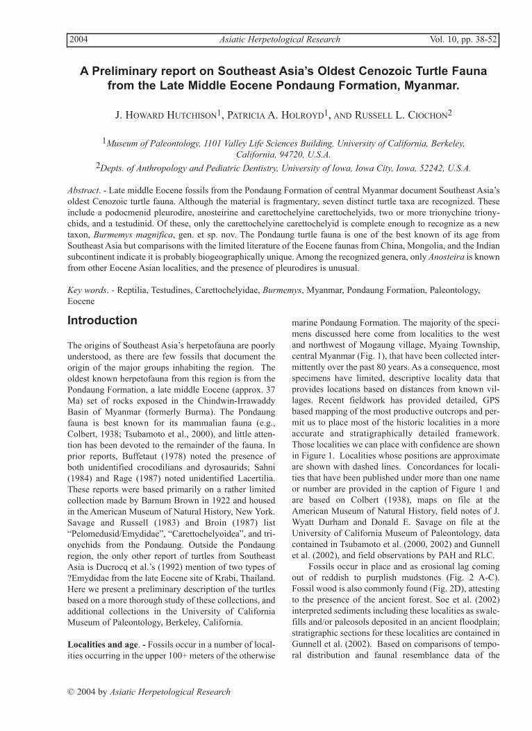
The presence of a prominent musk duct on theperipheral 3, absence of an axillary scale, and strongindication of a hyoplastral buttress rising onto the firstcostal is consistent with Neochelys-like pleurodires. Thehypoplastron fragment may be referable to the sametaxon, but the area of the pelvic sutures is broken off.
The general similarity to at least some Neochelysfavors a placement of the Pondaung Formation speci-mens in the Podocnemididae.
Cryptodira Cope, 1868Testudinidae Gray, 1825
Testudinidae undet.
Referred Material. - UCMP locality V6204: UCMP149166, right xiphiplastron fragment. UCMP localityV96009: UCMP 142226, partial right epiplatron.
Description. - The epiplastron (UCMP 142226, Fig.3D) lacks the gular region. The remaining part of thefree margin is greatly thickened along the anterior edgeof the dorsal scale covered portion. The posterior rim ofthis thickened gular area overhangs the visceral surface.The ventral surface is longitudinally convex. Thesutures are moderately thick and dentate.
The anterolateral part of a right xiphiplastron
(UCMP 149166, Fig. 3E) is referred to the Testudinidaeon the basis of the strong overlap of the femoral scaledorsally, its inflated appearance, and fairly porous sur-face texture.
Discussion. - The morphology of the epiplastron is typ-ical of testudinids and a few batagurids. The ratherporous bone, inflation of the gular area, and generalnature of the sutures and surface texture agrees best withthat of a testudinid. The overhang of the posterior gularrim is derived in testudinids and absent or poorly devel-oped in such tortoises as Hadrianus Cope, 1872,Stylemys Leidy, 1851, Sharemys Gilmore, 1931,Kansuchelys Yeh, 1963, and Ergilemys Ckhikvadze,1972. The epiplastron thus resembles more derived tor-toises such as Testudo Linnaeus, 1858.
Testudinoidea Fitzinger, 1826, indet.
Referred material. - UCMP locality V96019: UCMP147051, posterior part of left hypoplastron. UCMPlocality V78090: UCMP 170495, partial neural. UCMPlocality V98109: UCMP 170522, shell fragments.
Description. - The hypoplastron is represented by afragment (UCMP 147052) that preserves the portion
Figure 4. Anosteira sp., A. UCMP 131736, right hypoplastron, ventral view. B. UCMP 131737, lateral fragment of lefthypoplastron, ventral view. C. UCMP 147030, left peripheral 6, external, posterior, visceral, and anterior views. Scalebars equals 1 cm.
Vol. 10, p. 43 Asiatic Herpetological Research 2004
Figure 5. Burmemys magnifica gen. et sp. n. A. UCMP 61212, adult left hypoplastron (type), ventral view. B. UCMP131745. juvenile left hypoplastron, ventral view. C. UCMP 154993, posterior part of juvenile left epiplastron, ventralview. D. UCMP 131747, anterior part of juvenile left xiphiplastron, dorsal and ventral view. E. UCMP 157444, juvenileleft costal 2, external view. F. UCMP 157443, neural, external view. G. UCMP 157442, suprapygal, external view. Scalebars equals 1 cm.
2004 Asiatic Herpetological Research Vol. 10, p. 44
Figure 6. Burmemys magnifica gen. et sp. n. A. AMNH 14196, left peripheral 1, anterior suture, external and visceralviews. B. UCMP 147021, left peripheral 1, external, posterior suture and visceral views. C. UCMP 147027, left periph-eral 2, external and visceral views. D. UCMP 147002, left peripheral 3, external, visceral and posterior suture views.E. UCMP 61211, left peripheral 4, external and visceral views. F. UCMP 147001, left peripheral 4 fragment, visceraland posterior suture views (arrow points to flat hyoplastral suture). G. UCMP 131756, juvenile left peripheral 5, sutureview. H. UCMP 61218, left peripheral 6, external, posterior suture, visceral and anterior suture views. I. UCMP 142223,right peripheral 7, external, anterior suture, visceral, posterior suture views. J. UCMP 157445, juvenile left peripheral7, external, visceral, and anterior suture views. K. UCMP 142244, right peripheral 8, external, anterior suture, viscer-al and posterior views. L. AMNH 1911, left peripheral 10 and pygal, external view. Scale bars equal 1 cm.
Vol. 10, p. 45 Asiatic Herpetological Research 2004
posterior to the buttress. The free margin is slightly con-vex. The femoral scale distinctly but narrowly overlapsthe dorsal side. The margin dorsal margin of the scale ismarked by a shallow sulcus and the bone continues tothicken medially before thing nearer the midline. A par-tial neural (UCMP 170495) has a distinct carina with arounded top.
Discussion. - The referred fragmentary specimens donot appear to belong to other known taxa in the faunaand agree in general morphology with testudinoids,probably testudinids or batagurids. The neural resemblesthose of carinate batagurids.
Referred material. - UCMP locality UCMP V78090:UCMP 131752, peripheral 9 or 10; UCMP 131754,hypoplastron fragment; UCMP 131755, posterior frag-ment of nuchal; UCMP 147115, neural. UCMP localityV83106: UCMP 131736, medial right hypoplastronfragment; UCMP 131737, lateral hypoplastron frag-ment; UCMP 131741, peripheral 7; UCMP 131742,peripheral 8; UCMP 131744, costal fragments; UCMP131746, anterior fragment of a right peripheral 6. UCMPlocality V96001: UCMP 147005, hypoplastron frag-ment; UCMP 147011, left peripheral 7. UCMP localityV96002: UCMP 147030, left peripheral 6. UCMP local-ity V96008: UCMP 147024, right peripheral 2. UCMPlocality V96009: UCMP 142225, peripheral 9 or 10.Description: The hyoplastron resembles those seen intypical Anosteira and Pseudanosteira Clark, 1932, andlacks the truncated anteromedial articulation of the newgenus described below. This specimen differs fromAllaeochelys Noulet, 1867, in having a narrower poste-rior lobe and narrower bridge area.
The peripherals are referred to Anosteira on thebasis of their small size and well-formed sutures. Mostalso show the presence of weekly-defined sulci on theexternal surface. All the peripherals have sharp margin-al carina, and the surface is finely pustulate. The gom-photic pits for reception of the plastron on peripheral 6(UCMP 147030, Fig. 4C) lie within a longitudinaltrough that traverses the peripheral. The latter is 12.6mm along the free margin carina and 12.5 mm from thecarina to the costal suture. A partial peripheral 6 (UCMP131746) has the trough on the plastral suture filled with8-9 vertically elongated pits and a sharp lateral carina.The two gomphotic pits on peripheral 7 also occur with-in a trough, but on peripheral 7 the trough is onlyapproximately two-thirds the length of the bone. The
peripheral 7 (UCMP 147011) is 12.0 mm along the cari-na. The specimen tentatively identified as peripheral 9 or10 (UCMP 131752) is deeper than long (16 mm alongthe margin, 18 mm in depth).
The posterior nuchal fragment has the typical caret-tochelyid nuchal pedicle. A faint transverse sulcus ispresent, and another faint longitudinal sulcus near themidline is visible.
A small neural (UCMP 147115) is also referred toAnosteira on the basis of the small size and patternedsurface, narrow length to width ratio, and low and broadcentral carina.
Discussion. - Anosteira is known from both Asia (5species) and North America (1 species) in the Eocene.The closely related genus Pseudanosteira is limited toNorth America and distinguishable from Anosteira onlyby details of the top of the carapace. No elements in thePondaung collection resemble Pseudanosteira. Thepresence of sulci on the peripherals, nuchal, and costalfragments indicates it should be assigned to Anosteira.The Pondaung specimen is most parsimoniouslyreferred to Anosteira in the absence of any evidence thatPseudanosteira occurs anywhere in Asia. Previousrecords of Anosteira are confined to China andMongolia.
Carettochelyinae Boulenger, 1887
Burmemys magnifica gen. et sp. nov.
Holotype. - UCMP 61212, adult left hypoplastron (Fig.5A) from UCMP Locality V6204 (near Myaing), foundby J. Wyatt Durham, late Professor of Paleontology atthe University of California, Berkeley.
Paratypes. - AMNH locality “1 mile northeast of Gyat,Magwe Province”: AMNH 1911, pygal, peripheral 10fragment, and costal fragment. AMNH locality “1 milenorth of Koniwa”: AMNH 1919, left first peripheral;AMNH 1928, distal half of right first peripheral; AMNH14196, partial left peripheral 1; AMNH 14197, plastronfragment. UCMP locality V6204: UCMP 61211, leftperipheral 4; UCMP 61218, left peripheral 6. UCMPlocality V78090: UCMP 131750, juvenile lateralhypoplastron fragment, UCMP 131751, juvenile periph-eral fragment; UCMP 131753 juvenile xiphiplastronfragment; UCMP 154994, proximal costal fragment.UCMP locality V83106: UCMP 131738, juvenile righthypoplastron; UCMP 131739, juvenile hypoplastronfragment; UCMP 131745, juvenile left hypoplastron.UCMP locality V83111: UCMP 128406, right peripher-al 2. UCMP locality V83116: UCMP 131748, hypoplas-tron fragment. UCMP locality V83143: UCMP 131747,
Vol. 10, p. 46 Asiatic Herpetological Research 2004
anterior part of left xiphiplastron. UCMP localityV96001: UCMP 147001, left peripheral 4 fragment;UCMP 147002, partial left peripheral 3; UCMP 147003,anterior peripheral fragment; UCMP 147009, neural;UCMP 147010, peripheral fragment; UCMP 147012,juvenile medial hypoplastron fragment. UCMP localityV96002: UCMP 142244, right peripheral 8; UCMP154984, anterior peripheral fragment. UCMP localityV96008: UCMP 147021, left first peripheral; UCMP147023, posterior peripheral fragment; UCMP 147027,left peripheral 2; UCMP 147028, partial right peripheral1; UCMP 147029 distal fragment of a costal. UCMPlocality V96009: UCMP 142223, right peripheral 7.UCMP locality V99498: UCMP 157443, neural; UCMP157446, shell fragments.
Referred material. - UCMP locality V96001: UCMP147004, plastron fragment. UCMP locality V83106:UCMP 131740, hyoplastron fragment. UCMP localityV78090: UCMP 131756, juvenile left peripheral 5;UCMP 154993, posterior fragment of left epiplastron.UCMP locality V99498: UCMP 157442, suprapygal;UCMP 157444, left costal 2; UCMP 157445, left periph-eral 7.
Diagnosis. - Burmemys is distinguished from othercarettochelyines by the combination of asymmetricalarticulation of the hyo-hypoplastra, narrow hypoplastralbridge, and large size (estimated carapace length greaterthan 1000 mm).
Description. - The holotype hypoplastron (UCMP61212, Fig. 5A) is massive. The anterior suture of theleft hypoplastron consists of two sutures. The suturewith the left hyoplastron is sinusoidal, curving antero-medially, and joins a distinct, straight and anteromedial-ly-facing suture, presumably for articulation with theright hyoplastron. The ventral sculpture consists of apattern of irregular, closely-spaced tubercles that radiatefrom a focal point lateral to the middle of the medialmoiety. Laterally, the tubercles coalesce into ridges radi-ating laterally. The sutures are finely dentate and thick(13 mm). The lateral margin and posterior half of themedial part is broken away in the type, but these are pre-served in the juvenile specimen (UCMP 131745, Fig.5B). The width of the medial part of the hypoplastronmeasured from the apex of the inguinal notch to theplastral midline is only one-half or less of the maximumhypoplastral width. The inguinal notch is open and notconfined as in Carettochelys Ramsey, 1887. The anteri-or-posterior width of the bridge area is one-half or lessthe width of the xiphiplastral lobe of the hypoplastron.The referred juvenile specimens exhibit the same sutur-
al shapes as the adult (type) but the inguinal notches areshallower, sculpture less organized, and lateral extent ofthe lateral arm of the bridges are shorter.
The posterior part of a juvenile epiplastron (UCMP154993, Fig. 5C) is referred to Burmemys on the basis ofthe convex curvature of the lateral margin that indicatesa short and rounded anterior lobe, and an obtuse anglebetween the entoplastral and hyoplastral sutures indicat-ing a short and broad entoplastron.
Two xiphiplastra (UCMP 131747, Fig. 5D; UCMP131753) are referred to Burmemys on the basis of rela-tively larger size, converging (non-parallel) medial andlateral margins of the anterior moiety, and thinningrather than thickening toward the midline suture. Bothspecimens are small (proximal width of UCMP 131747is 20 mm) and thus considered as juveniles.
The juvenile left costal (UCMP 157444, Fig. 5E) isnearly uniformly thin, parallel sided, and sculpturedwith a subdued and random pattern of low pustules andshort ridges. The distal margin forms about a 45 degreeangle to the sides. There are no sulci. The parallel sidesand high angle of the distal margin indicate a secondcostal.
The distal end of an adult costal (UCMP 147029) issubtlety sculptured with longitudinal irregular ridges.The distal suture is weakly dentate but patent exceptabove the rib. The rib ends protrudes prominently.Although damaged, the distal width is about 70 mm.
A large neural (UCMP 147009, Figs. 5F) is relative-ly narrow, lacks a midline carina, and has subtle sculp-ture of very shallow dimples. It has a midline length of51 mm, maximum width of 32 mm, and maximumthickness of the lateral side of 15.4 mm.
The suprapygal (UCMP 157442, Fig. 5G) is trian-gular with a distinct medial carina. The surface sculptureconsists of irregular vermiform ridges that radiate fromthe central area of the posterior margin. It is longer thanwide (32.2 mm long, 31.5 mm wide).
At least nine peripheral positions are represented.The sculpture is variable consisting of distinct tuberclesat one extreme to anastomosing pits and ridges at theother. The free margins of adult specimens are roundedbut may be acute in juveniles. There are no indicationsof scale sulci.
The first peripheral exhibits distinct sutures with thefirst costal, nuchal and second peripheral. The free mar-gin perimeter is asymmetrically curved. The largestspecimens (AMNH 1919, Fig. 6A; UCMP 147021, Fig.6B) have perimeter lengths of 114 and 119 mm, maxi-mum depths of 90 and 81 mm, maximum thicknesses atthe posterior suture of 33 and 27 mm, and maximumthicknesses at anterior suture of 28 and 25 mm respec-tively.
2004 Asiatic Herpetological Research Vol. 10, p. 47
The two second peripherals differ in size. UCMP147026 (Fig. 6C) is massive with a free margin length of82 mm, maximum depth of 67 mm, and maximum thick-ness of the anterior suture of 20 mm. Comparable meas-urements of UCMP 128406 are 45, 40, and 11 mmrespectively. The second peripheral is roughly rectangu-lar in external view.
The only specimen referred to the third peripheral(UCMP 147002, Fig. 6D) is lacking the anteroventraland posterodorsal corners. The anterior part of the dor-sal suture is a semi-scarf joint – probably for the rib endof the first costal. The peripheral thickens noticeablytowards the posterior suture and reaches a thickness of32 mm at the suture.
The fourth peripheral (UCMP 61211, Fig. 6E) isdamaged anteriorly and dorsally and locally abraded. Itslength along the lateral carina is 75 mm. The free mar-gin curves posteromedially on the posterior moiety toform a plastral articulation. The plastral articular surfaceis relatively flat but deep (up to 16 mm) and without pitsfor the hyoplastral buttress or normal dentations, thusindicating a weakly ligamental and kinetic joint. The lat-eral carina is broadly rounded. The peripheral 4 frag-ment (UCMP 147001, Fig. 6F) also shows this rather flatand deep (19 mm) hyoplastral suture.
A small peripheral, probably a left peripheral 5(UCMP 131756, Fig. 6G) is considered a juvenile of thisspecies. The plastral arm is very short with a longitudi-nal trough enclosing a series of gomphotic pits. The lat-eral carina is slightly rounded and broadly upturned. Thelength of the lateral carina is 22 mm and has a posteriorthickness of about 9 mm.
A relatively complete left peripheral 6 (UCMP61218, Fig. 6H) has a damaged plastral margin and lacksthe dorsal suture. The plastral and costal arms convergeposteriorly. The plastral articulation is broken anteriorlybut posteriorly has a longitudinal trough indicating inter-digitation with the hypoplastron. The lateral carina isrounded and slightly upturned. The length along the lat-eral carina is 82 mm.
A peripheral 7 (UCMP 142223, Fig. 6I) of an adultmeasures 99 mm along the marginal carina, 100 mmfrom the carina to costal margin, posterior thickness of29 mm and an anterior thickness of more than 45 mm.The hypoplastral suture is damaged but trough-like,extends about half-way along the medial side, andappears to have housed one or two recessed pits. Theisolated peripheral 7 (UCMP 157445, Fig. 6J), a pre-sumed juvenile, closely resembles Carettochelys withthe hypoplastral buttress rising up the central part of themedial side. The free margin is sharp and broadlyupturned. The length of the free margin is about 34 mm.
The adult peripheral 8 (UCMP 142244, Fig. 6K) ismassive and slightly shorter than deep (97 mm along thelateral carina and 105 mm from the carina to costal
suture). An anteriorly-deepening trough divides themedial surface into dorsal and ventral arms anteriorly.
The pygal (AMNH 1911, Fig. 6L) is distinctlytrapezoidal with a short anterior side and a low but sharpmedial crest. The sculpture consists of widely spacedirregular tubercles that fade out near the medial crest andfree margin. An associated posterior peripheral 10 frag-ment has a sculpture of irregular ridges and tuberclesthat radial from a central focus.
Discussion. - The absence of scales and large size placeBurmemys in the Carettochelyinae. Of the three Eocenegenera of Carettochelyinae, Burmemys differs from allin the presence of two distinct anterior articular sutureson one of the hypoplastra. This most likely represents anasymmetrical articulation with the hyoplastra, with oneof the hyoplastra extending well across the midline toform an angled articulation with the opposite hypoplas-tron. Even where the hyo-and hypoplastra are not mirrorimages with one of the hypoplastra contacting the oppo-site hyoplastron (e.g., Anosteira in Hay, 1908, Fig. 353),the midline suture remains straight as in other Paleogenecarettochelyids and the plastral midline suture usuallyexhibits some limited kinesis. This asymmetry is notunusual in turtles, but within carettochelyids was knownonly to a lesser degree in some Carettochelys. Thehypoplastron in extant Carettochelys insculpta Ramsey,1887 may cross the midline to form a short angled suturewith the opposite hyoplastron (AMNH 84212, andRooij, 1915, fig. 123a). This occurs on the left hyoplas-tra on both of these.
Burmemys resembles anosteirines and differs fromextant Carettochelys, Hemichelys Lydekker, 1887, (pl.XII, fig. 2) from the Eocene of the Punjab, andChorlakkichelys Broin, 1987 (pl. 1, fig. 2) from theEocene of Pakistan in the relatively broad inguinalnotch. Burmemys additionally differs fromChorlakkichelys and Carettochelys in having a distinct-ly narrow bridge area. The general proportions of thehypoplastron resemble those of Allaeochelys from theEocene of Europe (Broin, 1977, Pl. XV1, Fig. 3). Thesuprapygal differs from Allaeochelys, Carettochelys,Hemichelys and probably Chorlakkichelys in beinglonger than wide.
Burmemys is also the largest carettochelyiddescribed to date. Based on scaling up of the elementsin comparison to other carettochelyids, we can estimatethat the shell length of Burmemys exceeded 1000 mm.This estimate suggests that Burmemys is among thelargest turtles known, but is smaller than estimates Headet al. (1999) provided for Eocene trionychids fromPakistan, which may have reached more than 2000 mmin length, and is smaller than the giant Bridgerian triony-chid from Wyoming (Gaffney, 1979).
Vol. 10, p. 48 Asiatic Herpetological Research 2004
Trionychidae Gray, 1825Trionychinae Gray, 1825Trionychinae genus indet.Trionychinae, large form
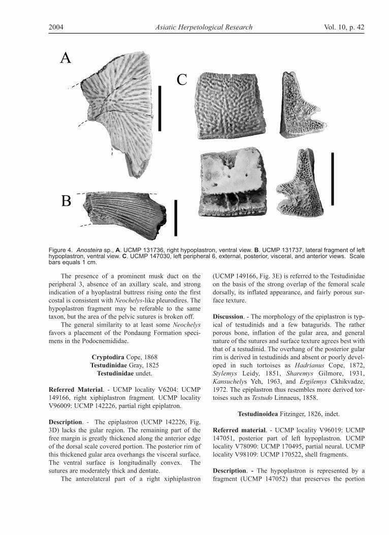
Description. - The hypoplastron (UCMP 61213, Fig.7A) lacks the projecting spines (present in UCMP153799, Fig. 7B) but is otherwise relatively complete.The calloused area has well defined edges and coversmost of the ventral surface, except for the long offsetshelf on the medial edge. The calloused area is sculp-tured with distinct pits and ridges, while the shelf isamorphously roughened. The buttress is composed oftwo protruding spikes. The posteromost of these is bro-ken off at the base, but the larger anterior one is divided
into three fluted points at its tip.The isolated neural (UCMP 147022, Fig. 7C), prob-
ably 6 or 7, is hexagonal and narrows distinctly posteri-orly. The dorsal surface is weakly sculptured, lack sulci,and is flat anteriorly but is formed into a central carinaposteriorly. The neural is 46 mm long and 50 mm wide.
Discussion. - The large size of the hypoplastron andgeneral conformation indicates a trionychine and gener-ally resembles Pelochelys Gray 1864, Chitra Gray 1844,and Pelodiscus Fitzinger 1835 in these features, but dif-fers in having a wide, unsculptured medial shelf.Additional material would be needed to refine identifi-cation.
Trionychinae, small form
Referred material. - UCMP locality V6204: UCMP61210 plastron fragment. UCMP locality V83106:UCMP 147116, two costal fragments.
Description. - Two distal costal fragments exhibit awell-defined sculpture of pits and ridges with indica-tions of longitudinal welts. The pattern extends to thefree margin with only a slight sculpture-free zone at thefree margin, suggesting an adult turtle of relatively small
Figure 7. Trionychinae. A. UCMP 61213, right hypoplastron, ventral view. B. UCMP 1537993, fragment of righthypoplastron, ventral view. C. UCMP 147022, neural, external view. D. UCMP 170520, costal fragment, external view.Scale bars equals 1 cm.
2004 Asiatic Herpetological Research Vol. 10, p. 49
size in comparison with the preceding taxon. The sculp-ture resembles that of a trionychine rather than acyclanorbine such as Lissemys Smith, 1931.
Trionychinae, ornate form
Referred material. - UCMP locality V98109: UCMP170520, two costal fragments.
Description. - The costal fragments (UCMP 170520,Fig. 7D) exhibit a striking sculpture of large, elongatetubercles rising above a surface composed of a general-ly organized pattern of longitudinal rows of shallow pitsand low rides. The longitudinal axes of the raised tuber-cles vary from anterior-posterior to medial-lateral andare large (11-15 mm) relative to the overall size of thelarger costal fragment (maximum preserved width of 32mm). Carapace sculpturing varies between individualsand also ontogenetically, but the peculiar sculptureshows some resemblance to that seen in some extantAspideretes Hay, 1904.
Discussion
In addition to the turtles, a variety of other lower verte-brates are present in the Pondaung Formation includinga carcharhinid shark, Galeocerdo Müller and Henle1837 (UCMP 142238), a clariid catfish (UCMP128411), at least four species of agamid lizards (UCMP128410, 130290, 142227, 142232), paleophid and colu-broid snakes (see Head et al, in prep.), and a minimumof two crocodilians, including a pristichampsine croco-dylian (UCMP 147127) and a dyrosaurid (Buffetaut,1978).
Unfortunately, reports on Asian lower vertebrates ofcomparable age (Sharamurunian Asian Land MammalAge or late middle Eocene) are few. Thus, the limited lit-erature, combined with the fragmentary nature of thePondaung fossils themselves, make detailed compar-isons with other faunas difficult. Nonetheless, compar-isons with known Sharamurunian lower vertebrate fau-nas reveal only a few similarities between the Pondaungand any other locality. Pleurodires are previously unde-scribed from Asia, although Broin (1987) notes the pres-ence of “Pelomedusidae and/ or Emydidae” from themiddle Eocene of Pakistan and Oligocene of India. Anadocid was described by Gilmore (1931) from the latemiddle Eocene of Mongolia, but none were identified inthe Pondaung assemblage. The carettochelyid genus,Anosteira, has been reported from age-equivalents inManchuria (Zangerl, 1947) and Guangdong, China (SunAiling et al., 1992) and from slightly younger sedimentsin Shandong and Guangdong provinces (Yeh, 1963).The only other carettochelyines described from Asia are
Chorlakkichelys and Hemichelys from the early middleEocene of Pakistan (Lydekker, 1887; Broin, 1987), andBurmemys is the most easterly and southerly Eocenerecord of the subfamily. Trionychids, as elsewhere, arean important part of the fauna, but our material is notsufficiently diagnostic to make any meaningful biogeo-graphic comparisons. Testudinids are widely reported inChinese and Mongolian Eocene faunas (Gilmore, 1931;Ye, 1963), but all of these appear to be more generalizedforms similar to Hadrianus or Kansuchelys. ThePondaung form appears to be more like the modernTestudo, and thus distinct from contemporaneousChinese and Mongolian taxa.
Among other reptiles, the only other agamid lizardknown in the Asian Sharamurunian is Tinosaurus yuan-quensis from the Heti Formation (Li, 1991), but it is adiminutive form that bears no resemblance to thePondaung agamids. Crocodilians are known elsewhere,but not in detail. In overall diversity, the Pondaungfauna shares more general resemblances to the better-known Irdinmanhan faunas, especially that of theKuldana Formation of Pakistan (Broin, 1987).
Based on the fragmentary evidence available todate, several observations can be made regarding thePondaung lower vertebrate fauna. Faunal endemicity issupported by the number of unique taxa, and the compo-sition of the turtle fauna is unusual with trionychoids(especially carettochelyids) dominating. Faunal compo-sition and the large size of these turtles are consistentwith an interpretation of these sites as representing awarm, tropical floodplain environment, deposited fairlynear shore. The aquatic habits of most of the lower ver-tebrates suggest that during late middle Eocene time thePondaung region was a well-drained floodplain environ-ment, a finding consistent with previous geologicalinterpretations (e.g., Bender, 1983, Soe et al., 2002).
Acknowledgments
We are grateful to Donald E. Savage, Ba Maw, andThaw Tint for reinitiating fieldwork in the Pondaungregion in the late 1970’s, and Brig.-Gen Than Tun andMajor Bo Bo from the Office of Strategic Studies,Ministry of Defense for arranging our visits toMyanmar. Tin Thein, Aye Ko Aung, Aung Naing Soe,and other members of the Pondaung Fossil Expeditionteam are thanked for their assistance during our staysand their help while in the field. Eugene Gaffney,Charlotte Hotton, and Mark Norell kindly providedaccess to and assistance in use of the AMNH collectionsand associated records. Walter Joyce reviewed the man-uscript and provided constructive criticisms. Fundingfor fieldwork in Myanmar has been provided by theSmithsonian Foreign Currency Program (from Public
Vol. 10, p. 50 Asiatic Herpetological Research 2004
Law 480 funds), the L.S.B. Leakey Foundation, theUniversity of California Museum of Paleontology, theUniversity of North Carolina at Charlotte Foundation,the University of Iowa Center for Pacific and AsianStudies, the Office of the Vice President for Researchand the Human Evolution Research Fund of theUniversity of Iowa Foundation. This is UCMP contribu-tion no. 1836.
Literature Cited
Batsch, A. J. G. C. 1788. Versuch einer Anleitung zurKenntniss und Geschichte der Thiere undMineralien. Vol. 1. Akademische Buchhandlung,Jena. viii + 528 pp.
Bender, F. 1983. The Geology of Burma. Beiträge zurRegionalen Geologie der Erde, Band 16, 293 pp.
Boulenger, G. A. 1887. On a new family of pleurodiranturtles. Annals and Magazine of Natural History19:170-172.
Broin, F. de. 1977. Contribution à l’étude deschéloniens. Chéloniens continentaux du Crétacé etdu Tertiaire de France. Mémoires du MuséumNational d’Histoire Naturelle, N. Ser. C, 38:1-423.
Broin, F. de. 1987. Lower vertebrates from the early-middle Eocene Kuldana Formation of Kohat(Pakistan): Chelonia. Contributions from theMuseum of Paleontology, University of Michigan27(7):169-185.
Buffetaut, E. 1978. A dyrosaurid (Crocodilia,Mesosuchia) from the upper Eocene of Burma.Neues Jahrbuch für Geologie und Paläontologie,Monatshefte 5:273-281.
Ckhikvadze, V. M. 1972. O sistematichekom polozheniitretichnykh gigantskikh sukhoputnykh cherepakhPalearktiki. [On the systematic positions of theTertiary gigantic land turtles of Palearctic]. Bulletinof the Academy of Sciences of the Georgian R.S.S.65(3):745-748.
Clark, J. 1932. A new anosteirid from the Uinta Eocene.Annals of the Carnegie Museum 21:161-170.
Colbert, E. 1938. Fossil mammals from Burma in theAmerican Museum of Natural History. Bulletin ofthe American Museum of Natural History 74:255-436.
Cope, E D. 1865. Third contribution to the herpetologyof tropical America. Proceedings of the Academy ofNatural Sciences of Philadelphia 1865:185-198.
Cope, E D. 1868. On the origin of genera. Proceedingsof the Academy of Natural Sciences, Philadelphia1868:92-93.
Cope, E. D. 1872. Second account of new vertebratafrom the Bridger Eocene of Wyoming Territory.Proceeding of the American Philosophical Society12:466-468.
De Rooij, N. 1915. Reptiles of the Indo-AustralianArchipelago, Lacertilia, Chelonia, Emydosauria. E.J. Brill, Leiden. 1: xiv + 384 pp.
Ducrocq, S., E. Buffetaut, H. Buffetaut-Tong, R. Helmcke-Ingavat, J.-J. Jaeger, Y. Jongkanjanasoontorn, and V. Suteethorn. 1992. ALower Tertiary vertebrate fauna from Krabi (South Thailand). Neues Jahrbuch für Geologie und Paläontologie, Abhandlungen 184(1): 101-122.
Fitzinger, L. J. 1826. Neue Classification der Reptiliennach ihren natürlichen Verwandtschaften nebsteiner Verwandtschafts-Tafel und einenVerzeichnisse der Reptilien-Sammlung des k. k.Zoologischen Museum’s zu Wien. J. G. Hübner,Wien, viii + 66 p.
Fitzinger, L., 1835. Entwurf einer systematischenAnordnung der Schildkröten nach den Grundsätzender natürlichen Methode. Annalen der WienerMuseums des Naturgeschichte 1:103-128.
Gilmore, C. W. 1931. Fossil turtles of Mongolia.Bulletin of the American Museum of NaturalHistory 59:213-257.
Gray, J. E. 1825. A synopsis of the genera of reptiles andAmphibia, with a description of some new species.Annals of Philosophy, London. Ser. 2 10:193-217.
Gray, J. E. 1844. Catalogue of tortoises, crocodiliens,and amphisbaenians in the collection of the BritishMuseum. British Museum (Natural History),London. viii + 80 pp.
Gray, J. E. 1864. Revision of the species Trionychidaefound in Asia and Africa, with description of somenew species. Proceedings of the Zoological Societyof London 1864: 76-98.
2004 Asiatic Herpetological Research Vol. 10, p. 51
Gunnell, G. F., R. L. Ciochon, P. D. Gingerich, and P. A.Holroyd. 2002. Re-assessment of Pondaungia andAmphipithecus (Primates) from the late middle Eocene of Myanmar with comments on “Amphipithecidae.” Contributions from the Museum of Paleontology, University of Michigan 30: 337-372.
Hay, O. P. 1904. On the existing genera of theTrionychidae. Proceedings of the AmericanaPhilosophical Society 42:268-249.
Hay, O. P. 1908. Fossil turtles of North America.Carnegie Institute of Washington, Publication 75:568 pp.
Head, J. J., P. A. Holroyd, J. H. Hutchison, and R. L.Ciochon. in prep. First report of snakes (Serpentes)from the late middle Eocene Pondaung Formation,Myanmar.
Head, J. J., S. M. Raza, and P. D. Gingerich. 1999.Drazinderetes tethyensis, a new large trionychid(Reptilia: Testudines) from the marine EoceneDrazinda Formation of the Sulaiman Range, Punjab(Pakistan). Contributions from the Museum ofPaleontology, University of Michigan 30:199-214.
Holroyd, P. A. and Ciochon, R. L. 1994. Relative ages ofEocene primate-bearing deposits of Asia. Pp. 123-141. In J. G. Fleagle and R. F. Kay (eds.),Anthropoid Origins. Plenum Press, New York.
Hutchison, J. H., and D. M. Bramble. 1981. Homologyof the plastral scales of the Kinosternidae and relat-ed turtles. Herpetologica 37:73-85.
Jiménez, E., M. A. Cuesta, and S. G. Tudanca. 1994.Vertebrados fósiles del Eoceno de Fuentesaúco(Zamora). Studia Geologica Salamanticensia29:7-21.
Leidy, J. 1851. [Fossil tortoises from NebraskaTerritory]. Proceedings of the Academy of NaturalSciences, Philadelphia 5: 326-327.
Leidy, J. 1871. [Remarks on extinct turtles fromWyoming Territory, Anosteira ornata andHybemys arenarius.] Proceedings of theAcademy of Natural Sciences, Philadelphia 1871:102-103.
Linnaeus, C. 1758. Systema Naturae 10th ed., vol. 1.Stockholm, 824 pp.
Lydekker, R. 1887. Eocene chelonians from the Salt-Range. Paleontologica Indica 4:59-65.
Lydekker, R. 1889. Chapter III. Class Reptilia – contin-ued. Orders Anomodontia, Sauropterygia, andChelonia. Pp. 1053-1118. In: H. A. Nicholson andR. Lydekker, Manual of Paleontology. Vol 2. W.Blackwood and Sons, Edinburgh. 3rd ed.
Müller, J., and J. Henle. 1837. Gattungen der Haifischeund Rochen nach einer von ihm mit Hrn. Henleunternommenen gemeinschaftlichen Arbeit über dieNaturgeschichte der Knorpelfische. Bericht derAkademie der Wissenschaften, Berlin 1:111-118.
Noulet, J. B. 1867. Nouveau genre de Tortues fossilessous le nom d’Allaeochelys. Mémoires de l’Académie des sciences, Toulouse (6e sér.) 5:172-177.
Ramsey, E. P. 1886 [1887]. On a new genus and speciesof fresh water tortoise from the Fly River, NewGuinea. Proceedings of the Linnean Society of NewSouth Wales (2)1:158-162.
Rage, J.-C. 1987. Lower vertebrates from the early-middle Eocene Kuldana Formation of Kohat(Pakistan): Squamata. University of MichiganContribution from the Museum of Paleontology27:187-193.
Sahni, A. 1984. Upper Cretaceous-early Palaeogenepalaeobiogeography of India based on terrestrialvertebrate faunas. Mémoires de la Sociéte´géologique de France, N. S. 147: 125-137.
Savage, D. E., and D. E. Russell. 1983. MammalianPaleofaunas of the World. Addison-Wesley,Reading Massachusetts. 432 pp.
Smith, M. A. 1931. The fauna of British India, includ-ing Ceylon and Burma. Reptilia and Amphibia.Vol. 1. Loricata, Testudines. Taylor and Francis,London. 185 pp.
Soe, Aung Naing, Myitta, Soe Thura Tun, Aye Ko Aung,Tin Thein, B. Marandat, S. Ducrocq, & J.-J. Jaeger.2002. Sedimentary facies of the late Middle EocenePondaung Formation (central Myanmar) and the
Vol. 10, p. 52 Asiatic Herpetological Research 2004
paleoenvironments of its anthropoid primates.Comptes Rendus Palevol 1:153-160.
Sun, A., J. Li, X. Ye, Z. Dong, L. Lou. 1992. TheChinese fossil reptiles and their kins. Science Press,Beijing. 242 pp.
Tsubamoto, T., N. Egi, M. Takai, N. Shigehara, Aye KoAung, Tin Thein, Aung Naing Soe and Soe ThuraTun. 2000,: A preliminary report on the Eocenemammals of the Pondaung fauna, Myanmar. AsianPaleoprimatology 1: 29-101.
Tsubamoto, T., Takai, M., Shigehara, N., Egi, N., SoeThura Tun, Aye Ko Aung, Maung Maung, Danhara,T., and Suzuki, H. 2002. Fission-track zircon age ofthe Eocene Pondaung Formation, Myanmar. Journalof Human Evolution 42: 361-369.
Wood, R. C. 1983. Kenyemys williamsi, a fossil pelome-dusid turtle from the Pliocene of Kenya. Pp. 74-85.In: A. G. Rhodin et al. (eds). Advances inHerpetology and Evolutionary Biology.
Ye (Yeh), X. 1963. Fossil turtles of China.Palaeontologica Sinica, new Series C (18):112 pp.
Zangerl, R. 1947. A new anosteirine turtle fromManchuria. Fieldiana, Geology 10:13-21. Effective Date of Publication: June 1, 2004