Page 1
Graduate Theses, Dissertations, and Problem Reports
2014
A preliminary study evaluating potential probiotic use in A preliminary study evaluating potential probiotic use in
endodontics endodontics
Khaled Seifelnasr West Virginia University
Follow this and additional works at: https://researchrepository.wvu.edu/etd
Recommended Citation Recommended Citation Seifelnasr, Khaled, "A preliminary study evaluating potential probiotic use in endodontics" (2014). Graduate Theses, Dissertations, and Problem Reports. 588. https://researchrepository.wvu.edu/etd/588
This Thesis is protected by copyright and/or related rights. It has been brought to you by the The Research Repository @ WVU with permission from the rights-holder(s). You are free to use this Thesis in any way that is permitted by the copyright and related rights legislation that applies to your use. For other uses you must obtain permission from the rights-holder(s) directly, unless additional rights are indicated by a Creative Commons license in the record and/ or on the work itself. This Thesis has been accepted for inclusion in WVU Graduate Theses, Dissertations, and Problem Reports collection by an authorized administrator of The Research Repository @ WVU. For more information, please contact [email protected] .
Page 2
A PRELIMINARY STUDY EVALUATING POTENTIAL PROBIOTIC USE IN ENDODONTICS
By
Khaled Seifelnasr, BDS, DDS
Thesis submitted to the School of Dentistry at West Virginia University in partial fulfillment of the requirements for the degree of
Master of Science In
Endodontics
John G. Thomas, PhD, Chair Anthony T. Borgia, DDS, MHA
Richard Jurevic, DDS, MSD, PhD
Department of Endodontics
Morgantown, West Virginia
2014
Keywords:
Probiotics, Dental, Endodontics, Candida albicans, Enterococci faecalis
Page 3
Abstract
A PRELIMINARY STUDY EVALUATING POTENTIAL PROBIOTIC USE IN ENDODONTICS
Khaled Seifelnasr, BDS, DDS
Introduction:
The main goal of endodontics is the prevention of apical periodontitis. This condition is the result of persistent pathogenic microorganisms such as Enterococcus faecalis (E. f) and Candida albicans (C. a) remaining in the root canal systems of teeth, and the ability of those organisms to directly cause acute and chronic inflammation in the periapical tissues. The concept of the use of probiotics has not yet been evaluated in addressing endodontic disease, but probiotics have proven successful in treating periodontal disease. Taking these findings into account, this preliminary work was performed to evaluate the possible effectiveness of a probiotic cocktail in preventing the growth of two potential endodontic patogens, Enterococcus faecalis and Candida albicans.
Materials and methods:
Five groups (I, II, III, IV and V) of commercial probiotics were selected and evaluated based upon numbers and concentration of organisms. Pathogenic test organisms were C.albicans (WVU Isolate Ca1028) and E. faecalis (WVU Isolate Ef01).
Phase 1 of the study was conducted by a disc diffusion assay test to evaluate zones of inhibition (ZOI) in millimeters (mm) of the selected probiotics against the E. f and C.a.Microorganisms from probiotic samples were extracted via manufacturer’s recommendations and mixed by weight. Thirty (30) microliters were then placed on sterile discs. Pathogenic organisms were set to a 1 McFarland standard challenge. A five probiotic disc template on blood agar plates were inoculated with a lawn of either E. F or C. a and incubated at 37° C for 48 hours and 1 week. Two, five sterile disc templates with a lawn of either pathogenic organism were run parallel as a control.
Phase 2 was conducted by mixing 9 ml of 30% poloxamer 407 and MRS broth in a test tube with 500 ml of either E. f or C. a set at a 1 McFarland standard, together with 500ml of either Group I or Group IV probiotic mixtures, set at a 2 McFarland standard. Samples were incubated at 37°C for 48 hours, followed by serial dilutions of 10-2, 10-4, and 10-6 for evaluation of CFU/ml counts. Controls were E. f or C. a 30% poloxamer with MRS broth and no probiotics.
Page 4
Results:
Phase 1: Based on a One-Way ANOVA analysis, Groups I, IV and V showed the most statistically significant results (P< 0.05) with a Mean of 7.4mm,10.05mm,11.2mm for C. a and a Mean of 6.7 mm,11.1 mm, and 12.5 mm for E. f respectively at 1 week, compared with no ZOI for the control. L. acidophilus, L. casei , L. rhamnosus and B. longum were all common strains in the probiotic cocktails selected.
Phase 2: Initial results showed a decrease to a 2 log difference and a 1 log difference for groups I and IV respectively regarding CFU/ml counts for C. a and for E. f. Probiotic Groups Iand IV showed complete elimination of E. f and only probiotic colonies were present on observation.
Conclusion:
Recognizing that probiotics may act differently based on their composition and concentration, this study suggests that organisms such as L. acidophillus, L. rhamnosus, L.caseiand B. Longum are effective for preventing the growth of E. faecalis and C. albicans in vitro against both their planktonic and biofilm morphological stages. Further evaluations for possible use in treating endodontic infection is suggested and warranted. Additionally, Phase 2 results of the study suggest that poloxamer 407 could be utilized as an ideal probiotic delivery vehicle when mixed with appropriate probiotics and utilized as an endodontic intra-canal medicament for treating teeth that have presented with non-vital pulps.
Page 5
iv
Dedication
I would like to dedicate this to my mother Amal, father Seifalla, wife Sumaya and children Aminah and Ammar, who have always instilled in me the idea that nothing is impossible and one can achieve anything in life as long as he puts thought and effort to accomplish it. Their love and care has always been the reason why I strive for success.
I would also like to dedicate this to both Dr.Borgia and Dr.Thomas for their mentorship, guidance, wisdom and most important of all, dedication, love and support.
Acknowlegments
I would like to thank Dr.Jurevic for his support.
I would like to thank Chris waters for his help as well as the endodontic staff for their help during residency.
I would like to thank Dr’s Nicholson, Parsa and Xu for making this residency fun and delightful, I wish all of them a successful and happy future.
Page 6
v
TABLE OF CONTENTS
ABSTRACT..................................................................................................................... ...............................................................II
DEDICATION................................................................................................................................................................................IV
ACKNOWLEDGEMENTS............................................................................................................. ..................................….…...IV
TABLE OF CONTENTS ........................................................................................................... ........................................………V
LIST OF TABLES...................................................................................................................................... ........................…….. VI
LIST OF FIGURES......................................................................................................... ............................................................. VI
CHAPTER 1 ................................................................................................................... .................................................................1
Introduction ............................................................................................................................. ..................................................….....1
Statement of the problem ............................................................................................................................. .....................................4
Significance of the problem ………………..................................................................................................................................... .4
Null Hypothesis ……….......................................................................................................................... ............................................5
Assumptions …............................................................................................................................ .......................................................5
Limitations ……............................................................................................................................. ...................................................6
Delimitations ……............................................................................................................................................................................. 6
CHAPTER 2 ............................................................................................................................. ........................................................7
REVIEW OF LITERATURE................................................................................................................... ..................................... 7
CHAPTER 3 ............................................................................................................................. ..................................................... 12
Materials and Methods…………......................................................................................................................................... ..............12
Probiotic Strain Selection …………........................................................................................................................... .................. 12
Pathogenic organism Strain Selection…........................................................................ .................................................................. 16
Extraction of Probiotic Organisms from commercial products…………………………………………………………..……...…16
Phase 1: Testing for Probiotic efficacy against E.faecalis and C.albicans; Planktonic Stage evaluation…………......................... 24
Phase 2: Biofilm stage testing; Intra-canal delivery vehicle for probiotics ………………………………..……….……............29
CHAPTER 4 ........................................................................................... ................................................................……………….35
Results ............................................................................................................................. ................................................................ 35
Discussion……............................................................................................................................. .................................................... 55
CHAPTER 5................................................................................................................................................. ................................... 60
Conclusion ……............................................................................................................................. .................................................60
WORKS CITED ............................................................................................................................................................………….61
Page 7
vi
List of Tables
Table (1). ZOI Candida albicans 48Hours ………………………………………………………………………………………………………………… 36
Table (2). ZOI Candida albicans 1 week ………………………………………………………………………………………………………………… 38
Table (3). Analysis of variance (ANOVA) for Zone of Inh by Group =CA …………………………………………………………………. 39
Table (4). Effect Tests for Zone of Inh by Group =CA ……………………………………………………………………………………… 40
Table (5).Least Squares Means Whole Model=CA ……………………………………………………………………………………………… 40
Table (6) .Least Squares Means by Day=CA……………………………………………………………………………………………………………… 40
Table (7). ZOI Enterococci faecalis 48Hours………………………………………………………………………………………………………… 42
Table (8). ZOI Enterococci faecalis 1 week…………………………………………………………………………………………………………… 44
Table (9). Analysis of variance (ANOVA) for Zone of Inh by Group =EF………………………………………………………………… 46
Table (10).Effect Tests for Zone of Inh by Group =EF…………………………………………………………………………………………… 46
Table (11). Least Squares Means Whole Model=EF……………………………………………………………………………………………… 47
Table (12). Least Squares Means by Day=EF……………………………………………………………………………………………………… 47
Table (13). CFU/mL Candida albicans -72hrs………………………………………………………………………………………………………… 53
Table (14). CFU/mL Enterococci faecalis -72hrs…………………………………………………………………………………………………… 54
List of Figures
Figure 1: Healing RCT with no canal……………………………………………………………………………………………………………………… 3
Figure 2: Probiotic Group 1 (Yellow)……………………………………………………………………………………………………………………… 13
Figure 3: Probiotic Group 2 (Red)…………………………………………………………………………………………………………………………… 13
Figure 4: Probiotic Group 3 (Purple)……………………………………………………………………………………………………………………… 14
Figure 5: Probiotic Group 4 (Blue)………………………………………………………………………………………………………………………… 14
Figure 6: Probiotic Group 5 (Green)………………………………………………………………………………………………………………………… 15
Figure 7: Color coding of probiotics …………………………………………………………………………………………………………………… 15
Figure 8: Commercial Probiotics utilized………………………………………………………………………………………………………………… 15
Page 8
vii
Figure 9: (A)L-spreader, (B) Micro-Pipette, (C) Sterile pliers and Loops, (D) Vortex machine…………………………………… 17
Figure 10: (A) Probiotics in TSB broth (posterior) and MRS broth(anterior), (B)Probiotics in TSB broth…………………….18
Figure 11: Cultered proibiotics on Muller Hinton plates ………………………………………………………………………………………… 18
Figure 12: (A) C.albicans lawn on blood agar plate, (B) E.faecalis on blood agar plate…………………………………………… 19
Figure 13: Gram Stain……………………………………………………………………………………………………………………………………………… 20
Figure 14: Gram stained probiotic slides………………………………………………………………………………………………………………… 20
Figure 15: Bright field microscope………………………………………………………………………………………………………………………… . 20
Figure 16: Live/Dead Stain slides prepared for fluorescent microscope…………………………………………………………………… 21
Figure 17: X-Cite® 120q wide field fluorescence microscope excitation source…………………………………………………………22
Figure 18: Zeiss ® Axiovert 40 CFL™ inverted microscope……………………………………………………………………………………… 22
Figure 19: C.albicans Budding under fluorescent microcsopy……………………………………………………………………………… 22
Figure 20:Clusters of probiotic group 5 under fluorescent microcsopy………………………………………………………………………23
Figure 21: Cluster of probiotic group 4 under fluorescent microcsopy……………………………………………………………………….23
Figure 22: Five disc probiotic template on plate…………………………………………………………………………………………………………25
Figure 23: Digital Micrometer ………………………………………………………………………………………………………………………………….26
Figure 24: Light Spectrophotometer……………………………………………………………………………………………………………………… 26
Figure 25: Illustration of five disc probiotic template on plate ……………………………………………………………………………….27
Figure 26: Five disc probiotic template on blood agar plate ………………………………………………………………………………...27
Figure 27: Six plates with five disc probiotic template on blood agar plates ..…………………………………………………….. 28
Figure 28: Control four disc template on Muller Hinton plate …………………………………………………………………………………28
Figure 29: 30 % Poloxamer 407 (Pluronic F-127) with MRS broth………………………………………………………………………………29
Figure 30: Magnetic Stirrer………………………………………………………………………………………………………………………………………..29
Figure 31: (A) and (B) Control E.f and C.a in poloxamer 407……………………………………………………………………………………….30
Figure 32: Serial Dilutions of E.faecalis and C.albicans……………………………………………………………………………………………….30
Figure 33: CFU/mL C.albicans 10-4 dilution ………………………………………………………………………………..……………………………31
Figure 34: CFU/mL E.faecalis 10-4 dilution ………………………………………………………………………………………………………………31
Figure 35: Poloxamer with pathogenic organisms and test probiotics 4 and 1 mixed together .…………………………..33
Figure 36: Serial dilutions of poloxamer/probiotic/pathogenic organisms mixed together ……………………………………..33
Figure 37: Serial dilutions of poloxamer/probiotic/pathogenic organisms mixed together on blood agar plates…….…34
Page 9
viii
Figure 38: (A) and (B) ZOI’s for Candida albicans 48-72hrs with five disc template on blood agar………………………..…………….36
Figure 39 :Control Candida albicans on blood agar plate………………………………………………………………………..…………..………………37
Figure 40: C.albicans and E.faecalis ZOI’s (multiple plates)………………………………………………………………………………………………….37
Figure 41: ZOI’s for Candida albicans 1 week with five disc templates on blood agar……………………………………………………..……37
Figure 42: Zone of growth 24hrs for Candida albicans on blood agar …………………………………………………………………….……38
Figure 43: (A) and (B) ZOI’s for Candida albicans one week with five disc template on blood agar ……………………………………..39
Figure 44: LS Means Plot Candida albicans ……………………..…………………………………………………………………………………………….……41
Figure 45: (A) and (B) LS Means bar Graph C.albicans……………………………………………………………………………………………………….....41
Figure 46: (A), (B) and (C) ZOI’s for Enterococci faecalis 48-72hrs with five disc template on blood agar………………………….....42
Figure 47: E.faecalis and C.albicans ZOI’s (multiple plates)……………………………………………………………………………………………….…..43
Figure 48: Zone of growth 24hrs for Enterococci faecalis on blood agar…………………………………………………………………………….....43
Figure 49: (A) and (B) ZOI’s for Enterococci faecalis 48-72hrs with five disc template on blood agar………………………...............44
Figure 50: (A), (B), (C) and (D) ZOI’s for Enterocci faecalis one week with five disc template on blood agar……………………….....45
Figure (51): LS Means plot E.faecalis……………………………………………………………………………………………………………………………………….48
Figure 52: (A) and (B) LS Means bar Graph E.faecalis………………………………………………………………………………………………………………48
Figure 53: Poloxamer with pathogenic organisms and test probiotics 4 and 1 mixed together ……………………………………………..50
Figure 54: CFU/mL Group 1 and C.albicans 10-4 dilution-72hrs……………………………………………………………………………………………….50
Figure 55: CFU/mL Group 4 and C.albicans 10-4 dilution-72hrs……………………………………………………………………………………………...51
Figure 56: CFU/mL C.albicans 10-4 dilution (Control)-72hrs…………………………………………………………………………………………………….51
Figure 57: CFU/mL Group 4 and E.faecalis 10-4 dilution-72hrs………………………………………………………………………………………………..52
Figure 58: CFU/mL Group 4 and E.faecalis 10-4 dilution-72hrs………………………………………………………………………………………………..52
Figure 59: CFU/mL E.faecalis 10-4 dilution (Control)-72hrs……………………………………………………………………………………………………..53
Figure 60: CFU/ml For Candida albicans (control), Group 1 and Group 4 mixed with Poloxamer-72hrs-Bar Graph…… …………..54
Figure 61: CFU/ml For Enterococcus faecalis (control), Group 1 and Group 4 mixed with Poloxamer-Bar Graph …………………….54
Figure 62: Poloxamer gel with group 1 in a syringe for intra-canal delivery……………………………………………………………………………...55
Page 11
1
CHAPTER 1
Introduction
Apical periodontitis is defined as inflammation and destruction of periradicular tissues
caused by the presence of etiological agents of endodontic origin. (1) It has long been known that
these agents have been recognized as being either microorganisms or their metabolic products.(1,2,3,6,7,8) It has been shown experimentally that no apical periodontitis develops in germ free
rats, even when their mechanically exposed molar pulps are left in direct contact with the oral
cavity, as opposed to control specimens with a conventional oral microflora, and in which
massive periapical radiolucencies were observed. (2)
According to multiple studies, (1, 3, 4, 5, 6, 7) bacteria that normally inhabit the oral cavity
have the ability to invade root canal systems during and after pulp necrosis. Microorganisms
present in infected root canals are known to include a restricted group of species when compared
to the rest of the normal flora found in the oral cavity. (1, 3) Most of the species that have been
found in infected root canals have also been identified in periodontal pockets. (3) Conditions
exist in root canal systems that permit growth of anaerobic bacteria because they are capable of
fermenting amino acids and peptides for metabolic needs. Bacteria that obtain energy mainly
through the fermentation of carbohydrates have more restricted growth potential due to the lack
of sufficient, appropriate and available nutrients in that specific environment. During the course
of infection, interrelationships develop between microbial species, and microbial population
shifts are produced as a result of these interactions. (1, 3, 4) These microbial interactions play a
significant role in the ecological regulation and eventual development of an endodontic habitat
adapted polymicrobial flora. (1)
It has long been held that there are three basic principles that must be adhered to in
clinical endodontics in order to achieve success in endodontic therapy. Also known as the
“endodontic triad”, these three principles are (1) thorough debridement of the root canal system,
(2) sterilization of the root canal system, and finally (3) complete obturation of the root canal
Page 12
2
system. A key question in endodontics which continues to remain unanswered is, “Can
pathogenic microorganisms actually be eliminated from an infected root canal”?
It has been proposed that it is impossible to obtain complete sterility within any given
root canal system. Microscopic examination of serial sections of roots of many teeth have
demonstrated the prevalence of multiple accessory and lateral canals.(9) It is currently believed
that these branches and ramifications can never be either completely debrided of tissue or
properly rendered sterile. It is recognized and acknowledged that all that can be achieved is a
reduction in the number of microorganisms in the main canal, or in other words, a reduction in
the so called “bioload”. Any perceived clinical success obtained from treatment of teeth with
known positive root canal cultures can probably be ascribed to a reduction in the number of
microorganisms, removal of most inflamed or necrotic tissue, and a favorable systemic
background. (1, 9) Findings from multiple studies lead to speculation that there is a missing link
or some unknown etiological factor in endodontic theory and practice.
This “missing link” could be an ongoing misunderstanding and even possibly incorrect
concept of endodontic infection, with that thinking being restricted to the belief that all
microorganisms must be removed from the root canal system, regardless of their pathogenicity or
other characteristics. Rather, and in light of current and emerging findings in microbiology, it
now seems reasonable that a better approach to addressing and dealing with microbial infection
should be to maintain a state of equilibrium within the “Human Microbiome”. The “Human
Microbiome” is defined as “the recognized, normal microbial component of all humans and
animals which is needed for health.”(27,28) Multiple studies have demonstrated that the human
microbiome is a necessary component for the health of the host, and that alterations in its
ecological equilibrium can lead to disease; therefore, logic suggests that it is necessary to
maintain a continuous state of equilibrium between these diverse microbial communities in order
to maintain health.(27,28)
Accepting that the dentition is a part of the Oral Human Microbiome, it is proposed that
there should be, of necessity, healthy organisms (probiotics) associated with the teeth in order to
establish endodontic health, since complete sterility is impossible anywhere within the oral
cavity. Therefore, to maintain or restore the equilibrium of the endodontic infrastructure, the
Page 13
3
host could be provided with microbiota which would then produce beneficial effects, shifting any
deficiencies to a more favorable ecological system.
“Probiotics”, as defined by the World Health Organization, are, live microorganisms
which, when administered in adequate amounts, confer a health benefit on the host”. Probiotics
have been successfully used to control gastrointestinal diseases and appear to act through
colonization resistance and/or immune modulation. (27, 28)
Recently, probiotics have been introduced to dentistry for the treatment or prevention of
disease. Experimental studies and clinical trials have demonstrated that certain gastrointestinal
bacteria may control the growth of some oral microorganisms, including those cariogenic species
associated with dental decay. Probiotics might potentially provide a means of preventing dental
caries.(10,11,14,17,19) The oral administration of probiotics has also been explored in the control of
periodontal disease by reducing plaque levels and gingival inflammation.(11, 24)
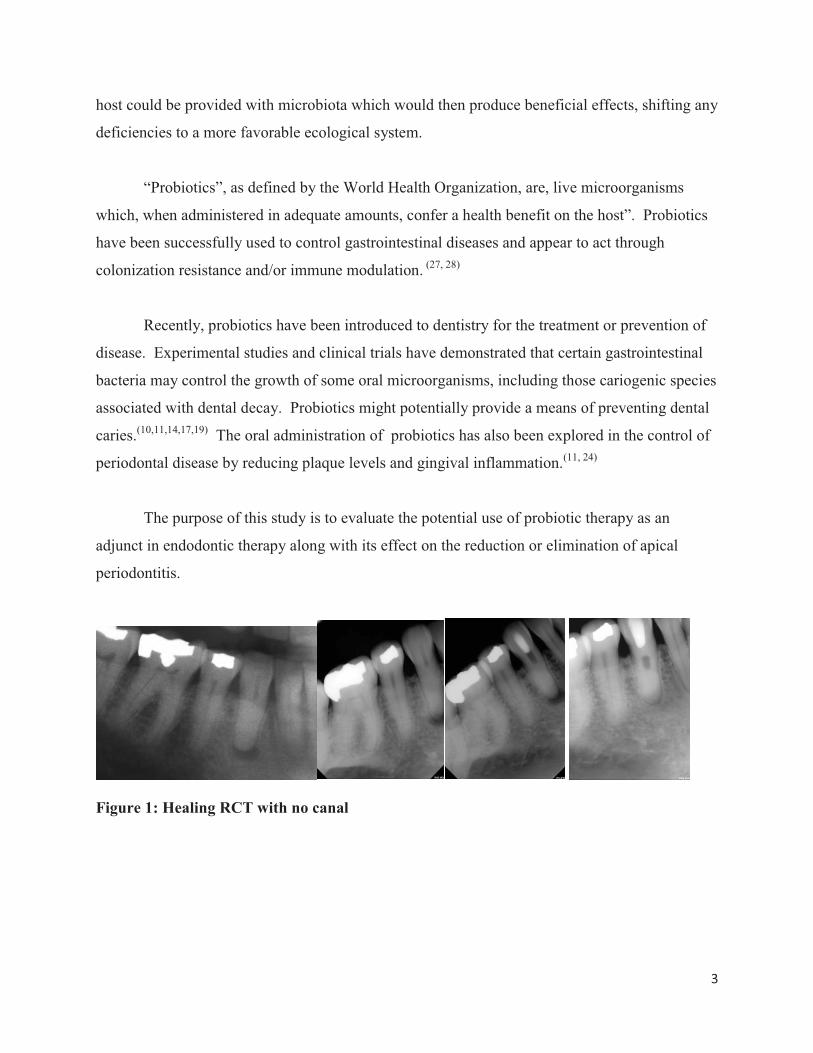
The purpose of this study is to evaluate the potential use of probiotic therapy as an
adjunct in endodontic therapy along with its effect on the reduction or elimination of apical
periodontitis.
Figure 1: Healing RCT with no canal
Page 14
4
Statement of the Problem
Does the use of probiotics have the potential to outcompete or eliminate pathogenic
microorganisms in endodontic therapy?
Significance of the Problem
There is a need for an innovative or novel approach to the current treatment modalities
which can possibly result in a higher, long term success in endodontic therapy. Despite the
universal and widely spread advancement of technology throughout all facets of dentistry, the
same basic approach, in conjunction with the same materials, has been employed over the past
several decades in providing endodontic treatment. The concept of the “Human Microbiome”
has been neglected in the development of new and better strategies used in endodontic therapy,
even though it is now known to be an integral and undeniable part of human health. The novel
concept in endodontics that the intentional establishment of a microbial equilibrium inside the
root canal system by utilizing probiotics, a procedure which might result in improved overall
success rates in the reduction of apical periodontitis, needs to be carefully examined and
explored. As previously mentioned, the use of probiotics has gained universal acceptance by the
gastroenterology community and some probiotics have been shown to be successful in treating
certain gastrointestinal diseases. The concept of using probiotics has recently been utilized for
the prevention of both dental caries and periodontal disease. Periodontal disease is known to
have a number of pathogenic microorganisms in common with those found in endodontic
infections, and therefore utilizing probiotics within the root canal system may also be found to be
beneficial in endodontics.
Page 15
5
Null-Hypothesis
There is no significant difference between probiotic therapy and no therapy in eliminating
or decreasing the amount of Enterococci faecalis and Candida albicans in planktonic and
biofilm microbial stages when tested in an in vitro model.
Assumptions
1. The Human Microbiome theory is gaining acceptance in medicine but has not yet been
evaluated in endodontics.
2. Maintaining or restoring equilibrium with probiotics may show promising results in
endodontic therapy.
3. Although sterility of the endodontic system is deemed necessary for endodontic success,
achieving complete sterility is currently impossible under normal conditions.
4. Probiotics used against test organisms evaluated in vitro in both planktonic and biofilm
stages in terms of measurement of zones of inhibition (ZOE) is an acceptable method of
evaluation of the efficacy of the probiotics against pathogenic organisms.
5. Synergistically acting probiotics tested against pathogenic organisms may give a broader
understanding of which particular species to select when conducting future studies.
6. Gram staining is an effective method to evaluate which organisms are present in blood
agar/MRS or Muller Hinton plates.
7. Live/dead staining is an appropriate method of identifying probiotic activity.
8. Poloxamer 147 mixed with MRS broth and probiotics is a novel delivery vehicle for the
introduction of probiotics into the root canal system as an intra-canal medicament.
Page 16
6
Limitations
1. Due to financial limitations, commercial probiotics were utilized, and probiotic
organisms were extracted either in groups or individually, according to manufacturer’s
instruction.
2. Due to time limitations, experiments were not performed in teeth.
3. As in any experimentation, human error may exist.
4. There is no known standardized method to identify different strains of microorganisms
mixed together in a single group.
5. There have been no studies involving the use of probiotics in endodontics. The effective
CFU count needed to eliminate or out compete the pathogenic organisms was unknown.
Delimitations
1. Manufacturer was contacted to determine the method for extraction of probiotic species
from commercial samples.
2. 50 Muller Hinton plates were utilized for initial evaluation.
3. 100 blood agar plates were utilized for evaluation of probiotic mixture against pathogenic
organisms.
4. Probiotics and pathogenic organisms E. feacalis and C. albicans were tested in both
planktonic and biofilm stages.
Page 17
7
Chapter II
Review of Literature
The microbial component of humans and animals has been termed the indigenous
microbiota. Experimental evidence shows that the microbiome is necessary for the health of the
host, and that alterations in the ecological equilibrium of these microbes can lead to disease. (26)
It is therefore logical to expect that the introduction of microbes that are also members of the
microbiome into an area of interest could help restore an ecological balance. (26)
A proposed solution to maintaining or restoring equilibrium would be to provide the host
with microbiota which would produce perceived beneficial effects, shifting any deficiencies to a
more favorable ecological system. The term “probiotics” is defined by the World Health
Organization as live microorganisms which, when administered in adequate amounts, confer a
health benefit on the host. The word “probiotics” was derived from the Greek, meaning “for
life”. The concept of probiotics is not new, but rather dates back to 1908, when Nobel Prize
winner, Ukrainian bacteriologist Ilya Metchnikoff, suggested that the long life of Bulgarian
peasants resulted from their consumption of fermented milk products which contained
lactobacillus. (11,12,13,14,15,16,17,18,19,20,21,22,23,24)
Another solution to maintaining and/or restoring microbial equilibrium would be to
administer substrates that improve the growth or metabolic activities of specific indigenous
organisms, or so called, “prebiotics”. The term prebiotic was introduced by Gibson and
Roberfroid who exchanged “pro” for “pre”, meaning “before”. They defined prebiotics as a
“non-digestible food ingredient that beneficially affects the host by selectively stimulating the
growth and/or activity of one or a limited number of bacteria in the colon”. (18) Experimental
models, along with several human studies, have shown that food ingredients, products, and
supplements demonstrating a prebiotic effect, have been shown to modulate certain
Page 18
8
immunological biomarkers and affect activities of the immune system by inducing change in the
gut microbiota. (29)
The term “synbiotic” is used when a product contains both probiotics and prebiotics.
According to this approach, a food or food supplement will include not only live cells of the
beneficial bacteria, but also their selective substrates, with the idea being that beneficial bacterial
cells can grow quickly and competitively because of the presence of the selective substrate which
allows it to predominate in the proposed environment.(18)
The introduction of these probiotics, prebiotics and synbiotics are coined as
“Bacteriotherapy”. Bacteriotherapy has been investigated in multiple studies to control
infectious diseases, especially gastrointestinal disease, with the objective being the restoration
and balance of the human microbiome. (14)
The suggested mechanisms of probiotic action on oral health are drawn from
gastrointestinal studies. These several mechanisms include but are not limited to, immune
modulation, down regulation of inflammatory responses, production of antimicrobial substances
such as peroxides, organic acids and bacteriocins, mucin production, inhibition of epithelial
invasion by inhibition of pathogens mucosal adherence, stimulation of IgA, and competition with
other flora, including potential pathogens. (12, 13, 14, 17, 18, 19)
Ideal features of a good probiotic would be:
(a) It should be a strain which is capable of exerting beneficial effect to the host
(b) It should be non-pathogenic and non-toxic
(c) It should be present as viable cells, preferably in large numbers
(d) It should be capable of surviving in the host environment
(e) It should also be able to maintain genetic stability in oral micro flora
(f) It should be stable and capable of remaining viable for periods under storage and field
conditions. (12, 13, 14, 17, 18, 19)
The most common probiotic bacteria belong to the Lactobacilli and Bifidobacteria
genera, but certain strains of Streptococci have also been investigated. (22) The reasoning behind
Page 19
9
why Lactobacillus species were chosen for this experiment is because they aide in producing
those enzymes which digest and metabolize proteins and carbohydrates. They also aid in the
synthesis of vitamins B and K, facilitating the breakdown of bile salts. Additionally, they have
the ability to help enhance innate and acquired immunity, along with inhibiting pro-
inflammatory mediators. Lactobacilli are considered to be a genus of gram positive facultative
anaerobic microorganisms, with more than 100 species identified. Most notable are the strains
L. acidophilus, L. salivarius, L. rhamnous, L. brevis, and L. casei which are utilized as
probiotics. (14)
Another organism as mentioned above is the Bifidobacterium species which are strictly
gram positive anaerobes and which are the dominant organism found in the large intestine. Over
30 species of Bifidobacterium have been identified. Their characteristics include metabolism of
lactose, generation of lactic ions from lactic acid, vitamin synthesis, fermentation of indigestible
carbohydrates, and production of beneficial short chain fatty acids. (10, 12, 14, 17, 18, 19) Other species
such as Streptococcus thermophillus are the organisms used as the chief cultures in yogurt
production, owing to their distinguishing benefits of metabolism of lactose and improving
lactose intolerance, while also possessing antimicrobial activity. Saccharomyces boulardii is a
non-colonizing lactic acid producing yeast. Their most preeminent feature is that they secrete
proteases and other substances that break down bacterial enterotoxins. They also help in the
enhancement of immune function and have been shown to be beneficial in helping with C.
difficile management. (30)
Recently, probiotics have been introduced in dentistry as an adjunct for the treatment or
prevention of oral diseases. Currently, probiotic therapy has been investigated in experimental
studies and clinical trials in an attempt to establish equilibrium in the oral component of the
human microbiome. This philosophy would eliminate microorganisms associated with disease
by allowing others associated with health to evolve and predominate.
Experimental studies and clinical trials are beginning to show advancement in multiple fields
of dentistry such as:
Page 20
10
� Caries Control:
In saliva, caries associated microbes such as Streptococcus mutans have been
shown to be reduced in number after the consumption of products containing the
probiotics Lactobacillus and Bifidobacteria. (11, 21)
� Periodontal disease:
Initial studies suggested that the use of probiotics could enhance oral health by
decreasing periodontal inflammation. (31) Subsequent studies evaluating patients who
presented with various forms of periodontal disease such as gingivitis, pregnancy
gingivitis and periodontitis, showed significant recovery after treatment with a culture of
the L. acidophilus strain in most patients. (32) Another study evaluated probiotic strains
including L. reuteri , L. brevis and L. casei which revealed an improvement in gingival
health, as measured by decreased gum bleeding. (33) Further studies evaluated L. reuteri,
L. brevis and L. salivarius probiotic strains against inflammatory markers where L.
reuteri showed decreased levels of pro-inflammatory cytokines in gingival crevicular
fluid(33) and the use of L. brevis and L. salivarius decreased matrix metalloproteinase
activity along with other inflammatory markers in saliva.(34)
� Oral Candidiasis:
A preliminary study investigated the probiotic bacteria L. acidophilus and L.
fermentum in oral cavities, which resulted in a rapid decline in C. albicans after the
intake of the probiotics. Further consumption led to an almost undetectable number of
fungi in the oral cavity. (35) Another study evaluated L. rhamnosus and
Propionibacterium freudenreichii ssp. shermanii for the effect on oral candida infection
in humans. After 16 weeks of therapy, the number of high oral yeast counts decreased,
but no changes were observed in mucosal lesions. (35).
� Halitosis:
A few clinical studies have found probiotic strains effective for the treatment of
oral or gut associated halitosis. The studied strains included Lactobacillus, E. coli Nisle,
S. salivarius and Weissella confusa isolates.(36) In endodontics, it has long been held that
there are three basic principles that must be adhered to in order to achieve success. This is
known as the endodontic triad where, if followed, the end result of endodontic treatment
should be both clinical and radiographic success. These three “principles” are (1)
Page 21
11
thorough debridement of the root canal system, (2) sterilization of the root canal system,
and (3) complete obturation of the root canal system. As previously stated, the question
which still remains today is whether or not microorganisms can be eliminated from an
infected root canal system.
It has been proposed that attaining complete sterility in any part of the human oral cavity
is impossible. Histological examination of serial sections of the roots of many teeth have
revealed the prevalence of multiple accessory and lateral canals.(9) It is inconceivable that these
branches can be either debrided properly or made completely free of bacteria. All that can be
reasonably expected and achieved in conventional root canal therapy is a reduction in the number
of microorganisms within the main canal. Any success obtained from treatment of teeth with
positive root canal cultures can be ascribed to a reduction in the number of microorganisms,
removal of most inflamed or necrotic tissue, and a favorable systemic background. (1, 9) Multiple
pathogenic organisms have been attributed to endodontic failure, but the two organisms in
particular most commonly associated with treatment failure are E. faecalis and C. albicans.(8)
E. faecalis is resistant to most of the intra-canal medicaments, particularly calcium
hydroxide dressings. This is due to its ability to regulate internal pH with an efficient proton
pump, as well as its ability to survive prolonged starvation. Although endodontic infections are
considered to be polymicrobial in nature, it has been shown that E. faecalis is the pathogen of
significance in most failing endodontic treatment cases. (37, 38)
Microbiological and correlative electron microscopic studies have shown the presence of
yeasts in canals of teeth with apical periodontitis.(1) Candida albicans is the most frequently
isolated fungus from root filled teeth with apical periodontitis.(3)
The literature shows that there is a need for an innovative method of handling endodontic
infections other than the currently used methods. A promising approach would be to manage
endodontic treatment as part of the human microbiome and utilize probiotics in the same manner
that they are used for other oral conditions to reestablish equilibrium of healthy flora.
Probiotics testing should be done against both planktonic and biofilm stages, the rationale
being that although planktonic organisms represent free floating and homogeneous microbial
Page 22
12
cells, there has been a paradigm shift showing a link between surface attached, heterogeneous
microbial cells (biofilms) and microbial pathogenesis, which then leads to human infections.(39)
Biofilms are defined as highly structured communities of microorganisms that are either surface
associated or attached to one another and which are enclosed within a self-produced, protective
extracellular matrix (ECM). Biofilm formation provides protection from the environment,
resistance of physical and chemical stress, metabolic cooperation, and a community based
regulation of gene expression. (39, 40, 41) These features allow organisms (bacteria and fungi) in
biofilms to assume a stronger pathogenic potential than those solely in a planktonic state. (40)
There is also evidence showing a major role of fungi in biofilm formation and disease. (41)
Chapter III
Materials and Methods
� Probiotic Strain Selection:
Due to financial limitations, individual probiotic strains were not able to be purchased
from ATCC (American Type Culture Collection). Alternatively, commercial probiotic cocktails
were purchased for utilization in this study, decreasing financial burden. After extensive
research was conducted about effective probiotic strains used to establish equilibrium of the gut
flora, five probiotic cocktails were purchased from Klaire Labs®. The probiotic cocktails were
delivered in wrapped ice packaging in order to preserve viability of the organisms. Upon
arrival, the probiotics were stored in a refrigerator at 30º F. Each group of probiotic blend was
designated a specific color to easily identify the group for the study. The five groups (G I, GII,
GIII, GIV and GV) of commercial probiotics were selected and evaluated based upon numbers
and concentration of organisms.
Page 23
13
The five commercial groups of probiotics were as follows:
I. Group 1: Designated Color =YELLOW
Vital-Immune Biotic® Caps
Amount Per Capsule % Daily Value
Probiotic Blend (5+ billion CFUs) in a baseof inulin (derived from chicory root)
460 mg *
Lactobacillus rhamnosus 2.0+ billion CFUs
*
Lactobacillus casei 1.5+ billion CFUs
*
Lactobacillus acidophilus 1.0+ billion CFUs
*
Bifidobacterium longum 0.5+ billion CFUs
*
Figure 2: Probiotic Group 1 (Yellow)
II. Group 2: Designated Color =RED
ABx Support™
Amount Per Capsule % Daily Value
Probiotic Blend (10+ billion CFUs) in abase of inulin (derived from chicoryroot)
430 mg *
Saccharomyces boulardii 5.0+ billion CFUs
*
Lactobacillus rhamnosus 2.5+ billion CFUs
*
Bifidobacterium bifidum 1.25+ billion CFUs
*
Bifidobacterium breve 1.25+ billion CFUs
*
Figure 3: Probiotic Group 2 (Red)
S
Page 24
14
III. Group 3: Designated Color = PURPLE
Sacchromyces Boulardii
Amount Per Capsule % Daily Value
Probiotic Blend in a base of cellulose 320 mg *
Saccharomyces boulardii 3+Billion CFUs*
Figure 4: Probiotic Group 3 (Purple)
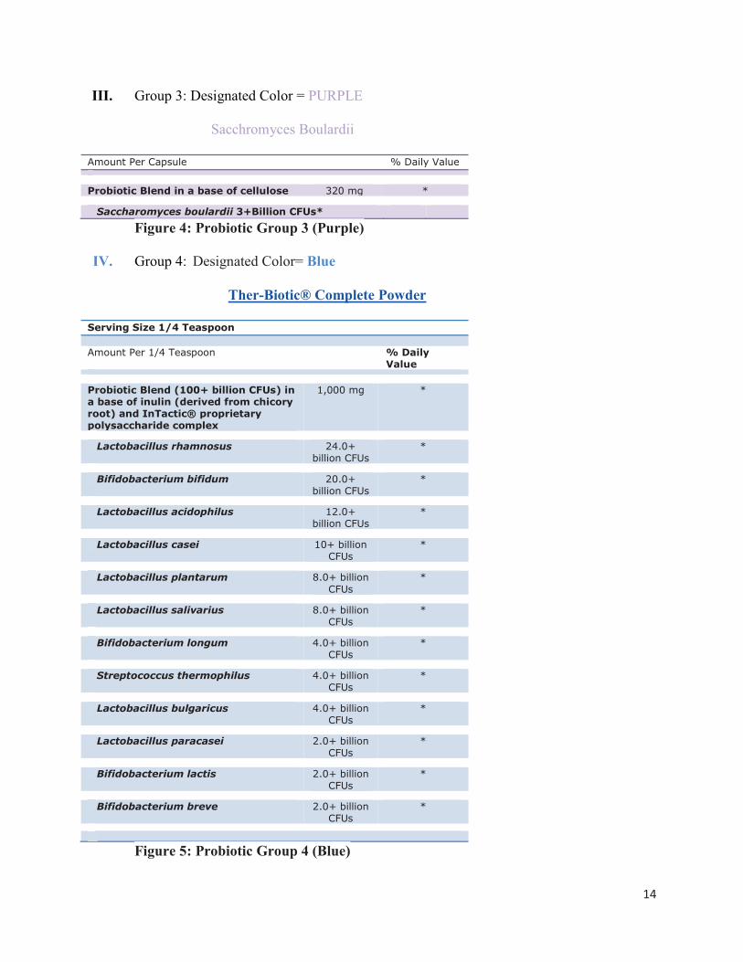
IV. Group 4: Designated Color= Blue
Ther-Biotic® Complete Powder
Serving Size 1/4 Teaspoon
Amount Per 1/4 Teaspoon % Daily Value
Probiotic Blend (100+ billion CFUs) in a base of inulin (derived from chicory root) and InTactic® proprietary polysaccharide complex
1,000 mg *
Lactobacillus rhamnosus 24.0+ billion CFUs
*
Bifidobacterium bifidum 20.0+ billion CFUs
*
Lactobacillus acidophilus 12.0+ billion CFUs
*
Lactobacillus casei 10+ billion CFUs
*
Lactobacillus plantarum 8.0+ billion CFUs
*
Lactobacillus salivarius 8.0+ billion CFUs
*
Bifidobacterium longum 4.0+ billion CFUs
*
Streptococcus thermophilus 4.0+ billion CFUs
*
Lactobacillus bulgaricus 4.0+ billion CFUs
*
Lactobacillus paracasei 2.0+ billion CFUs
*
Bifidobacterium lactis 2.0+ billion CFUs
*
Bifidobacterium breve 2.0+ billion CFUs
*
Figure 5: Probiotic Group 4 (Blue)
Page 25
15
V. Group 5: Designated Color = GREEN
Vital-10® Powder
Amount Per 1/4 Teaspoon % Daily Value
Probiotic Blend (10+ billion CFUs) in a base of inulin (derived from chicory root)
1,000 mg *
Lactobacillus acidophilus 3.7+ billion CFUs
*
Bifidobacterium bifidum 1.4+ billion CFUs
*
Proprietary Blend of: 4.9+ billion CFUs
*
Lactobacillus bulgaricus
Lactobacillus rhamnosus
Lactobacillus brevis
Streptococcus thermophilus
Lactobacillus casei
Lactobacillus salivarius
Lactobacillus plantarum
Bifidobacterium lactis
Figure 6: Probiotic Group 5 (Green)
Figure 7: Color coding of probiotics Figure 8: Commercial Probiotics utilized
Page 26
16
� Pathogenic Strain Selection:
E. faecalis was chosen for this study after extensive literature review which had revealed that this
organism possesses multiple properties leading to its key role as an endodontic pathogen. Some
of these features: (37, 38):
1. It is resistant to most of the intra-canal medicaments, particularly calcium hydroxide
dressing due to its ability to regulate internal pH with an efficient proton pump.
2. E. faecalis can survive prolonged starvation.
3. Controlled studies have shown that E. faecalis is the pathogen of significance in most
cases of failing endodontic treatment.
C. albicans was chosen as another pathogenic test organism due to: (1, 3)
1. It’s biphasic nature which allows it to be the universal co-aggregate in biofilms.
2. It is the most frequently isolated fungus from root filled teeth with apical periodontitis.
� Extraction of Probiotic Organisms From Commercial Products:
Klaire® Labs, a division of Prothera Inc®, was contacted after purchasing and receipt of
the probiotics that were intended for experimentation. The lab forwarded an extraction method
for re-growing and culturing the microorganisms.
The following protocol was followed for the extraction/culturing method:
1. An aseptic protocol was followed for every extraction method via spraying the
operative fields with 99% ethyl alcohol, followed by Cavicide™ spray or wipes. The operative
fields were then left to dry. For manipulation of probiotics and extraction processes, sterile
gloves were utilized to decrease any cross-contamination. All instruments such as sterile plastic
pliers/loops were discarded after single use.
2.1.1 Grams of the dried probiotic powder of groups 1 through 5 were weighed
aseptically on a lab scale. The measured powder was aseptically placed into sterile 15 ml
test tubes containing 10 ml of sterile MRS broth.
Page 27
17
3. The tubes were then vortexed at room temperature on a vortex mixer (Fisher
Scientific™ Digital mixer) for two minutes until the mixture was homogenous.
4. The samples were then kept at room temperature for 30 minutes to assure rehydration
of the freeze-dried powder.
5. The samples were then returned to the vortex machine and vortexed for an additional
two minutes.
6. Samples where incubated at 37° C in an incubator for 48 hours (an anaerobic chamber
would have been preferred for growth of the microorganisms, but was unavailable).
7. Samples were then placed in the lab refrigerator at 4° C for no longer than two weeks
before being used for testing to avoid any mutation in the test species. New probiotic
stock was made every two weeks.
8. Samples that were to be utilized for testing were transferred via a sterile pipette from
the stock solution into 9 ml of MRS broth in sterile tubes and were adjusted to a
McFarland Standard of 1 for standardization (1 McFarland = 3 X 10⁸ CFU/ml) .
9. To insure growth of microorganisms, 0.5 ml of the 1 McFarland mixture of the
probiotic groups was transferred via a micropipette and spread on a blood agar plate or Mueller
Hinton plates with an L-spreader, followed by incubation for 24 hours, 48 hours and 1 week.
This was followed by gram staining and growth observation (CFU observation).
(A) (B) (C) (D)
Figure 9: (A)L-spreader, (B) Micro-Pipette, (C) Sterile pliers and Loops, (D) Vortex
machine
Page 28
18
(A) (B)
Figure 10: (A) Probiotics in TSB broth (posterior) and MRS broth(anterior), (B)Probiotics in TSB broth
Figure 11: Cultered proibiotics on Muller Hinton plates
Page 29
19
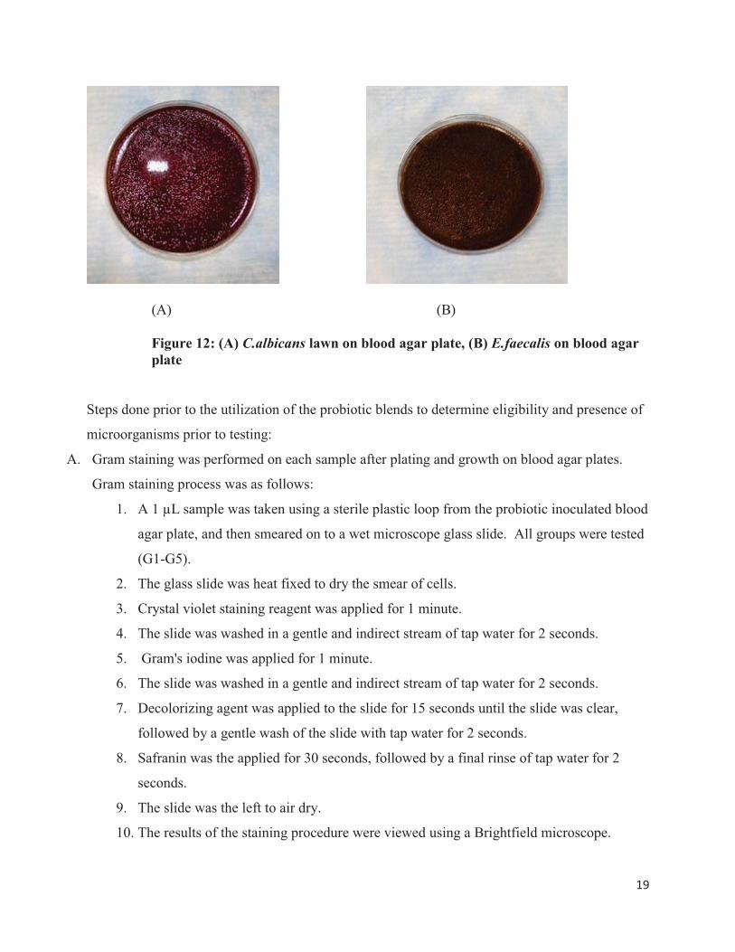
(A) (B)
Figure 12: (A) C.albicans lawn on blood agar plate, (B) E.faecalis on blood agar plate
Steps done prior to the utilization of the probiotic blends to determine eligibility and presence of
microorganisms prior to testing:
A. Gram staining was performed on each sample after plating and growth on blood agar plates.
Gram staining process was as follows:
1. A 1 µL sample was taken using a sterile plastic loop from the probiotic inoculated blood
agar plate, and then smeared on to a wet microscope glass slide. All groups were tested
(G1-G5).
2. The glass slide was heat fixed to dry the smear of cells.
3. Crystal violet staining reagent was applied for 1 minute.
4. The slide was washed in a gentle and indirect stream of tap water for 2 seconds.
5. Gram's iodine was applied for 1 minute.
6. The slide was washed in a gentle and indirect stream of tap water for 2 seconds.
7. Decolorizing agent was applied to the slide for 15 seconds until the slide was clear,
followed by a gentle wash of the slide with tap water for 2 seconds.
8. Safranin was the applied for 30 seconds, followed by a final rinse of tap water for 2
seconds.
9. The slide was the left to air dry.
10. The results of the staining procedure were viewed using a Brightfield microscope.
Page 30
20
11. At the completion of the gram stain, it is noted that gram negative bacteria stain
pink/red whereas gram positive bacteria stain blue/purple.
Figure 13: Gram Stain
Figure 14: Gram stained probiotic slides
Figure 15: Bright field microscope
Page 31
21
B. Live/dead staining was performed by live/dead Baclight™ bacterial viability kits for
evaluation of cell viability for all test organisms.
The following protocol was followed: Staining Bacteria in suspension with kit L13152
1. A 2X stock solution of the live/deadD BacLight staining reagent mixture was prepared
by dissolving the contents of one component A pipet (containing yellow-orange solids)
and one component B pipet (containing red solids) in a common 5 mL–volume of filter-
sterilized H2O.
2. A sample of the 2X stock solution was combined with an equal volume of the bacterial
suspension. The final concentration of each dye will be 6 μM SYTO 9 stain and 30 μM
propidium iodide.
3. The resulting solution was mixed thoroughly and incubated at room temperature in the
dark for 15 minutes.
4. 5 μL of the stained bacterial suspension was trapped between a slide and an 18 mm
square coverslip.
5. Fluorescence was observed under a Zeiss ® Axiovert 40 CFL™ inverted microscope
with X-Cite® 120q wide field fluorescence microscope excitation source.
Figure 16: Live/dead stain slides prepared for fluorescent microscope
Page 32
22
Figure 17: X-Cite® 120q wide field fluorescence microscope excitation source
Figure 18: Zeiss ® Axiovert 40 CFL™ inverted microscope
Figure 19: C. albicans budding under fluorescent microcsopy
Page 33
23
Figure 20:Clusters of probiotic group 5 under fluorescent microcsopy
Figure 21: Cluster of probiotic group 4 under fluorescent microcsopy
Page 34
24
Phase 1: Testing for Probiotic efficacy against E. faecalis and C. albicans;
Planktonic Stage Evaluation.
A disc diffusion assay test was conducted. The purpose of the Kirby-Bauer disc diffusion
susceptibility test is to determine the sensitivity or resistance of pathogenic aerobic and
facultative anaerobic bacteria to various antimicrobial compounds. In this study, probiotics
where used to determine their antimicrobial effect against pathogenic organisms. The
pathogenic organisms were grown on blood agar in the presence of test probiotic group
impregnated filter paper discs. Observation of the presence or absence of growth around the
discs is an indirect measure of the ability of that probiotic group to inhibit growth or out compete
the pathogenic organisms E. faecalis or C. albicans.
Testing: An aseptic technique (sterile pipette carriers, alcohol and Cavicide wipes for surfaces,
sterile cotton pliers, sterile loops and sterile L-spreaders) was utilized throughout the procedure
to insure sterile environment, as well as prevention of cross contamination.
1. The five probiotic groups were extracted according to manufacturer’s instructions and
incubated for 48 hours.
2. Probiotic samples were then placed in 9ml MRS broth (De Man, Rogosa and Sharpe
broth) and vortexed to insure a homogenous mixture, then set to a 2 McFarland standard
via laser spectrophotometry (average 6 x 10⁸ CFU/ml).
3. The pathogenic organisms E. faecalis and C. albicans were freshly stocked, placed in 9
ml TSB (tryptic soy broth) and vortexed to insure homogenous mixture, then set to a 1
McFarland standard via laser spectrophotometry (average 3 x 10⁸ CFU/ml).
4. 500 microliters of E. faecalis was plated on 100 mm diameter blood agar plates and
spread with a sterile L-Loop. The sample was incubated for 24 hours to allow growth of
a bacterial lawn.
5. 500 microliters of C. albicans was plated on 100 mm diameter blood agar plates and
spread with a sterile L-Loop. The sample was incubated for 24 hours to allow growth of a
bacterial lawn.
Page 35
25
6. 20 microliters of probiotic G I through G V were placed on sterile blank paper discs and
left for 15 seconds to allow the discs to saturate with the probiotic cocktails. The discs
were then transferred to the previously grown lawns of the pathogenic test organisms, E.
faecalis and C. albicans, according to a 5 group template as seen in figure (22).
7. The blood agar plates were then incubated at 37 ° C and evaluated at 48 hours and 168
hours (1 week), respectively.
8. The test was conducted three times per group against the organisms, E. faecalis and C.
albicans to allow proper statistical analysis.
9. The control was conducted by growing E. faecalis and C. albicans lawns at a 1
McFarland standard with empty sterile discs placed on the lawns.
10. Results for ZOI’s were measured with a digital micrometer in mm increments at 48 hours
and 168 hours (1 week).
Figure 22: Five disc probiotic template on plate
Page 36
26
Figure 23: Digital Micrometer
Figure 24: Light Spectrophotometer
Page 37
27
Figure 25: Illustration of five disc probiotic template on plates
Figure 26: Five disc probiotic template on blood agar plate
1
5 2
3
4
Page 38
28
Figure 27: Six plates with five disc probiotic template on blood agar plates
Figure 28: Control four disc template on Muller Hinton plate
Phase 2: Biofilm stage testing; Intra-canal delivery vehicle for probiotics
In this phase of the study, we evaluated biofilm staging by mixing pathogenic organisms
with probiotic cocktails from individual groups, followed by serial dilutions and colony forming
unit counts per ml. Only G I and G IV were evaluated for this part of the study, since they
demonstrated the most significant zones of inhibition. G V was excluded from the study due to
patent pending status of the probiotic cocktail.
Stage 1: Making of the Delivery Vehicle
Poloxamer F127 (407) was prepared by adding dry powder to the appropriate type
and amount of broth (MRS and TSB were both utilized). Prebiotic mixtures (Inulin,
Page 39
29
Oligofructose , beta-glucan, larch arabinogalactan, glycerin and trechalose) were initially
mixed and were considered to be included as part of the formulation, but due to time
limitations of the study and to avoid having too many variables, it was decided to remove
these prebiotics from the mixture as well as to prove the concept of probiotics
outcompeting pathogenic organisms in the biofilm stage. This suggests that further
studies be conducted with prebiotics added to the mixture and possibly enhancing the
probiotic effect.
The poloxamer was dissolved in cold MRS broth at a concentration of 30% by a
magnetic stirrer for 10 to 15 minutes until a homogenous mixture was obtained. The poloxamer
was then sterilized and placed in the refrigerator at 4° C until testing was conducted. The MRS
broth mixture was utilized for this study.
Figure 29: 30 % Poloxamer 407 (Pluronic F-127) with MRS broth
Figure 30: Magnetic Stirrer
Control testing:
Page 40
30
E. faecalis and C. albicans stocks were both prepared in TSB broth to a McFarland
Standard of 1 via light spectrophotometry. A total of 9 mls of poloxamer was placed in a test
tube and 500 ml of pathogenic organisms were added and vortexed at 4 °C in a refrigerated
environment to allow homogenous mixture of poloxamer and microorganisms. Samples were
then incubated for 48 hours in an incubator at 37° C.
After 48 hours, serial dilutions of the pathogenic biofilm samples were prepared and
plated on blood agar plates to evaluate colony forming units of the organisms. Serial dilutions
were made by adding 0.1 ml of poloxamer mix to 9.9 ml sterile saline, followed by serially
diluting the mixture by 0.1ml into 9.9 ml sterile saline three times, reaching dilutions of 10-2,
10-4,10-6 respectively. Plating was conducted by adding 500 ml of dilutions onto blood agar
plates, followed by incubation at 37° C for 72 hours. CFU counts were evaluated.
Figure 31: (A) and (B) Control E.f and C.a in poloxamer 407
(A) (B)
Figure 32: Serial Dilutions of E.faecalis and C.albicans
Page 41
31
Figure 33: CFU/mL C.albicans 10-4 dilution
Figure 34: CFU/mL E.faecalis 10-4 dilution
Testing for probiotic/pathogenic organism Poloxamer mixture:
1. 9 ml of the poloxamer mixture was placed in a test tube along with 500 ml of E. faecalis
at a 1 McFarland standard. Following that, 500 ml of test probiotic group 1 was added at
1.5 to 2 McFarland standards.
Page 42
32
2. 9ml of the poloxamer mixture was placed in a test tube and 500 ml of E. faecalis was
added at a 1 McFarland standard. Following that, 500 ml of test probiotic group 4 was
added at 1.5 to 2 McFarland standards.
3. 9ml of the poloxamer mixture was placed in a test tube and 500 ml of C albicans at a 1
McFarland standard was added. Following that, 500 ml of test probiotic group 4 was
added at 1.5 to 2 McFarland standards.
4. 9ml of the poloxamer mixture was placed in a test tube and 500 ml of C. albicans at a 1
McFarland standard was added. Following that, 500 ml of test probiotic group 4 was
added at 1.5 to 2 McFarland standards.
5. After 48 hours serial dilutions of the pathogenic biofilm samples were prepared and
plated on blood agar plates to evaluate colony forming units of the organisms.
6. Serial dilutions were made by adding 0.1 ml of poloxamer mix to 9.9 ml sterile saline ,
followed by serially diluting the mixture by 0.1ml into 9.9 ml sterile saline three times,
reaching dilutions of 10-2, 10-4 ,10-6 respectively.
7. Plating was conducted by adding 500 ml of the dilutions onto blood agar plates followed
by incubation at 37° C for 72 hours. CFU (colony forming units) were evaluated for all
test groups and compared to controls based upon the dilutions that were performed to
reflect the actual number of probiotics and pathogenic organisms in each group.
Page 43
33
Figure 35: Poloxomer with pathogenic organisms and test probiotics 4 and 1 mixed together
Figure 36: Serial dilutions of poloxamer/probiotic/pathogenic organisms mixed together.
Page 44
34
Figure 37: Serial dilutions of poloxamer/probiotic/pathogenic organisms mixed together on blood agar plates.
Page 45
35
Chapter IV
Results
Phase 1: ZOI
Probiotics were screened for their antimicrobial/outcompeting activity against the
selected microbes, E. faecalis and C. albicans.
The preliminary screening showed that G I, IV and V had antimicrobial activity against
the specific pathogenic microbes tested. Clear zones of inhibition were seen around G I, IV and
V with valid statistical significance (P<0.0001). Groups II and III showed variable results and
therefore it could not be concluded that they are fully effective against the specific pathogenic
organisms tested, and were subsequently eliminated from the biofilm stage testing. One
observation in this study was that G I, IV and V all contained the common organisms L.
acidophilus, L. casei , L. rhamnosus and B .longum in their groups, suggesting that further
studies should focus on these particular organisms. Another observation was that prior to zones
of inhibition being seen, a zone of growth was noted 24 hours after plating, which was then
followed by the observation of zones of inhibition beginning at 48 hours. G III (Sachromyces
boulardii) demonstrated a continuous ring or zone of growth around the disc with a small zone of
inhibition against either of the pathogenic organisms. G IV and V showed the largest ZOI
diameters.
Page 46
36
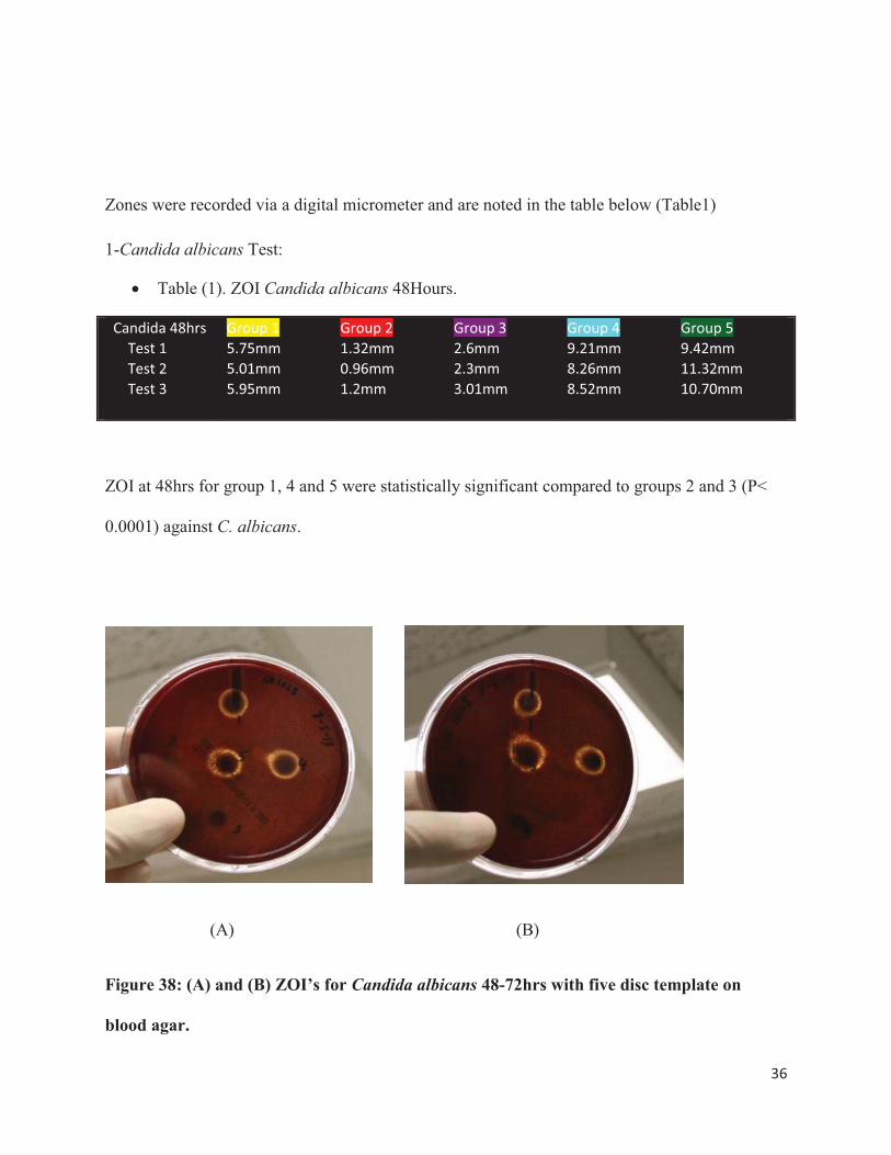
Zones were recorded via a digital micrometer and are noted in the table below (Table1)
1-Candida albicans Test:
� Table (1). ZOI Candida albicans 48Hours.
Candida 48hrs Group 1 Group 2 Group 3 Group 4 Group 5 Test 1 5.75mm 1.32mm 2.6mm 9.21mm 9.42mm Test 2 5.01mm 0.96mm 2.3mm 8.26mm 11.32mm Test 3 5.95mm 1.2mm 3.01mm 8.52mm 10.70mm
ZOI at 48hrs for group 1, 4 and 5 were statistically significant compared to groups 2 and 3 (P<
0.0001) against C. albicans.
(A) (B)
Figure 38: (A) and (B) ZOI’s for Candida albicans 48-72hrs with five disc template on
blood agar.
Page 47
37
Figure 39 :Control Candida albicans on blood agar plate.
Figure 40: C.albicans and E.faecalis ZOI’s (multiple plates).
Figure 41: ZOI’s for Candida albicans 1 week with five disc templates on blood agar.
Page 48
38
Figure 42: Zone of growth 24hrs for Candida albicans on blood agar.
� Table (2). ZOI Candida albicans 1 week.
Candida 1 week
Group 1 Group 2 Group 3 Group 4 Group 5
Test 1 7.05 mm 2.1mm 3.21mm 9.94mm 10.79mm Test 2 7.24mm 2.13mm 3.26mm 9.82mm 12.64mm Test 3 7.95mm 1.89mm 3.3mm 10.4mm 12.53mm
ZOI at 1 week for group 1, 4 and 5 were statistically significant compared to groups 2 and 3 (P< 0.0001) against C. albicans.
Statistical analysis was conducted by least squares means and one way ANOVA for comparing all groups.
Page 49
39
(A) (B)
Figure 43: (A) and (B) ZOI’s for Candida albicans one week with five disc template on blood agar.
� Whole Model for Candida albicans:48hrs and 1 week
� Table ( 3 ) .Analysis of variance (ANOVA) for Zone of Inh by Group =CA
Analysis of VarianceSource DF Sum of
SquaresMean Square F Ratio
Model 9 415.49452 46.1661 153.8561Error 20 6.00120 0.3001 Prob > FC. Total
29 421.49572 <.0001*
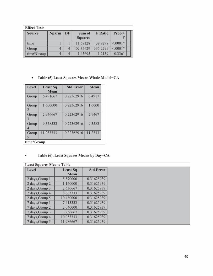
� Table (4). Effect Tests for Zone of Inh by Group =CA
Page 50
40
Effect TestsSource Nparm DF Sum of
SquaresF Ratio Prob >
Ftime 1 1 11.68128 38.9298 <.0001*Group 4 4 402.35629 335.2299 <.0001*time*Group 4 4 1.45695 1.2139 0.3361
� Table (5).Least Squares Means Whole Model=CA
Level Least Sq Mean
Std Error Mean
Group 1
6.491667 0.22362916 6.4917
Group 2
1.600000 0.22362916 1.6000
Group 3
2.946667 0.22362916 2.9467
Group 4
9.358333 0.22362916 9.3583
Group 5
11.233333 0.22362916 11.2333
time*Group
• Table (6) .Least Squares Means by Day=CA
Least Squares Means TableLevel Least Sq
MeanStd Error
2 days,Group 1 5.570000 0.316259392 days,Group 2 1.160000 0.316259392 days,Group 3 2.636667 0.316259392 days,Group 4 8.663333 0.316259392 days,Group 5 10.480000 0.316259397 days,Group 1 7.413333 0.316259397 days,Group 2 2.040000 0.316259397 days,Group 3 3.256667 0.316259397 days,Group 4 10.053333 0.316259397 days,Group 5 11.986667 0.31625939
Page 51
41
� Figure 44: LS Means Plot Candida albicans
(A) (B) Figure 45: (A) and (B) LS Means bar Graph C .albicans
Page 52
42
2-E.faecalis test:
� Table (7). ZOI Enterococci faecalis 48Hours.
E.faecalis 48 Hrs
Group 1 Group 2 Group 3 Group 4 Group5
Test 1 5.08mm 2.8mm 4.97mm 8.96mm 10.23mm Test 2 5.36mm 1.56mm 4.03mm 9.84mm 9.55mm Test 3 6.04mm 2.09mm 3.6mm 10.13mm 9.9mm
ZOI at 48hrs for group 1, 4 and 5 were statistically significant compared to groups 2 and 3 (P<
0.0001) against E. faecalis.
(A) (B) (C)
Figure 46: (A), (B) and (C) ZOI’s for Enterococci faecalis 48-72hrs with five disc templates
on blood agar.
Page 53
43
Figure 47: E.faecalis and C.albicans ZOI’s (multiple plates).
Figure 48: Zone of growth 24hrs for Enterococci faecalis on blood agar.
Page 54
44
(A) (B)
Figure 49: (A) and (B) ZOI’s for Enterococci faecalis 48-72hrs with five disc template on
blood agar.
� Table (8). ZOI Enterococci faecalis 1 week.
E.faecalis 1 week
Group 1 Group 2 Group 3 Group 4 Group 5
Test 1 6.01mm 4.61mm 5.3mm 9.9mm 12.8mm Test 2 6.2mm 4.47mm 5.4mm 11.92mm 12.2mm Test 3 7.9mm 3.89mm 4.2mm 11.6mm 12.72mm
ZOI at 1 week for group 1, 4 and 5 were statistically significant compared to groups 2 and 3 (P< 0.0001) against E. faecalis.
Page 55
45
(A) (B)
(C) (D)
Figure 50: (A), (B), (C) and (D) ZOI’s for Enterocci faecalis one week with five disc template on blood agar.
Page 56
46
Statistical analysis was conducted by least squares means and one way ANOVA for comparing all groups.
� Whole Model for Enterococci faecalis:48hrs and 1 week
� Table (9). Analysis of variance (ANOVA) for Zone of Inh by Group =EF
Analysis of VarianceSource DF Sum of Squares Mean Square F RatioModel 9 325.38508 36.1539 79.1870Error 20 9.13127 0.4566 Prob > FC. Total 29 334.51635 <.0001*
� Table (10). Effect Tests for Zone of Inh by Group =EF
Effect Tests
Source Nparm DF Sum of Squares
F Ratio Prob > F
time 1 1 20.80001 45.5578 <.0001*Group 4 4 301.08858 164.8668 <.0001*time*Group 4 4 3.49649 1.9146 0.1473
Page 57
47
� Table(11) .Least Squares Means Whole Model=EF
Least Squares Means TableLevel Least Sq
MeanStd Error Mean
Group 1
6.098333 0.27585121 6.0983
Group 2
3.236667 0.27585121 3.2367
Group 3
4.583333 0.27585121 4.5833
Group 4
10.391667 0.27585121 10.3917
Group 5
11.233333 0.27585121 11.2333
Time*Group
Table (12). Least Squares Means by Day=EF
Level Least Sq Mean
Std Error
2 days,Group 1 5.493333 0.39011252
2 days,Group 2 2.150000 0.39011252
2 days,Group 3 4.200000 0.39011252
2 days,Group 4 9.643333 0.39011252
2 days,Group 5 9.893333 0.39011252
7 days,Group 1 6.703333 0.39011252
7 days,Group 2 4.323333 0.39011252
7 days,Group 3 4.966667 0.39011252
7 days,Group 4 11.140000 0.39011252
7 days,Group 5 12.573333 0.39011252
Page 58
48
Figure (51): LS Means plot E. faecalis
(A) (B)
Figure 52: (A) and (B) LS Means bar Graph E. faecalis
Based on the statistical analysis, G I , IV and V were all effective in inhibiting and/or
outcompeting endodontic pathogenic organisms E. faecalis and C. albicans showing a tangible
ZOI (P<0.0001).
Page 59
49
Phase 2: Biofilm staging and CFU counts
Colony forming unit counts were conducted manually by dividing the blood agar plates
into four quadrants and counting the colonies in each segment with a click counter and a pen.
Controls of E. faecalis and C. albicans in the poloxamer mixture were plated after serial
dilutions of 10-2, 10-4, and 10-6. Dilutions of (10-2) both pathogenic organisms showed colonies
which were too numerous to count (10-4). Dilutions of (10-4) showed 7.5 x 10⁵ (75) colonies for
C. albicans, and 1.75 x 10⁶ (175) colonies for E. faecalis. (10-6) dilutions of (10-6) showed 3
colonies for C. albicans and 7 colonies for E. faecalis.
After CFU count was conducted for the controls, test probiotic groups I and IV were
mixed with the pathogenic organisms in the poloxamer mixture, plated and counted.
Colonies of the probiotics were less, compared to colonies of the controls. Due to financial
limitations of the study, PCR (DNA or RNA sequencing) testing was not conducted, but random
sampling by a sterile loop was taken from the colonies on the blood agar test plate, followed by
gram staining and observation under a light microscope for evaluation of type of organisms
present. None of the probiotic groups had yeast or cocci; therefore any observations of yeast or
cocci during the random sampling would have indicated incomplete elimination of either of the
pathogenic organisms.
Multiple random samples were taken from the plates. No cocci were found in the random
samples, indicating that E. faecalis was completely eliminated in the biofilm stage when mixed
with either groups I or IV.
Yeast (C. albicans) colonies were found in the samples, but were limited in colony
numbers and were easily distinguished from probiotics due to the size of the yeast colony.
Results for the probiotic/pathogenic organism poloxamer mixture were as follows:
Yeast (C. albicans) colonies were found in the samples but were limited in colony numbers and
were easily distinguished from probiotics due to the size of the yeast colony. Results for the
probiotic /pathogenic organism poloxamer mixture were as follows:
Page 60
50
Figure 53: Poloxomer with pathogenic organisms and test probiotics 4 and 1 mixed together
Figure 54: CFU/mL Group I and C.albicans 10-4 dilution -72hrs
Page 61
51
Figure 55: CFU/mL Group IV and C.albicans 10-4 dilution -72hrs
Figure 56: CFU/mL C.albicans 10-4 dilution (Control) -72hrs
Page 62
52
Figure 57: CFU/mL Group IV and E.faecalis 10-4 dilution -72hrs
Figure 58: CFU/mL Group 4 and E.faecalis 10-4 dilution -72hrs
Page 63
53
Figure 59: CFU/mL E.faecalis 10-4 dilution (Control) -72hrs
Poloxamer results (Biofilm stage testing):
� Table (13). CFU/mL Candida albicans -72hrs
Test organism Group 1 test ( Candida and Group 1)
Group 4 test (Candida and Group 4)
Control (Candida alone)
Candida colonies 4.0 x10³
( 2 log difference) 1.5x 10⁴ (1 log difference)
7.5x10⁵
Page 64
54
Figure 60: CFU/ml For Candida albicans (control), Group 1 and Group 4 mixed with Poloxamer-72hrs-Bar Graph
� Table (14). CFU/mL Enterococci faecalis -72hrs
Test Organism Group 1 test (E. f and Group 1)
Group 4 test (E. f and Group 4)
Control(E .faecalis alone)
E.Faecalis colonies 10-1 10-1 1.75 x10⁶
Figure 61: CFU/ml For Enterococcus faecalis (control), Group I and Group IV mixed with Poloxamer-Bar Graph
Page 65
55
Figure 62: Poloxamer gel with group 1 in a syringe for intra-canal delivery.
Discussion
The main goal of endodontic therapy is to obtain an effective cleaning and
decontamination of the root canal system. Traditional endodontic techniques employ mechanical
instrumentation, chemical irrigation and irrigant activation devices such as sonics, ultrasonics
and lasers, to assist in the attempt to clean, shape and decontaminate all areas of the root canal.
Even with all the advancements in technology, endodontic therapy still falls short of successfully
removing all of the infective microorganisms and debris. This is due to the complexity of the
root canal anatomy and the inability of common irrigants to penetrate into lateral canals and
apical ramifications. Therefore, it seems appropriate to search for new materials, techniques and
technologies that can improve the cleaning and decontamination of these anatomical areas.
In this study, an innovative approach which might aid in increasing success of endodontic
therapy was investigated. This innovative approach involves Bacteriotherapy by allowing
probiotic organisms to eliminate pathogenic organisms, either by outcompeting/immune-
modulation or by secreting antimicrobial substances such as peroxides. The rationale behind this
innovative Bacteriotherapy model evolved after extensive research was done to uncover any
missing links in endodontic treatment. Due to the fact that we cannot sterilize a root canal
system because of its complex anatomical structure, it was hypothesized that microorganisms
existing after treatment and which are considered healthy co-existents at a level where the human
body is able to resist damage or destruction to its tissues, might decrease the incidence of
endodontic failure. To date, total elimination of bacteria from the root canal system has been
the focus of all endodontic procedures, but the fact that some organisms are beneficial
throughout the human body has been ignored. That same condition might also exist within root
canals of human teeth. In addition to eliminating and out competing the pathogens that
Page 66
56
originally entered from the carious process, probiotic organisms could well not only eliminate
disease causing bacteria, but might also prevent their re-establishment after treatment has been
completed. It must be understood that the first life forms on earth were bacteria, appearing over
4 billion years ago, and we continue to live in the age of bacteria dominance. All subsequent
forms of life evolved to their present states, interacting and integrating with them. Human
beings appear to be free of bacteria until they pass through the heavily colonized birth canal, and
arrive in the microbial world in which we reside. Our exposed organs and digestive tract become
niches for adapted microbes (probiotics). These spaces reflect in part, the exterior environment
of our bodies. The contents of these organs are kept separated from the ‘‘interior’’ of the body
by barriers that effectively cordon off the luminal microbes. Humans have a developmental
process for the expression of antimicrobial peptides which modulate the microbial ecosystem
that begins to form at birth. The process of colonization is dynamic, and creates the structured
populations reached in the climax community. (28) This aggregate of organisms that resides are
found in various areas such as the oral cavity, saliva, conjuctival fluid, skin, gastrointestinal tract,
as well as numerous other areas of the body is defined as “The Human Microbiome”. (27, 28)
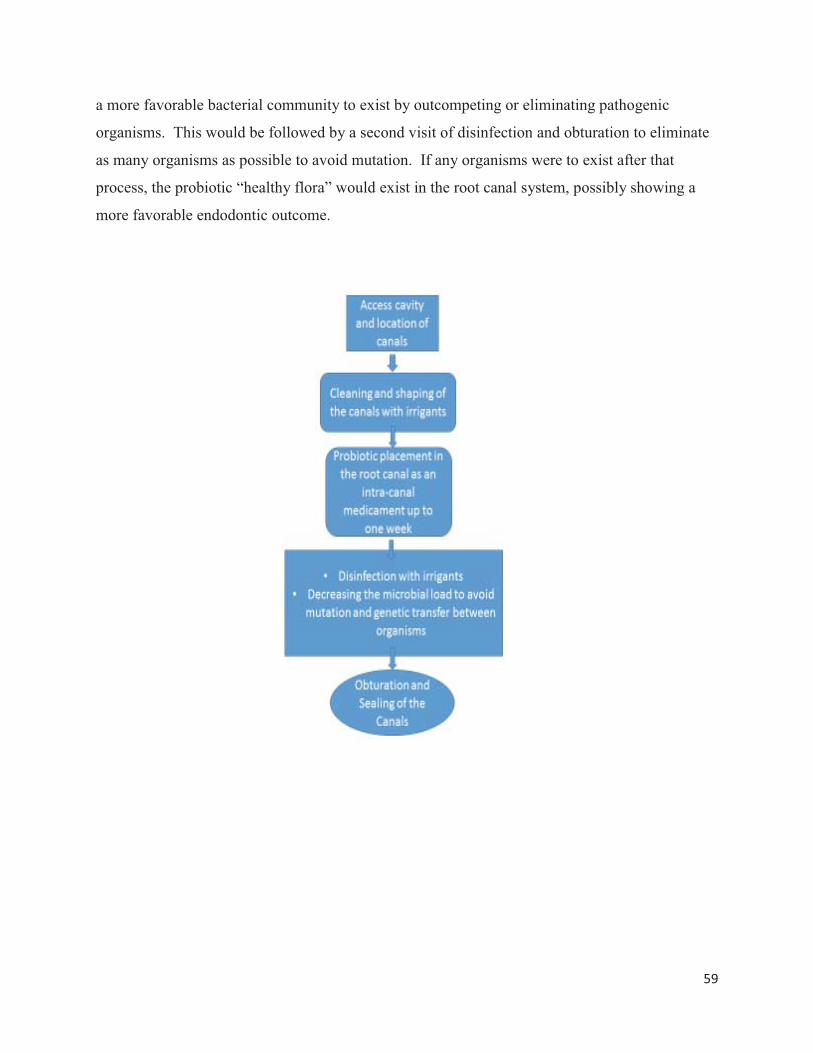
One of the most studied organs which contain microbial communities is the human
gastrointestinal tract which contains a very complex and dynamic microbial community, specific
for each person depending upon environmental and genetic factors. Each individual holds
approximately 1.5 kg of bacteria within in their gastrointestinal tract. . (12, 13, 14, 17, 18, 19, 27, 28)
The gut microbiota plays an important role in human health, not only due to its
participation in the digestion process, but also for its critical function in the development of the
gut and the immune system, along with regulation of absorption and storage of lipids. Recent
studies have suggested that specific aberrations in gut microbiota composition are associated
with different diseases. The rationale for modulating the gut microbiota by means of probiotics
is derived from the importance of gut microbiota on host health. (27)
It was suggested in this study that this very same concept be applied to endodontic
therapy owing to the fact that the oral cavity is part of the “Oral Human Microbiome”, which in
turn is part the “Human Microbiome”. (27, 28)
Page 67
57
This study involved two phases: phase one (discovery phase) and phase two (application
phase). Phase one was a Kirby-Bauer disc diffusion susceptibility test, utilized to determine the
sensitivity or resistance of the pathogenic bacteria (E .faecalis or C. albicans) to various
probiotic challenges. The pathogenic organisms were grown on blood agar in the presence of
test probiotic groups impregnated filter paper discs. The absence of growth around the discs was
an indirect measure of the ability of the test probiotic groups to inhibit growth/out compete the
pathogenic organism’s E. faecalis or C .albicans.
The results of phase one suggested that organisms such as Lactobacillus rhamnosus,
Bifidobacterium bifidum, Lactobacillus acidophilus, Lactobacillus casei, Lactobacillus
plantarum, Lactobacillus salivarius, Bifidobacterium longum, Streptococcus thermophilus,
Lactobacillus bulgaricus, Lactobacillus paracasei, Bifidobacterium lactis and Bifidobacterium
breve possess antimicrobial activity against Enterococcus faecalis and Candida albicans. An
observation from this study is that groups 1, 4 and 5 all contained common probiotic organisms
including L.acidophillus,L.rhamnosus,L.casei and B.Longum, suggesting future focus involve on
these organisms for future studies.
Phase two was considered the application portion of the study, suggesting a novel
delivery vehicle for probiotics into the root canal system by utilizing 30% poloxamer 407 (also
known as pluronic F-127) mixed with MRS or TSB broth containing the probiotics. Poloxamer
has been utilized in biofilm stage testing of microorganisms. Although this secondary purpose
was not the main focus of this study, it investigated poloxamer as a delivery mechanism.
Poloxamers are non-ionic polyethylene oxide (PEO) and polypropylene oxide (PPO)
copolymers. They are biocompatible and are used in pharmaceutical formulations such as
surfactants, emulsifying agents, solubilizing agents, dispersing agents, and in vivo absorbance
enhancer 1; therefore they are considered as “functional excipients” because they are essential
components, and play an important role in the formulation (42, 43, and 44) of multiple therapeutic
agents.
Page 68
58
Two polymers from this class, poloxamer 188 and poloxamer 407, show inverse
thermosensitivity. They are soluble in aqueous solutions at low temperatures (mainly 4°C), but
will gel at higher temperatures (42, 44). These properties make them an ideal delivery vehicle for
use as an intra-canal medicament between inter-appointment visits.
Other advantages of poloxamer 407, which make it ideal for use in the root canal system
is that it has already been employed in topical dosage forms of other surface applied
formulations. Poloxamer 407 has been used in vehicles for fluorinated dentifrices, eye
applications and contraceptive gels. A poloxamer based dental gel product has been used for
treating patients with sensitive gums and teeth. Other properties relevant to this study are that a
poloxamer based gel has showed many favorable characteristics for use as a burn dressing by
providing a non-toxic detergent covering to the wound. Other studies suggest that the gel itself
may accelerate wound healing, making P 407 a suitable vehicle for gels intended to be applied
for ulcers and traumatic lesions. It would be an ideal solution for apical periodontitis which is the
main cause for endodontic failure (42, 43, 44).
The CFU/ml count results for this study revealed a complete elimination of E. faecalis
colony counts for probiotic groups 1 and 4 when mixed in equal amounts with E. faecalis in the
30 % poloxamer/MRS broth formula when compared with controls, and a significant inhibition
in CFU/ml counts for C. albicans in probiotic groups 1 and 4 when mixed in equal amounts with
C. albicans in the 30 % poloxamer/MRS broth formula when compared with controls.
The suggested protocol for the use of a poloxamer probiotic based inter-appointment
medicament would be to replace the current use of calcium hydroxide. Calcium hydroxide
possesses favorable antimicrobial properties, yet it still cannot eliminate all pathogenic
organisms such as E. faecalis. The protocol for the probiotic medicament would be, instead of a
microbial “elimination therapy” by the use of antimicrobial agents, substituted by a microbial
“replacement therapy”. Instead of attempting to eliminate pathogenic organisms by
antimicrobial agents, probiotics would eliminate the endodontic pathogenic flora, allowing a
more favorable/biocompatible environment after standard root canal therapy procedures were
followed. This suggested new protocol for therapy would include cleaning, shaping, irrigation
and activation of irrigants to decrease the microbial load and remove organic tissue, followed by
introducing the poloxamer based probiotic mixture for a period of one week. This would allow
Page 69
59
a more favorable bacterial community to exist by outcompeting or eliminating pathogenic
organisms. This would be followed by a second visit of disinfection and obturation to eliminate
as many organisms as possible to avoid mutation. If any organisms were to exist after that
process, the probiotic “healthy flora” would exist in the root canal system, possibly showing a
more favorable endodontic outcome.
Page 70
60
Chapter V
Conclusion
The null hypothesis for this study was rejected, since groups I, IV and V showed
statistically significant results (P< 0.05) for ZOI for evaluating planktonic stages, and a reduction
in CFU/ml counts for biofilm stage testing against E. faecalis and C. albicans. Within the
limitations of this study, it was concluded in phase one that L. acidophilus, L. casei , L.
rhamnosus and B. longum were all strains found in common in the probiotic cocktails selected
against the endodontic pathogenic organisms E. faecalis and C. albicans in planktonic stages,
exhibiting antimicrobial activity which was reflected by the zones of inhibition (ZOI) seen. In
phase two it was concluded that probiotic groups I and IV demonstrated a decrease in CFU/ml
count of the pathogenic organisms E. faecalis and C. albicans in the biofilm stage when mixed
together with the probiotics. Another conclusion is that poloxamer 407 may be used as a
delivery vehicle for probiotics in the root canal system as an inter-appointment medicament. This
is due to its unique reverse gel physical properties as well as its biocompatibility. This pilot
study demonstrated that probiotics show a potential in root canal therapy, but further in vitro and
in vivo studies are needed to determine the full potential of “Bacteriotherapy” and its application
in endodontics. This model, if successful, may well be introduced and adapted in wound care
management, since apical periodontitis can be considered a chronically existing wound, similar
in nature to soft tissue wounds that are recalcitrant to standard antimicrobial therapies.
Page 71
61
References
Endodontic Microbiology:
1. Nair, P. N. R. (2004). Pathogenesis of apical periodontitis and the causes of endodontic
failures. Critical Reviews in Oral Biology & Medicine, 15(6), 348-381.
doi:10.1177/154411130401500604
2. KAKEHASHI, S., STANLEY, H. R., & FITZGERALD, R. J. (1965). The effects of
surgical exposures of dental pulps in germ-free and conventional laboratory rats. Oral
Surgery, Oral Medicine, and Oral Pathology, 20, 340-349.
3. Sundqvist, G. (1994). Taxonomy, ecology, and pathogenicity of the root canal flora. Oral
Surgery, Oral Medicine, and Oral Pathology, 78(4), 522-530.
4. Gomes, B. P., Pinheiro, E. T., Gade-Neto, C. R., Sousa, E. L., Ferraz, C. C., Zaia, A. A
.Souza-Filho, F. J. (2004). Microbiological examination of infected dental root canals.
Oral Microbiology and Immunology, 19(2), 71-76.
5. Chavez de Paz, L. (2004). Gram-positive organisms in endodontic infections. Endodontic
Topics, 9(1), 79-96.
6. Fabricius, L., Dahlen, G., Holm, S. E., & Moller, A. J. (1982). Influence of combinations
of oral bacteria on periapical tissues of monkeys. Scandinavian Journal of Dental
Research, 90(3), 200-206.
7. Fabricius, L., Dahlen, G., Sundqvist, G., Happonen, R. P., & Moller, A. J. (2006).
Influence of residual bacteria on periapical tissue healing after chemomechanical
treatment and root filling of experimentally infected monkey teeth. European Journal of
Oral Sciences, 114(4), 278-285. doi:10.1111/j.1600-0722.2006.00380.x
Page 72
62
8. Fujii, R., Saito, Y., Tokura, Y., Nakagawa, K. Okuda, K., & Ishihara, K. (2009).
Characterization of bacterial flora in persistent apical periodontitis lesions. Oral
Microbiology and Immunology, 24(6), 502-505. doi:10.1111/j.1399-302X.2009.00534.
9. Seltzer, S., & Bender, I. B. (2003). Cognitive dissonance in endodontics. 1965. Journal of
Endodontics, 29(11), 714-9; discussion 713.
Probiotics:
10. Al-Dajani, M., & Limeback, H. (2012). Emerging science in the dietary control and
prevention of dental caries. Journal of the California Dental Association, 40(10), 799-804.
11. Allaker, R. P., & Douglas, C. W. (2009). Novel anti-microbial therapies for dental plaque-
related diseases. International Journal of Antimicrobial Agents, 33(1), 8-13.
doi:10.1016/j.ijantimicag.2008.07.014; 10.1016/j.ijantimicag.2008.07.014
12. Borchers, A. T., Selmi, C., Meyers, F. J., Keen, C. L., & Gershwin, M. E. (2009).
Probiotics and immunity. Journal of Gastroenterology, 44(1), 26-46. doi:10.1007/s00535-
008-2296-0; 10.1007/s00535-008-2296-0
13. Elavarasu, S., Jayapalan, P., & Murugan, T. (2012). Bugs that debugs: Probiotics. Journal
of Pharmacy & Bioallied Sciences, 4(Suppl 2), S319-22. doi:10.4103/0975-7406.100286;
10.4103/0975-7406.100286
14. Grover, H. S., & Luthra, S. (2012). Probiotics–the nano soldiers of oral health. Journal,
Indian Academy of Clinical Medicine, 13(1), 49.
15. Harini, P. M., & Anegundi, R. T. (2010). Efficacy of a probiotic and chlorhexidine mouth
rinses: A short-term clinical study. Journal of the Indian Society of Pedodontics and
Page 73
63
Preventive Dentistry, 28(3), 179-182. doi:10.4103/0970-4388.73799; 10.4103/0970-
4388.73799
16. Khanafari, A., Porgham, S. H., & Ebrahimi, M. T. (2012). Investigation of probiotic
chocolate effect on streptococcus mutans growth inhibition. Jundishapur Journal of
Microbiology, 5(4), 590-597. doi:10.5812/jjm.3861
17. Meurman, J. H. (2005). Probiotics: Do they have a role in oral medicine and dentistry?
European Journal of Oral Sciences, 113(3), 188-196. doi:10.1111/j.1600-
0722.2005.00191.x
18. Reddy, J. J., Sampathkumar, N., & Aradhya, S. (2010). Probiotics in dentistry: Review of
the current status; probiótica em odontologia: Revisão do estado atual da questão.
Rev.Clín.Pesq.Odontol.(Impr.), 6(3), 261-267.
19. Reddy, S., Swapna, L., Ramesh, T., Singh, R., Vijayalaxmi, N., & Lavanya, R. (2011).
Bacteria in oral health–probiotics and prebiotics a review.
20. Salminen, S., von Wright, A., Morelli, L., Marteau, P., Brassart, D., de Vos, W. M.,
Mattila-Sandholm, T. (1998). Demonstration of safety of probiotics -- a review.
International Journal of Food Microbiology, 44(1-2), 93-106.
21. Slawik, S., Staufenbiel, I., Schilke, R., Nicksch, S., Weinspach, K., Stiesch, M., &
Eberhard, J. (2011). Probiotics affect the clinical inflammatory parameters of
experimental gingivitis in humans. ( No. 65). doi:10.1038/ejcn.2011.45
22. Twetman, S. (2012). Are we ready for caries prevention through bacteriotherapy?
Brazilian Oral Research, 26 Suppl 1, 64-70.
23. Gill, H. S., & Guarner, F. (2004). Probiotics and human health: A clinical perspective. Postgraduate
Medical Journal, 80(947), 516-526. doi:10.1136/pgmj.2003.008664
Page 74
64
24. Krasse, P., Carlsson, B., Dahl, C., Paulsson, A., Nilsson, A., & Sinkiewicz, G. (2006). Decreased gum
bleeding and reduced gingivitis by the probiotic lactobacillus reuteri. Swedish Dental Journal,
30(2), 55-60.
25. Haukioja, A. (2010). Probiotics and oral health. European Journal of Dentistry, 4(3), 348.
26. Caglar, E., Kargul, B., & Tanboga, I. (2005). Bacteriotherapy and probiotics’ role on oral
health. Oral diseases, 11(3), 131-137.
27. Gueimonde, M., & Collado, M. D. C. (2012). Metagenomics and probiotics.Clinical
Microbiology and Infection, 18(s4), 32-34.
28. Dominguez-Bello, M. G., & Blaser, M. J. (2008). Do you have a probiotic in your
future?. Microbes and Infection, 10(9), 1072-1076.
29. Roberfroid, M. (2007). Prebiotics: the concept revisited. The Journal of nutrition,137(3),
830S-837S.
30. Surawicz, C. M., Elmer, G. W., Speelman, P. I. E. T. E. R., McFarland, L. V., Chinn, J., &
Van Belle, G. (1989). Prevention of antibiotic-associated diarrhea by Saccharomyces
boulardii: A prospective study. Gastroenterology, 96(4), 981-988.
31. Kragen H. The treatment of inflammatory affections of the oral mucosa with a lactic acid
bacterial culture preparation. Zahnarztl Welt 1954;9:306-308.
32. Krasse P, Carlsson B, Dahl C, Paulsson A, Nilsson A, Sinkiewicz G. Decreased gum
bleeding and reduced gingivitis by the probiotic Lactobacillus reuteri. Swed Dent J
2006;30:55-60.
33. Della Riccia DN, Bizzini F, Perilli MG, Polimeni A, Trinchieri V, Amicosante G, et al.
Anti-inflammatory effects of Lactobacillus brevis (CD2) on periodontal disease. Oral Dis
2007;13:376-385.
Page 75
65
34. Twetman S, Derawi B, Keller M, Ekstrand K, Yucel-Lindberg T, Stecksen-Blicks C.
Short-term effect of chewing gums containing probiotic Lactobacillus reuteri on the levels
of inflammatory mediators in gingival crevicular fluid. Acta Odontol Scand 2009;67:19-
24.
35. Ahola AJ, Yli-Knuuttila H, Suomalainen T, Poussa T, Ahlstrom A, Meurman JH, et al.
Short-term consumption of probiotic-containing cheese and its effect on dental caries risk
factors. Arch Oral Biol 2002;47:799-804.
36. Delanghe G, Ghyselen J, van Steenberghe D, Feenstra L. Multidisciplinary breath-odour
clinic. The Lancet 1997;350:187-187.
37. Evans, M., Davies, J. K., Sundqvist, G., & Figdor, D. (2002). Mechanisms involved in the
resistance of Enterococcus faecalis to calcium hydroxide. International Endodontic
Journal, 35(3), 221-228.
38. Stuart, C. H., Schwartz, S. A., Beeson, T. J., & Owatz, C. B. (2006). Enterococcus
faecalis; Its Role in Root Canal Treatment Failure and Current Concepts in
Retreatment. Journal of endodontics, 32(2), 93-98.
39. Costerton, J. W., Lewandowski, Z., Caldwell, D. E., Korber, D. R., & Lappin-Scott, H. M.
(1995). Microbial biofilms. Annual Reviews in Microbiology, 49(1), 711-745.
40. Svensäter, G., & Bergenholtz, G. (2004). Biofilms in endodontic infections. Endodontic
topics, 9(1), 27-36.
41. Ramage, G., Mowat, E., Jones, B., Williams, C., & Lopez-Ribot, J. (2009). Our current
understanding of fungal biofilms. Critical reviews in microbiology, 35(4), 340-355.
Page 76
66
Poloxamer:
42. Patel, H. R., Patel, R. P., & Patel, M. M. (2009). Poloxamers: A pharmaceutical excipients
with therapeutic behaviors. International Journal of PharmTech Research, 1(2).
43. Cafaggi, S., Russo, E., Caviglioli, G., Parodi, B., Stefani, R., Sillo, G., ... & Bignardi, G.
(2008). Poloxamer 407 as a solubilising agent for tolfenamic acid and as a base for a gel
formulation. european journal of pharmaceutical sciences, 35(1), 19-29.
44. Nalbandian, R. M., Henry, R. L., Balko, K. W., Adams, D. V., & Neuman, N. R. (1987).
Pluronic F 127 gel preparation as an artificial skin in the treatment of third degree burns
in pigs. Journal of biomedical materials research, 21(9), 1135-1148.
45. Friedman, S., Abitbol, S., & Lawrence, H. P. (2003). Treatment outcome in endodontics: the
Toronto Study. Phase 1: initial treatment. Journal of Endodontics, 29(12), 787-793.
.