A Prominent Antigenic Surface Polypeptide Involved in the Biogenesisand Function of the Vaccinia Virus Envelope'
JAMES GORDON, 2 ANJANI MOHANDAS, SHARON WILTON, AND SAMUEL DALES
Cytobiology Group, Department of Microbiology and Immunology, University of Western Ontario, London, Ontario, Canada Ni 5C 1
Received October 22, 1990; accepted December 27, 1990
Polypeptides of the vaccinia virus envelope exposed on the surface were identified by means of sulfo-N-hydroxysuc-cinimidobiotin as a surface tag . Among surface expressed polypeptides is the 35-kDa antigen, previously designatedAg35. Both monoclonal (mAb) and monospecific affinity pure antibodies directed against Ag35 neutralized vacciniainfectiousness, indicating that this prominent surface antigen has a function during early virus-host cell interactions .The binding of several monoclonal antibodies to various regions of Ag35 was tested by reacting CNBr fragments,derived from the polypeptide, employing Western blotting . All mAbs tested reacted with the same region of Ag35 .Estimation of the molecular weights (MW), based on migration of the CNBr peptides in sodium dodecyl sulfate-polyac-rylamide gel electrophoresis, revealed that those partial digestion products which contained a proline-rich 99 aminoacid limit digest fragment were present at a position approximately 12 .5 kDa larger than that predicted from the DNAsequence. By contrast, partial and limit digest products lacking the praline-rich fragment migrated to the MW positionexpected from the length of the DNA sequence . This observation demonstrates that departure from a predicted 22 .3kDa to an anomalous MW of Ag35 is conferred by the proline-rich peptide . The surface location of Ag35 was confirmedby immune electron microscopy. In a competition test the binding specificity of mAb and affinity-purified antibodies atthe surface of virions could be demonstrated . Evidence for an association of Ag35 with the virus envelope at variousstages during biogenesis of vaccinia was obtained by immune electron microscopy of whole mounts and thin sections.Presence of Ag35 as an early component of immature and mature virions, probably residing in the bilayer membranestructure was detected . A distinction can, therefore, be made between Ag35 and several other vaccinia envelopepolypeptides which are synthesized as late functions and added during late stages of envelope assembly . x 1ss1
Academic Press, Inc .
INTRODUCTION
The orthopoxviruses as exemplified by vacciniavirushave been subjected to intensive study because theypossess great structural and functional complexity, fol-low a unique scheme of replication, and are versatileas recombinant vectors for transfer of foreign genesinto animal cells (Dales and Pogo, 1981 ; Fenner stall1989 ; Panicali et al., 1983; Smith et all 1983). Ortho-poxvirus replication is confined to the cytoplasm ofhost cells mandating the encoding by the virus of alarge spectrum of enzymatic activities to initiate un-coating and transcriptional events (Reviewed in Dalesand Pogo, 1981 ; Fenneretal ., 1989). The morphogen-esis of vaccinia can be subdivided into a temporallyregulated series of stages . One particular area of inter-est in this laboratory has been the biogenesis of thevaccinia viral envelope which occurs through a uniquede novo mechanism within the viroplasmic matrix alsotermed "factory ." We define as the envelope the mem-braneous lipoprotein tegument which encloses the vi-rion (Dales and Pogo, 1981) . Unlike the situation with
2Supported by the Medical Research Council of Canada .Recipient of an Ontario Graduate Scholarship .
671
other membrane-enclosed eukaryotic viruses, forma-tion of the vaccinia envelope is not connected withpreexisting cellular membranes (Dales and Mosbach,1968) . The first step in envelope assembly evident byelectron microscopy is the appearance of curved seg-ments consisting of a unit membrane configurationbacked by an external layer of spicules, which providerigidity and confer spherical shape to the immatureform of the virion (Dales and Mosbach, 1968) . Theearly stages of envelope formation can occur normallyin the absence of viral DNA synthesis, which may beblocked by inhibitors such as hydroxyurea (HU) (Pogoand Dales, 1971). During normal morphogenesis,when DNA synthesis is allowed to occur, nucleopro-tein complexes are packaged within the envelopes andthen become differentiated into characteristic bicon-cave cores and associated lateral bodies . At a latestage during maturation the virus surface is modifiedby exchange of the spicule layer for a coating of sur-face tubular elements (STE) (Stern and Dales, 1 976a ;Essani et al., 1982) .Although our understanding to date about the for-
mation of the vaccinia envelope is rudimentary, it isknown that the mixtures of phospholipids and glycolip-ids which become incorporated are different in compo-
0042-6822/91 $3.00Copyright 9 1991 by Academic Press, Inc .All rights of reproduction in any form reserved .
67 2
sition than those present in uninfected cells but corre-spond closely to the changed composition which issignificantly altered after infection (Stern and Dales,1974; Anderson and Dales, 1978) . Concerning enve-lope polypeptides, the spicules consist of a 65-kDapolypeptide which accumulates in large quantities dur-ing early stages of infection (Sarov and Joklik, 1973 ;Essani et al., 1982). Little information is available, how-ever, about the other proteins of the formative enve-lope .
Much attention is currently focused on the potentialuse of vaccinia virus recombinants as effective vac-cines against a wide variety of pathogens (reviewed byMackett and Smith, 1986; Moss and Flexner, 1987 ;Piccini and Paoletti, 1988) . The foreign polypeptidesexpressed by such recombinants are compartmenta-lized within the cell according to signal sequences en-coded by the gene inserted but do not become part ofthe virion . Analysis of the vaccinia envelope polypep-tides may identify structural motifs within these poly-peptides having a role in envelope assembly . Further-more, thorough understanding of the antigenic compo-nents of the viral envelope will have a bearing on futuredevelopments in creating safe and efficacious vac-cines capable of modulating the host's immune re-sponse, so as to circumvent a major limiting factor inrevaccination of individuals with such vaccinia recombi-nants .
One prominent envelope component of mature vac-cinia which becomes antigenically significant duringinfection in animals is a 35-kDa polypeptide designatedAg35. This polypeptide can be readily solubilized withthe nonionic detergent NP-40 (Wilton et al., 1986) .Ag35 was identified as an early virus function becauseit is synthesized when DNA replication was stopped byHU (Gordon etal., 1988) . The gene encoding Ag35 hasbeen mapped within the 8 .7-kbp HindIll fragment H(Gordon et al., 1988) and localized to the open readingframe (ORF) designated H6R according to the conven-tion of Rosel et al. (1986). The relevant ORF encodes apolypeptide with a predicted molecular weight (MW) ofonly 22 .3 kDa . The product of the gene when ex-pressed in Escherichia coil comigrates with the authen-tic polypeptide at 35 kDa implying that the observedanomalous electrophoretic migration in a conventionalgel system could result from some property of the poly-peptide itself and is not due to any post-translationalevent .
MATERIALS AND METHODS
Cells and viruses
HeLa and Mouse L2 cells were maintained in nu-trient media (NM) consisting of Eagle's modified MEM
GORDON ET ALL
supplemented with 10% fetal bovine serum . Vacciniavirus strain IHD-W and the temperature-sensitive mu-tant 1085 (Dales et at, 1978) were propagated andtitrated by plaque assay using L cells at 37 and 32°,respectively. Virus of high purity was obtained frominfected L cells by centrifugation through potassiumtartrate gradients, as previously described (Stern andDales, 1974) .
Protein gels and electroblotting
Sodium dodecyl sulfate-polyacrylamide gel electro-phoresis (SDS-PAGE) was carried out according toLaemmli (1970) . For immunoblotting, transfer of poly-peptides to nitrocellulose filters was performed as de-scribed by Towbin et al. (1979). Antigen was revealedby means of antibodies which were marked using "'kconjugated protein A (ICN) as described by Batteiger etal. (1982) and modified by Wilton et al. (1986) .
Labeling of vaccinia surface polypeptides
Polypeptides on the surface of vaccinia particleswere identified by reacting them with the sulfo-N-hy-droxysuccinimide ester of biotin (sulfo-NHS-biotin ;Pierce), using a modification of the procedurepreviously described for Dictyostelium discoideumplasma membranes (Ingalls et al., 1986 ; Goodloe-Hol-land and Luna, 1987) . Purified virions were suspendedin 10 mM NaHEPES, pH 8 .2, at a concentration of 250µg of protein/ml and reacted for 30 min on ice with thesulfo-NHS-biotin at a concentration of 12 .5 mg/ml . Toterminate the reaction, lysine was added at a final con-centration of 0 .1 M. The biotinylated virus polypeptideswere separated by SDS-PAGE and transferred to a ni-trocellulose filter as above . The material was exposedto 5% FBS in 10 mM Tris-HCl, pH 7 .3, 115 mM NaCl,0 .05% Tween-20 (TBS-Tw) for 1 hr as a blocking step,washed three times with TBS-Tw, then reacted with a1/3000 dilution of alkaline phosphatase-streptavidincomplex (Amersham) for 1 hr . After 3 washes withTBS-Tw, the biotinylated proteins were visualized us-ing an alkaline phosphatase color development sys-tem, as described by the manufacturer (Biorad) .
Affinity purification of antiserum
Antibody specific to Ag35 was separated from thepolyclonal antiserum, raised in rabbits against viral en-velope components released by NP-40 (Gordon et al.,1988), using affinity purification (Ball and Kovala,1988) . Briefly, an NP-40 extract from purified virus wasprepared as described (Wilton et at, 1986) and sepa-rated by preparative SDS-PAGE using a full-widthcomb . Following electrophoresis the proteins weretransferred to nitrocellulose and the band correspond-
ing to Ag35 was localized by staining with amido blackand excised from the nitrocellulose sheet . The excisedstrip was cut into small pieces and transferred to thebarrel of a 5-ml syringe . All subsequent steps werecarried out by drawing 1 ml of the appropriate bufferinto the syringe through an 18-g needle and mixinggently. The nitrocellulose was blocked by treating withphosphate-buffered saline + 0 .05% Tween 20 (PBS-Tw) containing 3% casein for 4 hr at 24° . Nitrocellulosepieces were washed extensively with PBS-Tw (six toeight washes) then reacted with the antiserum over-night at 4° . Once again the pieces were washed exten-sively with PBS-Tw . The bound antibodies were elutedfor 1 min with 0 .1 M glycine, pH 2 .4, 0 .15 M NaCl, theeluate was immediately neutralized with 1 M Tris-HCI,pH 8 .2, then dialyzed against PBS. The concentrationof immunoglobulln protein in the final sample was de-termined by the method of Lowry et al. (1951) .
Selection of hybridomas producing monoclonalantibodies
Proteins present in the envelope of vaccinia weresolubilized with NP-40 and 2-mercaptoethanol, as de-scribed previously (Stern and Dales, 1976) and usedfor immunizing 8-week-old Balb/c mice . For initial im-munizations the antigen was obtained from 300 ugviral protein and was administered intraperitoneally incomplete Freund's adjuvant. Two booster immuniza-tions were administered at 2-week intervals . Suspen-sions of splenocytes prepared 3 days after the last in-jection were fused with cells of the SP2/0 myeloma(10:1) in the presence of 40% polyethylene glycol (PEG)1000 in Iscoves modified D-MEM (IMDM) containing5% DMSO .Hybridomas were selected by the procedure of
Koehler and Milstein (1975) . Selection was carried outin HAT medium, culture fluids in which hybridoma colo-nies developed were screened for antibodies to vac-cinia virus polypeptides by means of ELISA, immunoflu-orescence, and Western blotting .
Established, antibody-producing, twice cloned hy-bridomas were grown intraperitoneally in syngeneicmice and ascites fluid was obtained . The Ig fractionwas purified from the ascites fluid by HPLC using aDEAE 5PW (Beckman) column employing in sequenceas binding buffer 20 mM Tris HCI (pH 8 .5) and 20 mMTris-HCI (pH 7 .0) + 0.5 M Na acetate as the elutingbuffer .
Neutralization assays
Neutralization of virus infectiousness was assayedby a plaque reduction test . Samples of the antiserum ormonoclonal antibody (mAb) being tested were diluted
A PROMINENT ANTIGENIC SURFACE POLYPEPTIDE 673
in PBS, then mixed with a standard number of plaqueforming units (pfu) of vaccinia virus in a final volume of36 Al . Following incubation for 30 min at 37° to bind theantibody, the mixture was serially diluted with NM andapplied to indicator monolayers of L cells for plaqueassay .
Cyanogen bromide (CNBr) cleavage of polypeptideAg35
The viral protein released by the method of Stern andDales (1976) with NP-40 and 2-mercaptoethanol (2-ME) from 2 mg of purified vaccinia virus protein wereseparated on a 11 % SDS-PAGE and identified by lim-ited staining with Coomassie blue . The Ag35 band wasexcised, destained, and cut into pieces 1 cm in length .Samples were reacted on a rotary mixer at room tem-perature for 30 min in sequential steps with 10 ml ofthe following solutions : H 2O, 0.1 N HCI, 0 .1 N HCI con-taining 0, 10, or 30 mg CNBr, two washes with H 20,0.25 M Tris-HCI, pH 6 .8. They were then equilibratedwith dissociation buffer for use with SDS-PAGE .
The reacted gel pieces and MW markers were elec-trophoresed in duplicate through 15% SDS-PAGE .One gel was stained with Coomassie blue and theother used for Western blot analysis .
Immunogold labeling of virions
Antigens were identified on virions and formativevirus intermediates by immunogold labeling . All reac-tions were carried out by floating formvar and carboncoated grids on droplets of the appropriate reagent di-luted in PBS containing 0 .5% bovine serum albumin(PBS-BSA). After attachment of virus material to thefilms, the grids were placed for 30 min in PBS-BSA toblock nonspecific binding of antibodies . The primaryantibody was diluted as desired and allowed to reactfor 30 min and then was removed by three washes of 2min each with PBS-BSA. The secondary antibodies,were goat anti-rabbit (Au-anti-rabbit) or goat anti-mouse (Au-anti-mouse) conjugated with 10 nm colloi-dal gold particles (Sigma) . They were diluted 1 :20 andapplied for a further 30 min . The grids were washed asabove with PBS-BSA and then 3 times with 10 mMHEPES, pH 7 .3. In some cases the material was nega-tively stained using 1 % phosphotungstic acid (PTA) .
Immunogold reactions on cell lysates
Cytoplasmic lysates from vaccinia virus infectedcells were prepared for immunogold EM by a modifica-tion of the procedure used by Stern and Dales (1974) .Infected monolayers were scraped, the cells pelletedat 700 g and resuspended in MEM diluted 1 :8 withwater to induce swelling . The cells were lysed by re-
674
GORDON ET AL .
peatedly (10 times) forcing them through a No . 25 nee-dle of a syringe . Remaining intact cells and large celldebris were removed by centrifugation at 200 g for 20min . The supernatant suspension was adjusted to0 .15 M NaCI to stabilize osmotically sensitive struc-tures . Dilutions of the supernatant fraction were ap-plied to EM grids, the material was then fixed for 2 minwith 0.19b glutaraldehyde in PBS, washed in PBS, andreacted with antibodies in the manner describedabove .
RESULTS
External polypeptides of vaccinia virus
Polypeptides exposed on the surface of vacciniavirus were identified by biotinylation of intact virions,using the sulfo derivative of N-hydroxysuccinimidobio-tin (sulfo-NHS-biotin) . This agent effects the transfer ofbiotin to primary amines, most notably the e group oflysine residues, within polypeptides . The charged sul-fate group on this compound prevents penetration ofthe compound into hydrophobic portion of mem-branes, thereby limiting reactivity to externally exposedregions of envelope polypeptides . The biotinylatedvirus was subjected to SDS-PAGE followed by electro-phoretic transfer to nitrocellulose . The biotinylatedpolypeptides were visualized by reaction with a strep-
Intracellular immunogold labeling
Intracellular virus components containing Ag35were localized by immunolabeling as described by
31.0-.Dales et al. (1983). Monolayers of HeLa cells grown inorgan culture dishes (Falcon 3037) pretreated withpoly-L-lysine, to enhance cell attachment, were in-fected with vaccinia virus at an m .o .i . of 10 PFU/cell .Cells undergoing infection were washed in 5 mM Na-PIPES, pH 7,2 mM MgCl2 , 2 mM EGTA, 0 .115 M NaCl(PiBS), fixed for 10 min in freshly prepared 1 % parafor-maldehyde in PiBS, permeated by exposure to threechanges of 0 .01% saponin in PiBS (PiBS-S) for 3 min .The PiBS-S was removed by washing twice with PiBS .The primary antibodies, either mAb 7C 1 , or the affinitypurified rabbit anti-Ag35 Ig, each diluted 1 in 10 inPiBS, were applied at room temperature for 1 hr . Afterremoval of unadsorbed antibodies by washing threetimes with PiBS of the appropriate secondary antibody-gold conjugates, diluted 1 in 10, were added for 1 hr .After five washes with PiBS, the cell material was fixedadditionally with 2% glutaraldehyde and 1% osmiumtetroxide and then was dehydrated through gradedethanol series and embedded in epoxy resin mixtures .Thin sections were prepared and stained by conven-tional methods .
AM 1 2 3 4 5
B1 2 3 4 5
FIG. 1 . Distribution of vaccinia polypeptides following surface bio-tinylation . Purified IHD-W vaccinia, at a protein concentration of 250pg/ml, was reacted for 30 min on ice with sulo-NHS biotin at aconcentration of 12 .5 mg/ml in 10 mM HEPES, pH 8 .2 . Followingbiotinylation, the virus was extracted with 196 NP-40 or 196 NP-40plus 125 roM 2-ME . Samples of whole virions, the soluble extractsand the insoluble material were subjected to separation by SDS-PAGE, then transferred to nitrocellulose membranes which werereacted for identification of biotinylated polypeptides (A), or pro-cessed for immunoblotting using rabbit antiserum directed againstthe NP-40-soluble envelope components (8) . (Lane M) biotinylatedMW standards (from Biorad) ; (lane 1) purified whole virus ; (lane 2)envelope fraction released with NP-40 ; (lane 3) NP-40-insoluble ma-terial ; (lane 4) envelopes solubilized with NP-40 and 2-ME ; (lane 5)core material insoluble in NP-40 and 2-ME .
tavidin-alkaline phosphatase (AP) complex and theuse of an AP color development system . This proce-dure identified six prominent polypeptides with molecu-lar weights of 58, 39, 35, 31, 24, 23, and 22 kDa (Fig1 A, lane 2) as derived by comparison with biotinylatedmarkers. One band present just above the 58-kDa poly-peptide and evident in all lanes is probably an artifact ofthe gel system and not an authentic viral polypeptidebecause it traversed the entire gel . When biotinylatedvirus polypeptides were separated on a 15% polyacryl-amide gel, an additional surface polypeptide of 14 kDawas evident (data not shown) ; this component corre-sponds to the 1 4-kDa external polypeptide describedby Rodriguez et al . (1987) .
Identification by Western blotting of vaccinia virusantigens associated with the envelope has been de-scribed previously (Wilton et al ., 1986); two groups ofantigens were identified, those released by the neutraldetergent NP-40 alone and those solubilized along
with the entire envelope by NP-40 plus 2-ME as de-scribed in Pogo and Dales (1968) . The biotinylatedvirus surface polypeptides were separated in the samemanner. Extraction with 196 NP-40 released the bulk ofthe 35- and 31-kDa polypeptides into the detergentphase (Fig . 1A, lane 2) leaving behind the 58- and 22-kDa biotinylated polypeptides with the NP-40 insolublepellet (Fig . 1A, lane 3). The 39-kDa antigen was aboutequally abundant in the NP-40 soluble and insolublefractions . After extraction of virions with NP-40 and125 mM 2-ME, the envelopes disappeared leaving be-hind cores with attached lateral bodies (See Fig . 6D) . Inthis preparation the 58-, 35-, 31-, and 22-kDa polypep-tides, appeared in the soluble fraction (Fig . 1A, lane 4) .The 31-kDa polypeptide released by NP-40 aloneshould not be confused with a similarly migrating 32-kDa polypeptide described previously (Wilton et at,1986). The latter was not synthesized by the IHD-Wstrain of vaccinia used in the current study but waspresent when the IHD-J strain was reacted with NHS-biotin (data not shown), indicating that the 32-kDa poly-peptide is also a surface exposed component . Oneshould note the faint signals of 60-, 24-, and 23-kDabiotinylated polypeptides which remained with the viralcores (Fig . 1A, lane 5) . Slight biotinylation of thesehighly abundant core polypeptides (Dales and Pogo,1981) could have been due to presence of a minorfraction of damaged virions in the purified suspen-sions .
The location of the prominent antigen of the enve-lope, Ag35 was compared on duplicate samples bymeans of Western blotting with that of other surface-la-beled polypeptides . The antigens were identified bypolyvalent rabbit antibodies raised against compo-nents of the viral envelope released by NP-40 (Gordonet a/., 1988). Distribution of Ag35 between detergentsoluble and insoluble fractions, detected by immuno-blotting (Fig . 1 B) was identical to that revealed by thebiotinylated polypeptides . More efficient solubilizationof Ag35 achieved here, as compared to that reportedby us previously (Wilton et a!, 1986) was probably dueto carrying out the extraction at a pH 8 .3 rather than7.3 and use as starting material of a lower concentra-tion (250 µg/ml not 1 mg/ml) of virus protein . Amongminor components observed by Western blotting em-ploying these polyvalent antibodies was an antigen mi-grating faster than Ag35 (Fig . 1B), which comigratedwith a biotinylated polypeptide (Fig . 1A) and a secondantigen, above Ag35 which did not have a biotinylatedcounterpart .
Characterization of Ag35 by monospecificantibodies
From data on the location of the biotinylated 35-kDapolypeptide and its ready solubilization by NP-40 it be-
A PROMINENT ANTIGENIC SURFACE POLYPEPTIDE
1 2 3 4 5 6 7 8
675
FIG . 2 . Specificity of affinity-purified rabbit antibodies against Ag35tested by Western blotting . Each strip contained polypeptides from 5µg of purified virus and the reactions were carried out in final vol-umes of 2 mi . 20 µl of affinity-purified antibodies (lane 1) was com-pared with (in lane 2) 0 .1 ul ; (3) 0 .5 µl ; (4) 1 Al ; (5) 5 µl ; (6) 10 µl ; (7) 50µl ; and (8) 100 µl of the rabbit antiserum against NP-40-soluble enve-lope components .
came evident that this surface component corre-sponds to Ag35, detected previously by mouse hyper-immune sera (Wilton et at, 1986) and rabbit serumagainst all vaccinia antigens solubilized by NP-40 (Gor-don at al., 1988). To define the antigen further affinitypurified rabbit antibodies were prepared, as describedunder Materials and Methods . Specificity of these anti-bodies is evident in immunoblotting analysis, illus-trated in Fig . 2, where only a single prominent band at35-kDa is evident . To obtain an approximate estimateon the amount of monospecific antibodies after affinitypurification, in relation to the starting material, serialdilutions of the original rabbit antiserum were reactedwith standard amounts of vaccinia antigen and com-pared with the affinity-purified antibodies (Fig . 2) . Itshould be noted that a very weak additional band at ahigher MW position is also evident in the lane corre-sponding to reaction with purified and nonpurified anti-bodies. The minor antigenic component may resultfrom retardation in the electrophoretic field due to anunreduced dimer or a contaminant associated withAg35 because following excision from the gel and ree-lectrophoresis, this antigen migrated to the 35-kDa po-sition (data not shown). As described below, similarobservations were made with MAb against Ag35 .
To ascertain whether Ag35 plays a role in infectivityduring early cell-virus interactions, the affinity-purified
67 6
10 10.001
001
0.1
1/ Antibody dilution
FIG. 3 . Reduction of vaccinia infectiousness by anti-envelope poly-specific and anti-Ag35 monospecific rabbit antibodies . Either PBSalone or varying dilutions of antisera in PBS were incubated in a 40 µlreaction volume at 37° for 1 hr with a standard number of PFUs ofvaccinia virus . Then the virus- antibody complexes were diluted andassayed on L cell monolayers . Open squares, antiserum to NP-40extract ; closed squares, prelmmune rabbit serum ; open circles, af-finity-purified monospecific antibodies ; closed circle, control in PBS,
monospecific antibodies were tested in a neutraliza-tion assay. For comparison we also used the serumdirected against the NP-40 extracted virion compo-nents. As evident from plaque reduction data seen inFig . 3, at an optimum dilution the polyvalent serum de-creased vaccinia virus infectivity approximately 400-fold, and the affinity-purified serum by about 10-fold .Preimmune control rabbit serum had no significant ef-fect on infectivity. As described above in Fig . 2, thelevel of reactivity specific for Ag35 was also evaluatedby Western blotting . Assessment of the comparativeantibody reactivity is based on affinity for SDS-dena-tured Ag35 antigen . From these data we estimatedthat the reactivity of 20 pl of the affinity-purified antibod-ies corresponded approximately to that obtained with5 ul of the polyvalent antiserum, suggesting that reac-tivity of purified anti-Ag35 antibodies could be several-fold higher than actually observed by the neutralizationassay . This still made them less efficient than the poly-valent serum . Loss of potency may occur because theaffinity purification procedure relied on immobilizationof antigen on Western blots . Therefore, the antibodiesreleased after binding must have been primarilyagainst linear epitopes, rather than some additional,
GORDON ET AL .
conformational epitopes, possessing perhaps greatersignificance for neutralization of infectiousness . A sec-ond and more important consideration is the presencein the serum, directed against the entire NP-40 extract,of antibodies to a spectrum of antigens interacting withthe host cell surface, including the important 14-kDapolypeptide described by Rodriguez et al . (1985) .
Specificity of monoclonal antibodies against Ag35
To further define antigenicity of the 35-kDa polypep-tide we selected a panel of mAbs, following immuniza-tion of mice with vaccinia virus components extractedwith NP-40 and 2-ME, as described under Materialsand Methods .
Among 158 colonies which grew into cultures 99proved to be positive against vaccinia antigens byELISA and/or immunofluorescence testing. Amongthese, 32 gave a signal by Western blotting, of which18/32 had affinity for Ag35 to a variable degree . mAbswhich gave the strongest signals with Ag35 were cho-sen for further studies .
Although in the NP-40/2-ME extract of virus threeprominent protein bands are usually present (see Fig .13), most of the antivaccinia mAbs selected wereagainst Ag35 . This finding can be attributed to eitheran abundance of Ag35 and/or to its effectiveness as anantigen. Table 1 summarizes the characterization ofsome selected and purified monoclonal antibodiesreactive against Ag35 . It should be noted that only onemAb (IF,) of all those tested was found to be of theIgM,k isotype. The degree of immunofluorescence
TABLE 1
PROPERTIES OF MONOCLONAL ANTIBODIES DIRECTED AGAINST Ag35
A Isotyping was carried out using INNO LIA mouse mAb isotypingkit (Innogenetics), as described by the manufacturer .
b Immunofluoresence was assayed on acetone permeabilized L .cells using rhodamine-conjugated goat anti-mouse immunoglobulinas the second antibody .
Tissue culture media from growing hyhridomas were assayed byplaque reduction on L cell monolayers, as described under Materialsand Methods .
° NA, not assayed .
°k PFUs neutralized'antibody dilutions
mAb lsotype°Intensity of
immunofluorescence° 1 1/10 1/100
7C, IgG„ k ++++ 70 62 534D5 IgG„ k ++ 0 0 01 F, IgM, k ++++ NA° NA NA6C, IgG, k ++++ NA NA NA
1 2 3 4 5 6
35KdS
Fic . 4 . Specificity of monoclonal antibodies for Ag35 demon-strated by immunoblotting . The reactions were conducted as de-scribed under Materials and Methods . (Lanes 1 and 4) extract fromuninfected L2 cells ; (lanes 2 and 5) extracts from vaccinia infectedcells ; (lanes 3 and 6) pure whole virus; lanes 1-3 reacted with mAb7C,, ; lanes 4-6 reacted with mAb 4D 6 .
was variable and only one of mAbs tested showed anyappreciable capacity for neutralization of infectivity .
The specificity of the mAbs giving the strongest reac-tions by immunofluorescence and Western blottingwas tested further following purification of ascites fluidby HPLC . As evident from Fig . 4, 4D 6 and 7C„ whenapplied to material from infected cells (lanes 2, 5) andpure virus (lanes 3, 6) gave a strong signal specificallywith Ag35, whereas uninfected cell lysates were nega-tive (lanes 1, 4) .
In view of the frequency with which mAbs againstAg35 were occurring in our selection we wished toascertain whether some particular domain(s) on thisantigen was immunogenic preferentially. The 35-kDapolypeptide could be broken selectively into a few frag-ments by CNBr because in the amino acid sequenceonly three internal methionines are available for cleav-age. The fragments derived from CNBrdigestion of pu-rified Ag35, as described in Materials and Methods,were resolved by PAGE separation . A graphic presenta-tion showing the location of N-terminal and internal me-thionines, as predicted from sequence of Ag35 (Gor-don et al., 1988) and the possible peptides which canbe derived therefrom, is provided in Fig . 5B . Thisshould be compared with the actual peptides resolvedby PAGE and stained by Coomassie blue (Fig . 5A) andbinding mAbs as revealed by immunoblotting (Fig . 5C) .Due to the anomalous migration of Ag35 which, based
A PROMINENT ANTIGENIC SURFACE POLYPEPTIDE
677
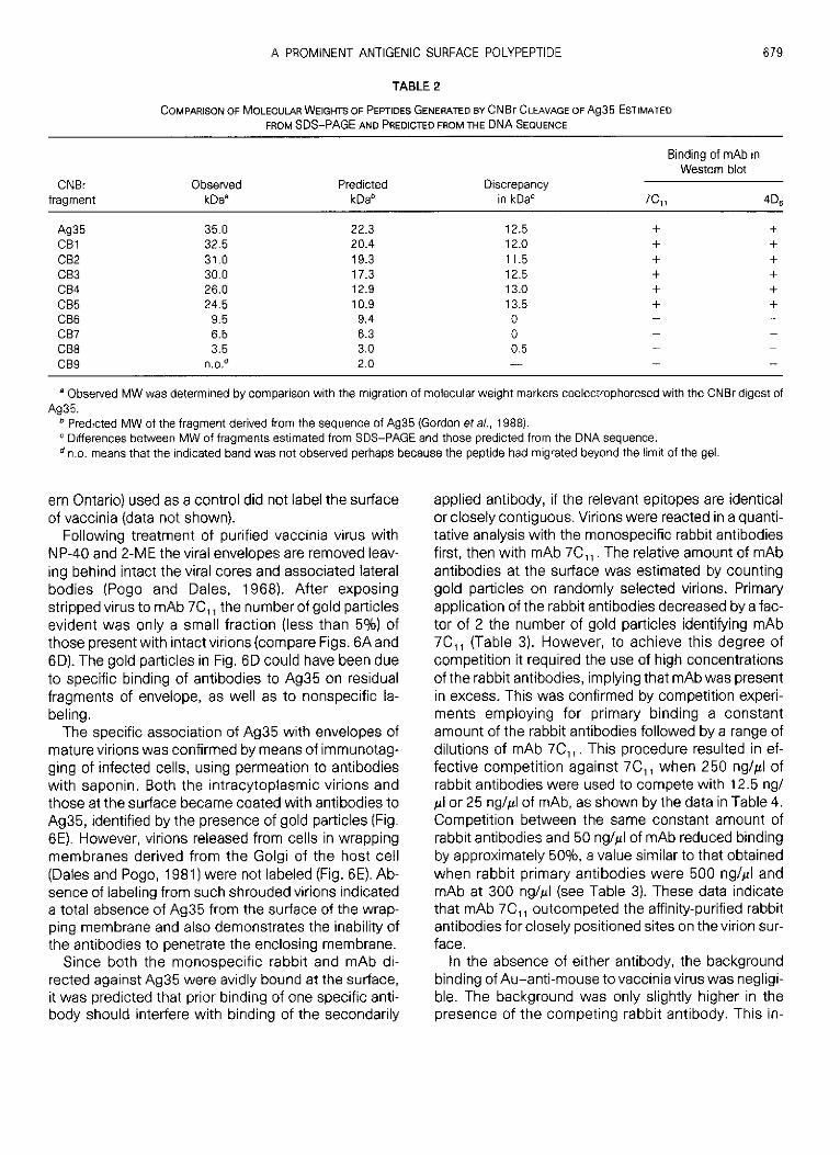
on the sequence data, should be 22 .3 kDa rather than35 kDa (Gordon et al., 1988) it is not possible to predictthe exact MWs for individual peptides obtained byCNBr cleavage. However, it is possible to relate thediminution in MW as a consequence of the removal ofone or more fragments . It is evident from data in Table1 that for CNBrfragments CB6, CB7, and CB8 the MWobtained from the gel (Fig . 5A), matches closely thatpredicted from the sequence. For the intact Ag35 andCB 1-5 fragments the apparent MWs in Fig . 5A andTable 2 are about 12-13 kDa greater than predictedfrom the sequence . Therefore, the anomalous migra-tion in PAGE must be due to a sequence motif residingin the limit digest peptide CBS, as this represents theonly fragment common to all peptides showing theanomalous migration . A prominent 20-kDa band in Fig .5A could be a cleavage or degradation product of oneof the larger fragments .
Affinity of mAb 7C„ for CNBr peptides is revealed inFig . 5C from which it is evident that this antibody iden-tifies an epitope on fragment CBS . Three other mAbs,4D6 , 1F, and 6F s among those tested showed thesame binding pattern as 7C„ (data not shown), indi-cating that CB5 contains a highly immunogenic se-quence of Ag35 .
Similar Western blotting utilizing the monospecificrabbit antibodies described above and hyperimmuneantisera from mice immunized with whole vacciniavirus (Wilton et al., 1986), gave identical or very similaraffinity-labeling patterns against the CB fragments(data not shown). This result confirms the importanceof the antigenic domain(s) in 0135 . Analysis by com-puter program for probable antigenic determinant(s)revealed, in Fig . 5D that there is a high correlation be-tween predicted regions and the amino acid sequencein fragment CB5 .
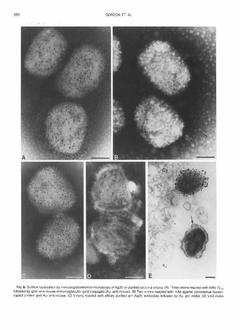
Localization of Ag35 on vaccinia virions by immuneelectron microscopy
The presence of the Ag35 in the envelope as deter-mined by its release with NP-40, as well as the pres-ence of a prominent surface reactive biotinylated poly-peptide at 35 kDa suggests that at least a part of Ag35is exposed. To confirm this assumption and ascertainwhether Ag35 is distributed randomly or at specific locion the virus surface we reacted highly purified intactvirions with antibodies and marked these antibodieswith immunoglobulin-colloidal gold conjugates . Ex-tensive labeling over the entire surface of virions wasobtained with mAb 7C„ (Fig . 6A). The apparent in-crease in concentration of gold particles near the pe-rimeter of virions which was often evident, was mostlikely due to the nature of the EM image which be-
6 7 8
GORDON ET AL .
97.4-
662-
42.7-
31.0-
21.5-
16.9-
14A-
2.5-
A1 2 3 4 5
D
B
C3 4 5
1 17
116
175 203
Ag35 + 1 ICB1CB2C83CB4CB5
CB6
C67
C88
CB5
comes a two-dimensional visualization of a three-di-mensional structure . As a control of the specificity, weapplied an irrelevant mAb, 46 B .2, against the corona-virus nucleocapsid protein (kindly provided by Dr. M .Buchmeier, Scripps Clinic and Research Fdn ., La Jolla,CA), which failed to bind to vaccinia virions (Fig . 6B) .
FiG. 5 . Epitope mapping of Ag35 by CNBr hydrolysis. Envelope material extracted with NP-40 and 2-ME was separated by preparativeSDS-PAGE and lightly stained with Coomasie blue . The band corresponding to Ag35 was excised from the gel, destained and either mock-reacted (lane 3) or reacted with 10 mg/ml (lane 4) or 30 mg/ml (lane 5) of CNBr, as described under Materials and Methcds . Following CNBrtreatment, the peptides were separated by SDS-PAGE on 15% gels . Duplicate samples were either stained with Coomassie blue (A) orsubjected to Western blotting, employing mAb 7C„ (C) . (B) diagrams CNBr cleavage sites and the sizes of all fragments which could possibly bederived by partial or complete digestion . MW markers in lane 2 included phosphorylase b (97 .4 kDa), bovine serum albumin (66 .2 kDa),ovalbumin (45 kDa), carbonic anhydrase (31 kDa), soybean trypsin inhibitor (21 .5 kDa), and lysozyme (14 .4 kDa) and in lane 1 CNBr digest ofmyoglobin (16.9, 14 .4, 8 .2, 6 .2, 2 .5 kDa) . (D) The sequence of Ag35 was analyzed for the presence of predicted regions of hydrophilicity, surfaceorientation, and flexibility (Devereux or al., 1984) and the plots of these analyses are illustrated . Note that the boxed region, spanning CB5fragment, residues 18 to 116, indicates the mapped antigenic determinant and shows a high degree of correlation with predicted regions of allthree parameters .
Parallel experiments conducted with affinity-purifiedmonospecific antibodies against Ag35, also showedspecific binding to the surface of virions in wholemounts (Fig . 6C) and thin sections (Fig . 6E), whereas,rabbit antiserum to tubulin of chicken (a gift of Dr . E .Ball, Department of Biochemistry, University of West-
em Ontario) used as a control did not label the surfaceof vaccinia (data not shown) .
Following treatment of purified vaccinia virus withNP-40 and 2-ME the viral envelopes are removed leav-ing behind intact the viral cores and associated lateralbodies (Pogo and Dales, 1968) . After exposingstripped virus to mAb 7C„ the number of gold particlesevident was only a small fraction (less than 5%) ofthose present with intactvirions (compare Figs . 6A and6D). The gold particles in Fig . 6D could have been dueto specific binding of antibodies to Ag35 on residualfragments of envelope, as well as to nonspecific la-beling .The specific association of Ag35 with envelopes of
mature virions was confirmed by means of immunotag-ging of infected cells, using permeation to antibodieswith saponin . Both the intracytoplasmic virions andthose at the surface became coated with antibodies toAg35, identified by the presence of gold particles (Fig .6E). However, virions released from cells in wrappingmembranes derived from the Golgi of the host cell(Dales and Pogo, 1981) were not labeled (Fig . 6E) . Ab-sence of labeling from such shrouded virions indicateda total absence of Ag35 from the surface of the wrap-ping membrane and also demonstrates the inability ofthe antibodies to penetrate the enclosing membrane .
Since both the monospecific rabbit and mAb di-rected against Ag35 were avidly bound at the surface,it was predicted that prior binding of one specific anti-body should interfere with binding of the secondarily
Observed MW was determined by comparison with the migration of molecular weight markers coelectrophoresed with the CNBr digest ofAg35 .
Predicted MW of the fragment derived from the sequence of Ag35 (Gordon er al., 1988) .Differences between MW of fragments estimated from SDS-PAGE and those predicted from the DNA sequence .
° n .o . means that the indicated band was not observed perhaps because the peptide had migrated beyond the limit of the gel .
applied antibody, if the relevant epitopes are identicalor closely contiguous . Virions were reacted in a quanti-tative analysis with the monospecific rabbit antibodiesfirst, then with mAb 7C„ . The relative amount of mAbantibodies at the surface was estimated by countinggold particles on randomly selected virions . Primaryapplication of the rabbit antibodies decreased by a fac-tor of 2 the number of gold particles identifying mAb7C„ (Table 3) . However, to achieve this degree ofcompetition it required the use of high concentrationsof the rabbit antibodies, implying that mAbwas presentin excess . This was confirmed by competition experi-ments employing for primary binding a constantamount of the rabbit antibodies followed by a range ofdilutions of mAb 7C„ . This procedure resulted in ef-fective competition against 7C, when 250 ng/µl ofrabbit antibodies were used to compete with 12 .5 ng/µl or 25 ng/µl of mAb, as shown by the data in Table 4 .Competition between the same constant amount ofrabbit antibodies and 50 ng/ul of mAb reduced bindingby approximately 500/0, a value similar to that obtainedwhen rabbit primary antibodies were 500 ng/ul andmAb at 300 ng/pl (see Table 3). These data indicatethat mAb 7C„ outcompeted the affinity-purified rabbitantibodies for closely positioned sites on the virion sur-face .
In the absence of either antibody, the backgroundbinding of Au-anti-mouse to vaccinia virus was negligi-ble . The background was only slightly higher in thepresence of the competing rabbit antibody . This in-
A PROMINENT ANTIGENIC SURFACE POLYPEPTIDE
679
TABLE 2
COMPARISON OF MOLECULAR WEIGHTS OF PEPTIDES GENERATED BY CNBr CLEAVAGE OF Ag35 ESTIMATEDFROM SDS-PAGE AND PREDICTED FROM THE DNA SEQUENCE
CNBrfragment
ObservedkDa`
PredictedkDa°
Binding of mAb inWestern blot
Discrepancyin kDa`
7C„
4Dg
Ag35 35 .0 22 .3 12.5
+
+Cal 32 .5 20 .4 12.0
+
+C82 31 .0 19.3 11 .5
+
+CB3 30.0 17.3 12.5
+
+CB4 26 .0 12.9 13.0
+
+CB5 24.5 10.9 13.5
+
+CB6 9 .5 9 .4 0
-
-CB7 6 .5 6 .3 0CBS 3 .5 3 .0 0 .5CB9 n.0 .° 2 .0
680 GORDON ET AL .
FIG . 6 . Surface localization by immunogold electron microscopy of Ag35 on purified vaccinia virions . (A), Three virions reacted with mAb 7C„followed by goat anti-mouse immunoglobulin-gold conjugate (Au-anti-mouse) . (B) Two virions reacted with mAb against coronavirus nucleo-capsid protein and Au-anti-mouse. (C) Virions reacted with affinity purified anti-Ag35 antibodies followed by Auanti rabbit . (D) Viral cores
creased background could have been due to someminor cross-reactivity between the goat anti-mouse Igand rabbit antibodies .
Efficient competition for Ag35 binding by antibodiesfrom different sources confirms the stringency of local-ization of Ag35 to the surface of the virion by the im-munogold technique .
Association of Ag35 with vaccinia envelopes duringvirus development
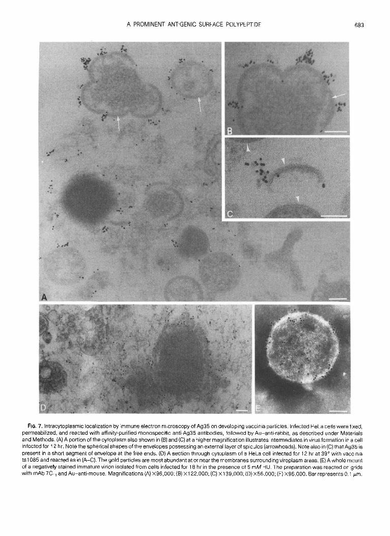
We took advantage of a procedure to introduce anti-bodies into infected cells, by permeation of the cellmembranes, to determine whether Ag35 is present onenvelopes during the formative stages as well as inmature particles. Although permeation of individualcells in cultures by means of saponin was highly vari-able, examples were found in which tagging of mAb7C t1 or the monospecific rabbit antibodies by gold-an-tibody conjugates was adequate to demonstrate in thinsections the specificity of binding to immature forms ofvaccinia . In one example of virus-infected cells sam-pled 12 hr after inoculation, numerous immature viri-ons labeled by gold particles were evident in the cyto-plasm (Figs . 7A and 7B) . Intermediates in envelopeassembly possessing the characteristic rigid, spicule-backed hemispherical conformation were also specifi-cally labeled, as evident in the examples shown in Figs .713 and 7C. Reactivity with Ag35 was frequently evidentat the free ends of segments of envelope, where thespicule-backed membrane structures are terminated,suggesting that specific antibodies became attachedto the bilayer structure of envelopes (Fig . 7C). It is quitepossible that an external layer of spicules, described indetail previously (Essani, et al., 1982) was a hindranceto the ready access of antibodies to the subjacent bi-layer. Very few gold particles were present within thenonmembraneous viroplasmic matrix material . Thespecificity of antigen-antibody interaction was shownby absence of gold particles when mAb directedagainst the variable surface glycoprotein of Trypano-soma bruceii (A gift of Dr. M . W. Clarke, Department ofMicrobiology, University of Western Ontario) was usedas a control (data not shown) .
Since Ag35 is known to be an early polypeptide,which can be synthesized despite an inhibition of DNAreplication by HU (Gordon at al., 1988), a drug causingarrest of virus assembly at the enveloped but immatureparticle stage (Pogo and Dales, 1971), we used thisdrug to ascertain whether Ag35 occurs in immature
A PROMINENT ANTIGENIC SURFACE POLYPEPTIDE 681
virions . Immature virions were released from cellswhich had either been treated with HU or were un-treated during infection . The intact particles were sub-jected to labeling with gold conjugates in the mannerused with isolated mature vaccinia . It was found thatanti-Ag35 antibodies became attached to the imma-ture virus particles regardless whether they originatedfrom controls or HU-treated cells (Fig . 7E) . This resultfurther demonstrated that Ag35 is, indeed, a compo-nent of the envelope .Association of Ag35 with the membrane bilayer of
the envelope was also investigated by means of thetemperature-sensitive mutant tsl 085 of vaccinia . Atthe restrictive temperature envelopes of this mutantare assembled as flexible bilayers lacking the attachedexternal spicule layer. Such membrane sheetssurround the viroplasmic assembly sites within the cy-toplasm (Dales et al ., 1978) . In order to determinewhether Ag35 could be closely associated with thesefree membranes, in situ, tracing by immunogold wascarried out on permeabilized cells which had been in-fected with is l 085 at 39 0 for 24 hr. The results, exem-plified by the illustration in Fig . 7D, demonstrated thatthe rabbit anti-Ag35 monospecific antibodies becamebound predominantly to membraneous structuressurrounding foci of viroplasm . This observation sup-ports the conclusion that Ag35 is an intimate compo-nent of the membrane bilayer of the envelope .
DISCUSSION
New data derived from the present study furthercharacterize the prominent vaccinia virus antigenAg35 (Gordon at al ., 1988), with respect to its involve-ment in the structure and formation of this virus . Local-ization of Ag35 by means of biotinylation of surfacepolypeptides and by immunoelectron microscopy re-vealed the unambiguous location to be on the externalsurface of the envelope .
Surface labeling in this study confirms and extendsthe work of Sarov and Joklik (1972), who used radioio-dination to identify surface exposed vaccinia polypep-tides. The use of a NHS-biotin type of probe for sur-face polypeptides has one major advantage over theuse of radioiodination of the target amino acid tyrosine ;the NHS-biotin reacts with primary amino groups mostnotably the a group of lysine, a much more prevalentand widely distributed amino acid in proteins . Sarovand Joklik (1972) observed a strong signal correspond-
produced by treatment with NP-40 and 2-ME reacted with mAb 7C„ and Au-anti-mouse . (E) Thin section of an infected HeLa cell illustrating twovirions near the surface. The cell monolayer was reacted with mAb 7C„ followed by Au-anti-mouse . Note the high concentration of goldparticles on the surface of one vaccinia particle and absence of labeling from the other virion enclosed by a wrapping membrane (arrow) . Cy,cytoplasm. Magnification (A, B, D) X151,000, (C) X131,000, (E) X78,000 . Bar represent 0 .1 µm .
682
TABLE 3
COMPETITION BETWEEN AFFINITY-PURIFIED RABBIT ANTIBODIESAND mAb 7C„ FOR BINDING TO Ag35 ON VIRIONS
a Vaccinia virions immobilized on EM grids were reacted withvarious concentrations of the affinity purified anti-Ag35 antibodiesfollowed by the 300 n9/µl of mAb and Au-anti-mouse . All reactionswere incubated at 24° for 30 min .
° The number of virions examined in the sample are given in paren-theses .
ing to low-MW polypeptides designated VP1 0 migrat-ing at 14-16 kDa, as well as weaker signals whichlikely correspond to the 58- and 35-kDa biotinylatedpolypeptides reported in the present study . In our in-vestigation we identified four additional surface ex-posed polypeptides migrating at 39, 31, 23, and 22kDa. The 58-kDa surface polypeptide most probablycorresponds to the component of the surface tubularelements or STE of Stern and Dales (1976a). UnlikeAg35 which can be solubilized by NP-40 alone, STEare only released when the entire envelope is dismem-bered by extraction with NP-40 and 2-ME .
Localization of Ag35 to the structure termed viral en-velope (Dales and Pogo, 1981) was confirmed by im-munogold labeling of intact virions using both affinitypurified antibodies, derived from a polyclonal anti-serum directed against envelope components re-leased by NP-40 and mAbs reacting with Ag35. Thespecificity of immunogold labeling of the vaccinia en-velope itself was confirmed in thin sections revealingthe absence of antigen reactivity from the wrapped,extracellular form of the virion . This result demon-strates that antibodies to Ag35 are unable to penetratethe bilayer of the wrapping membrane . Furthermore,the absence of reactivity from virions released by exo-cytosis after becoming wrapped in a membrane de-rived from the golgi (Dales, 1973) shows that Ag35 isnot a component of the membrane of cellular origin .
Neutralization of vaccinia infectivity by either affinity-purified rabbit monospecific antibodies or a mAb di-rected against Ag35, suggests that this polypeptideparticipates intimately in the cell-virus interaction . Notsurprisingly, the polyvalent affinity-purified antibodiescould diminish infectiousness more efficiently, to ap-proximately 1/10 the original value, than the mAbssince the affinity-purified serum would be likely to react
GORDON ET AL .
with more than one epitope on the polypeptide . Neu-tralization observed with the polyclonal anti-envelopeserum was much more effective than with the mono-specific antibodies indicating that in suppression of in-fectiousness several polypeptide antigens are proba-bly participants . The relatively low efficiency in plaquereduction tests of monospecific rabbit antibodiesagainst Ag35 is in keeping with neutralization data ob-tained using antibodies against other vaccinia enve-lope polypeptides (Rodriguez at at, 1985) and may bethe consequence of a very large surface area and anti-genic complexity of this virus . In addition, inefficientneutralization by mAb and monospecific antibodiesmay indicate that several polypeptides play a role at theinitial stage of the infection .As a consequence of the specific immunogold tag-
ging of Ag35 we could examine the involvement of thispolypeptide with the developing viral envelope . Ag35has been identified as an early protein (Gordon et al.,1988), and therefore one synthesized even in the ab-sence of viral DNA synthesis which can be blockedwith hydroxyurea . Thus Ag35 is in a different class ofvaccinia envelope polypeptides than the 14-kDa fuso-genic protein described by Rodriguez and Esteban(1987) and Rodriguez et al . (1987), the cell surface-binding 32-kDa polypeptide of Niles and Seto (1988)and Maa et al. (1990), and the 58-kDa STE componentdescribed by Stern and Dales (1974b, 1976b) all ofwhich are late proteins . This temporal distinction in reg-ulation of Ag35 expression is wholly compatible withthe mechanism of assembly of the vaccinia envelope
TABLE 4
COMPETITION FOR BINDING TO Ag35 ON VIRION SURFACE BETWEENVARIOUS CONCENTRATIONS OF mAb 7C„ AND MONOSPECIFic RABBIT AN-TIBODIES
' Affinity-purified rabbit anti-Ag35 antibodies (250 ng/µl) in PBS+ 0.5% BSA were reacted for 30 min with the virions immobilized ongrids for 30 min prior to the addition of the mAb . The uncompetedcontrols were handled in the same way except that rabbit antibodieswere omitted .
° MAb 7C„ was diluted in PBS + 0 .596 BSA and reacted with virus30 min after incubation with the competing rabbit antibody .
Concentration of competingantibody (ng/µl)
Gold particlesper virion'
0 226 ± 28 (15)°50 174±21 (13)
125 156 ± 34 (11)500 99 ± 22 (10)
Prior addition ofrabbit antibody'
Concentration of mAb(ng/µl) °
Number of goldparticles per virion`
50 230 ± 32+ 50 104 ± 26
25 197+ 37+ 25 62 ± 27
12 .5 246 ± 28+ 12 24±17
0 3± 3+ 0 12*- 5
A PROMINENT ANTIGENIC SURFACE POLYPEPTIDE
683
FIG . 7 . Intracytoplasmic localization by immune electron microscopy of Ag35 on developing vaccinia particles . Infected HeLa cells were fixed,permeabilized, and reacted with affinity-purified monospecific anti-Ag35 antibodies, followed by Au-anti-rabbit, as described under Materialsand Methods . (A) A portion of the cytoplasm also shown in (B) and (C) at a higher magnification illustrates intermediates in virus formation in a cellinfected for 12 hr . Note the spherical shapes of the envelopes possessing an external layer of spicules (arrowheads) . Note also in (C) that Ag35 ispresent in a short segment of envelope at the free ends . (D) A section through cytoplasm of a HeLa cell infected for 12 hr at 39° with vacciniais 1085 and reacted as in (A-C) . The gold particles are most abundant at or near the membranes surrounding viroplasm areas . (E) A whole mountof a negatively stained immature virion isolated from cells infected for 18 hr in the presence of 5 mM HU . The preparation was reacted on gridswith mAb 7C„ and Au-anti-mouse. Magnifications (A) X96,000 ; (B) X122,000 ; (C) X139,000 ; (D) X56,000 ; (E) X96,000 . Barrepresents 0 .1 µm .
684
GORDON ET AL .
which proceeds through a complex series of steps,commencing prior to the onset of viral DNA synthesiswith the formation of segments of membrane bilayerswith a backing of spicules . Ag35 was identified in theimmature, spherical vaccinia, which can be formed inthe presence of HU (Pogo and Dales, 1971), illustratingthat Ag35 participates during the earliest phases ofenvelope assembly .
Nydrophixc
In the normal assembly process, the nucleoproteinmaterial accumulated in viroplasmic foci undergoescomplex differentiation into mature virions within theenvelope. An additional late step in the assembly of thevaccinia virion is the replacement of the spicule layerby STE, characteristic of the mature envelope (Dalesand Pogo, 1981). Apparently, the late surface polypep-tides are incorporated at the time of virus maturationwhile Ag35 is part of the envelope throughout virusdevelopment .
During morphogenesis, in several vaccinia is mu-tants, the attachment of the external spicule layer ontothe envelope fails to occur (Dales et al., 1978). As aconsequence, accumulations of flexible pleomorphicmembranes surround viroplasmic foci . Demonstrationof an association of Ag35 with such membrane do-mains and in immature virions reveals that in normaldevelopment Ag35 is, indeed, associated with the en-velope bilayer and not with the spicules .
The mechanism(s) for integrating vaccinia polypep-tides into the envelope bilayer appears to be distinctfrom those which occur with conventional envelopedviruses . To date none of the recognized vaccinia enve-lope polypeptides for which DNA sequence data areavailable, among them the 14-kDa fusion protein(Rodriguez and Esteban, 1987), the 32-kDa compo-nent (Niles and Seto, 1988 ; Maa et al., 1990), andAg35 (Gordon et al., 1988) possess an amino-terminalhydrophobic signal sequence involved in insertion intoor through a bilayer membrane . Although vaccinia viri-ons contain two or more glycoproteins, none are pres-ent in the envelope (Holowczak, 1970 ; Garon andMoss, 1971). The absence of envelope glycoproteinsin vaccinia, in contrast with other enveloped viruses, isthe consequence of synthesis of envelope proteins inthe cytosol rather than on the endoplasmic reticulum .Hence the integration of nascent polypeptides mustoccur subsequent to their synthesis and not as a co-translational event .
Details of the integration of Ag35 into the envelopeare yet to be clarified . Although the C-terminal 1/3 por-tion of the polypeptide is hydrophobic (Gordon et al.,1988) it is devoid of a conventional transmembranesegment composed solely of hydrophobic aminoacids. Analyses by either theGOR(Gamier atal., 1978 ;Gamier and Robson, 1989) or Chou-Fassman (Chou
M5
wK+„
sip
\.
+
K13,
I/, 5
iI ,a
T,~
L
Fie. 8 . Helical wheel representation of amphipathic helix in Ag35 .
Hydrophobic
and Fassman, 1974, 78) protocols indicate that, interms of secondary structure, the hydrophobic portionpossesses an a-helical configuration . From a model ofthe helical region (Fig . 8) it becomes clear that there isamphipathicity whereby selectively hydrophobicamino acids occur on one side of the helix, providingsites for interaction with the lipids of the bilayer andpolar charged amino acids occur on the opposite sideof the helix . Involvement of amphipathic helices in lipidbilayers structures, analogous to the model presentedhere, has been reviewed by von Heijne (1988), whoconsiders two types of interactions with membranes :(1) as either homo- or heterodimers in which two ormore helices simultaneously span the membrane hav-ing the hydrophilic portions of the helices oriented in-ward away from the hydrophobic portion of the lipid or(2) as monomers "floating on the membrane surfacehaving the hydrophobic portion inserted within but notthrough the membrane" . The latter model fits the asso-ciation of myelin basic protein with myelin membranes(Mendz et al., 1990) .
One interesting characteristic of Ag35 is its anoma-lous migration by SDS-PAGE . Although the polypep-tide migrates to a position corresponding to 35 kDa,the open reading frame (ORF) encoding Ag35 is suffi-cient to encode a polypeptide of only 22 .3 kDa . Thisdiscrepancy was anticipated by Rosel etaL (1986) fromthe length of the ORF for HeR . The inconsistency can-not be due to post-translational modification of thepolypeptide since the ORF when linked to a prokaryoticpromoter and expressed in E. col/yielded a polypeptidethat retained the anomalous migration . When examin-ing the behavior of CNBr cleavage products of Ag35, inSDS-PAGE it became clear that fragments containingthe peptide CBS, corresponding to a unit peptide span-ning the region between methionines at residues 18
ElAEIRAHLKNSAE N K D K N E D I F P40
ED VIIPSTKPK60TKRATTPRKPA
ATKRSTKKEEV80E E E V V I E E Y H Q
TTEKNSPSPGV100
S D I V E S V A A V ELDDSDGDDEPM
FIG . 9 . Amino acid sequence of peptide CBS, residues 18-116, aspredicted from DNA sequence of Ag35 . Prolines are underlined .
and 116 in the predicted amino acid sequence (seeFig . 9), were found at positions 12-13 kDa larger thananticipated from the DNA sequence, while peptideswhich do not contain this fragment were positionedcorrectly, according to their predicted MW . Within C85is a stretch of 22 amino acids, residues 39-60, inwhich 5 prolines occur, consistent with the explana-tion we have suggested (Gordon et al., 1988), as thecause of the anomalous MW of this polypeptide inSDS-PAGE .
ACKNOWLEDGMENTS
Our gratitude to Dr . Michael Clarke of this Department for adviceabout protein analyses and providing the computer data in Fig . 5Dand to Dr . Eric Ball, Department of Biochemistry of this University, forhis valuable suggestions, and fruitful discussions throughout . An-drea Hanington provided assistance with cell cultures .
REFERENCES
ANDERSON, R ., and DALES, S . (1978) . Biogenesis of poxviruses: glyco-lipid metabolism in vaccinia-infected cells. Virology 84, 108-117 .
BALL, E . H ., and KovALA, T . (1988) . Mapping of caldesmon : Relation-ship between the high and low molecular weight forms . Biochemis-try 27, 6093-6098 .
BATTEIGER, B ., NEWHALL, W . J . V., and JONES, R . B . (1982) . The use ofTween-20 as a blocking agent in the immunological detection ofproteins transferred to nitrocellulose membranes . J. lmmunol.Methods 55, 297-307 .
CHOU, P . Y ., and FASSMAN, G . D . (1974). Prediction of protein confor-mation . Biochemistry 13, 222-245 .
CHOU, P. Y ., and FASSMAN, G . D . (1978). Prediction of secondarystructure of proteins from their amino acid sequence . Adv. Enzy-mol. 47, 45-148 .
DALES, S . (1973) . The structure and replication of poxviruses as ex-emplified by vaccine . In "Ultrastructure of Animal Viruses andBacteriophage : An Atlas" (F . Haguenau and A. Dalton, Eds .), pp.109-129. Academic Press, New York .
A PROMINENT ANTIGENIC SURFACE POLYPEPTIDE 685
DALES, S ., FUJINAMI, R. S ., and OLDSTONE, M . B . A . (1983) . Infectionwith vaccinia favors the selection of hybridomas synthesizing auto-antibodies against intermediate filaments one of them cross-reacting with the virus hemagglutinin . J. lmmunol. 131, 1546-1553 .
DALES, S., MILOVANOVITCH, V ., POGO, B . G . T ., WEINTRAUB, S . B.,HuIMA, T ., WILTON, S., and McFADDEN, G. (1978) . Biogenesis ofvaccinia : Isolation of conditional lethal mutants and electron micro-scopic characterization of their phenotypically expressed defects .Virology 84, 403-428 .
DALES, S ., and MosBACH, E . H . (1968). Vaccinia as a model for mem-brane biogenesis . Virology 35, 564-583 .
DALES, S ., and Poco, B . G . T . (1981) . Biology of poxviruses . In' virol-ogy Monographs" (D . W . Kingsbury and H . zur Hansen, Eds .), Vol .18, Springer-Verlag, New York.
DEVEREUX, J ., HAEBERLI, P., and SMITHIES, 0 . (1984) . A comprehen-sive set of sequence analysis programs for the VAX . Nucleic AcidRes . 12, 387-395 .
ESSANI, K ., DuGRE, R ., and DALES, S . (1982) . Biogenesis of vaccinia :Involvement of spicules of the envelope during virion assemblyexamined by means of conditional lethal mutants and serology .Virology 118, 279-292 .
FENNER, F ., WITTEK, R ., and DUMBELL, K . R. (1989) . "The Orthopoxvir-uses." Academic Press, N .Y .
GARNIER, J ., OSGUTHORPE, D . J ., and ROBSON, B . (1978) . Analysis ofthe accuracy and implications of a simple method for predictingthe secondary structure of globular proteins . ). Mol. Biol. 120,97-120 .
GARNIER, 1 ., and ROBSON, B . (1989) . The GOR method for predictingsecondary structures in proteins . In "Prediction of Protein Struc-ture and the Principles of Protein Conformation'' (G . D . Fassman,Ed .), pp. 417-465. Plenum, New York .
GARON, C . F ., and Moss, B . (1971) . Glycoprotein synthesis in cellinfected with vaccinia virus II . A glycoprotein component of thevirion . Virology 46, 233-246 .
GOOOLOE-HOLLAND, C . M., and LUNA, J . L. (1987) . Purification andcharacterization of Dicryostelium discoideum plasma membranes .In "Methods in Cell Biology" (J . A . Spudich, Ed .), Vol . 28, pp .103-128. Academic Press, San Diego .
GORDON . J ., KovALA, T ., and DALES, S . (1986) . Molecularcharacteriza-tion of a prominent antigen of the vaccinia virus envelope . Virology167,361-369 .
HOLOWCZAK, J . H . (1970) . Glycopeptides of vaccinia virus . I . Prelimi-nary characterization and hexosamine content . Virology 42, 87-99 .
INGALLS, H . M., GOODLOE-HOLLAND, C . M ., and LUNA, E . J . (1986) .Junctional plasma membrane domains isolated from aggregatingDictyostelium discoideum amebae . Proc. Nall. Acad. Sci. USA 83,4779-4783 .
KOHLER, G., and MILSTEIN, C . (1975) . Continuous cultures of fusedcells secreting antibody of predefined specificity . Nature (London)256,495-497 .
LAEMMLI, U. K. (1970) . Cleavage of structural proteins during theassembly of the head of bacteriophage T4 . Nature (London) 227,680-684 .
LOWRY, 0 . H., ROSERROUGH, N . J ., FARR, A . L, and RANDAIJ ., R . L(1951) . Protein measurement with the folin phenol reagent . J. Biol.Chem. 193, 265-275 .
MAA, J . S., RODRIGUEZ, J . F ., and ESTEBAN, M . (1990). Structural andfunctional characterization of a cell surface binding protein of vac-cinia virus . J. Biol. Chem . 265, 1569-1577 .
MACKETT, M ., and SMITH, G . L. (1986) . Vaccinia virus expressionvectors . J. Gen. Virol . 67, 2067-2082 .
MENDZ, G . L., BROWN, L . R ., and MARTENSON, R . E . (1990). Interac-
68 6
tions of myelin basic protein with mixed dodecylphosphocholine/palmitoyllysophosphatidic acid micelles . Biochemistry 29, 2304-2311 .
Moss, B ., and FLExNER, C . (1987) . Vaccinia virus expression vectors .Annu . Rev. lmmunol. 5, 305-324 .
NILES, E . G ., and SETO, J . (1988) . Vaccinia virus gene D8 encodes avirion transmembrane protein . J. Viral. 62, 3772-3778 .
PANICALI, D. L., DAMS, S . W., WEINBERG, R . L., and PAOLETTI, E .(1983) . Construction of live vaccines by using genetically engi-neered poxviruses : Biological activity of recombinant vacciniavirus expressing influenza virus hemagglutinin . Proc . Nat!. Acad.Sci. USA 80, 5364-5368 .
PICCINI, A ., and PAOLETnn, E . (1988) . Vaccinia : Virus vector vaccine .Adv. Virus. Res. 34,43-64 .
POGO, B . G . T ., and DALES, S . (1968) . Two deoxyhbonuclease activi-ties within purified vaccinia virions . Proc. Natl. Acad. Sci. USA 63,820-827 .
POGO, B . G . T ., and DALES . S . (1971) . Biogenesis of vaccinia : Separa-tion of early stages from maturation by means of hydroxyurea .Virology 43, 144-151 .
RODRIGUEZ, 1 . F., and ESTEBAN, M . (1987) . Mapping and nucleotidesequence of the vaccinia virus gene that encodes a 14-kilodaltonfusion protein .]. Viral. 61, 3550-3554 .
RODRIGUEZ, ) . F., JANECZKO, R ., and ESTEBAN, M . (1985) . Isolation andcharacterization of neutralizing monoclonal antibodies to vacciniavirus . J. Viral. 56, 482-488 .
RODRIGUEZ, J, F ., PAEZ, E ., and ESTEBAN, M . (1987) . A 14000-Mrenve-lope protein of vaccinia virus is involved in cell fusion and formscovalently linked trimers . I Viral 61, 395-404 .
ROSEL, J . L ., EARL, P . L ., WEIR, J . P ., and Moss, B . (1986) . Conserved
GORDON ET AL .
TAATG sequence at the transcriptional and translational initiationsites of vaccinia virus late genes deduced by structural and func-tional analysis of the Hindlll H genome fragment . J. Viral . 60, 436-449 .
SAROV, I ., and JOKLIK, W. K . (1972) . Studies on the nature and locationof the capsid polypeptides of vaccinia virions . Virology 50, 579-592 .
SAROV, I ., and JoKUK, W . K . (1973) . Isolation and characterization ofintermediates in vaccinia virus morphogenesis . Virology 52, 223-233 .
SMITH, G . L ., MACKETT, M., and Moss, B . (1983) . Infectious vacciniavirus recombinants that express hepatitis B surface antigen . Na-ture (London) 302, 490-495 .
STERN, W ., and DALES, S . (1974) . Biogenesis of vaccinia : Concerningthe origin of envelope phospholipids . Virology 62, 293-306 .
STERN, W ., and DALES, S . (1976a). Biogenesis of vaccinia : Isolationand characterization of a surface component that elicits antibodysupressing infectivity and cell cell fusion . Virology 75, 323-341 .
STERN, W ., and DALES, S . (1 976b). Biogenesis of vaccinia : Relation-ship of the envelope to virus assembly . Virology 75, 242-255 .
TowaIN, H . T., STAOHEUN, T ., and GORDON, 1 . (1979). Electrophoretictransfer of proteins from polyacrylamide gels to nitrocellulosesheets : procedure and some applications . Proc. Natl. Aced . Sci .USA 76, 4350-4354 .
VON HEUNE, G . (1988) . Transcending the impenetrable: How proteinscome to terms with membranes . Biochim. Biophys. Acta 947,307-333 .
WILTON, S., GORDON, J ., and DALES, S . (1986) . Identification of anti-genic determinants by polyclonal and hybridoma antibodies in-duced during the course of infection by vaccinia virus . Virology148, 84 -96 .