A PROPOSED INTERNATIONAL TERMINOLOGY FOR THE CLASSIFICATION OF CONGENITAL LIMB DEFICIENCIES* The Recommendations of a Working Group of the International Society for Prosthetics and Orthotics Hector W. Kay, Chairman (U.S.A.); H. J. B. Day (England); H.L. Henkel (West Germany); Leon M. Kruger Rapporteur (U.S.A.); Douglas W. Lamb (Scotland); Ernst Marquardt (West Germany); Ross Mitchell (Scotland); Alfred B. Swanson (U.S.A.); H.-G. Willert (West Germany). During the past twenty years the treatment of children with limb deficiencies has emerged as a recognizable subspecialty in both medicine and prosthetics. These children can be divided into two broad categories—those whose amputations are acquired as the result of trauma or disease; and those who are born with a limb defect or anomaly. With the first group, classification of the presenting condition usually poses little difficulty either nationally or internationally as the terms used are common throughout the world. For example, a partial limb loss described as a short- below-elbow stump in English would be reported as a kurzer Unterarmstumpf in German, and the translation is straightforward. However, in the case of congenital deficits or anomalies, the situation has been quite the reverse in that dif- ferent systems of nomenclature are used in dif- ferent parts of the world. In some cases there are even different systems in use within the same country. TWO PRIME SYSTEMS OF TERMINOLOGY The two mainstreams of nomenclature for congenital limb deficiencies are those developed and in vogue in the United States of America; and those which originated in Germany and are used extensively in European countries: U.S. TERMINOLOGY In the U.S.A. the classic work of Frantz and O'Rahilly (2), published in 1961, provided a clear, concise, and comprehensive system of nomencla- ture which was rapidly adopted by clinicians in that country. However, although this system offered many advantages, it did contain a num- ber of terms, chief among them hemimelia, which were unacceptable to European orthopedic sur- geons. Figure 1 shows the elbow-disarticulation and knee-disarticulation types of what might be called true forms of terminal transverse hemi- melia, or half a limb. Figure 2 shows the above- elbow and above-knee forms of the same defect. Figure 3 illustrates the deficiency classified as terminal transverse partial hemimelia. The ter- minal longitudinal defects identified as complete paraxial hemimelia are shown in Figure 4 and the incomplete forms of these deficiencies are shown in Figure 5. The complete and incomplete forms of the intercalary longitudinal type of paraxial hemimelia are indicated in Figures 6 and 7. Hemimelia literally means half a limb, which may be variously interpreted as being present, absent, or affected. Hence these terms admittedly could be somewhat confusing to the uninitiated. In an effort to eliminate features of the Frantz-O'Rahilly system that were objection- able to overseas clinicians, and to provide a means for classifying conditions not possible by that system, a proposed revision of the Frantz- O'Rahilly scheme was published in 1966 (1). This revision eliminated the term hemimelia by re- ferring to all partial-limb absences as meromelias, *Prepared for the Group by Hector W. Kay, As- sistant Executive Director, Committee on Prosthetics Research and Development, Division of Medical Sciences, National Research Council, Washington, D.C.
Transcript
A PROPOSED INTERNATIONAL TERMINOLOGY FOR THE CLASSIFICATION
OF CONGENITAL LIMB DEFICIENCIES*
The Recommendations of a Working Group of the International Society for Prosthetics and Orthotics Hector W. Kay, Chairman (U.S.A.); H. J. B. Day (England); H . L . Henkel (West Germany); Leon M. Kruger Rapporteur (U.S.A.); Douglas W. Lamb (Scotland); Ernst Marquardt (West Germany);
Ross Mitchell (Scotland); Alfred B. Swanson (U.S.A.); H.-G. Willert (West Germany).
During the past twenty years the treatment of children with limb deficiencies has emerged as a recognizable subspecialty in both medicine and prosthetics. These children can be divided into two broad categories—those whose amputations are acquired as the result of trauma or disease; and those who are born with a limb defect or anomaly.
With the first group, classification of the presenting condition usually poses little difficulty either nationally or internationally as the terms used are common throughout the world. For example, a partial limb loss described as a short-below-elbow stump in English would be reported as a kurzer Unterarmstumpf in German, and the translation is straightforward. However, in the case of congenital deficits or anomalies, the situation has been quite the reverse in that different systems of nomenclature are used in different parts of the world. In some cases there are even different systems in use within the same country.
TWO PRIME SYSTEMS OF TERMINOLOGY
The two mainstreams of nomenclature for congenital limb deficiencies are those developed and in vogue in the United States of America; and those which originated in Germany and are used extensively in European countries:
U.S. TERMINOLOGY
In the U.S.A. the classic work of Frantz and O'Rahilly (2), published in 1961, provided a clear, concise, and comprehensive system of nomenclature which was rapidly adopted by clinicians in that country. However, although this system offered many advantages, it did contain a number of terms, chief among them hemimelia, which were unacceptable to European orthopedic surgeons. Figure 1 shows the elbow-disarticulation and knee-disarticulation types of what might be called true forms of terminal transverse hemimelia, or half a limb. Figure 2 shows the above-elbow and above-knee forms of the same defect. Figure 3 illustrates the deficiency classified as terminal transverse partial hemimelia. The terminal longitudinal defects identified as complete paraxial hemimelia are shown in Figure 4 and the incomplete forms of these deficiencies are shown in Figure 5. The complete and incomplete forms of the intercalary longitudinal type of paraxial hemimelia are indicated in Figures 6 and 7. Hemimelia literally means half a limb, which may be variously interpreted as being present, absent, or affected. Hence these terms admittedly could be somewhat confusing to the uninitiated.
In an effort to eliminate features of the Frantz-O'Rahilly system that were objectionable to overseas clinicians, and to provide a means for classifying conditions not possible by that system, a proposed revision of the Frantz-O'Rahilly scheme was published in 1966 (1). This revision eliminated the term hemimelia by referring to all partial-limb absences as meromelias, but it did retain the four major Frantz-
*Prepared for the Group by Hector W. Kay, Assistant Executive Director, Committee on Prosthetics Research and Development, Division of Medical Sciences, National Research Council, Washington, D.C.
Fig. 1. Absence of forearm and hand, or leg and foot.
Fig. 2. Absence of part of arm and all of forearm and hand, or part of thigh and all of leg and foot.
Fig. 3. Absence of part of forearm and hand, or part of leg and foot.
Fig. 4. Absence of the radius and the corresponding skeletal elements of the wrists and hand, or absence of the tibia and the corresponding skeletal elements of the ankle and foot.
Fig. 5. Absence of part of the radius and the corresponding skeletal elements of the wrist and hand, or absence of part of the tibia and the corresponding skeletal elements of the ankle and foot.
Fig. 6. Absence of one of the skeletal elements of the forearm (or leg, not shown). Fig. 7. Partial absence of one of the skeletal elements of the leg (or forearm, not shown).
O'Rahilly categories (terminal transverse, terminal longitudinal, intercalary transverse, and intercalary longitudinal), as shown in the previously cited illustrations (Figs. 1 through 7). However, instead of serving to replace the original Frantz-O'Rahilly classification system in the U.S.A., the proposed revision came into use as an additional classification method, i.e., it has been adopted by some practitioners while others have continued to use the original Frantz-O'Rahilly terminology.
The third component in the U.S. picture is the classification procedure first proposed by Swan-son in 1964 (6) and amplified in 1966 (7). This system covers soft tissue as well as skeletal considerations, and such anomalies as duplications (Fig. 8), overgrowth (Fig. 9), and the congenital constriction band syndrome (Fig. 10) which are not included in the other (skeletal deficiency) classifications.
GERMAN TERMS
In Germany nomenclature for the classification of limb deficiencies followed a different
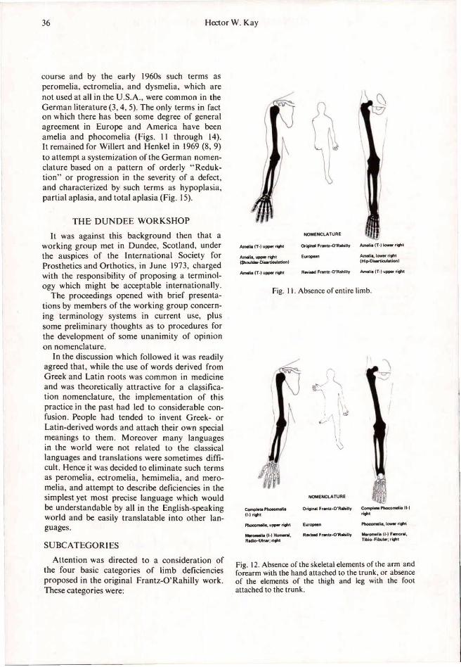
course and by the early 1960s such terms as peromelia, ectromelia, and dysmelia, which are not used at all in the U.S.A., were common in the German literature (3,4,5). The only terms in fact on which there has been some degree of general agreement in Europe and America have been amelia and phocomelia (Figs. 11 through 14). It remained for Willert and Henkel in 1969 (8, 9) to attempt a systemization of the German nomenclature based on a pattern of orderly "Reduktion" or progression in the severity of a defect, and characterized by such terms as hypoplasia, partial aplasia, and total aplasia (Fig. 15).
THE DUNDEE WORKSHOP
It was against this background then that a working group met in Dundee, Scotland, under the auspices of the International Society for Prosthetics and Orthotics, in June 1973, charged with the responsibility of proposing a terminology which might be acceptable internationally.
The proceedings opened with brief presentations by members of the working group concerning terminology systems in current use, plus some preliminary thoughts as to procedures for the development of some unanimity of opinion on nomenclature.
In the discussion which followed it was readily agreed that, while the use of words derived from Greek and Latin roots was common in medicine and was theoretically attractive for a classification nomenclature, the implementation of this practice in the past had led to considerable confusion. People had tended to invent Greek- or Latin-derived words and attach their own special meanings to them. Moreover many languages in the world were not related to the classical languages and translations were sometimes difficult. Hence it was decided to eliminate such terms as peromelia, ectromelia, hemimelia, and meromelia, and attempt to describe deficiencies in the simplest yet most precise language which would be understandable by all in the English-speaking world and be easily translatable into other languages.
SUBCATEGORIES
Attention was directed to a consideration of the four basic categories of limb deficiencies proposed in the original Frantz-O'Rahilly work. These categories were:
Fig. 11. Absence of entire limb.
Fig. 12. Absence of the skeletal elements of the arm and forearm with the hand attached to the trunk, or absence of the elements of the thigh and leg with the foot attached to the trunk.
Fig. 13. Absence of arm elements with forearm attached directly to the trunk, or absence of thigh elements with leg attached to the trunk.
Fig. 14. Absence of leg elements with foot attached directly to the thigh, or absence of forearm elements with the hand attached to the arm.
Fig. 15. Distal form of ectromelia. a, Tibia hypoplasia; b. Partial tibia aplasia; c, Subtotal tibia aplasia; d, Total tibia aplasia.
Unanimity of opinion was reached immediately concerning terminal transverse conditions, i.e., those presenting a congenital amputation-type stump. However, the existence of true intercalary deformities was questioned.
After considerable discussion and review of prior presentations involving both x-rays and diagrammatic representations of limb deficiencies, general agreement was reached that true intercalary deficiencies rarely if ever existed. It was postulated that all "phocomelias" or "intercalary deficiencies" had some terminal manifestation—be it a tarsal or carpal aberration, a defect of a finger or toe, or a deficiency of muscle, tendon, skin or nail. From this evolved an approach to classification which suggested that these intercalary defects were in reality variable degrees of longitudinal deficiencies. It was concluded that with the single exception of the previously mentioned transverse deficiencies all others were a manifestation of some longitudinal aberration in the formation of parts—thus even that condition described as "hypoplasia" of a limb or skeletal element in reality had a longitudinal (in the sense of the long axis of each bone) failure. Similarly, although "phocomelia" had a major manifestation of failure of formation in the long bones, there was also a lesser and perhaps minimal failure in the peripheral elements which, although present, were never truly normal. This concept of progressive longitudinal reduction can be carried to a point where only a single digital remnant of a limb remains and ultimately to the situation in which even this vestigial peripheral element failed to form—the true amelia. This, therefore, might be considered a maximum longitudinal deficiency although presenting as a transverse-type defect. For simplicity of designation, however, amelia might best be categorized as a transverse deficiency. It was recognized that in clinical practice the use of such well-established terms as amelia, phocomelia, and proximal
femoral focal deficiency (PFFD) would likely continue.
Based on this line of reasoning, a decision was reached to consolidate all limb deficiencies into two groups:
1. Transverse 2. Longitudinal
The transverse defects would encompass all so-called congenital amputation-type conditions and include what heretofore were referred to as terminal transverse deficiencies. The second major category would then become the longitudinal deficiencies which would encompass in effect all deficiencies which were not in the transverse category. It was agreed that the longitudinal category would require subdivision into 1) proximal longitudinal, 2) distal longitudinal, and 3) combined longitudinal deficiencies.
In further discussion of subdivisions under these two major categories, it was generally agreed that transverse deficiencies could be described and characterized by the level at which the limb terminated, but that in the longitudinal deficiencies such a description was unnecessary and that each deficiency could be described by naming the bone(s) affected and indicating whether they were completely or partially absent. It was recognized that conditions referred to as hypoplasia or underdevelopment in any one or all of the bones of the limb did exist, and could be described in the proximal, distal, or the combined form, again by naming the bone(s) affected and indicating the presence of hypoplasia.
OVERALL CLASSIFICATIONS OF MALFORMATIONS
At the request of Dr. Swanson the overall classification for congenital malformations,
which had been accepted previously by the American Hand Society, was considered. This system encompassed seven categories:
I. Failure of formation of parts II. Failure of differentiation (separation)
of parts III. Duplication IV. Overgrowth (Gigantism) V. Undergrowth
VI. Congenital constriction band syndrome VII. Generalized skeletal abnormalities.
While there was tacit acceptance of the rationality of these categories, it was unanimously agreed that the prime and virtually sole concern of the workshop was with nomenclature and classification in congenital skeletal limb deficiencies. In terms of the classification categories listed above these conditions would generally fall into the grouping designated as "failure of formation of parts," although some conditions might also involve failure of differentiation of parts, e.g., synostosis, or even occasionally undergrowth, e.g., a radius which was complete but hypoplastic.
PROPOSED INTERNATIONAL NOMENCLATURE
FOR CLASSIFICATION OF DEFICIENCIES
The workshop members then proceeded to the development of a schema which was unanimously proposed for international adoption, as follows:
FAILURE OF FORMATION OF PARTS
DISCUSSION
A few illustrations are presented to clarify the application of this classification procedure. It should be noted that in each case the designation indicates the level of absence; it being understood that all skeletal elements distal to that level are also absent. Each classification would, of course, include right (R), left (L), or bilateral (Bil).
1. Complete absence of an upper or lower limb (Figs. 16 and 17) would then be a transverse deficiency—arm (Ar), or thigh (Th), complete.
The term amelia (Fig. 11) would probably continue in clinical use to characterize this condition.
2. A transverse deficiency, Ar, upper, would indicate a short-above-elbow amputation-like limb which terminated in the upper third of the humerus (Fig. 18); a transverse deficiency, Th, lower, would be the term applied to a long above-knee amputation-like stump which terminated in the distal third of the femur (Fig. 19).
3. An elbow-disarticulation-type deficit (Fig. 20) would be classified as a transverse deficiency, forearm (Fo), complete; while below-elbow-type stumps would be designated Fo, upper, middle,
or lower, depending on the third of the forearm in which the limb terminated (Figs. 21, 22, and 23).
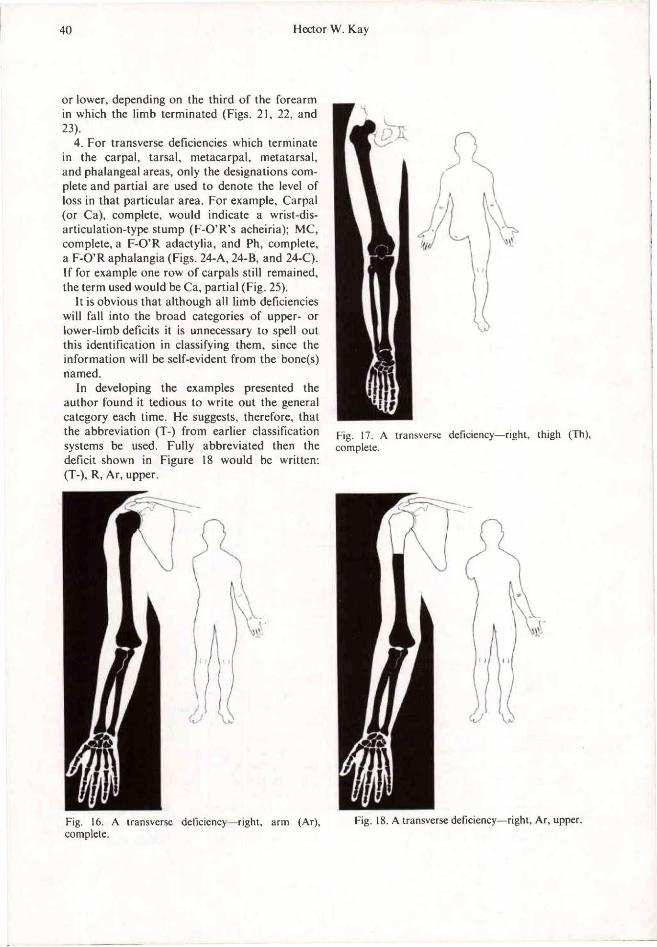
4. For transverse deficiencies which terminate in the carpal, tarsal, metacarpal, metatarsal, and phalangeal areas, only the designations complete and partial are used to denote the level of loss in that particular area. For example, Carpal (or Ca), complete, would indicate a wrist-disarticulation-type stump (F-O'R's acheiria); MC, complete, a F-O'R adactylia, and Ph, complete, a F-O'R aphalangia (Figs. 24-A, 24-B, and 24-C). If for example one row of carpals still remained, the term used would be Ca, partial (Fig. 25).
It is obvious that although all limb deficiencies will fall into the broad categories of upper- or lower-limb deficits it is unnecessary to spell out this identification in classifying them, since the information will be self-evident from the bone(s) named.
In developing the examples presented the author found it tedious to write out the general category each time. He suggests, therefore, that the abbreviation (T-) from earlier classification systems be used. Fully abbreviated then the deficit shown in Figure 18 would be written: (T-), R, Ar, upper.
Fig. 16. A transverse deficiency—right, arm (Ar), complete.
Fig. 17. A transverse deficiency—right, thigh (Th), complete.
Fig. 18. A transverse deficiency—right, Ar, upper.
Fig. 19. A transverse deficiency—right, Th, lower. Fig. 21. A transverse deficiency—right, Fo, upper.
Fig. 20. A transverse deficiency—right, forearm (Fo), complete.
Fig. 22. A transverse deficiency—right, Fo, middle.
Fig. 23. A transverse deficiency—right, Fo, lower.
Fig. 24-A. A transverse deficiency—right, carpal (Ca), complete.
Fig. 24-B. A transverse deficiency—right, metacarpal (MC), complete.
Fig. 24-C. A transverse deficiency—right, phalangeal (Ph), complete.
Fig. 25. A transverse deficiency—right, Ca, partial.
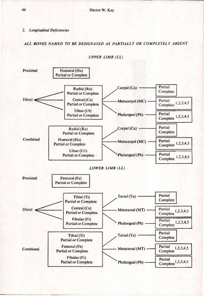
2. Longitudinal Deficiencies
ALL BONES NAMED TO BE DESIGNATED AS PARTIALLY OR COMPLETELY ABSENT
DISCUSSION
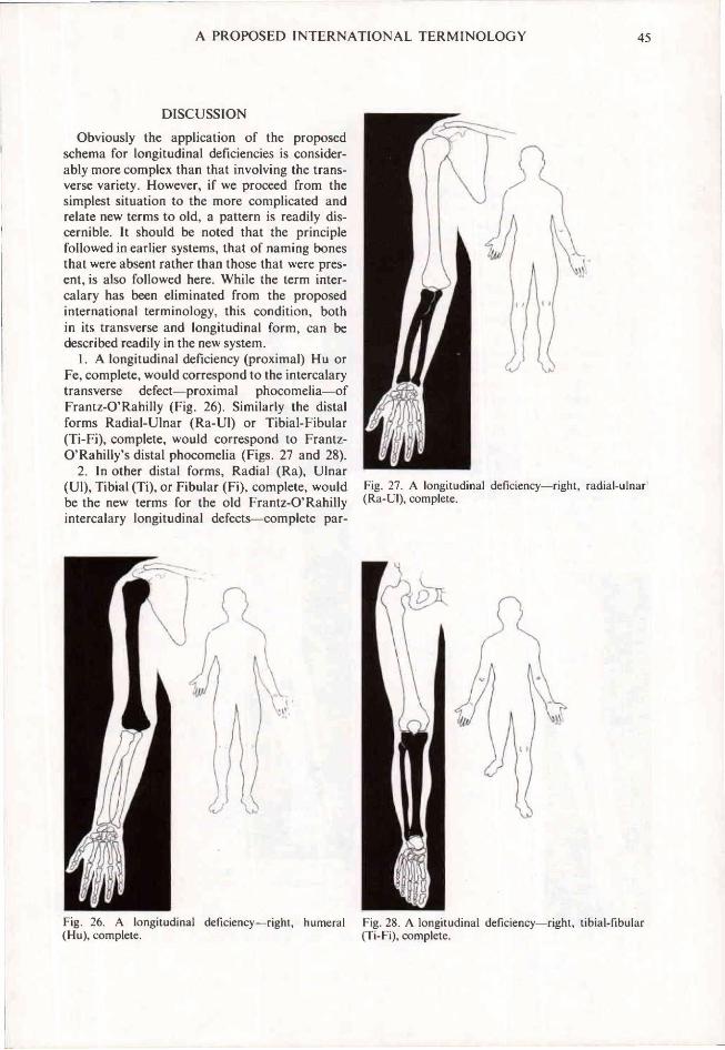
Obviously the application of the proposed schema for longitudinal deficiencies is considerably more complex than that involving the transverse variety. However, if we proceed from the simplest situation to the more complicated and relate new terms to old, a pattern is readily discernible. It should be noted that the principle followed in earlier systems, that of naming bones that were absent rather than those that were present, is also followed here. While the term intercalary has been eliminated from the proposed international terminology, this condition, both in its transverse and longitudinal form, can be described readily in the new system.
1. A longitudinal deficiency (proximal) Hu or Fe, complete, would correspond to the intercalary transverse defect—proximal phocomelia—of Frantz-O'Rahilly (Fig. 26). Similarly the distal forms Radial-Ulnar (Ra-Ul) or Tibial-Fibular (Ti-Fi), complete, would correspond to Frantz-O'Rahilly's distal phocomelia (Figs. 27 and 28).
2. In other distal forms, Radial (Ra), Ulnar (Ul), Tibial (Ti), or Fibular (Fi), complete, would be the new terms for the old Frantz-O'Rahilly intercalary longitudinal defects—complete par-
Fig. 26. A longitudinal deficiency—right, humeral (Hu), complete.
Fig. 27. A longitudinal deficiency—right, radial-ulnar (Ra-Ul), complete.
Fig. 28. A longitudinal deficiency—right, tibial-fibular (Ti-Fi), complete.
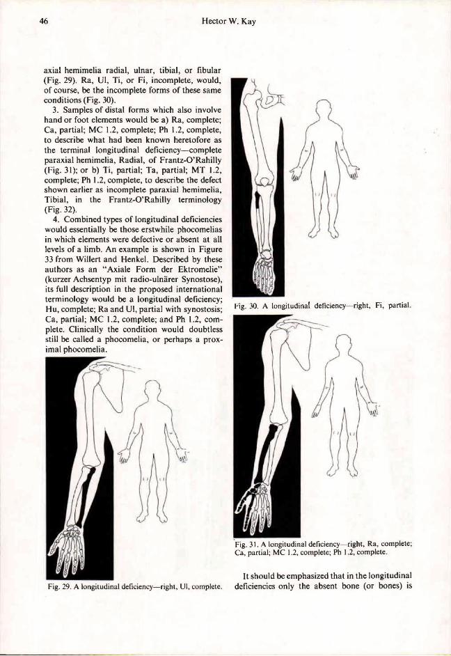
axial hemimelia radial, ulnar, tibial, or fibular (Fig. 29). Ra, Ul, Ti, or Fi, incomplete, would, of course, be the incomplete forms of these same conditions (Fig. 30).
3. Samples of distal forms which also involve hand or foot elements would be a) Ra, complete; Ca, partial; MC 1.2, complete; Ph 1.2, complete, to describe what had been known heretofore as the terminal longitudinal deficiency—complete paraxial hemimelia, Radial, of Frantz-O'Rahilly (Fig. 31); or b) Ti, partial; Ta, partial; MT 1.2, complete; Ph 1.2, complete, to describe the defect shown earlier as incomplete paraxial hemimelia, Tibial, in the Frantz-O'Rahilly terminology (Fig. 32).
4. Combined types of longitudinal deficiencies would essentially be those erstwhile phocomelias in which elements were defective or absent at all levels of a limb. An example is shown in Figure 33 from Willert and Henkel. Described by these authors as an "Axiale Form der Ektromelie" (kurzer Achsentyp mit radio-ulnarer Synostose), its full description in the proposed international terminology would be a longitudinal deficiency; Hu, complete; Ra and Ul, partial with synostosis; Ca, partial; MC 1.2, complete; and Ph 1.2, complete. Clinically the condition would doubtless still be called a phocomelia, or perhaps a proximal phocomelia.
It should be emphasized that in the longitudinal deficiencies only the absent bone (or bones) is Fig. 29. A longitudinal deficiency—right, Ul, complete.
Fig. 30. A longitudinal deficiency—right, Fi, partial.
Fig. 31. A longitudinal deficiency—right, Ra, complete; Ca, partial; MC 1.2, complete; Ph 1.2, complete.
Fig. 32. A longitudinal deficiency—right, Ti, partial; Ta, partial; MT 1.2, complete; Ph 1.2, complete.
Fig. 33. A longitudinal deficiency—left, Hu, complete; Ra and Ul, partial, with synostosis; Ca, partial; MC 1.2, complete; and Ph 1.2, complete.
cited no assumptions being made that more distal elements are also absent.
Again it is evident that, while the categories of UL and LL, and proximal, distal, or combined would be useful in organizing statistical or census data, these terms are not essential for classification. As in the transverse deficiencies this information is self-evident from the bone(s) named.
In preparing the examples for this paper the author again found it tedious to have to write out
"a longitudinal deficiency" each time. Use of an abbreviation L slash in parentheses (L/)-to avoid confusion with L for left-is proposed.
It should also be emphasized that the examples presented in this section are for illustrative purposes only. Some of the deficiencies classified may not exist clinically in as "pure" a form as depicted here.
FURTHER RECOMMENDATIONS
Following the development of the proposed international nomenclature as described, members of the working group tried out the new system by reclassifying a number of the deficiencies presented in slides earlier in the workshop. No difficulties were experienced. However, it was recognized that more extensive trials were desirable. Members of the workshop agreed to continue trying out the new system themselves but also recommended that field trials be carried out internationally under the auspices of the International Society for Prosthetics and Orthotics. These trials would be conducted in the U.S.A. through the medium of the Subcommittee on Child Prosthetics Problems of the Committee on Prosthetics Research and Development; and elsewhere in the world through selected child amputee clinics. Plans are now being made to implement these recommendations.
It was further recommended that the proposed international terminology for the classification of congenital limb deficiencies, as described in this paper, be brought to the attention of the World Health Organization for possible inclusion in the revision of standard nomenclature now being undertaken by that body. This recommendation has since been followed.
LITERATURE CITED
1. Burtch, Robert L., Nomenclature for Congenital Skeletal Limb Deficiencies, a Revision of the Frantz and O'Rahilly Classification. Artif. Limbs, 10:1:24-35, Spring 1966.
2. Frantz, C.H., and R O'Rahilly, Congenital Skeletal Limb Deficiencies. J. Bone and Joint Surg., 43-A:8:1202-1224, December 1961.
3. Hepp, Oscar, Frequency of the Congenital Defect—Anomalies of the Extremities in the Federal Republic of Germany, Inter-Clin. Inform. Bull., 1:10:3-12, July-August 1962.
4. Jentschura, G., E. Marquardt, and E.-M. Rudel, Behandlung und Versorgung bei Fehlbildungen und Amputationen der oberen Extremitat. Georg Thieme Verlag, Stuttgart, Germany, 1963.
5. Jentschura, G., E. Marquardt, and E.-M. Rudel, Malformations and Amputations of the Upper Extremity: Treatment and Prosthetic Replacement. Grune & Stratton, New York, 1967.
6. Swanson, A.B., A Classification for Congenital Malformations of the Hand. New Jersey Bull., Academy of Medicine, 10:166-169, September 1964.
7. Swanson, A.B., Classification of Limb Malformations on the Basis of Embryological Failures: A Preliminary Report. Inter-Clin. Inform. Bull., 6:3:1-15, December 1966.
8. Willert, H.-G., and H.-L. Henkel, Klinik und Pathologie der Dysmelie: Die Fehlbildungen an den oberen Extremitaten bei der Thalidomid-Embryopathie. Springer-Verlag, Heidelberg, New York, 1969.
9. Henkel, H.-L., and H.-G. Willert, Dysmelia, a Classification and a Pattern of Malformation of Congenital Limb Deficiencies. J. Bone and Joint Surg., 51-B:3:399-414, August 1969.
ILLUSTRATION CREDITS
Figs. 1-7, 11-14—Adapted from illustrations used in "Limb Development and Deformity: Problems of Evaluation and Rehabilitation" by Chester A. Swinyard, M.D. , Ph.D., (ed.). Charles C Thomas Publisher, Springfield, III., 1969. Figs. 8-10—Based on illustrations used in "Classification of Limb Malformations on the Basis of Embryo-logical Failures: A Preliminary Report" by A. B. Swanson, Inter-Clin. Inform. Bull., 6:3:1-15, December 1966. Drawings redone by George Rybczynski.
Figs. 15 and 33—Adapted from illustrations used in "Klinik und Pathologie der Dysmelie: Die Fehlbildungen an den oberen Extremitaten bei der Thalidomid-Embryopathie" by H.-G. Willert, and H.-L. Henkel. Springer-Verlag, Heidelberg, New York, 1969.