Aim of Study. To assess the feasibility of a new proposedmaneuver “RATLe-90” using real-time three-dimensional transesophagealechocardiography (RT-3DTEE) for anatomically oriented visualization of the interatrial septum (IAS) in guiding the transseptalpuncture TSP.Methods. The study included 20 patients (mean age, 60.2 ± 6.7 years; 60% males) who underwent TSP for differentindications. RT-3DTEEwas used to guide TSP.The proposedmaneuver RATLe-90 (Rotate-Anticlockwise-Tilt-Left-90) was appliedin all cases to have the anatomically oriented en face view of the IAS from the right atrial (RA) aspect. Having this anatomicallyoriented view, we guided the TSP catheter towards the proper puncture site according to the planned procedure. Results. Using theRATLe-90 maneuver, the anatomically oriented en face view of the IAS from the RA was obtained in all patients. We were ableto guide the puncture catheter to the proper puncture site on the IAS. The 3D images obtained were clearly understood by bothechocardiographers and interventionists. The RATLe-90 maneuver acquisition time was 19.9 ± 1.6 seconds. The time-to-tent was64.8 ± 16.3 seconds. Less TEE probe manipulations were needed while guiding the TSP. Conclusions. Application of RT3D-TEEduring TSP using RATLe-90 maneuver is feasible with shorter fluoroscopy time and minimizing TEE probe manipulations.
1. Introduction
Transseptal puncture (TSP) is a common step in variouspercutaneous structural cardiac interventions such as balloonmitral valvuloplasty [1], percutaneous mitral valve edge-to-edge repair [2], and percutaneous left atrial appendage (LAA)closure [3]. Two-dimensional transesophageal echocardiog-raphy (2D-TEE) is a widely used method for guidance ofTSP [4]. However, 2D-TEE has the inherent disadvantage ofbeing only two-dimensional whichmakes it very common forthe echocardiographer to lose the view if the catheter wentfew millimeters out of the very thin imaging plane. With theadvent of real-time three-dimensional TEE (RT3D-TEE) weare now capable of getting 3D anatomically oriented en faceview for the interatrial septum (IAS) in real time [5].
Till now there are no standardized maneuvers or proto-cols to get the anatomically oriented en face view for the IASduring guiding the septal puncture.
The aim of this pilot study was to propose a simple andstandardized maneuver to get the anatomically oriented enface view of the IAS from the right atrial (RA) perspective touse for guiding the TSP.
2. Methods
Using a commercially available ultrasound system (PhilipsiE33; Philips Medical Systems, Andover, MA) with a matrix-array 3D-TEEprobe (X7-2 t; PhilipsMedical Systems), twentypatients were included in this study (mean age, 60.2 ± 6.7
Hindawi Publishing CorporationCardiology Research and PracticeVolume 2015, Article ID 174051, 4 pageshttp://dx.doi.org/10.1155/2015/174051
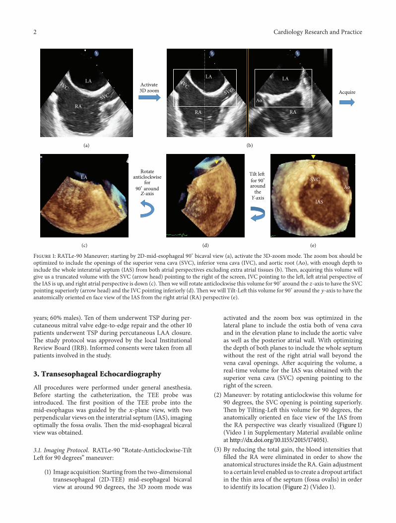
Figure 1: RATLe-90 Maneuver; starting by 2D-mid-esophageal 90∘ bicaval view (a), activate the 3D-zoom mode. The zoom box should beoptimized to include the openings of the superior vena cava (SVC), inferior vena cava (IVC), and aortic root (Ao), with enough depth toinclude the whole interatrial septum (IAS) from both atrial perspectives excluding extra atrial tissues (b). Then, acquiring this volume willgive us a truncated volume with the SVC (arrow head) pointing to the right of the screen, IVC pointing to the left, left atrial perspective ofthe IAS is up, and right atrial perspective is down (c).Then we will rotate anticlockwise this volume for 90∘ around the 𝑧-axis to have the SVCpointing superiorly (arrow head) and the IVC pointing inferiorly (d).Then we will Tilt-Left this volume for 90∘ around the 𝑦-axis to have theanatomically oriented en face view of the IAS from the right atrial (RA) perspective (e).
years; 60% males). Ten of them underwent TSP during per-cutaneous mitral valve edge-to-edge repair and the other 10patients underwent TSP during percutaneous LAA closure.The study protocol was approved by the local InstitutionalReview Board (IRB). Informed consents were taken from allpatients involved in the study.
3. Transesophageal Echocardiography
All procedures were performed under general anesthesia.Before starting the catheterization, the TEE probe wasintroduced. The first position of the TEE probe into themid-esophagus was guided by the 𝑥-plane view, with twoperpendicular views on the interatrial septum (IAS), imagingoptimally the fossa ovalis. Then the mid-esophageal bicavalview was obtained.
3.1. Imaging Protocol. RATLe-90 “Rotate-Anticlockwise-TiltLeft for 90 degrees” maneuver:
(1) Image acquisition: Starting from the two-dimensionaltransesophageal (2D-TEE) mid-esophageal bicavalview at around 90 degrees, the 3D zoom mode was
activated and the zoom box was optimized in thelateral plane to include the ostia both of vena cavaand in the elevation plane to include the aortic valveas well as the posterior atrial wall. With optimizingthe depth of both planes to include the whole septumwithout the rest of the right atrial wall beyond thevena caval openings. After acquiring the volume, areal-time volume for the IAS was obtained with thesuperior vena cava (SVC) opening pointing to theright of the screen.
(2) Maneuver: by rotating anticlockwise this volume for90 degrees, the SVC opening is pointing superiorly.Then by Tilting-Left this volume for 90 degrees, theanatomically oriented en face view of the IAS fromthe RA perspective was clearly visualized (Figure 1)(Video 1 in Supplementary Material available onlineat http://dx.doi.org/10.1155/2015/174051).
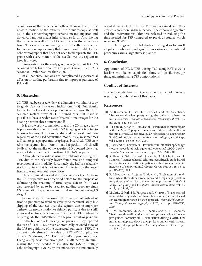
(3) By reducing the total gain, the blood intensities thatfilled the RA were eliminated in order to show theanatomical structures inside the RA. Gain adjustmentto a certain level enabled us to create a dropout artifactin the thin area of the septum (fossa ovalis) in orderto identify its location (Figure 2) (Video 1).
Cardiology Research and Practice 3
(a) (b)
SVC
Ao
EV
IAS
CSIVC
Superior
Anterior
Inferior
Posterior
Figure 2: Reducing the gain will remove the blood signals and will allow clear identification of the right atrial structures, for example, SVCopening, IVC opening, Eustachian valve (EV), coronary sinus (CS) opening, and aortic root (Ao). Further gain reduction will cause dropoutartifact in the thin area of the fossa ovalis (arrow head) that will help determining its location to guide septal puncture.
Ao
SVC
EVIVC CS
∗
(a)
AoSVC
EV
∗
(b)
AoSVC
EV
∗
(c)
Figure 3: RT3DTEE anatomically oriented view of the IAS from the RA perspective during guidance of transseptal puncture. The puncturecatheter (asterisk) was introduced through the IVC into the SVC (a).Then the catheter (asterisk) was pulled inferiorly and anteriorly towardsthe thin part of the IAS (arrow head) “Fossa” (b and c).
3.2. Guiding Transseptal Puncture (TSP). The interventionistpassed the catheter from the femoral vein through the IVCinto the SVC. Direct real-time visualization of the needle tipand the fossa using 3D-TEE was obtained to navigate thecatheter till it reached the fossa ovalis for tenting (Figure 3).
3.2.1. Time-to-Tent. We calculated the time from the firstmotion of the TSP catheter from the SVC downwards till theseptal tenting at the required position.
Retrospectively, we calculated the “time-to-tent” in acontrol group of twenty patients who underwent TSP for itsvarious indications in our center using fluoroscopy as well as2D-TEE for guidance.
4. Results
We could get the en face anatomically oriented view of theIAS from the RA perspective in all of the 20 patients included
in this study through using the RATLe-90 maneuver withina mean time of 19.5 ± 1.6 seconds starting from a standardmid-esophageal bicaval view.
Using this maneuver, probe manipulations were mini-mized regardless of the motion of the equipment used byinterventionist (needles, sheaths, and catheters). Comparedto 2D-TEE, the acquisition time was reduced.
In patients who underwent TSP for LAA closure, thelocation of fossa ovalis was clearly visualized and the orien-tation of the whole IAS helped the interventionist to directthe needle and catheter to the proper TSP site, that is,posteroinferior portion of the fossa ovalis.
In patients who underwent MV repair, the location offossa ovalis was clearly visualized and the orientation of thewhole IAS helped the interventionist to direct the needleand catheter to the proper TSP site, that is, posterosuperiorportion of the fossa ovalis.
Better communication between the echocardiographerand the interventionist was achieved in terms of directions
4 Cardiology Research and Practice
of motions of the catheter as both of them will agree thatupward motion of the catheter in the fluoroscopy as wellas in the echocardiography screens means superior anddownward motion means inferior and so forth. Also, havingthe catheter as well as the IAS and fossa in the same real-time 3D view while navigating with the catheter over theIAS is a unique opportunity that is more comfortable for theechocardiographer that does not need to manipulate the TEEprobe with every motion of the needle over the septum tokeep it in view.
Time-to-tent for the study group was (mean, 64.8 ± 16.3seconds), while for the control group was (mean, 139.9±29.3seconds); 𝑃 value was less than 0.0001.
In all patients, TSP was not complicated by pericardialeffusion or cardiac perforation due to improper puncture ofRA wall.
5. Discussion
2D-TEE had been used widely as adjunctive with fluoroscopyto guide TSP for its various indications [1–3]. But, thanksto the technological development, now we have the fullysampled matrix array 3D-TEE transducers that made itpossible to have a wider sector live/real-time images for thebeating heart in three dimensions [5].
It is also worthy to mention that if the 2D image qualityis poor one should not try using 3D imaging as it is going tobe worse because of the lower spatial and temporal resolutionregardless of the used acquisition mode. It is also sometimesdifficult to get a proper mid-esophageal bicaval 2D-TEE viewwith the septum in a more-or-less flat position which willbadly affect the quality of the acquired 3D-zoomed view thatmay not show the inferior portion of the IAS properly.
Although tachycardia is usually problematic during 3D-TEE due to the relatively lower frame rate and temporalresolution of this modality, fortunately, the IAS is a relativelystatic structure that is not too much affected by the lowerframe rate and temporal resolution.
The anatomically oriented en face view for the IAS fromthe RA perspective was described before for the purpose ofdelineating the anatomy of atrial septal defects [6]. It wasalso reported by us to be used for guiding coronary sinusCS cannulation in percutaneousmitral annuloplasty usingCSring [7].
In our study we measured the time-to-tent instead oftime-to-puncture to avoid bias related to technical issues likeslipping of the catheter over the septum due to impropercatheter on needle motion or delayed puncture due to a stiffabnormal septum, believing that the role of TEE guidance isonly to guide the TSP catheter to the proper tenting position.
To the best of our knowledge, no previous study reportedthe use of RT3D-TEE driven anatomically oriented view ofthe IAS for guidance of the transseptal puncture (TSP). Thecurrent study showed the value of RT3D-TEE applicationduring TSP during LAA closure and MV repair procedures.Using a step wise maneuver (RATLe-90) helped in mini-mizing the time needed to visualize the IAS in multipleechocardiographic views. By this maneuver, the anatomically
oriented view of IAS during TSP was obtained and thuscreated a common language between the echocardiographerand the interventionist. This was reflected in reducing thetime needed for TSP compared to previous studies whichrelied on 2D-TEE.
The findings of this pilot study encouraged us to enrollall patients who will undergo TSP in various interventionalprocedures and a large study is planned.
6. Conclusion
Application of RT3D-TEE during TSP using RATLe-90 isfeasible with better acquisition time, shorter fluoroscopytime, and minimizing TSP complications.
Conflict of Interests
The authors declare that there is no conflict of interestsregarding the publication of this paper.
References
[1] W. Bussmann, H. Sievert, N. Reifart, and M. Kaltenbach,“Transfemoral valvuloplasty using the balloon catheter inmitral stenosis,” Deutsche Medizinische Wochenschrift, vol. 112,no. 21, pp. 842–844, 1987.
[2] T. Feldman, S. Kar,M. Rinaldi et al., “Percutaneousmitral repairwith the MitraClip system: safety and midterm durability inthe initial EVEREST (Endovascular Valve Edge-to-Edge REpairStudy) cohort,” Journal of the American College of Cardiology,vol. 54, no. 8, pp. 686–694, 2009.
[3] J. Saw and M. Lempereur, “Percutaneous left atrial appendageclosure: procedural techniques and outcomes,” JACC: Cardio-vascular Interventions, vol. 7, no. 11, pp. 1205–1220, 2014.
[4] K. Hahn, R. Gal, J. Sarnoski, J. Kubota, D. H. Schmidt, and T.K. Bajwa, “Transesophageal echocardiographically guided atrialtransseptal catheterization in patients with normal-sized atria:incidence of complications,” Clinical Cardiology, vol. 18, no. 4,pp. 217–220, 1995.
[5] R. J. Housden, A. Arujuna, Y. Ma et al., “Evaluation of a real-time hybrid three-dimensional echo and X-ray imaging systemfor guidance of cardiac catheterisation procedures,” MedicalImage Computing and Computer-Assisted Intervention, vol. 15,no. 2, pp. 25–32, 2012.
[6] M. Saric, G. Perk, J. R. Purgess, and I. Kronzon, “Imaging atrialseptal defects by real-time three-dimensional transesophagealechocardiography: step-by-step approach,” Journal of the Amer-ican Society of Echocardiography, vol. 23, no. 11, pp. 1128–1135,2010.
[7] H. M. Mahmoud, M. A. Al-Ghamdi, and A. E. Ghabashi,“Real time three-dimensional transesophageal echocardiogra-phy guided coronary sinus cannulation during CARILLONmitral annuloplasty device therapy for a patient with chronicsevere mitral regurgitation,” Echocardiography, vol. 32, no. 1, pp.181–183, 2015.