Page 1
REPORTS OF ORIGINAL INVESTIGATIONS
A randomized comparison between costoclavicular andparacoracoid ultrasound-guided infraclavicular block for upperlimb surgery
Comparaison randomisee entre les blocs sous-claviculairesechoguides costoclaviculaires et paracoracoıdes pour la chirurgiedu membre superieur
Prangmalee Leurcharusmee, MD . Maria Francisca Elgueta, MD .
Worakamol Tiyaprasertkul, MD . Thitipan Sotthisopha, MD . Artid Samerchua, MD .
Aida Gordon, MD, FRCPC . Julian Aliste, MD . Roderick J. Finlayson, MD, FRCPC .
De Q. H. Tran, MD, FRCPC
Received: 28 November 2016 / Revised: 21 December 2016 / Accepted: 7 February 2017 / Published online: 15 February 2017
� Canadian Anesthesiologists’ Society 2017
Abstract
Background This two-centre randomized trial compared
costoclavicular and paracoracoid ultrasound-guided
infraclavicular brachial plexus block in patients
undergoing upper limb surgery. We hypothesized that
both techniques would result in similar onset times and
designed the study as an equivalence trial.
Methods Ninety patients undergoing upper limb surgery
at or distal to the elbow were randomly allocated to receive
a costoclavicular (n = 45) or paracoracoid (n = 45)
ultrasound-guided infraclavicular brachial plexus block.
Both groups received a 35-mL mixture of 1% lidocaine–
0.25% bupivacaine with epinephrine 5 lg�mL-1. In the
costoclavicular group, local anesthetic was injected into
the costoclavicular space in the middle of the three cords of
the brachial plexus. In the paracoracoid group, local
anesthetic was deposited dorsal to the axillary artery in the
lateral infraclavicular fossa. A blinded observer recorded
the block onset time (primary endpoint), success rate (i.e.,
surgical anesthesia), block-related pain scores, as well as
the incidence of hemidiaphragmatic paralysis.
Performance time and the number of needle passes were
also recorded during the performance of the block. The
total anesthesia-related time was defined as the sum of the
performance and onset times.
Results The mean (SD) onset times were comparable
between the costoclavicular and paracoracoid groups [16.0
(7.5) min vs 16.8 (6.2) min, respectively; mean difference,
0.8; 95% confidence interval, -2.3 to 3.8; P = 0.61].
Furthermore, no intergroup differences were found in terms
of performance time (P = 0.09), total anesthesia-related
time (P = 0.90), surgical anesthesia (P[ 0.99), and
hemidiaphragmatic paralysis (P[ 0.99). The paracoracoid
technique required marginally fewer median [interquartile
range] needle passes than the costoclavicular technique (2
[1-4] vs 2 [1-6], respectively; P = 0.048); however,
procedural pain was comparable between the two study
groups.
Conclusion Costoclavicular and paracoracoid
ultrasound-guided infraclavicular blocks resulted in
similar onset times. Furthermore, no intergroup
differences were found in terms of performance times and
success rates. Future dose-finding trials are required to
elucidate the minimum effective volume of local anesthetic
for costoclavicular infraclavicular blocks. This trial was
registered at www.clinicaltrials.in.th (Study ID:
TCTR20160525001).
Resume
Contexte Cette etude randomisee menee dans deux
centres a compare les blocs sous-claviculaires
echoguides du plexus brachial costoclaviculaires et
P. Leurcharusmee, MD � W. Tiyaprasertkul, MD
Department of Anesthesia, Maharaj Nakorn Chiang Mai
Hospital, Chiang Mai University, Chiang Mai, Thailand
M. F. Elgueta, MD � T. Sotthisopha, MD � A. Samerchua, MD �A. Gordon, MD, FRCPC � J. Aliste, MD �R. J. Finlayson, MD, FRCPC � D. Q. H. Tran, MD, FRCPC (&)
Department of Anesthesia, Montreal General Hospital, McGill
University, Montreal, QC, Canada
e-mail: [email protected]
123
Can J Anesth/J Can Anesth (2017) 64:617–625
DOI 10.1007/s12630-017-0842-z
Page 2
paracoracoıdes chez des patients devant subir une
chirurgie du membre superieur. Nous avons formule
l’hypothese que les deux techniques obtiendraient des
resultats dans les memes delais et avons concu cette
recherche comme une etude d’equivalence.
Methodes Quatre-vingt-dix patients subissant une
chirurgie du membre superieur au niveau du coude ou en
plus distal ont ete repartis pour recevoir un bloc sous-
claviculaire echoguide du plexus brachial par voie
costoclaviculaire (n = 45) ou paracoracoıde (n = 45).
Les deux groupes ont recu un melange de 35 mL de
lidocaıne a 1 % - bupivacaıne a 0,25 % et d’epinephrine a
raison de 5 lg�mL-1. Dans le groupe costoclaviculaire,
l’anesthesique local a ete injecte dans l’espace
costoclaviculaire au milieu des 3 branches du plexus
brachial. Dans le groupe paracoracoıde, l’anesthesique
local a ete depose en arriere de l’artere axillaire dans la
fosse sous-claviculaire externe. Un observateur tenu dans
l’insu de la technique a enregistre le temps d’installation
du bloc (critere d’evaluation principal), le taux de succes
(c’est-a-dire, l’anesthesie chirurgicale), les scores de
douleur lies au bloc ainsi que l’incidence de la paralysie
hemidiaphragmatique. Le temps de realisation et le
nombre de passages de l’aiguille ont egalement ete
consignes pendant la realisation du bloc. Le temps total
lie a l’anesthesie a ete defini comme la somme du temps de
realisation et du delai d’installation du bloc.
Resultats Les delais d’installation moyens (E-T) des
groupes costoclaviculaires et paracoracoıdes ont ete
comparables (respectivement 16,0 [7,5] minutes contre
16,8 [6,2] minutes; difference des moyennes, 0,8;
intervalle de confiance a 95 % : -2,3 a 3,8; P = 0,61).
En outre, aucune difference n’a ete constatee entre les
groupes pour ce qui concerne le temps de realisation
(P = 0,09), le temps total lie a l’anesthesie (P = 0,90),
l’anesthesie chirurgicale (P[ 0.99) et la paralysie
hemidiaphragmatique (P[ 0.99). La technique
paracoracoıde a necessite un nombre median
marginalement moindre (ecart interquartile) de passages
d’aiguille que la technique costoclaviculaire
(respectivement 2 [1 a 4] contre 2 [1 a 6]); P = 0,048).
Neanmoins, la douleur liee a la procedure a ete
comparable dans les deux groupes de l’etude.
Conclusion Les blocs sous-claviculaires echoguides
costoclaviculaires et paracoracoıdes ont abouti a des
temps d’installation similaires. De plus, aucune difference
n’a ete trouvee entre les groupes concernant les temps de
realisation et les taux de succes. Des essais futurs de
determination de dose sont necessaires pour preciser le
volume efficace minimum d’anesthesique local pour les
blocs sous-claviculaires costoclaviculaires. Cette etude a
ete enregistree sur le site www.clinicaltrials.in.th (ID de
l’etude : TCTR20160525001).
Ultrasound (US)-guided infraclavicular block (ICB)
anesthetizes the brachial plexus at the level of its cords
and provides reliable anesthesia and analgesia for upper
extremity surgery.1 The conventional (paracoracoid) ICB
technique aims to deposit local anesthetic (LA) dorsal to
the axillary artery in the lateral infraclavicular fossa.2,3
Karmakar et al.4 have recently described a new
costoclavicular ICB method whereby the brachial plexus
is targeted immediately caudal to the clavicle in the
costoclavicular space. In the latter method, the three cords
of the brachial plexus are tightly clustered together.
Consequently, Karmakar et al. speculated that the more
compact costoclavicular topography would result in a
‘‘very rapid onset of brachial plexus blockade similar to
that seen with a supraclavicular approach’’.4–6
Despite the potential anatomical benefits associated with a
costoclavicular technique, our general experience reveals a
similar block onset time between the latter and its
paracoracoid counterpart (unpublished data). Thus, in this
two-centre randomized-controlled trial, we set out to conduct
a formal comparison between US-guided costoclavicular and
paracoracoid ICBs. Our primary endpoint was onset time
(defined as the time required to reach a minimal score of 14/16
points using a sensorimotor composite scale). The secondary
outcomes included block performance time and success rate
(i.e., surgical anesthesia), number of needle passes, overall
procedural pain, and the incidence of adverse events (e.g.,
vascular breach, paresthesia, hemidiaphragmatic paralysis).
In keeping with our clinical experience, we hypothesized that
costoclavicular and paracoracoid ICBs would provide a
similar onset time; thus, we designed the study as an
equivalence trial.
Methods
After securing Ethics Committee approval (August, 2016
for the McGill University Health Centre, Montreal,
Canada and August, 2016 for the Maharaj Nakorn
Chiang Mai Hospital, Chiang Mai, Thailand) and
obtaining written informed consent, we enrolled 90
patients undergoing surgery of the elbow, forearm,
wrist, or hand. Inclusion criteria were: age between 18-
80 yrs of age, American Society of Anesthesiologists
physical status I- III, and body mass index 18-35 kg�m-2.
Exclusion criteria included the inability to consent to the
study, coagulopathy, sepsis, hepatic or renal failure,
allergy to LA, preexisting musculocutaneous/median/
radial/ ulnar neuropathy, and prior surgery in the
infraclavicular fossa.
After arrival in the induction room, intravenous
premedication (midazolam 0.03 mg�kg-1 and fentanyl 0.6
618 P. Leurcharusmee et al.
123
Page 3
lg�kg-1) was administered to all patients. Supplemental
oxygen (nasal cannulae at 4 L�min-1) and pulse oximetry
were applied throughout the procedure. Operators
(residents, fellows, or staff anesthesiologists) were
considered experts for a given technique if, prior to the
start of the study, they possessed an experience level of at
least 60 US-guided ICBs. Otherwise, they were considered
trainees.7 Three investigators supervised all the blocks
(P.L., M.F.E., D.Q.H.T.). Using a computer-generated
sequence of random numbers and a sealed envelope
technique, patients were randomly allocated to receive
costoclavicular (n = 45) or paracoracoid (n = 45) US-
guided ICB. Subjects were randomized in blocks of ten to
ensure equal distribution between the two study groups.
The Chiang Mai and Montreal centres were assigned five
and four blocks of ten patients, respectively. A 6-13 MHz
linear US transducer (SonoSite M-Turbo�, SonoSite� Inc,
Bothell, WA, USA), 22G 9-cm block needle (StimuQuik�ECHO, Arrow� International Inc, Reading, PA, USA), and
a lidocaine/bupivacaine mixture (1% lidocaine-0.25%
bupivacaine with epinephrine 5 lg�mL-1, obtained by
mixing equal parts of 2% lidocaine and 0.5% bupivacaine
before adding epinephrine 5 lg�mL-1) were used for all
subjects.
The paracoracoid ICB was performed according to a
previously described technique.1,2 The US probe was
applied in a sterile fashion in the lateral infraclavicular
fossa medially to the coracoid process in order to obtain a
short-axis view of the axillary artery (Fig. 1A). After skin
infiltration, using an in-plane technique and a cephalad-to-
caudad direction, the block needle was advanced until its
tip was located dorsal to the axillary artery. Thirty-five mL
of the LA mix were incrementally injected.
In the costoclavicular ICB group, patients were placed
in a supine position with the surgical limb in 90�abduction.6 The US probe was initially placed directly on
top of the middle third of the clavicle. Subsequently, the
probe was translocated off the inferior border of the
clavicle and positioned in the medial infraclavicular fossa.
In the costoclavicular space, the axillary artery was
identified underneath the subclavius muscle. The three
cords of the brachial plexus were visualized lateral to the
artery (Fig. 1B). If the cephalic vein (or thoracoacromial
artery) was visualized, the US transducer was tilted slightly
cephalad in order to provide an adequate sonographic view
of the costoclavicular space and the three cords of the
brachial plexus. After skin infiltration, using an in-plane
technique and a lateral-to-medial direction, the block
needle was advanced until its tip was located in the
middle of all three cords.4,6 Thirty-five mL of the LA mix
were incrementally injected.
For both groups, the imaging time was defined as the
temporal interval between contact of the US probe with the
patient and the acquisition of a satisfactory image. The
needling time (defined as the temporal interval between
skin infiltration and the end of LA injection through the
block needle) was also recorded. Thus, performance time
equalled the sum of imaging and needling times. The
number of needle passes was also recorded. The initial
needle insertion counted as the first pass. Any subsequent
needle advancement that was preceded by a retraction of at
least 10 mm counted as an additional pass.8 The
investigator supervising the ICB recorded the imaging/
needling times, number of passes, and occurrence of
adverse events (e.g., vascular puncture).
After LA injection through the block needle,
measurements of brachial plexus blockade were carried
out by a blinded observer every 5 min until 30 min.
Fig. 1 A. Sonographic view of the brachial plexus in the lateral
infraclavicular fossa; B. Sonographic view of the brachial plexus in
the costoclavicular space. L = lateral cord; M = medial cord; P =
posterior cord; PM = pectoralis major muscle; Pm = pectoralis minor
muscle; SCM = subclavius muscle; V = axillary vein
Costoclavicular infraclavicular block 619
123
Page 4
Sensory blockade of the musculocutaneous, median, radial,
and ulnar nerves was graded according to a three-point
scale using a cold test: 0 = no block; 1 = analgesia (patient
can feel touch, not cold); 2 = anesthesia (patient cannot feel
touch).1,2,9 Sensory blockade of the musculocutaneous,
median, radial, and ulnar nerves was assessed on the lateral
aspect of the forearm, the volar aspect of the thumb, the
lateral aspect of the dorsum of the hand, and the volar
aspect of the fifth finger, respectively.1,2,9 Motor blockade
was also graded on a three-point scale: 0 = no block; 1 =
paresis; 2 = paralysis.1,2,9 Motor blockade of the
musculocutaneous, radial, median, and ulnar nerves was
evaluated by elbow flexion, thumb abduction, thumb
opposition, and thumb adduction, respectively.1,2,9
Overall, the maximal composite sensorimotor score was
16 points. We considered the block a success and the
patient ready for surgery when a minimal composite score
of 14 points was achieved, provided the sensory block
score was equal or superior to 7 out of 8 points. This scale
has been used in previous studies.1,2,9 Thus, the block onset
time (i.e., the primary endpoint) was defined as the time
required to obtain C14 points after the end of LA injection
through the block needle. Total anesthesia-related time was
defined as the sum of performance and onset times. If the
composite score was inferior to 14 points after 30 min, the
patient was transferred to the operating room for the start of
the surgery. For these subjects, we did not record an onset
time and did not perform supplemental blocks. The same
blinded observer recorded surgical anesthesia, defined as
the capacity to undergo surgery without the need for
intravenous narcotics, general anesthesia, rescue blocks, or
LA infiltration by the surgeon.1,2,9 Nevertheless, subjects
could receive a propofol infusion (25-80 lg�kg-1�min-1)
intraoperatively provided response to verbal stimulus was
maintained. The blinded observer also recorded the
patient’s anthropometric data as well as the incidence of
paresthesia, Horner syndrome, hoarseness, and the level of
block-related pain (0 = no pain; 10 = worst imaginable
pain). Furthermore, 30 min after the ICB, the blinded
investigator assessed diaphragmatic motion with M-mode
US. Hemidiaphragmatic paralysis was defined as absent
diaphragm movement with inspiration coupled with absent
or paradoxical (i.e., downward deflection) movement when
the patient sniffs.10
One week after the surgery, the blinded investigator
contacted patients by telephone to inquire about
complications such as persistent numbness or paresthesia
and motor deficits.
Statistical analysis
Our experience with paracoracoid US-guided ICB revealed
a mean (SD) onset time of 17.1 (6.3) min.11 We deemed
that a 30% relative difference in onset time (i.e., 5.1 min)
carries minimal clinical significance and therefore set our
equivalence margin at ± 5.1 min. Thus, a calculated
sample size of 40 patients per group was required to
provide a statistical power of 0.90 and a type I error of
0.025. Since onset time can only be calculated for
successful blocks, 90 subjects were recruited to account
for an anticipated 10% failure rate.9
We elected to design the current protocol as an
equivalence instead of a superiority trial because our
preliminary experience with costoclavicular ICB led us to
suspect that its onset time would be similar to the one
provided by paracoracoid ICB. From a conceptual
standpoint, we also eschewed the non-inferiority design
since we aimed to show that costoclavicular ICB is no
better or no worse than its paracoracoid counterpart. In
contrast, a non-inferiority trial would have been indicated
had we wanted to demonstrate only that the new treatment
(i.e., costoclavicular ICB) is no worse than the active
control (i.e., paracoracoid ICB).12
Statistical analysis was performed using SPSS� version
21 statistical software (IBM Armonk, NY, USA). For
continuous data, normality was first assessed with
Lilliefors test, and if normal distribution was not rejected,
we employed the Student’s t test. Data that did not have a
normal distribution, as well as ordinal data, was analyzed
with the Mann-Whitney U test. For categorical data,
Pearson’s Chi square test was used. When the expected
count was less than five, Fisher’s exact test was employed.
All P values presented are two-sided and values \ 0.05
were considered significant.
Results
The 90 subjects were recruited from mid-August 2016 to
mid-October 2016 (Fig. 2). As planned, 50 and 40 patients
were enrolled in Chiang Mai and Montreal, respectively.
The costoclavicular and paracoracoid groups were
comparable in terms of anthropometric data and surgical
procedures (Table 1).
Our primary endpoint, mean (SD) onset time, was
similar between the costoclavicular and paracoracoid
groups [16.0 (7.5) min vs 16.8 (6.2) min, respectively;
mean difference, 0.8; 95% CI, -2.3 to 3.8; P = 0.61]
(Table 2). Furthermore, no intergroup differences were
found in terms of performance time (P = 0.09), total
anesthesia-related time (P = 0.90), surgical anesthesia
(P[ 0.99), and hemidiaphragmatic paralysis (P[ 0.99).
The proportions of blocks achieving minimal composite
scores of 14 points at the different five-minute intervals
were also similar between the two groups (Fig. 3). The
paracoracoid technique required marginally fewer median
620 P. Leurcharusmee et al.
123
Page 5
[interquartile range] needle passes compared with the
costoclavicular technique (2 [1-4] vs 2 [1-6], respectively;
P = 0.048); however, procedural pain was comparable
between the two study groups (Table 2).
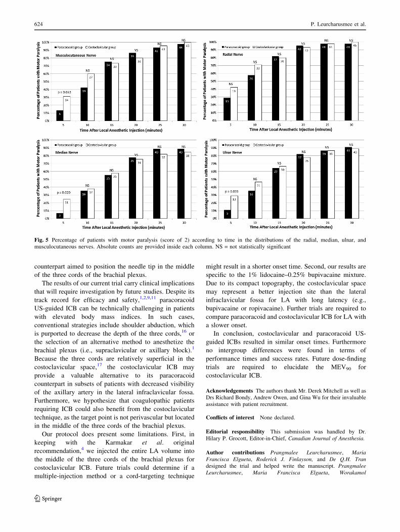
In the first five minutes after the ICB, more patients in
the costoclavicular group displayed complete sensory and
motor block in the territories of the musculocutaneous/
radial/ulnar nerves and musculocutaneous/median/ulnar
nerves, respectively. After ten minutes, however, sensory
and motor blockade of the musculocutaneous, median, and
ulnar nerves were comparable between the two groups.
Sensory blockade of the radial nerve was more prevalent in
the costoclavicular group until 20 min. No intergroup
differences were found thereafter (Figs 4 and 5).
Patient follow-up one week after the surgery revealed no
persistent sensory or motor deficits.
Discussion
In this randomized observer-blinded trial, we compared
costoclavicular and paracoracoid US-guided ICBs. No
intergroup differences were observed in terms of block
onset times (primary endpoint) or overall success rates (i.e.,
surgical anesthesia). We speculate that these findings might
be partially explained by the LA volume used. Although
the brachial plexus displays a more compact topography in
the costoclavicular space (where the costoclavicular ICB is
performed) than in the lateral infraclavicular fossa (where
the paracoracoid ICB is performed), these anatomical
differences were most likely compensated by the large
injectate (35 mL). This may explain why the
costoclavicular technique initially provided better
sensorimotor blockade, but after ten minutes, LA spread
Fig. 2 CONSORT diagram of
patient flow through the study.
The primary outcome (onset
time) and total anesthesia-
related time could not be
recorded for patients with
minimal composite scores\ 14
points at 30 min. However,
imaging/ needling/ performance
times, number of needle passes,
procedural pain, operator’s
experience level, adverse events
(e.g., vascular breach,
paresthesia, hemidiaphragmatic
paralysis) and surgical
anesthesia were recorded for
these subjects
Table 1 Patient characteristics
Paracoracoid
group
(n = 45)
Costoclavicular
group
(n = 45)
Age (yr) 40.9 (16.2) 42.8 (16.4)
Sex (M/ F) 19/ 26 28/ 17
BMI (kg�m-2) 24.1 (3.9) 23.4 (3.5)
ASA physical status (I/ II/ III) 29/ 15/ 1 28/ 14/ 3
Types of surgery (hand/ wrist/
forearm/ elbow)
20/ 18/ 7/ 0 25/ 12/ 7/ 1
Continuous variables are presented as means (standard deviation);
categorical variables are presented as counts. ASA = American
Society of Anesthesiologists; BMI = body mass index
Costoclavicular infraclavicular block 621
123
Page 6
in the paracoracoid group started to minimize differences
between the two methods. In the current study, due to the
absence of dose-finding trials for the 1.0% lidocaine–
0.25% bupivacaine mixture, we selected a 35-mL injectate
because this volume corresponds to the documented
minimum effective volume of 1.5% lidocaine in 90% of
patients (MEV90) for paracoracoid ICB.9 In their original
description, Karmakar et al.4 used only 20 mL for
costoclavicular ICB. Thus, we cannot rule out the
possibility that the costoclavicular technique may
outperform its paracoracoid counterpart with smaller LA
volumes. Therefore, future dose-finding trials should
attempt to elucidate the MEV90 for costoclavicular ICB.
Our costoclavicular ICB technique deserves special
mention. Karmakar et al.4,6 advocated dual guidance with
US and neurostimulation ‘‘until the operator is familiar
with the sonoanatomy of the [costoclavicular space]’’.
Although most of our operators were novices, we decided
to forego neurostimulation altogether and relied solely on
US guidance. In two previous randomized trials (combined
n = 182), Dingemans et al.13 and Gurkan et al.14 have
shown that, compared with US alone, combined US-
neurostimulation unnecessarily increased the performance
time13,14 and resulted in a lower success rate13 for
paracoracoid ICB. Thus, we similarly reasoned that nerve
stimulation would confer no benefits for US-guided
costoclavicular ICB. Furthermore, we believe that the
sole use of US did not impact our primary outcome (onset
time), as review of Karmakar et al.’s description clearly
reveals that the optimal target corresponded to needle tip
placement in the middle of the three cords of the brachial
plexus.4–6 In these studies, neurostimulation was not used
to shorten onset time. Instead, it served only as a failsafe
mechanism for the lateral-to-medial needle path—in other
words, the occurrence of evoked motor response acted as a
backstop and ensured that inexperienced operators would
not introduce the needle tip past the cords into the pleura.4,6
The incidences of ipsilateral phrenic nerve block,
vascular puncture, and paresthesia require discussion. In
both groups, 9% of subjects displayed complete
hemidiaphragmatic paralysis. In a recent trial, Petrar
et al.15 reported a mere 3% rate of diaphragmatic paresis
at 30 min after paracoracoid ICB. We hypothesize that the
discrepancy can be explained by a difference in LA volume
(and spread), as we used 35 mL whereas Petrar et al.15
employed 30 mL. Our findings also reveal a higher
incidence of vascular breach (8.9% vs 2.2%) with
paracoracoid ICB and a higher risk of paresthesia (4.4%
vs 0%) with costoclavicular ICB. Intergroup differences
did not reach statistical significance because our trial was
Table 2 Block performance data
Paracoracoid
group
(n = 45)
Costoclavicular
group
(n = 45)
Difference of the
means
95% CI P value
Imaging time (sec) 24.8 (18.4) 43.7 (29.1) -18.9 -29.1 to -8.7 \0.001*
Needling time (min) 5.6 (2.1) 6.0 (1.7) -0.4 -1.2 to 0.4 0.29�
Performance time (min) 6.0 (2.1) 6.7 (2.0) -0.7 -1.6 to 0.1 0.09�
Onset time (min) 16.8 (6.2) 16.0 (7.5) 0.8 -2.3 to 3.8 0.61�
Total anesthesia-related time (min) 22.6 (6.2) 22.8 (7.4) -0.2 -1.6 to 0.1 0.90�
Blocks with a minimal composite score of 14
points
40 (88.9) 41 (91.1) NA NA 0.76 }
Surgical anesthesia 44 (97.8) 43 (95.6) NA NA [0.99 }
Operator’s experience level (expert/trainee) 7 /38 11 /34 NA NA 0.43 }
Number of passes 2 [1-4] 2 [1-6] NA NA 0.048*
Block-related pain (scale 0-10) 0 [0-7] 0 [0-8] NA NA 0.85*
Vascular puncture 4 (8.9) 1 (2.2) NA NA 0.36§
Paresthesia 0 (0) 2 (4.4) NA NA 0.49§
Hemidiaphragmatic paralysis 4 (8.9) 4 (8.9) NA NA [0.99§
Horner syndrome 2 (4.4) 0 (0) NA NA 0.49§
Hoarseness 0 (0) 0 (0) NA NA [0.99§
Continuous variables are presented as mean (standard deviation); categorical variables are presented as count (percentage). Ordinal variables
(number of passes, block-related pain) are presented as median [range]. Onset and total anesthesia-related times are calculated only for patients
with a minimal composite score of 14 points at 30 min. CI = confidence interval; NA = not applicable
* Wilcoxon-Mann-Whitney test; �Student’s t test; }Chi square test; § Fisher’s exact test
622 P. Leurcharusmee et al.
123
Page 7
insufficiently powered to investigate the incidence of these
adverse events. Nonetheless, we attribute their occurrence
to the physical target (and needle path) required by each
technique—i.e., the paracoracoid method necessitated
placement of the needle tip contiguous to the axillary
artery (6 o’clock position), while its costoclavicular
Fig. 4 Percentage of patients with sensory anesthesia (score of 2) according to time in the cutaneous distributions of the radial, median, ulnar,
and musculocutaneous nerves. Absolute counts are provided inside each column. NS = not statistically significant
Fig. 3 Percentage of patients
with a minimal composite score
of 14 points according to time.
Absolute counts are provided
inside each column. NS = not
statistically significant
Costoclavicular infraclavicular block 623
123
Page 8
counterpart aimed to position the needle tip in the middle
of the three cords of the brachial plexus.
The results of our current trial carry clinical implications
that will require investigation by future studies. Despite its
track record for efficacy and safety,1,2,9,11 paracoracoid
US-guided ICB can be technically challenging in patients
with elevated body mass indices. In such cases,
conventional strategies include shoulder abduction, which
is purported to decrease the depth of the three cords,16 or
the selection of an alternative method to anesthetize the
brachial plexus (i.e., supraclavicular or axillary block).1
Because the three cords are relatively superficial in the
costoclavicular space,17 the costoclavicular ICB may
provide a valuable alternative to its paracoracoid
counterpart in subsets of patients with decreased visibility
of the axillary artery in the lateral infraclavicular fossa.
Furthermore, we hypothesize that coagulopathic patients
requiring ICB could also benefit from the costoclavicular
technique, as the target point is not perivascular but located
in the middle of the three cords of the brachial plexus.
Our protocol does present some limitations. First, in
keeping with the Karmakar et al. original
recommendation,4 we injected the entire LA volume into
the middle of the three cords of the brachial plexus for
costoclavicular ICB. Future trials could determine if a
multiple-injection method or a cord-targeting technique
might result in a shorter onset time. Second, our results are
specific to the 1% lidocaine–0.25% bupivacaine mixture.
Due to its compact topography, the costoclavicular space
may represent a better injection site than the lateral
infraclavicular fossa for LA with long latency (e.g.,
bupivacaine or ropivacaine). Further trials are required to
compare paracoracoid and costoclavicular ICB for LA with
a slower onset.
In conclusion, costoclavicular and paracoracoid US-
guided ICBs resulted in similar onset times. Furthermore
no intergroup differences were found in terms of
performance times and success rates. Future dose-finding
trials are required to elucidate the MEV90 for
costoclavicular ICB.
Acknowledgements The authors thank Mr. Derek Mitchell as well as
Drs Richard Bondy, Andrew Owen, and Gina Wu for their invaluable
assistance with patient recruitment.
Conflicts of interest None declared.
Editorial responsibility This submission was handled by Dr.
Hilary P. Grocott, Editor-in-Chief, Canadian Journal of Anesthesia.
Author contributions Prangmalee Leurcharusmee, Maria
Francisca Elgueta, Roderick J. Finlayson, and De Q.H. Tran
designed the trial and helped write the manuscript. Prangmalee
Leurcharusmee, Maria Francisca Elgueta, Worakamol
Fig. 5 Percentage of patients with motor paralysis (score of 2) according to time in the distributions of the radial, median, ulnar, and
musculocutaneous nerves. Absolute counts are provided inside each column. NS = not statistically significant
624 P. Leurcharusmee et al.
123
Page 9
Tiyaprasertkul, Thitipan Sotthisopha, Artid Samerchua, Aida Gordon,
Julian Aliste, Roderick J. Finlayson, and De Q.H. Tran helped
conduct the study as well as collect and analyze the data.
Funding None of the authors received funding for this study.
References
1. Tran DQ, Russo G, Munoz L, Zaouter C, Finlayson RJ. A
prospective, randomized comparison between ultrasound-guided
supraclavicular, infraclavicular, and axillary brachial plexus
blocks. Reg Anesth Pain Med 2009; 34: 366-71.
2. Tran DQ, Bertini P, Zaouter C, Munoz L, Finlayson RJ. A
prospective, randomized comparison between single- and double-
injection ultrasound-guided infraclavicular brachial plexus block.
Reg Anesth Pain Med 2010; 35: 16-21.
3. Desgagnes MC, Levesque S, Dion N, et al. A comparison of a
single or triple injection technique for ultrasound-guided
infraclavicular block: a prospective randomized controlled
study. Anesth Analg 2009; 109: 668-72.
4. Karmakar MK, Sala-Blanch X, Songthamwat B, Tsui BC. Benefits
of the costoclavicular for ultrasound-guided infraclavicular
brachial plexus block: description of a costoclavicular
approach. Reg Anesth Pain Med 2015; 40: 287-8.
5. Sala-Blanch X, Reina MA, Pangthipampai P, Karmakar MK.
Anatomic basis for brachial plexus block at the costoclavicular
space: a cadaver anatomic study. Reg Anesth Pain Med 2016; 41:
387-91.
6. Karmakar MK, Songthamwat B. Costoclavicular brachial plexus
block. In: Karmakar MK, editor. Musculoskeletal Ultrasound for
Regional Anesthesia and Pain Medicine. 2nd ed. Hong Kong,
China: CU Medicine; 2016 .
7. Konrad C, Schupfer G, Wietlisbach M, Gerber H. Learning
manual skills in anesthesiology: is there a recommended number
of cases for anesthetic procedures? Anesth Analg 1998; 86: 635-
9.
8. Casati A, Danelli G, Baciarello M, et al. A prospective,
randomized comparison between ultrasound and nerve
stimulation guidance for multiple injection axillary brachial
plexus block. Anesthesiology 2007; 106: 992-6.
9. Tran DQ, Dugani S, Diachenko A, Correa JA, Finlayson RJ.
Minimum effective volume of lidocaine for ultrasound-guided
infraclavicular block. Reg Anesth Pain Med 2011; 36: 190-4.
10. Loyd T, Tang YM, Benson MD, King S. Diaphragmatic paralysis:
the use of M mode ultrasound for diagnosis in adults. Spinal Cord
2006; 44: 505-8.
11. Leurcharusmee P, Aliste J, Van Zundert TC, et al. A multicenter
randomized comparison between intravenous and perineural
dexamethasone for ultrasound-guided infraclavicular block. Reg
Anesth Pain Med 2016; 41: 328-33.
12. Lesaffre E. Superiority, equivalence, and non-inferiority trials.
Bull NYU Hosp Jt Dis 2008; 66: 150-4.
13. Dingemans E, Williams SR, Arcand G, et al. Neurostimulation in
ultrasound-guided infraclavicular block: a prospective
randomized trial. Anesth Analg 2007; 104: 1275-80.
14. Gurkan Y, Tekin M, Acar S, Solak M, Toker K. Is nerve
stimulation needed during an ultrasound-guided lateral sagittal
infraclavicular block? Acta Anaesthesiol Scand 2010; 54: 403-7.
15. Petrar SD, Seltenrich ME, Head SJ, Schwarz SK.
Hemidiaphragmatic paralysis following ultrasound-guided
supraclavicular versus infraclavicular brachial plexus blockade:
a randomized clinical trial. Reg Anesth Pain Med 2015; 40: 133-
8.
16. Ruiz A, Sala X, Bargallo X, Hurtado P, Arguis MJ, Carrera A.
The influence of arm abduction on the anatomic relations of
infraclavicular brachial plexus: an ultrasound study. Anesth
Analg 2009; 108: 364-6.
17. Yoshida T, Watanabe Y, Furutani K. Proximal approach for
ultrasound-guided infraclavicular brachial plexus block. Acta
Anaesthesiol Taiwan 2016; 54: 31-2.
Costoclavicular infraclavicular block 625
123