CASE REPORT – OPEN ACCESSInternational Journal of Surgery Case Reports 7 (2015) 157–160
Contents lists available at ScienceDirect
International Journal of Surgery Case Reports
journa l homepage: www.caserepor ts .com
A rare case of chronic traumatic diaphragmatic hernia requiringcomplex abdominal wall reconstruction
Andrea Pakulaa,∗, Amber Jonesa, Javed Syedb, Ruby Skinnera
a Trauma Surgery, Critical Care, Acute Care General Surgery, Kern Medical Center, 1700 Mt. Vernon Ave. Bakersfield, CA 93306, USAb Radiology, Kern Medical Center, 1700 Mt. Vernon Ave. Bakersfield, CA 93306, USA
a r t i c l e i n f o
Article history:Received 5 January 2015Received in revised form 13 January 2015Accepted 13 January 2015Available online 15 January 2015
INTRODUCTION: Traumatic diaphragmatic hernia is a rare and often under recognized complication ofpenetrating and blunt trauma. These injuries are often missed or there is a delay in diagnosis which canlead to enlargement of the defect and the development of abdominal or respiratory symptoms.PRESENTATION OF CASE: We report a case of an otherwise healthy 37 year old male who was involvedin a motor vehicle accident at age twelve. He presented 25 years later with vague lower abdominalsymptoms and was found to have a large chronic left sided diaphragmatic hernia involving the major-ity of his intra-abdominal contents. Repair of the defect with a biologic mesh was undertaken and thepatient also required complex abdominal wall reconstruction due to loss of intra-abdominal domain fromthe chronicity of the hernia. A staged closure of the abdomen was performed first with placement of aWittmann patch. Medical management of intra-abdominal hypertension was successful and the midlinefascia was sequentially approximated at the bedside for three days. The final closure was performed witha component separation and implantation of a fenestrated biologic fetal bovine mesh to reinforce theclosure. In addition, a lightweight Ultrapro mesh was placed for additional lateral reinforcement. Thepatient recovered well and was discharged home.DISCUSSION: These injuries are rare and diagnosis is challenging. Mechanism and CT scan characteristicscan aid clinicians.CONCLUSION: Blunt diaphragmatic injury is rare and remains a diagnostic challenge. Depending on thechronicity of the injury, repair may require complex surgical decision making.
Traumatic diaphragmatic hernia is a rare and often under rec-ognized complication of penetrating and blunt trauma [1]. Theseinjuries are often missed or there is a delay in diagnosis, which canlead to enlargement of the defect and the development of abdomi-nal or respiratory symptoms [2,3]. We report a case of an otherwisehealthy 37 year old male who was involved in a motor vehicle acci-dent at age twelve. He presented 25 years later to us with vaguelower abdominal symptoms and was found to have a large chronicleft sided diaphragmatic hernia involving the majority of his intra-abdominal contents. Repair of the defect was undertaken and thepatient also required complex abdominal wall reconstruction as aresult. Blunt diaphragmatic injury is rare and remains a diagnosticchallenge. Clinicians must have a high index of suspicion if patientshave certain characteristics.
A 37 year old man presented to our emergency departmentwith complaints of lower left sided abdominal pain. He is an oth-erwise healthy man with no past medical or surgical history. Hestated the pain started one day prior but he denied any nausea,vomiting, shortness of breath or fever. He is a field worker anddenied any respiratory or abdominal symptoms in the past. Afterfurther questioning he did recall a motor vehicle accident when hewas twelve years old, twenty-five years earlier. His vital signs onpresentation were as follows: blood pressure 140/86 mmHg, heartrate 102 beats/min, respiratory rate 22 breaths/min, and oxygensaturation of 96% on room air. Laboratory findings were signif-icant for white blood cell count 18.8. A chest X-ray followedby CT scan of the chest, abdomen and pelvis were performedwhich demonstrated a left sided diaphragmatic hernia with stom-ach, pancreas, omentum, colon, and small bowel within the lefthemithorax (Fig. 1). Also noted were three left sided healed ribfractures. He was admitted to the surgical service and was takenfor exploratory laparotomy which revealed a 7 cm × 10 cm defectin the left diaphragm. The hernia defect was located in the medial
CASE REPORT – OPEN ACCESS158 A. Pakula et al. / International Journal of Surgery Case Reports 7 (2015) 157–160
Fig. 1. Preoperative chest X-ray and CT scan.
Fig. 2. Diaphragm defect before and after repair.
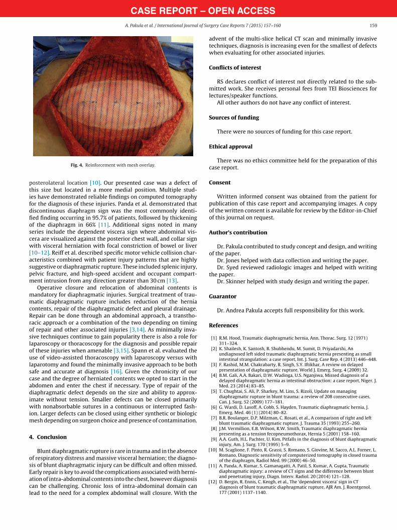
portion of the diaphragm abutting the GE junction. Though themajority of the intra-abdominal contents were within the chest,the GE junction remained in its usual anatomic location. Due to thesize of the defect we were unable to perform a primary repair andelected to use a fetal bovine dermal biologic mesh after the reduc-tion of contents (Fig. 2). We performed a staged abdominal wallreconstruction as the chronicity of the hernia led to loss of intra-abdominal domain. After reduction of the intrathoracic contentswe were unable to primarily close the midline fascia. A WittmannPatch was placed for serial advancement of the fascia and he wasreturned to the operating room for bilateral component separa-tion, closure and reinforcement with biologic mesh (Figs. 3 and 4).Postoperatively he did have evidence of intra-abdominal hyper-tension and poor compliance of the abdominal wall due to loss ofdomain. He required prolonged mechanical ventilator support withsedation and neuromuscular blockade. He developed mild acutekidney injury which resolved and was nutritionally supported withparenteral nutrition due to distension and ileus. Ultimately herecovered well from the abdominal portion of the surgery butdeveloped bronchiolitis obliterans organizing pneumonia (BOOP)of the left lung requiring a course of steroids.
3. Discussion
Traumatic diaphragmatic hernia is an underappreciated injuryin the acute setting and the majority of published literature isindividual reported cases. Trauma accounts for approximately0.8–8% of all diaphragmatic hernias [1]. These injuries are oftenmissed or there is a delay in diagnosis because they are associ-ated with other injuries, which require immediate attention; or thepatient does not present with typical symptoms. If an injury goes
undiagnosed, the hernia can grow in size over time but will oftenlead to the development of respiratory or abdominal symptoms[2–4]. As in our case, diagnosis of blunt diaphragmatic rupture canbe challenging if it is initially small and the patient does not requirelaparotomy for an associated injury. Left sided tears are most com-mon, as the liver tends to protect the right hemidiaphragm. Themost common reported mechanism of injury is motor vehicle col-lisions, accounting for approximately 88% of blunt diaphragmaticinjuries [5–9]. Tears in the diaphragm are usually more than tencentimeters in length, are radially orientated and occur at the weak-est part of the diaphragm, the musculotendinous junction in a
Fig. 3. Component separation and fascial closure.
CASE REPORT – OPEN ACCESSA. Pakula et al. / International Journal of Surgery Case Reports 7 (2015) 157–160 159
Fig. 4. Reinforcement with mesh overlay.
posterolateral location [10]. Our presented case was a defect ofthis size but located in a more medial position. Multiple stud-ies have demonstrated reliable findings on computed tomographyfor the diagnosis of these injuries. Panda et al. demonstrated thatdiscontinuous diaphragm sign was the most commonly identi-fied finding occurring in 95.7% of patients, followed by thickeningof the diaphragm in 66% [11]. Additional signs noted in manyseries include the dependent viscera sign where abdominal vis-cera are visualized against the posterior chest wall, and collar signwith visceral herniation with focal constriction of bowel or liver[10–12]. Reiff et al. described specific motor vehicle collision char-acteristics combined with patient injury patterns that are highlysuggestive or diaphragmatic rupture. These included splenic injury,pelvic fracture, and high-speed accident and occupant compart-ment intrusion from any direction greater than 30 cm [13].
Operative closure and relocation of abdominal contents ismandatory for diaphragmatic injuries. Surgical treatment of trau-matic diaphragmatic rupture includes reduction of the herniacontents, repair of the diaphragmatic defect and pleural drainage.Repair can be done through an abdominal approach, a transtho-racic approach or a combination of the two depending on timingof repair and other associated injuries [3,14]. As minimally inva-sive techniques continue to gain popularity there is also a role forlaparoscopy or thoracoscopy for the diagnosis and possible repairof these injuries when amenable [3,15]. Spann et al. evaluated theuse of video-assisted thoracoscopy with laparoscopy versus withlaparotomy and found the minimally invasive approach to be bothsafe and accurate at diagnosis [16]. Given the chronicity of ourcase and the degree of herniated contents we opted to start in theabdomen and enter the chest if necessary. Type of repair of thediaphragmatic defect depends on the size and ability to approx-imate without tension. Smaller defects can be closed primarilywith nonabsorbable sutures in a continuous or interrupted fash-ion. Larger defects can be closed using either synthetic or biologicmesh depending on surgeon choice and presence of contamination.
4. Conclusion
Blunt diaphragmatic rupture is rare in trauma and in the absenceof respiratory distress and massive visceral herniation; the diagno-sis of blunt diaphragmatic injury can be difficult and often missed.Early repair is key to avoid the complications associated with herni-ation of intra-abdominal contents into the chest, however diagnosiscan be challenging. Chronic loss of intra-abdominal domain canlead to the need for a complex abdominal wall closure. With the
advent of the multi-slice helical CT scan and minimally invasivetechniques, diagnosis is increasing even for the smallest of defectswhen evaluating for other associated injuries.
Conflicts of interest
RS declares conflict of interest not directly related to the sub-mitted work. She receives personal fees from TEI Biosciences forlectures/speaker functions.
All other authors do not have any conflict of interest.
Sources of funding
There were no sources of funding for this case report.
Ethical approval
There was no ethics committee held for the preparation of thiscase report.
Consent
Written informed consent was obtained from the patient forpublication of this case report and accompanying images. A copyof the written consent is available for review by the Editor-in-Chiefof this journal on request.
Author’s contribution
Dr. Pakula contributed to study concept and design, and writingof the paper.
Dr. Jones helped with data collection and writing the paper.Dr. Syed reviewed radiologic images and helped with writing
the paper.Dr. Skinner helped with study design and writing the paper.
Guarantor
Dr. Andrea Pakula accepts full responsibility for this work.
[2] K. Shailesh, K. Santosh, B. Shubhendu, M. Sumit, D. Priyadarshi, Anundiagnosed left sided traumatic diaphragmatic hernia presenting as smallintestinal strangulation: a case report, Int. J. Surg. Case Rep. 4 (2013) 446–448.
[3] F. Rashid, M.M. Chakrabarty, R. Singh, S.Y. Iftikhar, A review on delayedpresentation of diaphragmatic rupture, World J. Emerg. Surg. 4 (2009) 32.
[4] B.M. Gali, A.A. Bakari, D.W. Wadinga, U.S. Nganjiwa, Missed diagnosis of adelayed diaphragmatic hernia as intestinal obstruction: a case report, Niger. J.Med. 23 (2014) 83–85.
[5] T. Chughtai, S. Ali, P. Sharkey, M. Lins, S. Rizoli, Update on managingdiaphragmatic rupture in blunt trauma: a review of 208 consecutive cases,Can. J. Surg. 52 (2009) 177–181.
[6] G. Wardi, D. Lasoff, A. Cobb, S. Hayden, Traumatic diaphragmatic hernia, J.Emerg. Med. 46 (1) (2014) 80–82.
[7] B.R. Boulanger, D.P. Milzman, C. Rosati, et al., A comparison of right and leftblunt traumatic diaphragmatic rupture, J. Trauma 35 (1993) 255–260.
[8] J.M. Vermillion, E.B. Wilson, R.W. Smith, Traumatic diaphragmatic herniapresenting as a tension fecopneumothorax, Hernia 5 (2001) 158–160.
[9] A.A. Guth, H.L. Pachter, U. Kim, Pitfalls in the diagnosis of blunt diaphragmaticinjury, Am. J. Surg. 170 (1995) 5–9.
[10] M. Scaglione, F. Pinto, R. Grassi, S. Romano, S. Giovine, M. Sacco, A.L. Forner, L.Romano, Diagnostic sensitivity of computerized tomography in closed traumaof the diaphragm, Radiol Med. 99 (2000) 46–50.
[11] A. Panda, A. Kumar, S. Gamanagatti, A. Patil, S. Kumar, A. Gupta, Traumaticdiaphragmatic injury: a review of CT signs and the difference between bluntand penetrating injury, Diagn. Interv. Radiol. 20 (2014) 121–128.
[12] D. Bergin, R. Ennis, C. Keogh, et al., The ‘dependent viscera’ sign in CTdiagnosis of blunt traumatic diaphragmatic rupture, AJR Am. J. Roentgenol.177 (2001) 1137–1140.
CASE REPORT – OPEN ACCESS160 A. Pakula et al. / International Journal of Surgery Case Reports 7 (2015) 157–160
[13] D.A. Reiff, G. McGwin Jr., J. Metzger, et al., Identifying injuries and motorvehicle collision characteristics that together are suggestive of diaphragmaticrupture, J. Trauma 53 (2002) 1139–1145.
[14] R. DeBlasio, P. Maione, U. Avallone, M. Rossi, F. Pigna, C. Napolitano, Lateposttraumatic diaphragmatic hernia. A clinical case report, Minerva Chir. 49(1994) 481–487.
[15] M. Pross, T. Manger, L. Mirow, S. Wolff, H. Lippert, Laparoscopic managementof a late-diagnosed major diaphragmatic rupture, Laparoendosc. Adv. Surg.Tech. 10 (2) (2000) 111–114.
[16] J.C. Spann, F.E. Nwariaku, M. Wait, Evaluation of video-assisted thoracoscopicsurgery in the diagnosis of diaphragmatic injuries, Am. J. Surg. 170 (1995)628–631.
Open AccessThis article is published Open Access at sciencedirect.com. It is distributed under the IJSCR Supplemental terms and conditions, whichpermits unrestricted non commercial use, distribution, and reproduction in any medium, provided the original authors and source arecredited.