DEVELOPMENT 3427 RESEARCH ARTICLE INTRODUCTION The neurons and glial cells of the mature central nervous system (CNS) develop from the neuroepithelial progenitor cells that surround the lumen of the embryonic spinal cord and the ventricles of the brain – the so-called ventricular zone (VZ). The spinal cord VZ is a mosaic of progenitor cell domains, each of which generates one or more distinct subtypes of neurons followed by glial cells. The domain pattern is established in response to signals from local organising centres (Briscoe et al., 2000; Ericson et al., 1997). For example, sonic hedgehog diffusing from the notochord and floor plate forms a concentration gradient that specifies five ventral progenitor domains known as p3, pMN, p2, p1 and p0 (ventral to dorsal). This initial patterning phase is followed by the neurogenic phase, during which the progenitor domains give rise to particular combinations of differentiated neurons and glia (Rowitch, 2004). Motor neurons and several types of interneurons (INs) in the ventral spinal cord assemble into local networks that generate the rhythmic output required for locomotion (reviewed by Kiehn, 2006). To understand how locomotor circuits develop, it is necessary to understand the genetic and cellular mechanisms that determine neuron diversity. The p2 progenitor domain generates two distinct subtypes of INs, V2a and V2b (Karunaratne et al., 2002; Li et al., 2005; Smith et al., 2002; Zhou et al., 2000). Postmitotic V2a INs are characterised by expression of the homeodomain transcription factor Chx10 (Ericson et al., 1997), whereas V2b INs express transcription factors Gata2, Gata3 and Scl (Tal1) (Karunaratne et al., 2002; Muroyama et al., 2005; Smith et al., 2002). How V2 INs incorporate into the local spinal circuitry is not established, although V2a INs are thought to be excitatory (glutamatergic) and to project ipsilaterally (Kiehn, 2006; Kimura et al., 2006). The neurotransmitter phenotype of V2b INs is not known. V2a and V2b INs are derived from common progenitors that initially express the forkhead/winged helix transcription factor Foxn4 (Li et al., 2005) (this paper). How does this homogeneous progenitor pool generate two distinct neuronal subtypes? The Notch-Delta signalling pathway is often used to establish or to maintain differences between lineally related cells (Artavanis- Tsakonas et al., 1999; Louvi and Artavanis-Tsakonas, 2006). For example, signalling between Notch1 and its ligand delta-like 4 (Dll4) in endothelial cells is necessary for artery-vein discrimination and also for sprouting of lymphatic vessels from veins (Duarte et al., 2004; Seo et al., 2006). We thought it possible that the distinction between V2a and V2b INs might also be established through Notch- Delta signalling. Notch1, 2 and 3 are all expressed in the ventral VZ of the embryonic spinal cord (Lindsell et al., 1996), as are their ligands Dll1, Dll3, Dll4 and jagged 1 (Benedito and Duarte, 2005; Dunwoodie et al., 1997; Lindsell et al., 1996; Mailhos et al., 2001). Unlike Dll1 and Dll3, which are expressed widely throughout the VZ and/or in postmitotic neurons, Dll4 appears to be restricted to the p2 domain of the VZ, suggesting a specific role in V2 IN development (Benedito and Duarte, 2005). A regulatory network involving Foxn4, Mash1 and delta-like 4/ Notch1 generates V2a and V2b spinal interneurons from a common progenitor pool Marta G. Del Barrio 1 , Raquel Taveira-Marques 1 , Yuko Muroyama 2, *, Dong-In Yuk 2 , Shengguo Li 3 , Mary Wines-Samuelson 4 , Jie Shen 4 , Hazel K. Smith 1 , Mengqing Xiang 3 , David Rowitch 2,† and William D. Richardson 1,‡ In the developing central nervous system, cellular diversity depends in part on organising signals that establish regionally restricted progenitor domains, each of which produces distinct types of differentiated neurons. However, the mechanisms of neuronal subtype specification within each progenitor domain remain poorly understood. The p2 progenitor domain in the ventral spinal cord gives rise to two interneuron (IN) subtypes, V2a and V2b, which integrate into local neuronal networks that control motor activity and locomotion. Foxn4, a forkhead transcription factor, is expressed in the common progenitors of V2a and V2b INs and is required directly for V2b but not for V2a development. We show here in experiments conducted using mouse and chick that Foxn4 induces expression of delta-like 4 (Dll4) and Mash1 (Ascl1). Dll4 then signals through Notch1 to subdivide the p2 progenitor pool. Foxn4, Mash1 and activated Notch1 trigger the genetic cascade leading to V2b INs, whereas the complementary set of progenitors, without active Notch1, generates V2a INs. Thus, Foxn4 plays a dual role in V2 IN development: (1) by initiating Notch-Delta signalling, it introduces the asymmetry required for development of V2a and V2b INs from their common progenitors; (2) it simultaneously activates the V2b genetic programme. KEY WORDS: Notch1, Delta-like 4, Foxn4, Gata, Scl (Tal1), Chx10, Spinal cord, Neurogenesis, Chick, Mouse, V2 interneurons, Mash1 (Ascl1) Development 134, 3427-3436 (2007) doi:10.1242/dev.005868 1 Wolfson Institute for Biomedical Research and Department of Biology, University College London, Gower Street, London WC1E 6BT, UK. 2 Department of Pediatric Oncology, Dana-Farber Cancer Institute, Dana 640D, 44 Binney Street, Boston, MA 02115, USA. 3 Center for Advanced Biotechnology and Medicine, UMDNJ-Robert Wood Johnson Medical School, 679 Hoes Lane, Piscataway, NJ 08854, USA. 4 Center for Neurologic Diseases, Brigham and Women’s Hospital, Program in Neuroscience, Harvard Medical School, 77 Avenue Louis Pasteur, Boston, MA 02115, USA. *Present address: Department of Developmental Biology, Graduate School of Medicine, Chiba University, 1-8-1 Inohana, Chuo-ku, Chiba 260-8670, Japan † Present address: Department of Pediatrics, Institute for Regeneration Medicine, University of California at San Francisco, 513 Parnassus Avenue, San Francisco, CA 94143- 0525, USA ‡ Author for correspondence (e-mail: [email protected]) Accepted 25 July 2007
Transcript
DEVELO
PMENT
3427RESEARCH ARTICLE
INTRODUCTIONThe neurons and glial cells of the mature central nervous system(CNS) develop from the neuroepithelial progenitor cells thatsurround the lumen of the embryonic spinal cord and the ventriclesof the brain – the so-called ventricular zone (VZ). The spinal cordVZ is a mosaic of progenitor cell domains, each of which generatesone or more distinct subtypes of neurons followed by glial cells. Thedomain pattern is established in response to signals from localorganising centres (Briscoe et al., 2000; Ericson et al., 1997). Forexample, sonic hedgehog diffusing from the notochord and floorplate forms a concentration gradient that specifies five ventralprogenitor domains known as p3, pMN, p2, p1 and p0 (ventral todorsal). This initial patterning phase is followed by the neurogenicphase, during which the progenitor domains give rise to particularcombinations of differentiated neurons and glia (Rowitch, 2004).Motor neurons and several types of interneurons (INs) in the ventralspinal cord assemble into local networks that generate the rhythmicoutput required for locomotion (reviewed by Kiehn, 2006). To
understand how locomotor circuits develop, it is necessary tounderstand the genetic and cellular mechanisms that determineneuron diversity.
The p2 progenitor domain generates two distinct subtypes of INs,V2a and V2b (Karunaratne et al., 2002; Li et al., 2005; Smith et al.,2002; Zhou et al., 2000). Postmitotic V2a INs are characterised byexpression of the homeodomain transcription factor Chx10 (Ericsonet al., 1997), whereas V2b INs express transcription factors Gata2,Gata3 and Scl (Tal1) (Karunaratne et al., 2002; Muroyama et al., 2005;Smith et al., 2002). How V2 INs incorporate into the local spinalcircuitry is not established, although V2a INs are thought to beexcitatory (glutamatergic) and to project ipsilaterally (Kiehn, 2006;Kimura et al., 2006). The neurotransmitter phenotype of V2b INs isnot known. V2a and V2b INs are derived from common progenitorsthat initially express the forkhead/winged helix transcription factorFoxn4 (Li et al., 2005) (this paper). How does this homogeneousprogenitor pool generate two distinct neuronal subtypes?
The Notch-Delta signalling pathway is often used to establish orto maintain differences between lineally related cells (Artavanis-Tsakonas et al., 1999; Louvi and Artavanis-Tsakonas, 2006). Forexample, signalling between Notch1 and its ligand delta-like 4(Dll4) in endothelial cells is necessary for artery-vein discriminationand also for sprouting of lymphatic vessels from veins (Duarte et al.,2004; Seo et al., 2006). We thought it possible that the distinctionbetween V2a and V2b INs might also be established through Notch-Delta signalling. Notch1, 2 and 3 are all expressed in the ventral VZof the embryonic spinal cord (Lindsell et al., 1996), as are theirligands Dll1, Dll3, Dll4 and jagged 1 (Benedito and Duarte, 2005;Dunwoodie et al., 1997; Lindsell et al., 1996; Mailhos et al., 2001).Unlike Dll1 and Dll3, which are expressed widely throughout theVZ and/or in postmitotic neurons, Dll4 appears to be restricted to thep2 domain of the VZ, suggesting a specific role in V2 INdevelopment (Benedito and Duarte, 2005).
A regulatory network involving Foxn4, Mash1 and delta-like 4/Notch1 generates V2a and V2b spinal interneurons from acommon progenitor poolMarta G. Del Barrio1, Raquel Taveira-Marques1, Yuko Muroyama2,*, Dong-In Yuk2, Shengguo Li3,Mary Wines-Samuelson4, Jie Shen4, Hazel K. Smith1, Mengqing Xiang3, David Rowitch2,† andWilliam D. Richardson1,‡
In the developing central nervous system, cellular diversity depends in part on organising signals that establish regionally restrictedprogenitor domains, each of which produces distinct types of differentiated neurons. However, the mechanisms of neuronalsubtype specification within each progenitor domain remain poorly understood. The p2 progenitor domain in the ventral spinalcord gives rise to two interneuron (IN) subtypes, V2a and V2b, which integrate into local neuronal networks that control motoractivity and locomotion. Foxn4, a forkhead transcription factor, is expressed in the common progenitors of V2a and V2b INs and isrequired directly for V2b but not for V2a development. We show here in experiments conducted using mouse and chick that Foxn4induces expression of delta-like 4 (Dll4) and Mash1 (Ascl1). Dll4 then signals through Notch1 to subdivide the p2 progenitor pool.Foxn4, Mash1 and activated Notch1 trigger the genetic cascade leading to V2b INs, whereas the complementary set of progenitors,without active Notch1, generates V2a INs. Thus, Foxn4 plays a dual role in V2 IN development: (1) by initiating Notch-Deltasignalling, it introduces the asymmetry required for development of V2a and V2b INs from their common progenitors; (2) itsimultaneously activates the V2b genetic programme.
Development 134, 3427-3436 (2007) doi:10.1242/dev.005868
1Wolfson Institute for Biomedical Research and Department of Biology, UniversityCollege London, Gower Street, London WC1E 6BT, UK. 2Department of PediatricOncology, Dana-Farber Cancer Institute, Dana 640D, 44 Binney Street, Boston, MA02115, USA. 3Center for Advanced Biotechnology and Medicine, UMDNJ-RobertWood Johnson Medical School, 679 Hoes Lane, Piscataway, NJ 08854, USA. 4Centerfor Neurologic Diseases, Brigham and Women’s Hospital, Program in Neuroscience,Harvard Medical School, 77 Avenue Louis Pasteur, Boston, MA 02115, USA.
*Present address: Department of Developmental Biology, Graduate School ofMedicine, Chiba University, 1-8-1 Inohana, Chuo-ku, Chiba 260-8670, Japan†Present address: Department of Pediatrics, Institute for Regeneration Medicine,University of California at San Francisco, 513 Parnassus Avenue, San Francisco,CA 94143- 0525, USA‡Author for correspondence (e-mail: [email protected])
Accepted 25 July 2007
DEVELO
PMENT
3428
We have examined the relationship between Foxn4 and Notch-Delta signalling during development of V2a and V2b sub-lineages.We demonstrated that Foxn4 is a master regulator of the V2b sub-lineage, being necessary and sufficient to induce the V2bdeterminants Gata2, Gata3 and Scl, while repressing markers ofother neuronal lineages. We also found that Foxn4 controls Dll4and Mash1 (Ascl1) expression in p2. In gain-of-function assays,Dll4 inhibited the development of V2a INs and, conversely, whenNotch1 was conditionally inactivated, V2a INs were overproducedat the expense of V2b INs. Taken together, our data suggest thefollowing model: (1) Foxn4 activates Dll4 and Mash1 in commonV2a/V2b progenitors; (2) subsequent neighbour-to-neighboursignalling via Dll4 activates Notch1 in a subset of p2 progenitors,which then generate V2b INs under the combined action of
Notch1, Foxn4 and Mash1; (3) the complementary set ofprogenitors fails to activate Notch1 and consequently generatesV2a INs.
MATERIALS AND METHODSTransgenic miceWe used tissue from the following mutant mice: Foxn4–/– (Li et al., 2004),Scl conditional nulls (�Scl) (Muroyama et al., 2005), Notch1 conditionalnulls (Yang et al., 2006), Mash1–/– (Guillemot et al., 1993). The Scl andNotch1 conditional nulls were crossed to Nestin-Cre to eliminate the floxedalleles throughout the CNS.
Electroporation constructsThe complete coding sequence of mouse Foxn4 was cloned from an E15.5mouse eye cDNA library by PCR with the primers 5�-CTCCAG G -AAATGATAGAAAGTG and 5�-CTGCAGAAGATGGG TAGGTAGAG.The cloned sequence matched the published mouse Foxn4 mRNA (GenBankaccession AY039039), with the exception of nucleotide T288G, which doesnot change the translated protein sequence. The cDNA (from ATG to stopcodon) was cloned into the pCA�-LINK-IRESeGFPm5-ClaI bi-cistronicexpression vector (Schubert and Lumsden, 2005) by PCR.
The Mash1 vector (gift from Francois Guillemot, National Institute forMedical Research, London, UK) contains the coding sequence of mouseMash1 under transcriptional control of a synthetic �-actin promoter(CAGGS), followed by IRES-eGFP (with a nuclear localisation signal).
A human DLL4 (hDll4) expression vector was kindly provided by Ji-Liang Li (John Radcliffe Hospital, University of Oxford, UK). The full-length human DLL4 coding sequence was PCR-amplified from humanplacental cDNA, using primers 5�-GGATCCCATATGGCGGC AGC -GTCCCGTAGCGCCT and 5�-ACCGGTTCCCGCGGTACCTCC GTG -GCAATGACACATTCATTC. hDll4 was released from the pGEM-T Easyvector (Promega) by BamHI/SacII digestion and inserted intopcDNA3.1/myc-His (Invitrogen).
Electroporation of chick embryos in ovoFertilised chicken eggs were incubated at 38°C in a humidified incubator,opened and staged according to Hamburger and Hamilton (Hamburger andHamilton, 1951). Embryos were electroporated at st11-16 (Itasaki et al.,1999). The expression constructs [2-5 �g/�l in PBS and 0.8% (w/v) FastGreen] were injected into the lumen of the spinal cord and electroporatedusing an Intracel TSS20 Ovodyne electroporator with EP21 currentamplifier and 0.5 mm diameter home-made platinum electrodes (4-5 pulsesof 20-25 volts for 50 milliseconds each).
Tissue preparation and immunohistochemistryEmbryos were dissected in cold PBS and fixed in 4% (w/v)paraformaldehyde in PBS. They were then cryo-protected with 20% (w/v)sucrose in PBS, embedded in OCT and frozen for cryo-sectioning (10 �mnominal thickness). The antibodies used were: rabbit polyclonal anti-GFPat 1:8000 (ab290-50, Abcam), rabbit anti-Chx10 at 1:100 (provided by
RESEARCH ARTICLE Development 134 (19)
Fig. 1. Foxn4 is sufficient to induce V2b and suppress V2ainterneurons. In this and subsequent figure legends, consecutivesections are labelled A,A�,A� etc, and different fluorescence channels ofthe same micrograph are labelled A,A1,A2 etc. (A-J�) Chick embryoswere electroporated at st12-14 with �-actin-Foxn4-IRES-GFP andharvested after 24 or 48 hours. Expression of the vector was confirmedby in situ hybridisation (ISH) for Foxn4 or immunolabelling for GFP(panels marked Foxn4-GFP). Foxn4 induces robust ectopic expression ofGata2 at either 24 or 48 hours post-electroporation (A�,E�,F). Foxn4induced Gata3 (D�, I�) and Scl (J�, note ventral induction, arrowhead)only after 48 hours. Foxn4 does not induce ectopic expression of Chx10(B�,G,H) or Lhx3 (C�,E�). On the contrary, Foxn4 represses endogenousChx10 in the p2 domain (G,H).
DEVELO
PMENT
Thomas Jessell, Columbia University, NY and Connie Cepko, HarvardMedical School, Boston, MA), mouse monoclonal anti-Myc at 1:200(M4439, Sigma), mouse monoclonal anti-Gata3 at 1:100 (SC268, SantaCruz), rabbit anti-Olig2 1:8000 (provided by Charles Stiles, Dana FarberCancer Institute, Boston, MA), mouse monoclonal anti-Hb9 (DevelopmentalStudies Hybridoma Bank, DSHB), rabbit anti-�-gal at 1:2000 (Cappel, ICNPharmaceuticals), mouse anti-Lim1/2 (Lhx1/5) at 1:30 (DSHB), mouse anti-En1 at 1:5 (DSHB), mouse anti-�-gal (Promega) at 1:300 (with tyramideamplification, Molecular Probes). Some of the sections were incubated withDAPI in PBS in order to visualise cell nuclei before mounting.
In situ hybridisation (ISH)Our ISH protocols are as described (http://www.ucl.ac.uk/ ~ucbzwdr/richardson.htm). Some of the templates used to make in situ hybridisationprobes were cDNAs obtained by RT-PCR (Invitrogen Kit 11904-018) froma ventral spinal cord chicken cRNA library (Ivanova et al., 2004). PCRforward and reverse primers were as follows: Foxn4, 5�-CCCGATG-GCTGGAAAAACTC and 5�-AGAGTGTGGAGAGGAGGTGT; Lhx3,5�-AGACGCAGCTGGCCGAGAAGTG and 5�-TGTCCCATGATGCC-CAAACC; Chx10, 5�-AC AATCTTCACATCCTACCAACTG and 5�-GCTCCATATCTCAAACACCTCAAT; Gata2, 5�-TGCCGGCCTCATC -TTATCCAC and 5�-TTTGCCATCCCTACATTCTCCTCT; Gata3, 5�-AAGCTCTTTCCCCACCCCGACTC and 5�-GGACATCAGACCCATA -ACCACACG.
A longer chick Foxn4 template was cloned by RNA ligase-mediated rapidamplification of 5� and 3� cDNA ends (RLM-RACE) (Gene Racer Kit),using the supplied 5� upper primer and 5�-GG CAGAGTG TGG AG -AGGAGGTGTC. The cDNA product was 850 bp. The template for chickenDll4 was plasmid ChEST714c11 (ARK-Genomics) cut with NotI. Themouse Foxn4 probe includes the ORF minus the first 1000 bp, plus the entire3� UTR sequence (Gouge et al., 2001). The lacZ probe contained a 3.7 kbBamHI fragment of the lacZ gene (lacZ-pBlueSK). The mouse Scl probe hasbeen described previously (Muroyama et al., 2005).
RESULTSFoxn4 is a master regulator of V2b INsFoxn4 expression has been described in the developing mouse retinaand neural tube (Gouge et al., 2001; Li et al., 2004; Li et al., 2005).In the ventral neural tube it is expressed specifically in the p2progenitor domain (Li et al., 2005), which generates V2a and V2bINs. We analysed Foxn4 expression in chick embryos by in situhybridisation (ISH) during Hamburger-Hamilton stages 10 to 25(st10-25). We first detected small numbers of Foxn4-positive cellsin the rostral spinal cord at st13 (see Fig. S1A in the supplementarymaterial). At later stages, the number of Foxn4-positive cellsincreased. As in the mouse, a few cells were present in the VZ closeto the lumen, but most accumulated towards the outer margin of theVZ (see Fig. S1B,C in the supplementary material). They aregenerated exclusively in the p2 progenitor domain, within the regionof Nkx6.1 expression but immediately dorsal to the Olig2-expressingpMN domain (see Fig. S1D in the supplementary material and datanot shown).
We compared the expression of Foxn4 with Chx10, which marksV2a INs (Ericson et al., 1997), and with Gata2, which marks V2bINs (Karunaratne et al., 2002), in chick embryos. There was asignificant degree of overlap between Foxn4 and Gata2 (see Fig.S1E,G in the supplementary material) but no overlap between Foxn4and Chx10 (see Fig. S1F,H in the supplementary material),implicating Foxn4 in the development of V2b but not V2a INs. Insupport of this, we found that electroporation of �-actin-Foxn4-IRES-GFP into st12-14 chick spinal cord could induce ectopicexpression of the V2b markers Gata2, Gata3 and Scl, but was unableto induce ectopic V2a markers Chx10 (0/15 embryos) or Lhx3 (0/7embryos) (Fig.1A-F,I,J). Gata2 was induced robustly by 24 hourspost-electroporation (50/50 embryos), whereas Scl and Gata3
required longer (0/5 embryos after 24 hours versus 17/17 embryosafter 48 hours for Scl; 0/5 embryos after 24 hours versus 6/8 embryosafter 48 hours for Gata3). The number of Chx10-positive V2a INsgenerated from the p2 progenitor domain was reduced markedly inthese experiments (62±9% reduction, mean±s.e.; 107 cells on thecontrol side versus 33 on the electroporated side; 41 sections fromeight embryos) (Fig. 1G,H).
This suggested that Foxn4 might act as a master regulator of theV2b sub-lineage. In a further test of this idea we asked whetherFoxn4 can repress alternative IN fates in more-dorsal progenitordomains. We found that electroporated Foxn4 inhibitedexpression of engrailed 1 (En1), a marker of postmitotic V1 INs(Ericson et al., 1997), and of Lhx1/5, which marks postmitoticINs derived from dorsal progenitor domains dP1-dP6 with the
3429RESEARCH ARTICLEFoxn4, Dll4/Notch1 and Mash1 specify V2a/V2b spinal interneurons
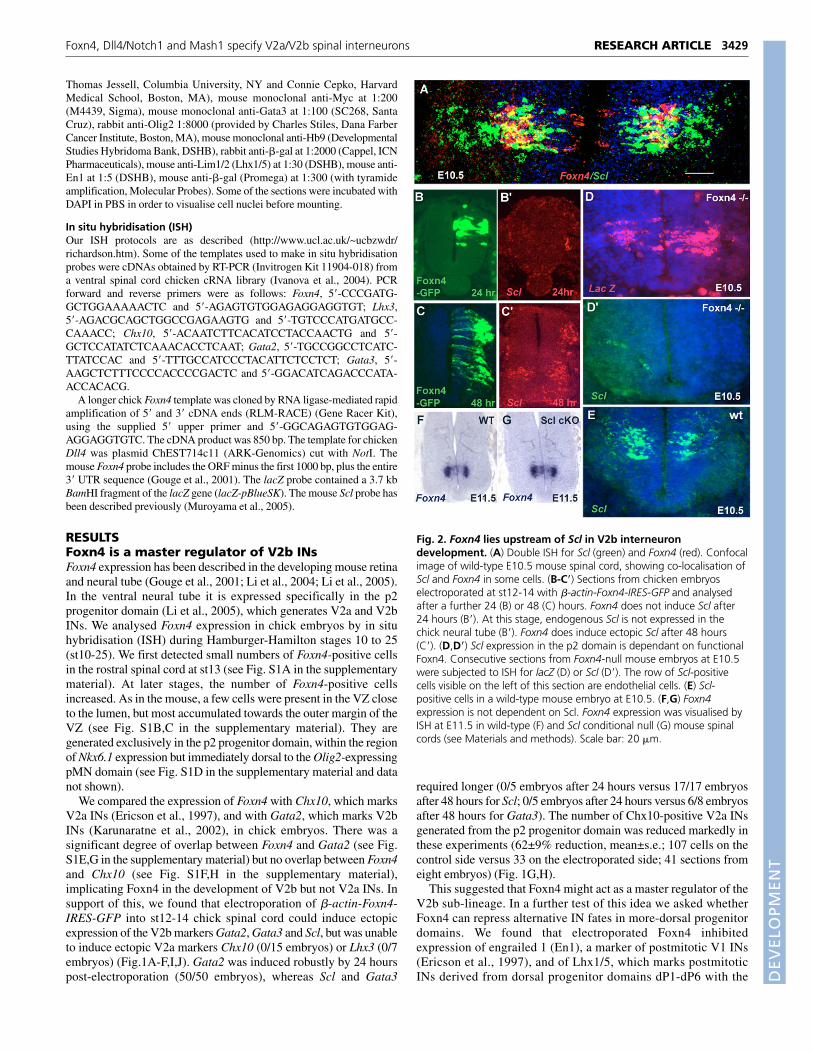
Fig. 2. Foxn4 lies upstream of Scl in V2b interneurondevelopment. (A) Double ISH for Scl (green) and Foxn4 (red). Confocalimage of wild-type E10.5 mouse spinal cord, showing co-localisation ofScl and Foxn4 in some cells. (B-C�) Sections from chicken embryoselectroporated at st12-14 with �-actin-Foxn4-IRES-GFP and analysedafter a further 24 (B) or 48 (C) hours. Foxn4 does not induce Scl after24 hours (B�). At this stage, endogenous Scl is not expressed in thechick neural tube (B�). Foxn4 does induce ectopic Scl after 48 hours(C�). (D,D�) Scl expression in the p2 domain is dependant on functionalFoxn4. Consecutive sections from Foxn4-null mouse embryos at E10.5were subjected to ISH for lacZ (D) or Scl (D�). The row of Scl-positivecells visible on the left of this section are endothelial cells. (E) Scl-positive cells in a wild-type mouse embryo at E10.5. (F,G) Foxn4expression is not dependent on Scl. Foxn4 expression was visualised byISH at E11.5 in wild-type (F) and Scl conditional null (G) mouse spinalcords (see Materials and methods). Scale bar: 20 �m.
DEVELO
PMENT
3430
exception of dP3 (reviewed by Lewis, 2006). A reduction of31±3% (mean±s.e., n=4) was observed for En1 (1173 cells on thecontrol side versus 776 on the electroporated side; 39 sectionsfrom four embryos; see Fig. S2A in the supplementary material)and a reduction of 45±13% (n=4) for Lhx1/5 (4239 cells on thecontrol side versus 2604 on the electroporated side; 30 sectionsfrom four embryos; see Fig. S2B in the supplementary material).These experiments suggest that ectopic expression of Foxn4 canreprogram progenitors to a V2b IN fate.
It has been reported that Scl function is necessary and sufficientfor V2b IN development and is required for the maintenance ofnormal levels of Gata2 expression (Muroyama et al., 2005). Wetherefore explored the genetic relationship between Scl and Foxn4.There was a small but significant overlap between Foxn4 and Scl inwild-type mice (Fig. 2A). Scl mRNA expression was abolished inFoxn4 mutant mouse spinal cords at E10.5 (3/3 embryos) and E11.5(2/2 embryos) (Fig. 2D,E and data not shown). Conversely, Foxn4was expressed as normal in Scl conditional null mice (2/2 embryos)(Fig. 2F,G). Also, as described above, Foxn4 induces Scl expressionafter 48 hours (17/17 embryos) (Fig. 1D, Fig. 2C). Therefore, itseems that Foxn4 lies upstream of Scl in the genetic hierarchyleading to V2b INs.
A negative-control vector with inverted Foxn4 sequences has beenused in parallel with all experiments reported above, without anyactivity (data not shown). Taken together, our data suggest thatFoxn4 is a master regulator of the V2b sub-lineage. Furthermore, wehave shown that Scl lies downstream of Foxn4 in the pathway thatgoverns development of V2b INs.
Foxn4 is expressed in the common progenitors ofV2a and V2b INsIt was previously reported that V2a and V2b INs share common,Foxn4-expressing progenitor cells in the VZ (Li et al., 2005). Weconfirmed this by following expression of �-galactosidase (�-gal) inmouse Foxn4+/– heterozygotes, which is possible because theknockout allele contains a functional copy of lacZ under Foxn4transcriptional control. By double immunohistochemistry we foundthat �-gal protein was present in cells that co-express Chx10 (Fig.3A), as well as in cells that express Gata3 (Fig. 3B). By contrast,Foxn4 transcripts or protein were never found in the same cells asChx10 or Gata3 (see Fig. S1H in the supplementary material) (Li etal., 2005). The most parsimonious interpretation is that there is acommon pool of Foxn4-positive progenitors that generates both V2aand V2b INs. The reason that �-gal can be detected in differentiatedV2a as well as V2b INs is presumably because it has a longer half-life than Foxn4. In further support of the existence of a common pool
of V2a/V2b progenitors, we found that those Foxn4-positive cells thatlie closest to the lumen (where neural progenitors undergo mitosis)co-express the V2a determinant Lhx3 (Fig. 3D), as well as Gata2 (seeFig. 1G in the supplementary material) and Mash1 (Fig. 3C, Fig. 6A).
Foxn4 activates delta-like 4 in p2 progenitorsmRNA encoding the Notch ligand delta-like 4 (Dll4) is expressed inscattered cells in mouse and chicken within the p2 progenitordomain (Fig. 4A,B and data not shown). Some of the Dll4-positivecells in the p2 domain co-expressed Foxn4 (Fig. 4A,B). Many ofthese Foxn4/Dll4 double-positive cells were found at the ventricularsurface, where mitosis occurs. Double-positive cells frequentlyoccurred as cell pairs (arrows in Fig. 4B, shown at highermagnification in C,D). These images strongly suggest that Dll4 andFoxn4 are co-expressed in cells that are dividing, or in recentlyseparated siblings that are still in contact.
To determine whether Dll4 and Foxn4 interact genetically, weperformed chick electroporation experiments at st11-12 with �-actin-Foxn4-IRES-GFP. Foxn4 induced ectopic expression of Dll4at 34 hours post-electroporation in 12/12 embryos analysed (Fig.4F). A control vector with inverted Foxn4 sequences had no sucheffect (data not shown). Consistent with these observations, Dll4expression was abolished in the p2 domain of Foxn4-null mice atE10.5 (3/3 embryos analysed) and E11.5 (2/2 embryos analysed)(Fig. 4E and data not shown). We conclude that Foxn4 is necessaryand sufficient for activation of Dll4 in p2 progenitors.
Dll4 inhibits V2a lineage progressionTo discover whether Dll4 is involved in the specification of V2 INs –possibly through its interactions with Notch – we performed gain-of-function experiments in chick neural tube by electroporating anexpression vector encoding human DLL4 (CMV-hDll4-Myc). Weperformed two sets of experiments. In the first, we electroporated atst11-13 and analysed the embryos after a further 44 hours (st19-20).In 16 embryos analysed, we found no ectopic induction of Chx10immunoreactivity or Gata2 mRNA. By contrast, a reduction ofChx10 and Gata2 expression was observed on the electroporatedversus the control side, Chx10 being more strongly repressed (~80%reduction) than Gata2 (~35% reduction) (63 Chx10-positive cells onthe control side versus 12 on the electroporated side, compared with90 Gata2-positive cells on the control side versus 58 on theelectroporated side; 24 sections from four embryos; data not shown).In the second set of experiments, we electroporated at st14-16 andanalysed the embryos after a further 48 hours (st21-23). In this set ofexperiments, 15 embryos were analysed for Chx10 immunoreactivityand Chx10, Scl and Gata2 mRNA (Fig. 5). As in the first experiment,
RESEARCH ARTICLE Development 134 (19)
Fig. 3. Foxn4 is expressed in common precursorsof V2a and V2b interneurons. (A,B) Foxn4+/– mouseembryos were labelled by doubleimmunohistochemistry for �-galactosidase (�-gal,green) and either Chx10 or Gata3 (red). Confocalmicroscopy reveals cells that are double labelled for �-gal and either Chx10 (A) or Gata3 (B), suggesting thatFoxn4-expressing progenitors give rise to both V2aand V2b interneurons (INs). (C-F) Consistent with thisconclusion, Foxn4-positive progenitors co-expressMash1 (C) and Lhx3 (D), markers that later segregateinto V2b and V2a INs respectively. IndividualFoxn4/Lhx3 double-positive cells (boxes E and F) arereproduced, with fluorescence channels separated, inthe lower left and lower right corners, respectively,of D.
DEVELO
PMENT
there was no ectopic expression of Chx10 protein or mRNA but astrong repression of Chx10 protein on the electroporated versuscontrol side (51±5% reduction, mean±s.e.; 137 sections from 13embryos; two-tail t-test=3.6 at P=0.001) (Fig. 5A,B). Gata2 mRNAwas expressed ectopically in some embryos (19/63 sections in fiveout of 15 embryos). In general, the induction of Gata2 was modestand always restricted to the p1-p0 domain (Fig. 5D�, white arrow).Despite this small amount of ectopic expression, the total amount ofGata2 signal (estimated by counting pixels with ImageJ) was notdetectably different on the electroporated versus control sides(594±83 versus 562±83 pixels, respectively; 80 sections from 7embryos; two-tail t-test=0.3 at P=0.8, not significant) (Fig. 5D�,E).The Scl signal was also not significantly different betweenelectroporated and control sides (396±64 pixels versus 361±57,respectively; 86 sections from 9 embryos; two-tail t-test=0.5 at P=0.6,not significant), nor was there any ectopic expression of Scl (Fig.5D�,E). These results suggest that at st14-16, Dll4 overexpressionspecifically represses the V2a fate with little or no effect on V2b fate.In Dll4 electroporations, some cells were Dll4-Myc/Chx10 doublepositive (Fig. 5C), indicating that expression of Dll4 is compatiblewith expression of Chx10 in the same cell.
Foxn4 induces Mash1 in the p2 domainThe extensive overlap of Mash1 and Foxn4 expression in the mousep2 domain (Fig. 3C, Fig. 6A) suggested some form of regulatoryrelationship. We therefore explored the interactions between Foxn4and Mash1 in more detail. We confirmed the finding of Li et al. (Liet al., 2005) that Foxn4 is expressed as normal in Mash1-null spinalcord (Fig. 6C,D). After electroporating �-actin-Foxn4-IRES-GFPin the chick spinal cord at st13-14, we found strong ectopic inductionof Cash1 (the chick homologue of Mash1) after 24 hours (6/6embryos) and 48 hours (3/5 embryos; Fig. 6B and data not shown).The negative control vector with inverted Foxn4 sequences had noactivity (data not shown). These experiments indicate that Foxn4 isupstream of and controls expression of Mash1 in p2, and fits withthe observation that Mash1 expression in p2 is lost in Foxn4-nullmice (Li et al., 2005).
Mash1 stimulates Dll4 expression but does notinduce V2b INsMash1 controls the expression of Dll1 in the ventral telencephalonand dorsal spinal cord (Casarosa et al., 1999), so we asked whetherMash1 can also induce Dll4. We electroporated full-length mouseMash1 (�-actin-Mash1-IRES-GFP) into st13-14 chick neural tube.After 24 hours of incubation, 8/8 embryos showed clear ectopicinduction of Dll4 on the electroporated side (Fig. 6E). After 48hours, 5/5 embryos displayed weaker but still clear induction of Dll4(data not shown). In none of the 13 embryos analysed did we findany ectopic expression of Chx10, Gata2 or Scl transcripts or Chx10immunoreactivity (Fig. 6F,G,H�,H� and data not shown). On theother hand, we observed a loss of endogenous Chx10-positive INsin the p2 domain of 5/5 embryos analysed (76±6% reduction, n=23)(Fig. 6F,F�,G), with little or no concomitant reduction of Gata2 orScl (Fig. 6H�,H�). These data suggest that induction of Dll4 andconsequent repression of Chx10-positive V2a INs by Foxn4 mightbe mediated indirectly via Mash1. However, we found that Dll4 isexpressed as normal at E10.5-11 in Mash1-null embryos (4/4embryos; Fig. 6I,J). Therefore, Mash1 might be involved inmaintaining or reinforcing Dll4 expression but is not required for itsinitiation. Although Mash1 is necessary to develop the V2b fate (Liet al., 2005), it is not sufficient to do so, judging by its inability toinduce ectopic Gata2 or Scl expression. Therefore, it appears thatthe V2b program of gene expression is absolutely dependent onFoxn4.
Notch1 is required for generation of V2b INsThe fact that Dll4 preferentially represses the V2a fate suggests thatthe Notch-Delta system might be responsible for the V2a-V2b binaryfate decision in p2 progenitors. To test this, we analysed Notch1mutant (cKO) mouse embryos at E10.5 and E11.5 by ISH for Foxn4or Scl, or by immunohistochemistry for Chx10, Gata3, Olig2 or Hb9(Hlxb9) (Fig. 7). Olig2 is a basic helix-loop-helix transcription factorthat is expressed in the progenitors of motor neurons (MNs) andoligodendrocytes but not in postmitotic MNs (Lu et al., 2000),whereas Hb9 is a transcription factor expressed in early committed
3431RESEARCH ARTICLEFoxn4, Dll4/Notch1 and Mash1 specify V2a/V2b spinal interneurons
Fig. 4. Foxn4 is necessary and sufficient to induceDll4 in the p2 domain. (A-D1) Double ISH for Dll4(green) and Foxn4 (red) in wild-type E10.5 mouseembryos, counter-stained with Hoechst to visualise cellnuclei. A and B are transverse and longitudinalsections, respectively, of spinal cord. Foxn4 isexpressed in some of the Dll4-positive cells within andoutside the VZ (arrows). A significant proportion ofdouble-labelled cells at the ventricular surface arepairs of cells in contact with each other, presumptivedaughters of a recent progenitor cell division (e.g.arrows in B). Examples of these are shown at highermagnification in C,D; note the paired nuclei in C1,D1.(E,E�) Foxn4-null mouse embryos at E10.5. (E) lacZexpression under Foxn4 control. (E�) Dll4 expression inthe p2 domain is abolished. (F,F1) Double ISH forFoxn4 (green) and Dll4 (red) showing that Foxn4induces ectopic expression of Dll4 in electroporatedst11-13 chick neural tube.
DEVELO
PMENT
3432
MNs (Thaler et al., 1999). At E11.5, no Gata3 (0 versus 99±3, n=14sections from three embryos; two-tail t-test=26, P<0.001) or Scl-positive cells were present in the ventral spinal cord of Notch1 cKOmice (3/3 embryos analysed) (Fig. 7A-D,M-N). Instead, twice thenormal number of Chx10-positive cells was observed (200±10 versus102±3, n=16 sections from three embryos; two-tail t-test=8.6 atP<0.001) (Fig. 7A-D), as previously reported (Yang et al., 2006).pMN progenitors that express Olig2 were drastically reduced at thisage (2±0.5 versus 39±2, n=14 sections from three embryos; two-tailt-test=17, P<0.001), but the number of Hb9-positive cells was notsignificantly affected in the Notch1 mutant (3/3 embryos analysed)by comparison with wild-type mice (175±16 versus 165±8, n=14sections from three embryos; two-tail t-test=0.6, P=0.6) (Fig. 7D,E-F). It seems that Notch1 signalling is necessary to prevent prematuredifferentiation of most progenitor cells in the ventral cord, judging bythe loss of the ventral VZ in the mutant (Yang et al., 2006). However,
loss of Notch1 does not seem to result in respecification of pMNprogenitors to p2 progenitors, as originally proposed (Yang et al.,2006). Rather, the phenotype is more consistent with respecificationof V2b to V2a INs, consistent with the idea that signalling throughNotch1 is required for V2b IN development.
Foxn4 is very much reduced in the E11.5 Notch1 conditional nullspinal cord (Fig. 7I,J, arrow). A simple interpretation is that theFoxn4-positive progenitors of V2a and V2b INs differentiateprematurely and completely into V2a INs in the mutant and, in doing
RESEARCH ARTICLE Development 134 (19)
Fig. 5. Dll4 inhibits V2a lineage progression. Chick embryos wereelectroporated with human DLL4 (hDll4) in the form of hDll4-Myc atst14-16 and analysed after 48 hours. (A-B) Double immunolabellingfor Chx10 (red) and hDll4-Myc (green) showing repression of Chx10-positive cells. (B) Quantification of Chx10-labelled cells showed a~50% decrease on the hDll4-electroporated side compared with thecontralateral, control side. (C) Some hDll4-electroporated cells co-express Chx10, consistent with the idea that Dll4 can suppress V2ageneration in a non-cell-autonomous fashion. (D) Immunolabellingfor hDll4-Myc (green). (D�) Dll4 exceptionally can induce Gata2(arrowhead). (D�) Dll4 does not affect Scl expression. (E) Dll4 doesnot greatly affect generation of V2b INs, judging by ISH.Quantification of V2b markers Gata2 and Scl by pixel-countingsoftware showed no significant effect on V2b production (see textfor statistics).
Fig. 6. Foxn4 controls Mash1 expression. (A) Double ISH for Foxn4(red) and Mash1 (green) in E10.5 mouse cord. Note the extensiveoverlap in the p2 domain. (B,B�) Foxn4 induces ectopic expression ofCash1 in chick electroporation experiments. (C,D) Foxn4 expressiondoes not depend on Mash1; there is no noticeable change in the Foxn4ISH signal in Mash1-null mice compared with wild type.(E,E�) Electroporation of �-actin-Mash1-IRES-GFP in the st13-14 chickneural tube induces Dll4 after 24 hours. Mash1 expression wasconfirmed by GFP immunolabelling (E) and Dll4 by ISH (E�). (F-G) Mash1did not induce ectopic Chx10, but repressed endogenous Chx10 V2aINs in the p2 domain. G is a magnified view of the ventral part of panelF�. (H-H�) Also, Mash1 did not induce ectopic V2b markers Gata2 orScl. (I,J) Despite the fact that Mash1 is sufficient to induce Dll4 in chick(see E,E�), Mash1 is not required for Dll4 expression in mice; Dll4 isexpressed as normal in the ventral spinal cord of Mash1-null mice.
DEVELO
PMENT
so, lose expression of Foxn4. Likewise, Scl and Dll4 transcripts werestrongly downregulated compared with wild type at E11.5,consistent with their demonstrated dependence on Foxn4 (Fig. 7M,Nand data not shown). Cre recombination is thought to be activated ator shortly before E10.5 in the Nestin-Cre line (Yang et al., 2006). Inkeeping with this, the morphology of the spinal cord was normal inthe Nestin-Cre/Notch1flox at E10.5 (i.e. the ventral VZ was stillpresent). Foxn4 and Dll4 were expressed at higher than normal levelsin the mutant at E10.5, consistent with the idea that V2a/V2bprogenitors are formed prematurely but have not yet had time todifferentiate (Fig. 7G,H and data not shown). By contrast, andconsistent with the above reasoning, Scl was expressed at a reducedlevel at E10.5 (Fig. 7K,L).
DISCUSSIONFoxn4 activates V2b interneuron developmentWe found that Foxn4 is both necessary and sufficient to activateGata2, Scl and Gata3, suggesting that it is near or at the top of thegenetic hierarchy that specifies V2b INs. This differs from ourprevious study, which found that co-electroporation of Foxn4together with Mash1 was necessary to induce ectopic V2b gene
expression, Foxn4 alone being insufficient (Li et al., 2005). Atpresent we are unable to explain this difference but it could perhapsrelate to differences in the level of Foxn4 expression achievedfollowing electroporation. We have ruled out functional differencesbetween the Foxn4 electroporation vectors used because in ourhands both constructs give the result reported here. In any case, bothour studies demonstrate that Foxn4 is a key determinant of the V2bsub-lineage.
It was shown previously that the transcription factor Scl isnecessary and sufficient to induce V2b INs (Muroyama et al., 2005).Foxn4 transcripts are detected before Scl during normal development– at st13 in chick/E9.5 mouse, compared with st16-17 chick/E10.5mouse (Li et al., 2005; Muroyama et al., 2005) (data not shown),suggesting that Foxn4 is upstream of Scl. Consistent with this, wehave now shown that: (1) Scl expression is lost in Foxn4–/– mice,whereas Foxn4 expression is unaffected in Scl–/– mice; and (2) Foxn4is able to induce Scl expression in chick electroporation experiments.Foxn4 induces robust expression of Gata2 in chick neural tubewithin 24 hours post-electroporation, whereas Scl and Gata3 are notdetectable until 48 hours post-electroporation. This temporal orderpresumably reflects the fact that Gata2 is required for Gata3expression (Karunaratne et al., 2002; Nardelli et al., 1999) andsuggests that Gata2 is genetically upstream of Scl. This is backed upby the fact that Gata2 is expressed ahead of Scl during normaldevelopment in both chick and mouse (Muroyama et al., 2005) (datanot shown). Gata3 expression is lost in Scl-null mice, placing Sclupstream of Gata3 (Muroyama et al., 2005). Taken together, theavailable data support a genetic cascade Foxn4 r Gata2 r Scl rGata3. The reduction of Gata2 expression that was observed in Scl-null mice (Muroyama et al., 2005) can be attributed to loss ofpositive feedback from Gata3 (Karunaratne et al., 2002). A diagramof the proposed network is shown in Fig. 8.
Foxn4 activates Dll4 and Mash1By loss- and gain-of-function experiments we found that Foxn4 isnecessary and sufficient to activate Dll4 and Mash1 expression. Wesubsequently showed that Mash1 can also induce ectopic expressionof Dll4 in chick spinal cord. This suggests that the conservedMash1/Brn binding site in the Dll4 upstream region, reported byCastro et al. (Castro et al., 2006), is functional in vivo and further
3433RESEARCH ARTICLEFoxn4, Dll4/Notch1 and Mash1 specify V2a/V2b spinal interneurons
Fig. 7. Notch1 is required for specification of V2b interneurons.Mice carrying a floxed allele of Notch1 and a Nestin-Cre transgene(Notch1 cKO mice) were analysed at E10.5 and E11.5 by ISH anddouble immunolabelling for V2a and V2b IN markers. (A-D) There is atwo-fold increase in the number of Chx10 immunopositive V2a INs inthe Notch1 cKO compared with wild type, whereas Gata3immunopositive V2b INs are abolished. In addition, the Chx10-positiveV2a INs accumulate near the midline of the spinal cord instead ofmigrating into the parenchyma. (E,F) Double immunolabelling for Olig2(magenta) and Hb9 (green). In the Notch1 cKO, Olig2-positive cells aremissing and the Hb9 population is similar to that in the control.Therefore, Notch1 activity is needed for V2b IN production; in theabsence of Notch1, V2b INs are respecified as V2a INs with little or noinfluence on MN fate. (G-J) In the Notch1 cKO, expression of Foxn4 isincreased at E10.5 relative to wild type (compare G with H), but isalmost extinguished by E11.5 (I,J). (K-N) Scl (V2b INs) is reduced atE10.5 (K,L) and absent at E11.5 (M,N) in the Notch1 cKO. Note that theventral half of the central canal (and the VZ) is lost in the Notch1 cKOmouse between E10.5 and E11.5.
DEVELO
PMENT
3434
suggested that Foxn4 might activate Dll4 indirectly through Mash1.However, we found that Mash1 is not required for initiation of Dll4expression in the mouse because Dll4 is expressed normally in thep2 domain of E10.5 Mash1-null spinal cord. It is possible thatMash1 might be required to maintain Dll4 expression after E10.5,but we have not examined older embryos. Alternatively, arequirement for Mash1 in the initiation of Dll4 expression might bemasked in Mash1 mutant mice through compensatory upregulationof a related proneural factor such as Ngn1 (Neurog1) or Ngn2(Neurog2). It is also possible that Foxn4 induces Dll4 directly; inendothelial cells, for example, Foxc1 and/or Foxc2 are known toactivate Dll4 by binding directly to a Fox-binding site in the Dll4gene upstream region (Seo et al., 2006).
Apart from regulating Dll4, Mash1 must have another role inpromoting V2b IN fate, because Mash1-null mice at E10.5 arereported to have ~50% less V2b INs than normal (Li et al., 2005),despite the fact that Foxn4 and Dll4 are both expressed normally(Fig. 6D and data not shown). More work needs to be done toestablish the precise role of Mash1 in V2b IN development.
Notch1 is required for V2b interneurondevelopmentThe connection between Foxn4, Dll4 and Mash1 led us to explorethe role of Notch-Delta signalling more directly. We previouslyreported that when Notch1 function was disrupted in the ventralspinal cord, the result was a ~30% overproduction of (Chx10,Lhx3) double-positive V2a INs and an ~18% loss of [Islet 1 (Isl1),Lhx3] double-positive MNs, although the total number of Islet-positive MNs was unchanged (Yang et al., 2006). This wasoriginally interpreted as a fate switch from MN to V2 INproduction. However, in the present study we found that Gata3-positive V2b INs were completely lost, whereas the number ofHb9-positive MNs was not changed significantly in the Notch1mutant. Therefore, we conclude that the increase in V2a INs ismore likely to result from respecification of V2b INs than from
respecification of MNs to V2a INs. Since the V2 phenotype of theconditional Notch1 mutant is analogous to that of the Foxn4-nullmouse, it appears that both Notch1 and Foxn4 activities arerequired for V2b IN production.
The default behaviour of p2 progenitors in the absence of Notch1or Foxn4 activity is to differentiate as V2a INs, suggesting that activeNotch1 acts cell-autonomously in collaboration with Foxn4 to driveV2b development. We have not been able to address directly thequestion of whether Notch1 acts in a cell-autonomous fashion inV2b INs. However, we observed that electroporated Dll4 is co-expressed with endogenous Chx10 in some V2a INs (Fig. 5C),suggesting that Dll4-mediated inhibition of V2a INs is non-cell-autonomous, as expected from the classical view of Notch-Deltaneighbour-to-neighbour signalling. This contrasts with Foxn4, whichwas never co-expressed with Chx10, in keeping with its expectedcell-autonomous role. A cell-autonomous role for Notch is indicatedby the requirement for presenilin 1 (Psen1) for V2b lineagedevelopment (Peng et al., 2007). Psen1 is involved in theintracellular cleavage of Notch (Wines-Samuelson and Shen, 2005).It also fits with the report that Notch1 binds to an enhancer in theupstream region of the Gata2 gene during hematopoiesis (Robert-Moreno et al., 2005).
Why does Dll4 electroporation inhibit V2a IN production withoutcausing a compensatory increase in V2b INs in p2? Perhaps dll4electroporation reduces the total number of V2 INs (V2a +V2b) byinhibiting production of V2 progenitors from their neuroepithelialprecursors, while simultaneously biasing the fate of the remainingV2 progenitors from V2a towards V2b. If so, the fact that there is nosignificant change in the number of V2b INs in our electroporationexperiments at st14-16 might be the result of two equal but opposingeffects. If this explanation is correct, then the precise outcome of theexperiment might depend critically on the time of electroporation,because this could alter the magnitude of one effect versus the other.Consistent with this idea, we found a small reduction in the numberof V2b INs (as well as a reduction in V2a INs) when weelectroporated at st11-12. Peng et al. (Peng et al., 2007) also founda reduction in total V2 INs in their electroporation experiments atst13. This model is necessarily speculative and other explanationsare possible.
Notch1/Dll4 signalling breaks symmetry and splitsthe V2 lineageWhat is the mode of action of Notch1 in V2 IN development? Onepossibility might be that p2 progenitors normally generate V2a INsfirst, before switching to V2b production, and that Dll4/Notch1 isneeded to keep some progenitors in cycle long enough to generateV2b INs. In that case, eliminating Notch signalling might beexpected to cause accelerated differentiation along the V2a pathwayand loss of V2b differentiation, as observed. However, there is noevidence that V2a INs are formed before V2b INs. Chx10 and Gata3are both expressed together for the first time at E10.5 in mouse (Liuet al., 1994; Nardelli et al., 1999). V2a and V2b subpopulations areformed simultaneously in chicken too (Karunaratne et al., 2002). Wetherefore propose that Notch1-Delta signalling has two consecutiveor parallel functions in the p2 progenitor domain: (1) it inhibitsneuroepithelial (radial) precursors from differentiating prematurelyinto V2 progenitors; and (2) it segregates V2 progenitors into V2aand V2b sub-lineages, inhibiting V2a and promoting V2bdevelopment.
The majority of cells that co-express Foxn4 and Dll4 are closelyapposed pairs of cells at the ventricular surface, and these are likelyto represent the products of recent progenitor cell divisions (Fig. 4A-
RESEARCH ARTICLE Development 134 (19)
Fig. 8. Genetic interactions in the V2 interneuron lineage. Redarrows represent positive intracellular interactions that wedemonstrated in the present study, except for Foxn4 r (?) Dll4, which isspeculative. Black arrows denote speculative interactions proposed inthe present study. Blue arrows depict interactions demonstrated inprevious studies (see below). The dashed red line represents theproposed intercellular Dll4/Notch1 interaction between sibling V2progenitors, which results in Notch1 being activated (yellow star) in thecells that develop subsequently as V2b INs. The grey arrow signifies therequirement of Mash1 for proper V2b development (Li et al., 2005);this role of Mash1 is ill-defined (see Discussion). Mash1 is sufficient (inchick) but not necessary (in mice) for Dll4 upregulation (see Discussion).This diagram incorporates observations from a number of studiesincluding the present one: Karunaratne et al. (Karunaratne et al., 2002)demonstrated reciprocal activation of Gata2 and Gata3 and repressionof Chx10 by Gata2; Muroyama et al. (Muroyama et al., 2005) showedthat Scl induces Gata2 and Gata3 and represses Chx10 in chick, andthat Gata3 is abolished and Gata2 severely reduced in Scl-null mice; Liet al. (Li et al., 2005) showed that Mash1 expression is abolished inFoxn4-null mice.
DEVELO
PMENT
D). This observation suggests that Dll4/Notch1 interactions involvesibling pairs of cells that have not yet separated after division, and isconsistent with the idea that a single V2a/V2b progenitor cell mightgenerate one V2a and one V2b neuron, as illustrated schematicallyin Fig. 9. Alternatively, bipotential V2a/V2b progenitors mightdivide asymmetrically to generate a dedicated V2a progenitor and adedicated V2b progenitor, which can undergo a further symmetricaldivision(s) before terminal differentiation. Either of these scenarioswould be consistent with our observations that approximately equalnumbers of V2a and V2b INs are formed under normalcircumstances and that twice the normal number of V2a INs form inthe absence of Notch1 (Fig. 7D).
Note that our proposed roles for Mash1 and Dll4/Notch1signalling in separating V2a and V2b lineages is closelyanalogous to the roles proposed for Mash1 and Dll1/Notch inspecifying excitatory and inhibitory (dILA and dILB) INs in thedorsal spinal cord (Mizuguchi et al., 2006). It is possible that V2aand V2b INs are also a complementary excitatory/inhibitory pair– V2a INs are known to be glutamatergic and excitatory inzebrafish (Kimura et al., 2006), but the neurotransmitter
phenotype of V2b INs has not yet been established. It is possiblethat Notch-Delta signalling might be a general mechanism forcreating complementary pairs of INs.
We thank our colleagues at UCL, especially Lisbeth Flores-Garcia, Huiliang Li,Nicoletta Kessaris and Marcus Fruttiger for helpful discussions and insights;Kamal Sharma for sharing data prior to publication; Ji-Liang Li for the humanDLL4 expression vector; Andrew Lumsden for the chick electroporation vector;Francois Guillemot for the Mash1 vector and Mash1 mutant mouse embryos;Thomas Jessell and Connie Cepko for anti-Chx10 antibodies; Charles D. Stilesfor anti-Olig2; and the following for DNA templates: Luis Puelles for chickNkx6.1, Graham Goodwin for chick Scl, Thomas Reh for Cash1, HenriqueDomingos for mouse Dll4, Janette Nardell for mouse Chx10 and Stuart Orkinfor mouse Gata2. Raquel Taveira-Marques is supported by a studentship fromthe Portuguese Fundação para a Ciência e a Tecnologia. This work wassupported by grants from the US National Institutes of Health [R01 NS047572(D.R.), R01 NS042818 (J.S.), EY015777 (M.X.)], the New Jersey Commission onSpinal Cord Research [05-3039-SCR-E-0 (M.X.)] the Wellcome Trust and theUK Medical Research Council (W.D.R.).
Supplementary materialSupplementary material for this article is available athttp://dev.biologists.org/cgi/content/full/134/19/3427/DC1
ReferencesArtavanis-Tsakonas, S., Rand, M. D. and Lake, R. J. (1999). Notch signaling:
cell fate control and signal integration in development. Science 284, 770-776.Benedito, R. and Duarte, A. (2005). Expression of Dll4 during mouse
Briscoe, J., Pierani, A., Jessell, T. M. and Ericson, J. (2000). A homeodomainprotein code specifies progenitor cell identity and neuronal fate in the ventralneural tube. Cell 101, 435-445.
Casarosa, S., Fode, C. and Guillemot, F. (1999). Mash1 regulates neurogenesisin the ventral telencephalon. Development 126, 525-534.
Castro, D. S., Skowronsky-Krawzyck, D., Armant, O., Donaldson, I. J.,Parras, C., Hunt, C., Critchley, J. A., Nguyen, L., Gossler, A., Göttgens, B. etal. (2006). Proneural bHLH and Brn proteins coregulate a neurogenic programthrough cooperative binding to a conserved DNA motif. Dev. Cell 11, 831-844.
Duarte, A., Hirashima, M., Benedito, R., Trindade, A., Diniz, P., Bekman, E.,Costa, L., Henrique, D. and Rossant, J. (2004). Dosage-sensitive requirementfor mouse Dll4 in artery development. Genes Dev. 18, 2474-2478.
Dunwoodie, S. L., Henrique, D., Harrison, S. M. and Beddington, R. S.(1997). Mouse Dll3: a novel divergent Delta gene which may complement thefunction of other Delta homologues during early pattern formation in themouse embryo. Development 124, 3065-3076.
Ericson, J., Rashbass, P., Schedl, A., Brenner-Morton, S., Kawakami, A., vanHeyningen, V., Jessell, T. M. and Briscoe, J. (1997). Pax6 controls progenitorcell identity and neuronal fate in response to graded Shh signaling. Cell 90, 169-180.
Gouge, A., Holt, J., Hardy, A. P., Sowden, J. C. and Smith, H. K. (2001). Foxn4– a new member of the forkhead gene family is expressed in the retina. Mech.Dev. 107, 203-206.
Guillemot, F., Lo, L. C., Johnson, J. E., Auerbach, A., Anderson, D. J. andJoyner, A. L. (1993). Mammalian achaete-scute homolog 1 is required for theearly development of olfactory and autonomic neurons. Cell 75, 463-476.
Hamburger, V. and Hamilton, H. L. (1951). A series of normal changes in thedevelopment of the chick embryo. J. Morphol. 88, 49-92.
Itasaki, N., Bel-Vialar, S. and Krumlauf, R. (1999). ‘Shocking’ developments inchick embryology: electroporation and in ovo gene expression. Nat. Cell Biol. 1,E203-E207.
Ivanova, A., Agochiya, M., Amoyel, M. and Richardson, W. D. (2004).Receptor tyrosine phosphatase zeta/beta in astrocyte progenitors in thedeveloping chick spinal cord. Gene Expr. Patterns 4, 161-166.
Karunaratne, A., Hargrave, M., Poh, A. and Yamada, T. (2002). GATA proteinsidentify a novel ventral interneuron subclass in the developing chick spinal cord.Dev. Biol. 249, 30-43.
Kiehn, O. (2006). Locomotor circuits in the mammalian spinal cord. Annu. Rev.Neurosci. 29, 279-306.
Kimura, Y., Okamura, Y. and Higashijima, S. (2006). alx, a zebrafish homologof Chx10, marks ipsilateral descending excitatory interneurons that participate inthe regulation of spinal locomotor circuits. J. Neurosci. 26, 5684-5697.
Lewis, K. E. (2006). How do genes regulate simple behaviours? Understandinghow different neurons in the vertebrate spinal cord are genetically specified.Philos. Trans. R. Soc. Lond. B Biol. Sci. 361, 45-66.
Li, S., Mo, Z., Yang, X., Price, S. M., Shen, M. M. and Xiang, M. (2004). Foxn4controls the genesis of amacrine and horizontal cells by retinal progenitors.Neuron 43, 795-807.
3435RESEARCH ARTICLEFoxn4, Dll4/Notch1 and Mash1 specify V2a/V2b spinal interneurons
Fig. 9. Generation of V2a and V2b INs from common progenitorsin the p2 domain. Multipotent neuroepithelial (radial) progenitors (A),which do not express Foxn4, generate a population of V2a/V2b (p2)progenitors (B). All V2a/V2b progenitors express Foxn4, which inducesthe expression of Dll4, Gata2 and Mash1. These common progenitorsalso start to express Lhx3 at their final division (C). Notch1 is expressedin all p2 progenitors (Lindsell et al., 1996), so Notch1/Dll4 reciprocalcell-cell interactions are initiated (opposing arrows in C). This situationresolves into two populations of progenitors, one with activatedNotch1 (Notch1*) and the other with Dll4 (D). Notch1* blocks the V2afate and, in cooperation with Foxn4 and Mash1, specifies V2b IN fate(E). The complementary set of p2 progenitors (Dll4-positive) that fails toactivate Notch1 adopts the V2a fate instead, possibly under the controlof Lhx3 (Tanabe et al., 1998) (E). In this way, V2a and V2b INs aregenerated in a salt-and-pepper fashion during the same time windowfrom a homogeneous population of p2 progenitors.
DEVELO
PMENT
3436
Li, S., Misra, K., Matise, M. P. and Xiang, M. (2005). Foxn4 acts synergisticallywith Mash1 to specify subtype identity of V2 interneurons in the spinal cord.Proc. Natl. Acad. Sci. USA. 102, 10688-10693.
Lindsell, C. E., Boulter, J., diSibio, G., Gossler, A. and Weinmaster, G. (1996).Expression patterns of Jagged, Delta1, Notch1, Notch2, and Notch3 genesidentify ligand-receptor pairs that may function in neural development. Mol.Cell. Neurosci. 8, 14-27.
Liu, I. S., Chen, J. D., Ploder, L., Vidgen, D., van der Kooy, D., Kalnins, V. I.and McInnes, R. R. (1994). Developmental expression of a novel murinehomeobox gene (Chx10): evidence for roles in determination of the neuroretinaand inner nuclear layer. Neuron 13, 377-393.
Louvi, A. and Artavanis-Tsakonas, S. (2006). Notch signalling in vertebrateneural development. Nat. Rev. Neurosci. 7, 93-102.
Lu, Q. R., Yuk, D., Alberta, J. A., Zhu, Z., Pawlitzky, I., Chan, J., McMahon, A.,Stiles, C. D. and Rowitch, D. H. (2000). Sonic hedgehog-regulatedoligodendrocyte lineage genes encoding bHLH proteins in the mammaliancentral nervous system. Neuron 25, 317-329.
Mailhos, C., Modlich, U., Lewis, J., Harris, A., Bicknell, R. and Ish-Horowicz,D. (2001). Delta4, an endothelial specific notch ligand expressed at sites ofphysiological and tumor angiogenesis. Differentiation 69, 135-144.
Mizuguchi, R., Kriks, S., Cordes, R., Gossler, A., Ma, Q. and Goulding, M.(2006). Ascl1 and Gsh1/2 control inhibitory and excitatory cell fate in spinalsensory interneurons. Nat. Neurosci. 9, 770-778.
Muroyama, Y., Fujiwara, Y., Orkin, S. H. and Rowitch, D. H. (2005).Specification of astrocytes by bHLH protein SCL in a restricted region of theneural tube. Nature 438, 360-363.
Nardelli, J., Thiesson, D., Fujiwara, Y., Tsai, F. Y. and Orkin, S. H. (1999).Expression and genetic interaction of transcription factors GATA-2 and GATA-3during development of the mouse central nervous system. Dev. Biol. 210, 305-321.
Peng, C. Y., Yajima, H., Burns, C. E., Zon, L. I., Sisodia, S. S., Pfaff, S. L. andSharma, K. (2007). Notch and MAML signaling drives Scl-dependentinterneuron diversity in the spinal cord. Neuron 53, 813-827.
Robert-Moreno, A., Espinosa, L., de la Pompa, J. L. and Bigas, A. (2005).RBPjkappa-dependent Notch function regulates Gata2 and is essential for theformation of intra-embryonic hematopoietic cells. Development 132, 1117-1126.
Rowitch, D. H. (2004). Glial specification in the vertebrate neural tube. Nat. Rev.Neurosci. 5, 409-419.
Schubert, F. R. and Lumsden, A. (2005). Transcriptional control of early tractformation in the embryonic chick midbrain. Development 132, 1785-1793.
Seo, S., Fujita, H., Nakano, A., Kang, M., Duarte, A. and Kume, T. (2006). Theforkhead transcription factors, Foxc1 and Foxc2, are required for arterialspecification and lymphatic sprouting during vascular development. Dev. Biol.294, 458-470.
Smith, E., Hargrave, M., Yamada, T., Begley, C. G. and Little, M. H. (2002).Co-expression of SCL and GATA3 in the V2 interneurons of the developingmouse spinal cord. Dev. Dyn. 224, 231-237.
Tanabe, Y., William, C. and Jessell, T. M. (1998). Specification of motor neuronidentity by the MNR2 homeodomain protein. Cell 95, 67-80.
Thaler, J., Harrison, K., Sharma, K., Lettieri, K., Kehrl, J. and Pfaff, S. L.(1999). Active suppression of interneuron programs within developing motorneurons revealed by analysis of homeodomain factor HB9. Neuron 23, 675-687.
Wines-Samuelson, M. and Shen, J. (2005). Presenilins in the developing, adultand ageing cerebral cortex. Neuroscientist 11, 441-451.
Yang, X., Tomita, T., Wines-Samuelson, M., Beglopoulos, V., Tansey, M. G.,Kopan, R. and Shen, J. (2006). Notch1 signaling influences V2 interneuron andmotor neuron development in the spinal cord. Dev. Neurosci. 28, 102-117.
Zhou, Y., Yamamoto, M. and Engel, J. D. (2000). GATA2 is required for thegeneration of V2 interneurons. Development 127, 3829-3838.