Page 1
REVIEW PAPER
A review of the antibacterial effects of silver nanomaterialsand potential implications for human healthand the environment
Catalina Marambio-Jones • Eric M. V. Hoek
Received: 29 July 2009 / Accepted: 6 March 2010 / Published online: 23 March 2010
� Springer Science+Business Media B.V. 2010
Abstract Here, we present a review of the antibac-
terial effects of silver nanomaterials, including pro-
posed antibacterial mechanisms and possible toxicity
to higher organisms. For purpose of this review, silver
nanomaterials include silver nanoparticles, stabilized
silver salts, silver–dendrimer, polymer and metal oxide
composites, and silver-impregnated zeolite and acti-
vated carbon materials. While there is some evidence
that silver nanoparticles can directly damage bacteria
cell membranes, silver nanomaterials appear to exert
bacteriocidal activity predominantly through release of
silver ions followed (individually or in combination)
by increased membrane permeability, loss of the
proton motive force, inducing de-energization of the
cells and efflux of phosphate, leakage of cellular
content, and disruption DNA replication. Eukaryotic
cells could be similarly impacted by most of these
mechanisms and, indeed, a small but growing body of
literature supports this concern. Most antimicrobial
studies are performed in simple aquatic media or cell
culture media without proper characterization of silver
nanomaterial stability (aggregation, dissolution, and
re-precipitation). Silver nanoparticle stability is gov-
erned by particle size, shape, and capping agents as
well as solution pH, ionic strength, specific ions and
ligands, and organic macromolecules—all of which
influence silver nanoparticle stability and bioavailabil-
ity. Although none of the studies reviewed definitively
proved any immediate impacts to human health or the
environment by a silver nanomaterial containing
product, the entirety of the science reviewed suggests
some caution and further research are warranted given
the already widespread and rapidly growing use of
silver nanomaterials.
Keywords Silver � Nanoparticle �Antimicrobial � Antibacterial � Nanotechnology �Nanotoxicology � Safety � EHS
Introduction
The broad-spectrum antimicrobial properties of silver
encourage its use in biomedical applications, water
and air purification, food production, cosmetics,
clothing, and numerous household products. With
the rapid development of nanotechnology, applica-
tions have been extended further and now silver is the
engineered nanomaterial most commonly used in
consumer products (Rejeski 2009). Clothing, respi-
rators, household water filters, contraconceptives,
antibacterial sprays, cosmetics, detergent, dietary
supplements, cutting boards, sox, shoes, cell phones,
laptop keyboards, and children’s toys are among the
C. Marambio-Jones � E. M. V. Hoek (&)
Department of Civil and Environmental Engineering,
California NanoSystems Institute, University
of California, Los Angeles, 5732G Boelter Hall,
PO Box 951593, Los Angeles, CA 90095-1593, USA
e-mail: [email protected]
123
J Nanopart Res (2010) 12:1531–1551
DOI 10.1007/s11051-010-9900-y
Page 2
retail products that purportedly exploit the antimi-
crobial properties of silver nanomaterials.
Different forms of silver nanomaterials already in
such products include: metallic silver nanoparticles
(Arora et al. 2008; Chi et al. 2009; Choi et al. 2008;
Hwang et al. 2008; Kim et al. 2007, 2008a, b; Kvitek
et al. 2008; Lok et al. 2006; Raffi et al. 2008; Schrand
et al. 2008; Sondi and Salopek-Sondi 2004; Vertelov
et al. 2008), silver chloride particles (Choi et al.
2008), silver-impregnated zeolite powders and acti-
vated carbon materials (Cowan et al. 2003; Inoue
et al. 2002; Yoon et al. 2008a, b), dendrimer–silver
complexes and composites (Balogh et al. 2001;
Lesniak et al. 2005; Zhang et al. 2008), polymer-
silver nanoparticle composites (Bajpai et al. 2007;
Damm and Munstedt 2008; Damm et al. 2008; Hlidek
et al. 2008; Jin et al. 2007; Kim et al. 2009a, b;
Kvitek et al. 2008; Naidu et al. 2008; Nita 2008;
Sambhy and Sen 2008; Sanpui et al. 2008; Xu et al.
2006), silver-titanium dioxide composite nanopow-
ders (Yeo and Kang 2008), and silver nanoparticles
coated onto polymers like polyurethane (Jain and
Pradeep 2005). While all of these forms of silver
exert antimicrobial activity to some extent through
release of silver ions, silver nanoparticles might
exhibit additional antimicrobial capabilities not
exerted by bulk or ionic silver (Chen and Schluesener
2008).
Already, silver nanoparticles have been shown to
be effective biocides against: (a) bacteria such as
Escherichia coli, Staphylococcus aureus, Staphylo-
coccus epidermis, Leuconostoc mesenteroides, Bacil-
lus subtilis, Klebsiella mobilis, and Klebsiella
pneumonia among others (Benn and Westerhoff
2008; Chen and Chiang 2008; Falletta et al. 2008;
Hernandez-Sierra et al. 2008; Ingle et al. 2008; Jung
et al. 2009; Kim 2007; Kim et al. 2007, 2009a, b;
Kvitek et al. 2008; Raffi et al. 2008; Ruparelia et al.
2008; Smetana et al. 2008; Sondi and Salopek-Sondi
2004; Vertelov et al. 2008; Yang et al. 2009; Yoon
et al. 2008a, b); (b) fungi such as Aspergillus niger,
Candida albicans, Saccharomyces cerevisia, Tricho-
phyton mentagrophytes, and Penicillium citrinum
(Kim et al. 2007, 2008a, b, 2009a, b; Roe et al.
2008; Vertelov et al. 2008; Zhang et al. 2008); and (c)
virii such as Hepatitis B, HIV-1, syncytial virus
(Elechiguerra et al. 2005; Lu et al. 2008; Sun et al.
2008; Zodrow et al. 2009). Hybrid silver nanocom-
posites with dendrimers and polymers have been
shown effective for S. aureus, Pseudomonas aeru-
ginosa, E. coli, B. subtilis, and K. mobilis (Balogh
et al. 2001; Zhang et al. 2008). Furthermore, silver
loaded in nanoporous materials such as silver-
exchanged zeolites exhibit antibacterial effects for
Pseudomonas putida, E. coli, B. subtilis, S. aureus,
and P. aeruginosa (Cowan et al. 2003; Inoue et al.
2002; Lind et al. 2009; McDonnell et al. 2005).
Despite the vast number of papers touting the
beneficial antimicrobial effects of silver nanomateri-
als, a relatively modest number of studies have
attempted to elucidate the mechanisms by which
silver nanomaterials exert this antimicrobial activity.
As a result, the mechanisms are not widely under-
stood or agreed upon. For bacteria, commonly
proposed mechanisms in the literature begin with
the release of silver ions (Hwang et al. 2008; Smetana
et al. 2008) followed by generation of reactive
oxygen species (ROS) (Hwang et al. 2008; Kim
et al. 2007) and cell membrane damage (Choi et al.
2008; Raffi et al. 2008; Smetana et al. 2008; Sondi
and Salopek-Sondi 2004), but there are many
contradictory findings reported.
The more widespread our use of silver nanoma-
terials becomes the more widespread will become the
potential for human and ecosystem exposure. Silver
nanoparticles may be released to the environment
from discharges at the point of production, from
erosion of engineered materials in household prod-
ucts (e.g., antibacterial coatings and silver-impreg-
nated water filters), and from washing or disposal of
silver-containing products (Benn and Westerhoff
2008). Silver released to both natural and engineered
systems will likely impact the lowest trophic levels
first, i.e., bacteria. However, little is known about
trophic transfer of silver and impacts to higher
organisms. Indeed, silver nanoparticles have already
been proven toxic to both aerobic and anaerobic
bacteria isolated from wastewater treatment plants
(Choi and Hu 2008), which we speculate could lead
to severe disruption of this critical environmental
infrastructure if the load of silver into wastewater
treatment plants increases significantly.
Given the vast number of products leveraging the
benefits of silver, it seems prudent to assess the
potential human and ecosystem hazards associated
with its increased utilization. The main routes of
human exposure would be the respiratory system,
gastrointestinal system, and skin, which are interfaces
1532 J Nanopart Res (2010) 12:1531–1551
123
Page 3
between the internal systems of the human body and
the external environment (Chen and Schluesener
2008). For example, silver nanomaterials may enter
through the respiratory tract due to inhalation of dust
or fumes containing silver nanomaterials at the point
of manufacture, it may be ingested from water,
children’s toys, or food containers treated with silver,
or it may penetrate the skin via silver-containing
textiles and cosmetics. Additionally, other potential
entryways could include the female genital tract (due
to incorporation of silver nanoparticles into numerous
female hygienic products), via systemic administra-
tion as it is used for some imaging and therapeutic
purposes (Chen and Schluesener 2008; Schrand et al.
2008; West and Halas 2003), or by incorporation into
medical implants, catheters, and wound dressings
(Furno et al. 2004; Galiano et al. 2008; Maneerung
et al. 2008; Roe et al. 2008).
In addition to broad-spectrum antimicrobial
effects, silver nanoparticles have produced toxic
effects in higher cell lines like zebra fish, clams,
rats, and humans (Arora et al. 2008, 2009; Asharani
et al. 2008; Braydich-Stolle et al. 2005; Hsin et al.
2008; Hussain et al. 2005; Kim et al. 2008a, b; Sung
et al. 2008; Yeo and Kang 2008; Yeo and Yoon
2009). Evidence in rodents shows that after entering
into the body silver nanoparticles can accumulate
and, in some cases, damage tissues such as the liver,
lungs, and olfactory bulbs, or penetrate the blood–
brain barrier (Arora et al. 2009; Braydich-Stolle et al.
2005; Hussain et al. 2005; Sung et al. 2008). A study
in human cells concluded that silver can be genotoxic
(Asharani et al. 2009). Additionally, a release of ionic
silver led to the sterility of Macoma balthica clams in
the San Francisco Bay during the 1980s (Brown et al.
2003). If silver nanomaterials exhibit similar or
stronger reactivity, the impacts of this isolated event
in San Francisco may foreshadow potential ecosys-
tem impacts of silver nanomaterials.
The various forms of silver nanomaterials are
among the most promising antimicrobial agents being
developed from nanotechnology, but the preliminary
evidence of effects on higher organisms alerts us to
remain cautious of its widespread utilization. This
cautiousness demands additional research to deter-
mine how to safely design, use, and dispose products
containing silver nanomaterials without creating new
risks to humans or the environment. Consequently,
the goal of this article is to provide a critical review
of the state-of-knowledge about silver nanomaterial
antibacterial effects with insights toward better
understanding potential implications for human
health and the environment.
Typical forms of silver nanomaterials
Herein, the term ‘‘silver nanomaterials’’ refers to any
silver-containing materials with enhanced activity
due to their nanoscale features. In some cases,
commercial products containing metallic silver nano-
particles in the range of 5–50 nm or ionic silver are
given the name ‘nanosilver’ (Panyala et al. 2008).
Silver nanoparticles are nanoscale clusters of metallic
silver atoms, Ag0, engineered for some practical
purpose—most typically antimicrobial and sterile
applications.
The most common method of producing silver
nanoparticles is chemical reduction of a silver salt
dissolved in water with a reducing compound such as
NaBH4, citrate, glucose, hydrazine, and ascorbate
(Gulrajani et al. 2008; Martinez-Castanon et al. 2009;
Panacek et al. 2006; Pillai and Kamat 2004). Strong
reductants lead to small monodisperse particles,
while generating larger sizes can be difficult to
control. Weaker reductants produce slower reduction
reactions, but the nanoparticles obtained tend to be
more polydisperse in size. In order to generate silver
nanoparticles with controlled sizes, a two-step
method is usually utilized. In this method, nuclei
particles are prepared using a strong reducing agent
and they are enlarged by a weak reducing agent
(Schneider et al. 1994; Shirtcliffe et al. 1999). Since
reducing agents for silver nanoparticle synthesis are
often considered toxic or hazardous, the use of green
synthesis methods is becoming a priority (Panacek
et al. 2006). A recent review of green synthesis
methods for silver nanoparticles discussed the use of
polysaccharides, polyphenols, Tollens agent, irradia-
tion, biological reduction, and polyoxometalate
(Sharma et al. 2009).
Polysaccharides and polyphenols are typically
used as capping agents during silver nanoparticle
synthesis, but they may also contribute to reduction
of silver ions through as yet poorly understood
mechanisms. For polysaccharides, the reduction of
silver may be linked to the oxidation of aldehyde
groups to carboxylic acid groups (Manzi and van
J Nanopart Res (2010) 12:1531–1551 1533
123
Page 4
Halbeek 1999). Typical polysaccharides used are
glucose, starch, and heparin (Batabyal et al. 2007;
Huang and Yang 2004; Manno et al. 2008; Singh
et al. 2009; Venediktov and Padokhin 2008). In the
Tollens method, a silver ammoniacal solution is
reduced by an aldehyde forming silver nanoparticles.
This method can be altered (i.e., ‘‘modified Tollens
method’’) by reducing Ag? using saccharides in the
presence of ammonia resulting in films with nano-
particles sizes ranging from 50 to 200 nm and silver
hydrosols ranging from 20 to 50 nm (Panacek et al.
2006; Saito et al. 2003; Yu and Yam 2004). Silver
nanoparticles can also be synthesized by irradiating
silver salts solutions containing reducing and capping
agents. Different sources of irradiation have been
used such as laser, microwave, ionization radiation,
and radiolysis (Abid et al. 2002; Ha et al. 2006; Li
et al. 2006; Long et al. 2007; Mahapatra et al. 2007;
Pillai and Kamat 2004; Sharma et al. 2007, 2008;
Yanagihara et al. 2001; Yin et al. 2004; Zeng et al.
2007).
Biological methods involve the production of
silver nanoparticles utilizing extracts from bio-organ-
isms as reductant, capping agents or both (Li et al.
2007; Sanghi and Verma 2009). Such extracts can
include proteins, amino acids, polysaccharides, and
vitamins (Eby et al. 2009; Sharma et al. 2009). Plant
extracts such as apiin (a glucoside compound) and
leaf extract from magnolia, Persimmon, geranium,
and Pine leaf have also been used as reducing agents
of Ag? to produce silver nanoparticles (Kasthuri
et al. 2009; Shankar et al. 2003; Song and Kim 2009).
Additionally, silver nanoparticles can be synthesized
by several microorganisms such as the bacterial
strains Bacillus licheniformis, K. pneumonia, and
fungi strains such as Verticillium and Fusarium
oxysporum, Aspergillus flavus (Ahmad et al. 2003;
Kalishwaralal et al. 2008; Mokhtari et al. 2009;
Mukherjee et al. 2001; Senapati et al. 2004; Vig-
neshwaran et al. 2007). It is not clear if these
microbes are impacted (favorably or unfavorably) by
exposure to engineered forms of nano-scaled silver,
but their seemingly favorable interactions with silver
suggest resistance may be fairly widespread.
Another procedure utilized to synthesize silver
nanoparticles is the solvated metal atom dispersion
(SMAD) method (Stoeva et al. 2002). In this method,
a metal is co-vaporized with a solvent onto a liquid
nitrogen cooled surface, as liquid nitrogen is removed
the metal atoms and solvent warms causing the
aggregation of metal atoms. SMAD can be performed
in conjunction with digestive ripening, in this way the
nanoparticles resulting from SMAD method are
further refined by heating them in inert atmosphere
in the presence of selected ligands that encourage the
particles to reach a narrow size range. As a result,
monodisperse spherical particles are obtained (Sme-
tana et al. 2005, 2008).
The use of silver ions as antimicrobial agents is
limited by the solubility of silver ions in biological
and environmental media containing Cl-, because
AgCl has a very low solubility and rapidly precip-
itates out of solution. In some cases, silver salts are
stabilized with hyperbranched polymers or dendri-
mers that act as nanoreactors, wherein silver ions
are initially complexed with a specific moiety in the
polymeric structure and then reduced to form silver
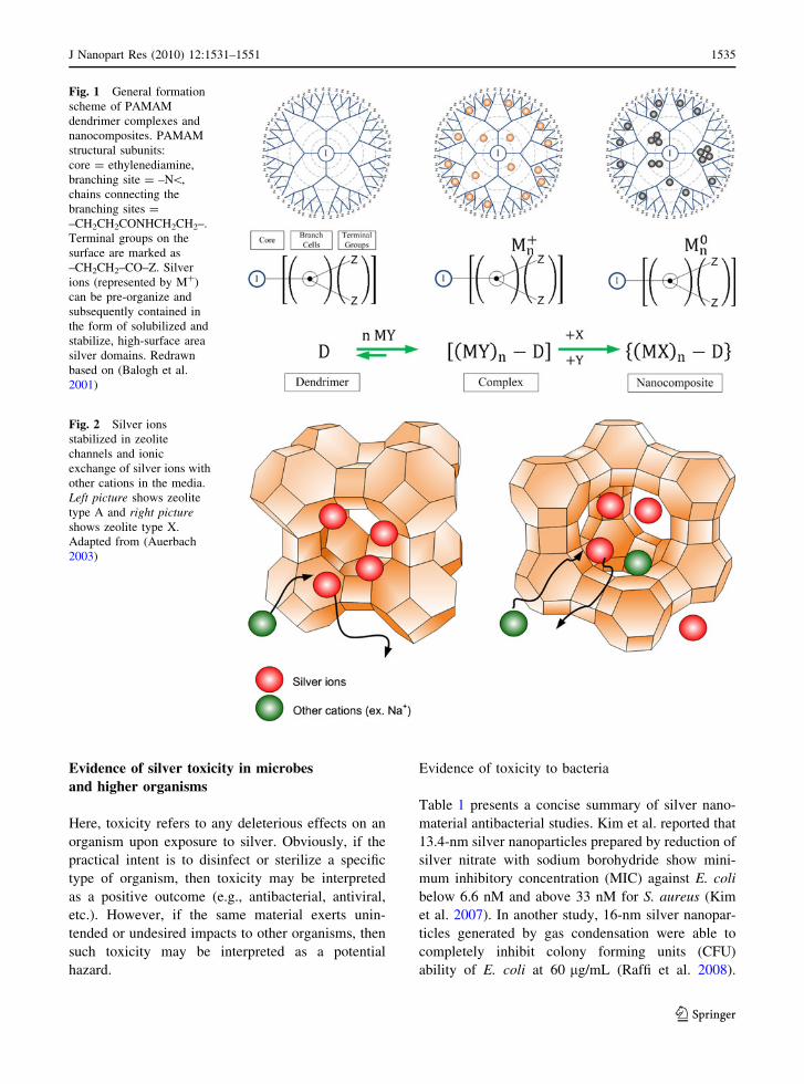
nanoparticles within the polymeric matrix (Fig. 1)
(Lesniak et al. 2005; Zhang et al. 2008). Dendri-
mer–silver complexes prevent silver ions from
precipitating and keep silver dispersed in the media
long enough to be delivered where it is desired
(Balogh et al. 2001; Lesniak et al. 2005; Zhang
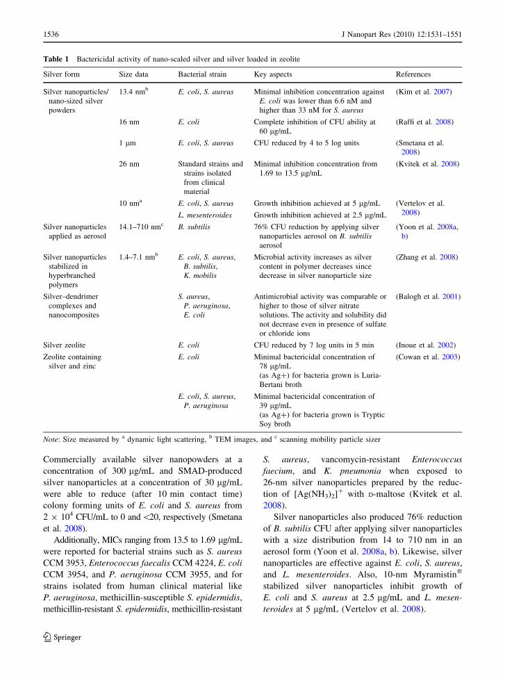
et al. 2008). Silver ions can also be stabilized in
zeolite channels (Fig. 2) or deposited in activated
carbon fibers (Inoue et al. 2002; Ogden et al. 1999;
Pal et al. 2009).
Composites of silver coatings over titanium diox-
ide nanoparticles are used in products such as baby
bottles and blood-clotting agents to produce antibac-
terial activity (Yeo and Kang 2008). Other hybrid
silver nanomaterials may include silver nanoparticles
coated onto polyurethane and silver–magnetite com-
posite nanoparticles (Fe3O4@Ag); both of these
hybrids are utilized for water disinfection (Gong
et al. 2007; Jain and Pradeep 2005). One of the
challenges of using silver (or any) nanoparticles for
water treatment is recovering the particles after the
treatment process. Silver–magnetite nanoparticles
offer the potential advantage of being removed by a
magnet, avoiding release to the environment, and
making possible direct reuse without additional
separation processes. For example, in related
research, magnetite particles proved effective for
removal of arsenic from water (Mayo et al. 2007;
Yavuz et al. 2006). However, for silver materials, an
additional concern is controlling the release of metal
ions into the final produce water.
1534 J Nanopart Res (2010) 12:1531–1551
123
Page 5
Evidence of silver toxicity in microbes
and higher organisms
Here, toxicity refers to any deleterious effects on an
organism upon exposure to silver. Obviously, if the
practical intent is to disinfect or sterilize a specific
type of organism, then toxicity may be interpreted
as a positive outcome (e.g., antibacterial, antiviral,
etc.). However, if the same material exerts unin-
tended or undesired impacts to other organisms, then
such toxicity may be interpreted as a potential
hazard.
Evidence of toxicity to bacteria
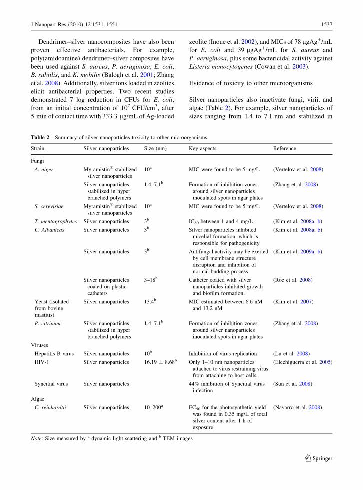
Table 1 presents a concise summary of silver nano-
material antibacterial studies. Kim et al. reported that
13.4-nm silver nanoparticles prepared by reduction of
silver nitrate with sodium borohydride show mini-
mum inhibitory concentration (MIC) against E. coli
below 6.6 nM and above 33 nM for S. aureus (Kim
et al. 2007). In another study, 16-nm silver nanopar-
ticles generated by gas condensation were able to
completely inhibit colony forming units (CFU)
ability of E. coli at 60 lg/mL (Raffi et al. 2008).
Fig. 1 General formation
scheme of PAMAM
dendrimer complexes and
nanocomposites. PAMAM
structural subunits:
core = ethylenediamine,
branching site = –N\,
chains connecting the
branching sites =
–CH2CH2CONHCH2CH2–.
Terminal groups on the
surface are marked as
–CH2CH2–CO–Z. Silver
ions (represented by M?)
can be pre-organize and
subsequently contained in
the form of solubilized and
stabilize, high-surface area
silver domains. Redrawn
based on (Balogh et al.
2001)
Fig. 2 Silver ions
stabilized in zeolite
channels and ionic
exchange of silver ions with
other cations in the media.
Left picture shows zeolite
type A and right pictureshows zeolite type X.
Adapted from (Auerbach
2003)
J Nanopart Res (2010) 12:1531–1551 1535
123
Page 6
Commercially available silver nanopowders at a
concentration of 300 lg/mL and SMAD-produced
silver nanoparticles at a concentration of 30 lg/mL
were able to reduce (after 10 min contact time)
colony forming units of E. coli and S. aureus from
2 9 104 CFU/mL to 0 and\20, respectively (Smetana
et al. 2008).
Additionally, MICs ranging from 13.5 to 1.69 lg/mL
were reported for bacterial strains such as S. aureus
CCM 3953, Enterococcus faecalis CCM 4224, E. coli
CCM 3954, and P. aeruginosa CCM 3955, and for
strains isolated from human clinical material like
P. aeruginosa, methicillin-susceptible S. epidermidis,
methicillin-resistant S. epidermidis, methicillin-resistant
S. aureus, vancomycin-resistant Enterococcus
faecium, and K. pneumonia when exposed to
26-nm silver nanoparticles prepared by the reduc-
tion of [Ag(NH3)2]? with D-maltose (Kvitek et al.
2008).
Silver nanoparticles also produced 76% reduction
of B. subtilis CFU after applying silver nanoparticles
with a size distribution from 14 to 710 nm in an
aerosol form (Yoon et al. 2008a, b). Likewise, silver
nanoparticles are effective against E. coli, S. aureus,
and L. mesenteroides. Also, 10-nm Myramistin�
stabilized silver nanoparticles inhibit growth of
E. coli and S. aureus at 2.5 lg/mL and L. mesen-
teroides at 5 lg/mL (Vertelov et al. 2008).
Table 1 Bactericidal activity of nano-scaled silver and silver loaded in zeolite
Silver form Size data Bacterial strain Key aspects References
Silver nanoparticles/
nano-sized silver
powders
13.4 nmb E. coli, S. aureus Minimal inhibition concentration against
E. coli was lower than 6.6 nM and
higher than 33 nM for S. aureus
(Kim et al. 2007)
16 nm E. coli Complete inhibition of CFU ability at
60 lg/mL
(Raffi et al. 2008)
1 lm E. coli, S. aureus CFU reduced by 4 to 5 log units (Smetana et al.
2008)
26 nm Standard strains and
strains isolated
from clinical
material
Minimal inhibition concentration from
1.69 to 13.5 lg/mL
(Kvitek et al. 2008)
10 nma E. coli, S. aureus Growth inhibition achieved at 5 lg/mL (Vertelov et al.
2008)L. mesenteroides Growth inhibition achieved at 2.5 lg/mL
Silver nanoparticles
applied as aerosol
14.1–710 nmc B. subtilis 76% CFU reduction by applying silver
nanoparticles aerosol on B. subtilisaerosol
(Yoon et al. 2008a,
b)
Silver nanoparticles
stabilized in
hyperbranched
polymers
1.4–7.1 nmb E. coli, S. aureus,
B. subtilis,
K. mobilis
Microbial activity increases as silver
content in polymer decreases since
decrease in silver nanoparticle size
(Zhang et al. 2008)
Silver–dendrimer
complexes and
nanocomposites
S. aureus,
P. aeruginosa,
E. coli
Antimicrobial activity was comparable or
higher to those of silver nitrate
solutions. The activity and solubility did
not decrease even in presence of sulfate
or chloride ions
(Balogh et al. 2001)
Silver zeolite E. coli CFU reduced by 7 log units in 5 min (Inoue et al. 2002)
Zeolite containing
silver and zinc
E. coli Minimal bactericidal concentration of
78 lg/mL
(as Ag?) for bacteria grown is Luria-
Bertani broth
(Cowan et al. 2003)
E. coli, S. aureus,
P. aeruginosaMinimal bactericidal concentration of
39 lg/mL
(as Ag?) for bacteria grown is Tryptic
Soy broth
Note: Size measured by a dynamic light scattering, b TEM images, and c scanning mobility particle sizer
1536 J Nanopart Res (2010) 12:1531–1551
123
Page 7
Dendrimer–silver nanocomposites have also been
proven effective antibacterials. For example,
poly(amidoamine) dendrimer–silver composites have
been used against S. aureus, P. aeruginosa, E. coli,
B. subtilis, and K. mobilis (Balogh et al. 2001; Zhang
et al. 2008). Additionally, silver ions loaded in zeolites
elicit antibacterial properties. Two recent studies
demonstrated 7 log reduction in CFUs for E. coli,
from an initial concentration of 107 CFU/cm3, after
5 min of contact time with 333.3 lg/mL of Ag-loaded
zeolite (Inoue et al. 2002), and MICs of 78 lgAg?/mL
for E. coli and 39 lgAg?/mL for S. aureus and
P. aeruginosa, plus some bactericidal activity against
Listeria monocytogenes (Cowan et al. 2003).
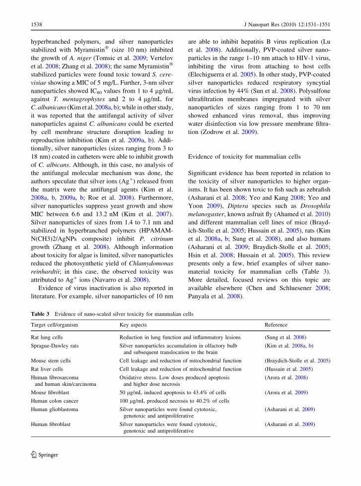
Evidence of toxicity to other microorganisms
Silver nanoparticles also inactivate fungi, virii, and
algae (Table 2). For example, silver nanoparticles of
sizes ranging from 1.4 to 7.1 nm and stabilized in
Table 2 Summary of silver nanoparticles toxicity to other microorganisms
Strain Silver nanoparticles Size (nm) Key aspects Reference
Fungi
A. niger Myramistin� stabilized
silver nanoparticles
10a MIC were found to be 5 mg/L (Vertelov et al. 2008)
Silver nanoparticles
stabilized in hyper
branched polymers
1.4–7.1b Formation of inhibition zones
around silver nanoparticles
inoculated spots in agar plates
(Zhang et al. 2008)
S. cerevisiae Myramistin� stabilized
silver nanoparticles
10a MIC were found to be 5 mg/L (Vertelov et al. 2008)
T. mentagrophytes Silver nanoparticles 3b IC80 between 1 and 4 mg/L (Kim et al. 2008a, b)
C. Albanicas Silver nanoparticles 3b Silver nanoparticles inhibited
micelial formation, which is
responsible for pathogenicity
(Kim et al. 2008a, b)
Silver nanoparticles 3b Antifungal activity may be exerted
by cell membrane structure
disruption and inhibition of
normal budding process
(Kim et al. 2009a, b)
Silver nanoparticles
coated on plastic
catheters
3–18b Catheter coated with silver
nanoparticles inhibited growth
and biofilm formation.
(Roe et al. 2008)
Yeast (isolated
from bovine
mastitis)
Silver nanoparticles 13.4b MIC estimated between 6.6 nM
and 13.2 nM
(Kim et al. 2007)
P. citrinum Silver nanoparticles
stabilized in hyper
branched polymers
1.4–7.1b Formation of inhibition zones
around silver nanoparticles
inoculated spots in agar plates
(Zhang et al. 2008)
Viruses
Hepatitis B virus Silver nanoparticles 10b Inhibition of virus replication (Lu et al. 2008)
HIV-1 Silver nanoparticles 16.19 ± 8.68b Only 1–10 nm nanoparticles
attached to virus restraining virus
from attaching to host cells.
(Elechiguerra et al. 2005)
Syncitial virus Silver nanoparticles 44% inhibition of Syncitial virus
infection
(Sun et al. 2008)
Algae
C. reinhardtii Silver nanoparticles 10–200a EC50 for the photosynthetic yield
was found in 0.35 mg/L of total
silver content after 1 h of
exposure
(Navarro et al. 2008)
Note: Size measured by a dynamic light scattering and b TEM images
J Nanopart Res (2010) 12:1531–1551 1537
123
Page 8
hyperbranched polymers, and silver nanoparticles
stabilized with Myramistin� (size 10 nm) inhibited
the growth of A. niger (Tomsic et al. 2009; Vertelov
et al. 2008; Zhang et al. 2008); the same Myramistin�
stabilized particles were found toxic toward S. cere-
visiae showing a MIC of 5 mg/L. Further, 3-nm silver
nanoparticles showed IC80 values from 1 to 4 lg/mL
against T. mentagrophytes and 2 to 4 lg/mL for
C. albanicans (Kim et al. 2008a, b); while in other study,
it was reported that the antifungal activity of silver
nanoparticles against C. albanicans could be exerted
by cell membrane structure disruption leading to
reproduction inhibition (Kim et al. 2009a, b). Addi-
tionally, silver nanoparticles (sizes ranging from 3 to
18 nm) coated in catheters were able to inhibit growth
of C. albicans. Although, in this case, no analysis of
the antifungal molecular mechanism was done, the
authors speculate that silver ions (Ag?) released from
the matrix were the antifungal agents (Kim et al.
2008a, b, 2009a, b; Roe et al. 2008). Furthermore,
silver nanoparticles suppress yeast growth and show
MIC between 6.6 and 13.2 nM (Kim et al. 2007).
Silver nanoparticles of sizes from 1.4 to 7.1 nm and
stabilized in hyperbranched polymers (HPAMAM-
N(CH3)2/AgNPs composite) inhibit P. citrinum
growth (Zhang et al. 2008). Although information
about toxicity for algae is limited, silver nanoparticles
reduced the photosynthetic yield of Chlamydomonas
reinhardtii; in this case, the observed toxicity was
attributed to Ag? ions (Navarro et al. 2008).
Evidence of virus inactivation is also reported in
literature. For example, silver nanoparticles of 10 nm
are able to inhibit hepatitis B virus replication (Lu
et al. 2008). Additionally, PVP-coated silver nano-
particles in the range 1–10 nm attach to HIV-1 virus,
inhibiting the virus from attaching to host cells
(Elechiguerra et al. 2005). In other study, PVP-coated
silver nanoparticles reduced respiratory syncytial
virus infection by 44% (Sun et al. 2008). Polysulfone
ultrafiltration membranes impregnated with silver
nanoparticles of sizes ranging from 1 to 70 nm
showed enhanced virus removal, thus improving
water disinfection via low pressure membrane filtra-
tion (Zodrow et al. 2009).
Evidence of toxicity for mammalian cells
Significant evidence has been reported in relation to
the toxicity of silver nanoparticles to higher organ-
isms. It has been shown toxic to fish such as zebrafish
(Asharani et al. 2008; Yeo and Kang 2008; Yeo and
Yoon 2009), Diptera species such as Drosophila
melanogaster, known asfruit fly (Ahamed et al. 2010)
and different mammalian cell lines of mice (Brayd-
ich-Stolle et al. 2005; Hussain et al. 2005), rats (Kim
et al. 2008a, b; Sung et al. 2008), and also humans
(Asharani et al. 2009; Braydich-Stolle et al. 2005;
Hsin et al. 2008; Hussain et al. 2005). This review
presents only a few, brief examples of silver nano-
material toxicity for mammalian cells (Table 3).
More detailed, focused reviews on this topic are
available elsewhere (Chen and Schluesener 2008;
Panyala et al. 2008).
Table 3 Evidence of nano-scaled silver toxicity for mammalian cells
Target cell/organism Key aspects Reference
Rat lung cells Reduction in lung function and inflammatory lesions (Sung et al. 2008)
Sprague-Dawley rats Silver nanoparticles accumulation in olfactory bulb
and subsequent translocation to the brain
(Kim et al. 2008a, b)
Mouse stem cells Cell leakage and reduction of mitochondrial function (Braydich-Stolle et al. 2005)
Rat liver cells Cell leakage and reduction of mitochondrial function (Hussain et al. 2005)
Human fibrosarcoma
and human skin/carcinoma
Oxidative stress. Low doses produced apoptosis
and higher dose necrosis
(Arora et al. 2008)
Mouse fibroblast 50 lg/mL induced apoptosis to 43.4% of cells (Arora et al. 2009)
Human colon cancer 100 lg/mL produced necrosis to 40.2% of cells
Human glioblastoma Silver nanoparticles were found cytotoxic,
genotoxic and antiproliferative
(Asharani et al. 2009)
Human fibroblast Silver nanoparticles were found cytotoxic,
genotoxic and antiproliferative
(Asharani et al. 2009)
1538 J Nanopart Res (2010) 12:1531–1551
123
Page 9
Evidence of silver nanoparticle toxicity for mam-
malian cells was presented in the in vivo studies
performed by Sung et al. (2008) and Kim et al.
(2008a, b). In the former, a 90-day inhalation study in
rats showed that silver nanoparticles reduce lung
function and produce inflammatory lesions in the
lungs. In the later, silver nanoparticles accumulated
in the olfactory bulbs of Sprague-Dawley rats and
also accumulated in the brain.
Evidence from in vitro studies is also available in the
literature. For example, silver nanoparticles have been
shown to reduce mitochondrial function and to
increase membrane leakage of mouse spermatogonial
stem cell and rat liver cells (Braydich-Stolle et al. 2005;
Hussain et al. 2005). Studies performed on human
fibrosarcoma and human skin/carcinoma cells with
silver nanoparticles used in a topical antimicrobial
agent concluded that in the presence of the nanopar-
ticles the cellular levels of glutathione are reduced,
indicating oxidative stress, that results in cellular
damage and lipid peroxidation (Arora et al. 2008).
However, in the study performed by Arora et al. the
dose required to induce apoptosis (0.78–1.56 lg/mL)
was much smaller than that required to produce
necrosis (12.5 lg/mL) in both cell types. Therefore,
the authors concluded that, after the required in vivo
studies, it would be possible to define a safe range for
the application of silver nanoparticles as a topical
antimicrobial agent. Similar differences of the required
concentration to cause apoptosis or necrosis were
found in a second study by the same authors in mouse
fibroblasts and liver cells (Arora et al. 2009). In this
second article, it was suggested that although silver
nanoparticles may enter into the cells, the cellular
antioxidant mechanisms would limit oxidative stress.
Mechanistic studies of silver nanoparticle toxicity
in mammalian cells have considered mouse fibroblast
and human colon cancer cells (Hsin et al. 2008). In
this study, silver nanoparticle doses of 50 lg/mL
induced apoptosis to 43.4% of fibroblast cells, while
100 lg/mL produced necrosis to 40.2% of the cancer
cells. The authors concluded that the apoptotic
mechanisms in fibrosblast cells are a mitochondrial
mediated pathway including the generation of ROS in
the cell, which activate the apoptosis regulators JNK
and p53 proteins inducing protein Bax to migrate to
the surface of the mitochondria. That subsequently
induces cytochrome C release from mitochondria and
cleavage of PARP. Additionally, in a study done by
Asharani, a possible mechanism of toxicity to human
cells was proposed (Asharani et al. 2009). Silver
nanoparticles would affect the mitochondrial respira-
tory chain, causing ROS generation and affecting the
production of ATP, which subsequently leads to DNA
damage. In this study, the authors also concluded that
‘‘even and small dose of Ag-NP (silver nanoparticles)
has the potential to cause toxicity’’ and that silver
nanoparticles are cytotoxic, genotoxic, and antipro-
liferative, being as toxic for human glioblastoma as
for normal human fibroblasts cells.
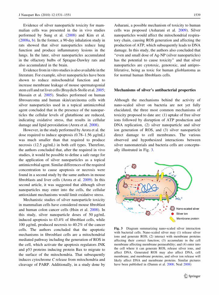
Mechanisms of silver’s antibacterial properties
Although the mechanisms behind the activity of
nano-scaled silver on bacteria are not yet fully
elucidated, the three most common mechanisms of
toxicity proposed to date are: (1) uptake of free silver
ions followed by disruption of ATP production and
DNA replication, (2) silver nanoparticle and silver
ion generation of ROS, and (3) silver nanoparticle
direct damage to cell membranes. The various
observed and hypothesized interactions between
silver nanomaterials and bacteria cells are conceptu-
ally illustrated in Fig. 3.
Fig. 3 Diagram summarizing nano-scaled silver interaction
with bacterial cells. Nano-scaled silver may (1) release silver
ions and generate ROS; (2) interact with membrane proteins
affecting their correct function; (3) accumulate in the cell
membrane affecting membrane permeability; and (4) enter into
the cell where it can generate ROS, release silver ions, and
affect DNA. Generated ROS may also affect DNA, cell
membrane, and membrane proteins, and silver ion release will
likely affect DNA and membrane proteins. Similar pictures
have been published in (Damm et al. 2008; Neal 2008)
J Nanopart Res (2010) 12:1531–1551 1539
123
Page 10
Free silver ion uptake
Silver nanoparticles have been reported to dissolve
generating silver ions and it is thought that in vivo this
release would be product of reactions of silver nano-
particles with H2O2 (Asharani et al. 2009). Asharani has
proposed the following reaction as a possible mecha-
nism for silver nanoparticle oxidative dissolution.
2AgþH2O2 þ 2Hþ ! 2Agþ þ 2H2O E0 ¼ 0:17 V:
Asharani suggests that in eukaryotic cells this
reaction could occur in the mitochondria, where
exists an important concentration of H?. Similarly,
we hypothesized that a similar mechanism could
occur in the bacterial cell membrane where proton
motive force takes place.
Another possible mechanism for the oxidative
dissolution of silver nanoparticles has been reported
by Choi et al., in this case silver is oxidized in the
presence of oxygen. Choi et al. speculated that the
observed changed of color of their silver nanoparti-
cles suspensions, over a week period, would be
attributed to this mechanism.
4Agþ O2 þ 2H2O$ 4Agþ þ 4OH�:
The amount of free silver ion measured in this case
was approximately 2.2% of the total silver content in
the silver nanoparticle suspension (Choi et al. 2008).
In other article, a 0.1% content of the total silver in
partially oxidized silver nanoparticles suspensions
was attributed to silver ions (Lok et al. 2007).
Ionic silver has known antibacterial properties;
thus, it is expected that eluted ions from silver
nanoparticles are responsible for at least a part of
their antibacterial properties. At sub-micromolar
concentrations, Ag? interacts with enzymes of the
respiratory chain reaction such as NADH dehydro-
genase resulting in the uncoupling of respiration from
ATP synthesis. Silver ions also bind with transport
proteins leading to proton leakage, inducing collapse
of the proton motive force (Dibrov et al. 2002; Holt
and Bard 2005; Lok et al. 2006). Silver inhibits the
uptake of phosphate and causes the efflux of intra-
cellular phosphate (Schreurs and Rosenberg 1982).
The interaction with respiratory and transport pro-
teins is due to the high affinity of Ag? with thiol
groups present in the cysteine residues of those
proteins (Holt and Bard 2005; Liau et al. 1997;
Petering 1976). Additionally, it has been reported that
Ag? increases DNA mutation frequencies during
polymerase chain reactions (Yang et al. 2009).
Bacterial cells exposed to milli-molar Ag? doses
suffer morphological changes such as cytoplasm shrink-
age and detachment of cell wall membrane, DNA
condensation and localization in an electron-light region
in the center of the cell, and cell membrane degradation
allowing leakage of intracellular contents (Feng et al.
2000; Jung et al. 2008). Physiological changes occur
together with the morphological changes. Bacterial cells
enter an active, but non-culturable state in which
physiological levels can be measured but bacteria are
not able to growth and replicate.
Several studies have linked the toxicity of silver
nanoparticles to the release of silver ions. For example,
Smetana et al. observed that silver ions eroded from
high-surface area silver powders prepared by SMAD
method interacted and destroyed bacterial cells (Sme-
tana et al. 2008). In the same study, a second
preparation of silver nanoparticles using water-soluble
ligands was used to obtain silver nanoparticles with
higher surface area to improve their antibacterial
efficacy. However, the second preparation of silver
nanoparticles showed lower toxicity toward bacterial
cells than uncoated powders, suggesting the ligands
prevented silver ion erosion; thus, diminishing the
resulting toxicity. Another possible reason for this
result is that the surface coatings prevented adhesion
of silver nanoparticles to the bacterial cell surface, but
the authors did not explore this option.
Hwang et al. observed that Ag? induced the same
effect in bioluminescence bacteria sensitive to mem-
brane protein damage and slightly less effect in a strain
sensitive to superoxides compared to silver nanopar-
ticles (Hwang et al. 2008). The authors suggested that
silver nanoparticles produce silver ions that move
inside the cell producing ROS through redox reactions
with oxygen. In other research, bacterial activity of
activated carbon fiber supported silver was attributed
to the synergistic action of silver ions, superoxides,
and hydrogen peroxide (Le Pape et al. 2004).
Generation of reactive oxygen species
Reactive oxygen species (ROS) are natural byprod-
ucts of the metabolism of respiring organisms. While
1540 J Nanopart Res (2010) 12:1531–1551
123
Page 11
small levels can be controlled by the antioxidant
defenses of the cells such glutathione/glutathione
disulfide (GSH/GSSG) ratio, excess ROS production
may produce oxidative stress (Nel et al. 2006). The
additional generation of free radicals can attack
membrane lipids and lead to a breakdown of mem-
brane and mitochondrial function or cause DNA
damage (Mendis et al. 2005).
Metals can act as catalysts and generate ROS in
the presence of dissolved oxygen (Stohs and Bagchi
1995). In this context, silver nanoparticles may
catalyze reactions with oxygen leading to excess free
radical production. Studies done in eukaryotic cells
suggest that silver nanoparticles inhibit the antioxi-
dant defense by interacting directly with GSH,
binding GSH reductase or other GSH maintenance
enzymes (Carlson et al. 2008). This could decrease
the GSH/GSSG ratio and, subsequently, increase
ROS in the cell. Silver ions eluted from nano-scaled
silver or chemisorbed on its surface may also be
responsible for the generation of ROS by serving as
electron acceptor. In bacterial cells, silver ions would
likely induce the generation of ROS by impairing the
respiratory chain enzymes through direct interactions
with thiol groups in these enzymes or the superoxide-
radical scavenging enzymes such as superoxide
dismutases (Park et al. 2009). ROS generation from
silver nanoparticles and silver ions may also be
induced photocatalytically; however, no correlations
have been reported between bacterial toxicity and
photocatalytic ROS concentration (Choi and Hu
2008).
Evidence of toxicity related to ROS generated
from silver nanoparticles and silver ions, released
from or chemisorbed on its surface, has been shown
previously. Kim et al. determined the existence of
free radicals from silver nanoparticles by means of
spin resonance measurements (Kim et al. 2007). They
observed that silver nanoparticles and silver nitrate
toxicity was abolished in the presence of an antiox-
idant, these results leaded them to suggest that the
antimicrobial mechanisms of silver nanoparticles
against S. aureus and E. coli was related to the
formation of free radicals from the surface of silver
nanoparticles and subsequent free radical induced
membrane damage.
Bactericidal activity of silver ions loaded in
nanoporous materials such as zeolites has also been
related to the generation of ROS. Using ROS
scavengers, it was determined that superoxide anions,
hydrogen peroxide, hydroxyl radicals, and singlet
oxygen contributed to the antibacterial activity
against E. coli (Inoue et al. 2002). In another article,
the bactericidal activity of activated carbon fiber
supported silver occurred only in the presence of
oxygen and could not be explained by the silver ions
eluted from the fiber (Yoon et al. 2008a, b). These
observations together with a study of gene expression
related to oxidative stress lead the authors to suggest
that the strong bactericidal activity of this materials
resulted from the combined effects of silver ions,
superoxides ions, and hydrogen peroxide (Le Pape
et al. 2004).
A study of the toxicity effects of different silver
forms on nitrifying bacteria showed that not only
Ag? and AgCl, but also silver nanoparticles are able
to induce intracellular ROS generation. Moreover, the
bacterial inhibition of each of these silver species
correlates with their individual generation of intra-
cellular ROS concentration. However, at the same
level of intracellular ROS concentration, silver
nanoparticles appeared more toxic than the other
species indicating that other factors besides the
generation of intracellular ROS played a role in the
overall toxicity (Choi et al. 2008). In other research, a
panel of recombinant bioluminescent bacteria was
used to test different pathways of silver nanoparticles
toxicity such as oxidative stress, DNA damage and
protein or membrane damage in bacterial strains. The
addition of silver nanoparticles to growing bacterial
cultures leaded to the production of superoxide
radicals, but not of hydroxyl radicals. The authors
conclude that toxicity occurs via membrane protein
damage and oxidative stress, but not DNA damage
(Hwang et al. 2008).
Direct cell membrane damage
Silver nanoparticles interact with the bacterial mem-
brane and are able to penetrate inside the cell.
Transmission electron microscopy data show that
silver nanoparticles adhere to and penetrate into
E. coli cells and also are able to induce the formation
of pits in the cell membrane (Choi et al. 2008; Raffi
et al. 2008; Sondi and Salopek-Sondi 2004). Silver
nanoparticles have been observed within E. coli cells
albeit at sizes much smaller than the original particles;
moreover, silver nanoparticles with oxidized surfaces
J Nanopart Res (2010) 12:1531–1551 1541
123
Page 12
induce the formation of ‘‘huge holes’’ in E. coli
surfaces after the interaction and large portions of the
cellular content seemed to be ‘‘eaten away’’ (Smetana
et al. 2008).
Silver nanoparticle accumulation on the cell mem-
brane and uptake within the cell has also been reported
for other bacteria such as V. cholera, P. aeruginosa,
and S. typhus. In these cases, only nanoparticles
smaller than 10 nm attached to bacteria cell mem-
branes or where observed inside the bacteria (Morones
et al. 2005). However, in other research silver
nanoparticles with sizes up to 80 nm were transported
through the inner and outer membrane of P. aerugin-
osa (Xu et al. 2004). Silver nanoparticle composites
and silver nanoparticles stabilized with surfactants are
also thought to interact with cell membranes. Myram-
istin� capped silver nanoparticles showed notable
antibacterial activity against gram-positive and
-negative bacteria, it was speculated that Myramistin�
anchors to the cell wall and weakens it allowing
penetration of silver nanoparticles inside the cells
(Vertelov et al. 2008). Composites of HPAMAM-
N(CH3)2 silver nanoparticle are thought to catch
bacteria by ionic interactions between the cationic
polymer and the negative cell membrane, followed by
release the silver nanoparticles (Zhang et al. 2008).
The detailed mechanism by which silver nanopar-
ticles interact with cytoplasmic membranes and are
able to penetrate inside cells is not fully determined.
One hypothesis is that the interaction between
nanoparticles and bacterial cells are due to electro-
static attraction between negatively charged cell
membranes and positively charged nanoparticles
(Raffi et al. 2008). However, this mechanism does
not likely explain the adhesion and uptake of
negatively charged silver nanoparticles. It has been
also proposed that the preferential sites of interaction
for silver nanoparticles and membrane cells might be
sulfur containing proteins—in a similar way as silver
interacts with thiol groups of respiratory chain
proteins and transport proteins, interfering with their
proper function (Morones et al. 2005). This seems
more likely than electrostatic attraction because
evidence of protein membrane damage on bacteria
after silver nanoparticle exposure has been demon-
strated with bioluminescence bacteria (Hwang et al.
2008).
Another proposed mechanism of E. coli membrane
damage by silver nanoparticles relates to metal
depletion, i.e., the formation of irregular-shaped pits
in the outer membrane and change in membrane
permeability by the progressive release of LPS
molecules and membrane proteins (Amro et al.
2000; Sondi and Salopek-Sondi 2004). This may be
fairly general for gram-negative bacteria. It has been
reported that extrusion pump systems such as Mex-
AB-OprM system of P. aeruginosa could also play an
important role in controlling the accumulation of
silver nanoparticles in living cells (Xu et al. 2004).
The same pump systems are responsible for the silver
resistance of several bacteria (see ‘‘Mechanisms of
silver resistance’’ section).
Despite the mechanism of interaction involved, it is
evident that silver nanoparticles attached to bacterial
cell membranes increase permeability and disturb
respiration. Proteomic data show the accumulation of
envelope protein precursors in E. coli cells after
exposure to silver nanoparticles (Lok et al. 2006).
Energy from ATP and proton motive force is required
in order to newly synthesize envelope proteins to be
translocated to the membrane, therefore cytoplasmic
accumulation of protein precursors suggests dissipa-
tion of proton motive force and depletion of intracel-
lular levels of ATP. In a similar way, silver ions have
also been linked to the collapse of proton motive force
and destabilization of the cell membrane, but the
concentrations at which this occur are much higher
than the ones of silver nanoparticles (micromolar vs.
nanomolar) (Dibrov et al. 2002; Lok et al. 2006).
Mechanisms of silver resistance
Bacterial strains resistant to specific toxicants are
naturally selected in environments where these agents
are present (Gupta and Silver 1998). In this way, the
widespread use of silver, whether in nano or bulk
form, could lead to selection of bacterial communities
exhibiting silver resistance. Since most commercial
uses of silver and silver nanoparticles relate to
fighting infection, widespread silver resistance is a
growing concern. In fact, it has been suggested that it
already occurs extensively, but it not recognized due
to a lack of testing for silver resistance (Silver et al.
2006). For example, 10% of the enteric bacteria
tested randomly in a hospital in Chicago, IL, showed
genes for Ag? resistance (Silver 2003).
Silver resistance in bacteria (and other heavy
metals) is often encoded by plasmids genes, as for
1542 J Nanopart Res (2010) 12:1531–1551
123
Page 13
example the Salmonella plasmid pMG101, within the
IncHI incompatibility group, as well as in other five
plasmids in the same group (Gupta et al. 2001).
Although resistance is mainly encoded by plasmids,
bacterial chromosomes have also been reported to
encode Ag? resistance genes (sil) in strains such as
E. coli K-12 and O157:H7 (Gupta et al. 2001; Silver
et al. 2006). The plasmid pMG101 has been studied in
detail (Gupta et al. 1999; Silver 2003; Silver et al.
2006), this plasmid has nine genes, whose products
have been identified to be proteins responsible for
heavy metal resistance. Here, the resistance is
achieved by two efflux systems SilCBA and SilP
acting in combination with two periplasmic binding
proteins SilE and SilF. The complex SilCBA is
constituted by three proteins SilC in the outer mem-
brane, SilA in the inner membrane and SilB that links
SilA and SilC. This complex acts as an antiporter that
transports Ag? out of the cell by pumping a proton
inside. In turn, SilC is a P-type ATPase that transports
Ag? from the cytoplasm to the perisplasm. These two
efflux pumps work jointly with proteins SilE and SilF,
which bind to Ag?. SilF is thought to act as a
‘‘chaperone’’ by taking one Ag? ion from its release
site in SilP to its uptake site in SilCBA while SilE
binds 5 Ag? ions to 10 histidines preventing silver ions
entrance to the cytoplasm (Gupta et al. 1999).
Limited evidence has been reported of the resis-
tance shown by silver-resistant strains to silver
nanoparticles. However, in one study, it was reported
that albumin-stabilized oxidized silver nanoparticles
were unable to inhibit growth of 116 AgNO3R and
J53 (pMG101) silver-resistant strains even when
apply at a concentration of up to 80 nM (Lok et al.
2007). Therefore, it seems that silver resistance may
also be of concern for the efficacy of silver nanopar-
ticles use as antibacterial agent.
Bacterial resistance and sensitivity to silver is
affected by environmental conditions such as the
presence of halides in the media due to mainly the
variation of silver bioavailability experienced at
different concentrations of Cl- and other halides
such as Br- and I- (Gupta et al. 1998). For moderate
Cl- concentration, silver is precipitated as AgCl,
which decreases silver bioavailability and increases
bacterial silver resistance, as observed for E. coli J53
(pMG101). Nevertheless, silver bioavailability rises
when ionic complexes such as AgCl2-, AgCl3
2- are
formed as product of a higher halide concentration.
This increase in bioavailability increases sensitivity
of the silver-resistant and silver-sensitive bacteria.
Similar, behavior is also observed for silver in the
presence of other halides (Gupta et al. 1998).
Further insights about potential toxicity to higher
organisms
Silver nanomaterials exhibit outstanding antibacterial
properties that give rise to many potentially beneficial
applications. However, misuses of silver nanoparti-
cles as a bactericide may also result in unintended
exposure to higher organisms, including humans.
This is of concern because silver nanoparticles appear
to have toxic effects in higher organisms (‘‘Evidence
of silver toxicity in microbes and higher organisms’’
section). While eukaryotic and prokaryotic cells are
different in many ways, understanding the funda-
mental mechanisms governing silver nanoparticle
toxicity in bacterial cells may shed light on the
potential effects of silver nanoparticles on important
organelles such as mitochondria.
Mitochondria are believed to have evolved from
bacteria (i.e., the ‘‘serial primary endosymbiosis
theory’’) and they share several characteristics
(Kutschera 2009). For example, the inner membrane
of a mitochondrion and the prokaryotic cell mem-
brane are structurally very similar. Respiration occurs
in bacterial cells similarly as in the mitochondria of
eukaryotes, including electron transport, ATP syn-
thesis, and proton motive force. Likewise mitochon-
drial DNA, the bacterial DNA are analogous, and the
division of the mitochondria is similar to fission of
prokaryotes (Slonczewski and Foster 2009). Based on
these similarities, it is hypothesized that silver
nanoparticles may affect mitochondria in higher
organisms via similar mechanisms as in bacterial
cells—assuming they can be internalized by the cell.
Factors affecting the toxicity of silver
nanomaterials
Several factors have been reported to influence silver
nanoparticle toxicity like particle size, shape, crys-
tallinity, surface chemistry, capping agents, as well,
as for environmental factors such as pH, ionic
strength, and the presence of ligands, divalent
cations, and macromolecules (Carlson et al. 2008;
J Nanopart Res (2010) 12:1531–1551 1543
123
Page 14
Choi and Hu 2008; Kvitek et al. 2008; Lok et al.
2007; Morones et al. 2005; Pal et al. 2007; Smetana
et al. 2008). Many publications have shown size-
dependent toxicities of silver nanoparticles (Carlson
et al. 2008; Choi and Hu 2008; Martinez-Castanon
et al. 2008; Morones et al. 2005). As particle size
decreases, the specific surface area increases leaving
a higher number of atoms exposed on the surface
available for redox, photochemical, and biochemical
reactions in addition to physicochemical interactions
with cells.
As discussed previously, one of the key mecha-
nisms for silver nanomaterials to exert biocidal
activity is through the release of silver ions. As the
rate of ion release, in general, is proportional to
particle surface area, nanoparticles can release ions
more rapidly than larger particles and macroscopic
materials. In effect, chemisorbed silver ions would be
the cause of the antibacterial activity observed by
Lok et al., who reported that only partially oxidized
silver nanoparticles exhibit antibacterial activities,
while zero valent silver nanoparticles do not (Lok
et al. 2007). This surface area effect also influences
RO generation. For example, at the same silver
concentration, silver nanoparticles of 15 nm gener-
ated higher levels of ROS in macrophages than 30
and 50 nm particles (Carlson et al. 2008).
High atom densities at h111i facets increased the
toxicity of silver nanoparticles to several bacterial
strains; the increased toxicity was the result of the
higher reactivity presented by the h111i facets
(Morones et al. 2005). Silver nanoparticle shape
may also be a factor. Truncated triangular nanoplates
exert stronger antibacterial activity than spherical-
and rod-shaped silver nanoparticles because they
contain more h111i facets; thus, they would be more
reactive (Pal et al. 2007). Hence, the antibacterial
properties of silver nanoparticles are related to both
size (i.e., reactive surface area) and crystallinity (i.e.,
surface reactivity).
Stability of silver nanoparticles also influences
toxicity since the formation of aggregates tends to
decrease biocidal activity (Kvitek et al. 2008; Shriv-
astava et al. 2007; Teeguarden et al. 2007). Different
surfactants and polymers (e.g., sodium dodecyl
sulfate, polyoxyethylene-sorbitan monooleate, poly-
vinylpyrrolidone, Na?-carrying poly(glutamic acid),
hydroxyl functionalized ionic liquids and hydroxyl
functionalized cationic surfactants) have been used to
stabilize silver nanoparticle dispersions and enhance
biocidal activity (Dorjnamjin et al. 2008; Kvitek et al.
2008; Yu 2007). However, some ligand-capped silver
nanoparticles—although highly stable and monodis-
perse in suspension—were less bioactive because the
capping agent hindered release of silver ions (Sme-
tana et al. 2008).
Environmental conditions such as pH, ionic
strength, presence of complexing agents, and natural
organic matter, also affect the toxicity of silver
nanoparticles. High salt concentrations and pH values
close to the isoelectric point promote nanoparticle
aggregation by screening electrical double layer
repulsion (Nel et al. 2009). However, water chemistry
also governs silver dissolution and/or re-precipitation
through various possible redox and precipitation
reactions (Lok et al. 2007). Dissolved Ag? ions are
sparingly soluble in the presence of various ligands
such as chloride (pK = 9.75), sulfate (pK = 4.9),
sulfide (pK = 49), hydroxide (pK = 7.8) and dis-
solved organic carbon (pK [ 7.5) (Choi et al. 2008;
Gao et al. 2009). The released Ag? can also form Ag0
-containing clusters through light or chemical reduc-
tion (Morones et al. 2005). Therefore, nanomaterials in
aqueous suspensions must be considered in a contin-
uous state of flux where the apparent speciation is
controlled by the aquatic media pH, redox potential,
ionic composition, and exposure to light.
Previously, cysteine ligands and chloride were
used to scavenge or precipitate eluted silver ions from
silver nanoparticles; thus, diminishing their toxicity
(Navarro et al. 2008). Sulfide ligands have also been
used to reduce the inhibition of silver nanoparticles to
nitrifying bacteria (Choi et al. 2009). More recently,
it was reported that halide ions act as precipitating
agents and profoundly affect the ‘‘bioavailability’’ of
Ag? in unexpected ways (Silver 2003). For example,
at low chloride concentrations, soluble Ag? binds to
cell membranes impacting respiration and producing
other toxics effects (Gupta et al. 1998). As chloride
concentration increases, silver becomes less bioavail-
able because the solubility of AgCl is very low.
However, further increase in halide concentration
results in the formation of water-soluble anionic
complexes in the form of AgCl2- and AgCl32-.
These anionic complexes are more bioavailable and
increase silver toxicity to both silver-sensitive and
silver-resistant bacteria. Other halides such as Br-
and I- have similar effects, but the concentrations at
1544 J Nanopart Res (2010) 12:1531–1551
123
Page 15
which ionic complexes form depends on their
respective solubility limits.
Finally, aerobic versus anaerobic conditions are
important when assessing the antibacterial properties
of silver loaded in zeolite or activated carbon fibers
(Inoue et al. 2002; Le Pape et al. 2002; 2004).
Dissolved oxygen is important in these cases because
ROS contributes significantly to bactericidal activity.
Dissolved organic carbon (DOC) seems also to
influence the toxicity of silver nanoparticles, specif-
ically, toxicity may decrease for aquatic invertebrates
as the concentration DOC increases (Gao et al. 2009).
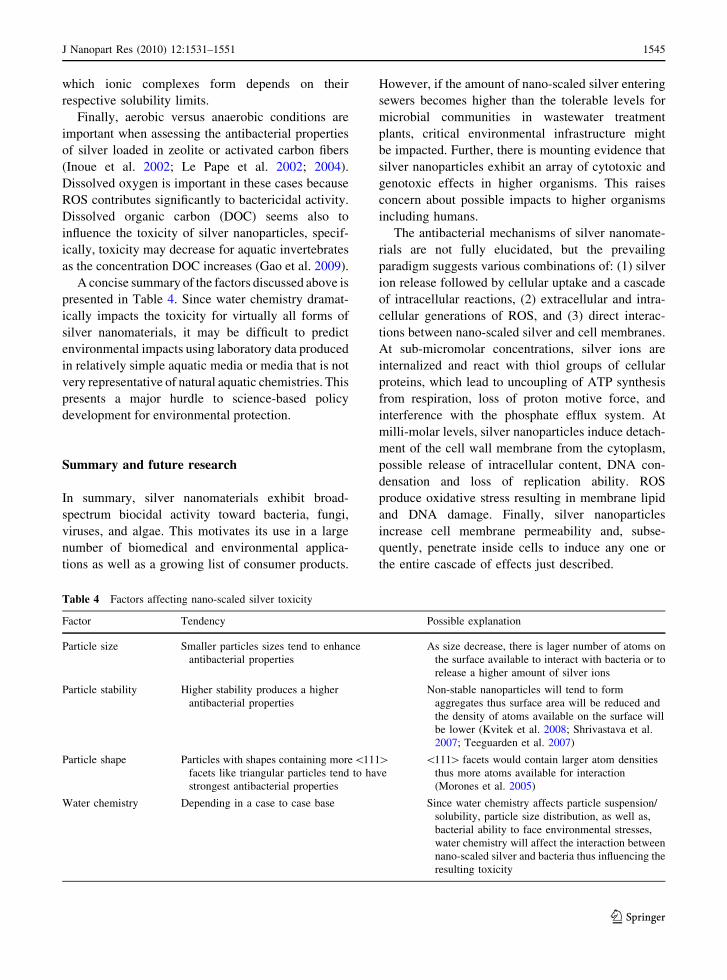
A concise summary of the factors discussed above is
presented in Table 4. Since water chemistry dramat-
ically impacts the toxicity for virtually all forms of
silver nanomaterials, it may be difficult to predict
environmental impacts using laboratory data produced
in relatively simple aquatic media or media that is not
very representative of natural aquatic chemistries. This
presents a major hurdle to science-based policy
development for environmental protection.
Summary and future research
In summary, silver nanomaterials exhibit broad-
spectrum biocidal activity toward bacteria, fungi,
viruses, and algae. This motivates its use in a large
number of biomedical and environmental applica-
tions as well as a growing list of consumer products.
However, if the amount of nano-scaled silver entering
sewers becomes higher than the tolerable levels for
microbial communities in wastewater treatment
plants, critical environmental infrastructure might
be impacted. Further, there is mounting evidence that
silver nanoparticles exhibit an array of cytotoxic and
genotoxic effects in higher organisms. This raises
concern about possible impacts to higher organisms
including humans.
The antibacterial mechanisms of silver nanomate-
rials are not fully elucidated, but the prevailing
paradigm suggests various combinations of: (1) silver
ion release followed by cellular uptake and a cascade
of intracellular reactions, (2) extracellular and intra-
cellular generations of ROS, and (3) direct interac-
tions between nano-scaled silver and cell membranes.
At sub-micromolar concentrations, silver ions are
internalized and react with thiol groups of cellular
proteins, which lead to uncoupling of ATP synthesis
from respiration, loss of proton motive force, and
interference with the phosphate efflux system. At
milli-molar levels, silver nanoparticles induce detach-
ment of the cell wall membrane from the cytoplasm,
possible release of intracellular content, DNA con-
densation and loss of replication ability. ROS
produce oxidative stress resulting in membrane lipid
and DNA damage. Finally, silver nanoparticles
increase cell membrane permeability and, subse-
quently, penetrate inside cells to induce any one or
the entire cascade of effects just described.
Table 4 Factors affecting nano-scaled silver toxicity
Factor Tendency Possible explanation
Particle size Smaller particles sizes tend to enhance
antibacterial properties
As size decrease, there is lager number of atoms on
the surface available to interact with bacteria or to
release a higher amount of silver ions
Particle stability Higher stability produces a higher
antibacterial properties
Non-stable nanoparticles will tend to form
aggregates thus surface area will be reduced and
the density of atoms available on the surface will
be lower (Kvitek et al. 2008; Shrivastava et al.
2007; Teeguarden et al. 2007)
Particle shape Particles with shapes containing more\111[facets like triangular particles tend to have
strongest antibacterial properties
\111[ facets would contain larger atom densities
thus more atoms available for interaction
(Morones et al. 2005)
Water chemistry Depending in a case to case base Since water chemistry affects particle suspension/
solubility, particle size distribution, as well as,
bacterial ability to face environmental stresses,
water chemistry will affect the interaction between
nano-scaled silver and bacteria thus influencing the
resulting toxicity
J Nanopart Res (2010) 12:1531–1551 1545
123
Page 16
Silver nanoparticle toxicity is influenced by intrin-
sic nanoparticle features like size, shape, chemistry,
crystallinity, and capping agents, but also by the
aquatic chemistry through such factors as solution
pH, redox state, ionic strength, and ionic composi-
tion. Most of the toxicity data presented in the
literature are obtained in relatively simple media like
distilled water or cell culture media, which do not
reflect the aquatic environment inside living organ-
isms or the natural environment. Hence, the surface
chemistry, reactivity, and state of dispersion achieved
in the laboratory may not be relevant for assessing
behavior in real systems. In natural waters, the wide
variation of pH, ionic strength, ionic composition,
and natural organic matter will induce widely varying
aggregation states of silver nanoparticles; thus,
resulting in widely varying antimicrobial activities
and toxicities. In addition, most toxicity data are
obtained by dispersing silver nanomaterials in or on
nutrient rich growth media; however, oligotrophic
conditions prevail in most natural environmental
systems. Microorganisms in these conditions may be
more or less vulnerable to silver than predicted by
laboratory studies.
Although significant progress has been made to
elucidate the mechanisms of silver nanomaterial
toxicity, further research is required to fully under-
stand the processes involved and to safely exploit the
tremendous antimicrobial properties of silver without
jeopardizing human health, critical infrastructure, and
the environment. Future in vivo, in vitro, and
environmental studies should consider more system-
atically the various effects of aquatic chemistry on
nano-scaled silver fate, transport, and toxicity.
Acknowledgments Financial support for this research was
provided by the University of California Toxic Substances
Research and Training Program: Lead Campus Program in
Nanotoxicology.
References
Abid J, Wark A, Brevet P, Girault H (2002) Preparation of
silver nanoparticles in solution from a silver salt by laser
irradiation. Chem Commun 792–793. doi: 10.1039/
b200272h
Ahamed M, Posgai R, Gorey TJ, Nielsen M, Hussain SM,
Rowe JJ (2010) Silver nanoparticles induced heat shock
protein 70, oxidative stress and apoptosis in Drosophila
melanogaster. Toxicol Appl Pharmacol 242:263–269. doi:
10.1016/j.taap.2009.10.016
Ahmad A, Mukherjee P, Senapati S, Mandal D, Khan M,
Kumar R, Sastry M (2003) Extracellular biosynthesis of
silver nanoparticles using the fungus Fusarium oxyspo-rum. Colloids Surf B 28:313–318. doi:10.1002/cbic.2007
00592
Amro N, Kotra L, Wadu-Mesthrige K, Bulychev A, Mobashery S,
Liu G (2000) High-resolution atomic force microscopy studies
of the Escherichia coli outer membrane: structural basis for
permeability. Langmuir 16:2789–2796. doi:10.1021/la9910
13x
Arora S, Jain J, Rajwade J, Paknikar K (2008) Cellular responses
induced by silver nanoparticles: in vitro studies. Toxicol
Lett 179:93–100. doi:10.1016/j.toxlet.2008.04.009
Arora S, Jain J, Rajwade JM, Paknikar KM (2009) Interactions of
silver nanoparticles with primary mouse fibroblasts and liver
cells. Toxicol Appl Pharmacol 236:310–318. doi:10.1016/
j.taap.2009.02.020
Asharani P, Wu Y, Gong Z, Valiyaveettil S (2008) Toxicity of
silver nanoparticles in zebrafish models. Nanotechnology
19:1–8. doi:10.1088/0957-4484/19/25/255102
Asharani PV, Mun GLK, Hande MP, Valiyaveettil S (2009)
Cytotoxicity and genotoxicity of silver nanoparticles in
human cells. ACS Nano 3:279–290. doi:10.1021/nn800
596w
Auerbach SM (2003) Zeolite science and technology. Marcel
Dekker, New York
Bajpai S, Mohan Y, Bajpai M, Tankhiwale R, Thomas V
(2007) Synthesis of polymer stabilized silver and gold
nanostructures. J Nanosci Nanotechnol 7:2994–3010. doi:
10.1166/jnn.2007.911
Balogh L, Swanson D, Tomalia D, Hagnauer G, McManus A
(2001) Dendrimer–silver complexes and nanocomposites
as antimicrobial agents. Nano Lett 1:18–21. doi:10.1021/
nl005502p
Batabyal S, Basu C, Das A, Sanyal G (2007) Green chemical
synthesis of silver nanowires and microfibers using starch. J
Biobased Mater Bioenergy 1:143–147. doi:10.1166/jbmb.
2007.016
Benn T, Westerhoff P (2008) Nanoparticle silver released into
water from commercially available sock fabrics. Environ
Sci Technol 42:4133–4139. doi:10.1021/es7032718
Braydich-Stolle L, Hussain S, Schlager J, Hofmann M (2005) In
vitro cytotoxicity of nanoparticles in mammalian germline
stem cells. Toxicol Sci 88:412–419. doi:10.1093/toxsci/
kfi256
Brown C, Parchaso F, Thompson J, Luoma S (2003) Assessing
toxicant effects in a complex estuary: a case study of
effects of silver on reproduction in the bivalve, Potamo-corbula amurensis, in San Francisco Bay. Hum Ecol Risk
Assess 9:95–119. doi:10.1080/713609854
Carlson C, Hussain SM, Schrand AM, Braydich-Stolle LK,
Hess KL, Jones RL, Schlager JJ (2008) Unique cellular
interaction of silver nanoparticles: size-dependent gener-
ation of reactive oxygen species. J Phys Chem B 112:
13608–13619. doi:10.1021/jp712087m
Chen C, Chiang C (2008) Preparation of cotton fibers with anti-
bacterial silver nanoparticles. Mater Lett 62:3607–3609. doi:
10.1016/j.matlet.2008.04.008
Chen X, Schluesener H (2008) Nanosilver: a nanoproduct in
medical application. Toxicol Lett 176:1–12. doi:10.1016/
j.toxlet.2007.10.004
1546 J Nanopart Res (2010) 12:1531–1551
123
Page 17
Chi Z, Liu R, Zhao L, Qin P, Pan X, Sun F, Hao X (2009) A
new strategy to probe the genotoxicity of silver nano-
particles combined with cetylpyridine bromide. Spectro-
chim Acta A 72:577–581. doi:10.1016/j.saa.2008.10.044
Choi O, Hu Z (2008) Size dependent and reactive oxygen species
related nanosilver toxicity to nitrifying bacteria. Environ
Sci Technol 42:4583–4588. doi:10.1021/es703238h
Choi O, Deng K, Kim N, Ross L, Surampalli R, Hu Z (2008)
The inhibitory effects of silver nanoparticles, silver ions,
and silver chloride colloids on microbial growth. Water
Res 42:3066–3074. doi:10.1016/j.watres.2008.02.021
Choi O, Cleuenger T, Deng B, Surampalli R, Ross L, Hu Z
(2009) Role of sulfide and ligand strength in controlling
nanosilver toxicity. Water Res 43:1879–1886. doi:10.1016/
j.watres.2009.01.029
Cowan M, Abshire K, Houk S, Evans S (2003) Antimicrobial
efficacy of a silver-zeolite matrix coating on stainless
steel. J Ind Microbiol Biotechnol 30:102–106. doi:10.1007/
s10295-002-0022-0
Damm C, Munstedt H (2008) Kinetic aspects of the silver ion
release from antimicrobial polyamide/silver nanocom-
posites. Appl Phys A 91:479–486. doi:10.1007/s00339-
008-4434-1
Damm C, Munstedt H, Rosch A (2008) The antimicrobial
efficacy of polyamide 6/silver-nano- and microcomposites.
Mater Chem Phys 108:61–66. doi:10.1016/j.matchemphys.
2007.09.002
Dibrov P, Dzioba J, Gosink K, Hase C (2002) Chemiosmotic
mechanism of antimicrobial activity of Ag? in Vibriocholerae. Antimicrob Agents Chemother 46:2668–2670.
doi:10.1128/AAC.46.8.2668-2670.2002
Dorjnamjin D, Ariunaa M, Shim Y (2008) Synthesis of silver
nanoparticles using hydroxyl functionalized ionic liquids
and their antimicrobial activity. Int J Mol Sci 9:807–819.
doi:10.3390/ijms9050807
Eby D, Schaeublin N, Farrington K, Hussain S, Johnson G
(2009) Lysozyme catalyzes the formation of antimicrobial
silver nanoparticles. ACS Nano 3:984–994. doi:10.1021/
nn900079e
Elechiguerra J, Burt J, Morones J, Camacho-Bragado A, Gao X,
Lara H, Yacaman M (2005) Interaction of silver nanopar-
ticles with HIV-1. J Nanobiotechnol 3:6. doi:10.1186/
1477-3155-3-6
Falletta E, Bonini M, Fratini E, Lo Nostro A, Pesavento G,
Becheri A, Lo Nostro P, Canton P, Baglioni P (2008)
Clusters of poly(acrylates) and silver nanoparticles: struc-
ture and applications for antimicrobial fabrics. J Phys Chem
C 112:11758–11766. doi:10.1021/jp8035814
Feng Q, Wu J, Chen G, Cui F, Kim T, Kim J (2000) A
mechanistic study of the antibacterial effect of silver ions
on Escherichia coli and Staphylococcus aureus. J Biomed
Mater Res 52:662–668
Furno F, Morley KS, Wong B, Sharp BL, Arnold PL, Howdle
SM, Bayston R, Brown PD, Winship PD, Reid HJ (2004)
Silver nanoparticles and polymeric medical devices: a
new approach to prevention of infection? J Antimicrob
Chemother 54:1019–1024. doi:10.1093/Jac/Dkh478
Galiano K, Pleifer C, Engelhardt K, Brossner G, Lackner P,
Huck C, Lass-Florl C, Obwegeser A (2008) Silver seg-
regation and bacterial growth of intraventricular catheters
impregnated with silver nanoparticles in cerebrospinal
fluid drainages. Neurol Res 30:285–287. doi:10.1179/
016164107x229902
Gao J, Youn S, Hovsepyan A, Llaneza VL, Wang Y, Bitton G,
Bonzongo JCJ (2009) Dispersion and toxicity of selected
manufactured nanomaterials in Natural River water sam-
ples: effects of water chemical composition. Environ Sci
Technol 43:3322–3328. doi:10.1021/es803315v
Gong P, Li HM, He XX, Wang KM, Hu JB, Tan WH, Zhang
SC, Yang XH (2007) Preparation and antibacterial activ-
ity of Fe3O4@Ag nanoparticles. Nanotechnology 18. doi:
10.1088/0957-4484/18/28/285604
Gulrajani M, Gupta D, Periyasamy S, Muthu S (2008) Prepa-
ration and application of silver nanoparticles on silk for
imparting antimicrobial properties. J Appl Polym Sci 108:
614–623. doi:10.1002/app.27584
Gupta A, Silver S (1998) Molecular genetics—silver as a bio-
cide: Will resistance become a problem? Nat Biotechnol 16:
888. doi: 10.1038/nbt1098-888
Gupta A, Maynes M, Silver S (1998) Effects of halides on
plasmid-mediated silver resistance in Escherichia coli.
Appl Environ Microbiol 64:5042–5045
Gupta A, Matsui K, Lo JF, Silver S (1999) Molecular basis for
resistance to silver cations in Salmonella. Nat Med 5:183–
188. doi:10.1038/5545
Gupta A, Phung LT, Taylor DE, Silver S (2001) Diversity of
silver resistance genes in IncH incompatibility group
plasmids. Microbiology 147:3393–3402
Ha H, Xu W, An J, Li D, Zhao B (2006) A simple method to
synthesize triangular silver nanoparticles by light irradi-
ation. Spectrochim Acta A 64:956–960. doi:10.1016/
j.saa.2005.09.004
Hernandez-Sierra J, Ruiz F, Pena D, Martinez-Gutierrez F,
Martinez A, Guillen A, Tapia-Perez H, Castanon G
(2008) The antimicrobial sensitivity of Streptococcusmutants to nanoparticles of silver, zinc oxide, and gold.
Nanomed Nanotechnol 4:237–240. doi:10.1016/j.nano.
2008.04.005
Hlidek P, Biederman H, Choukourov A, Slavinska D (2008)
Behavior of polymeric matrices containing silver inclu-
sions. 1—Review of adsorption and oxidation of hydro-
carbons on silver surfaces/interfaces as witnessed by FT-
IR spectroscopy. Plasma Process Polym 5:807–824. doi:
10.1002/ppap.200800083
Holt K, Bard A (2005) Interaction of silver(I) ions with the
respiratory chain of Escherichia coli: an electrochemical
and scanning electrochemical microscopy study of the
antimicrobial mechanism of micromolar Ag. Biochemis-
try 44:13214–13223. doi:10.1021/bi0508542
Hsin Y, Chena C, Huang S, Shih T, Lai P, Chueh P (2008) The
apoptotic effect of nanosilver is mediated by a ROS- and
JNK-dependent mechanism involving the mitochondrial
pathway in NIH3T3 cells. Toxicol Lett 179:130–139. doi:
10.1016/j.toxlet.2008.04.015
Huang H, Yang X (2004) Synthesis of polysaccharide-stabilized
gold and silver nanoparticles: a green method. Carbohydr
Res 339:2627–2631. doi:10.1016/j.carres.2004.08.005
Hussain S, Hess K, Gearhart J, Geiss K, Schlager J (2005) In vitro
toxicity of nanoparticles in BRL 3A rat liver cells. Toxicol
In Vitro 19:975–983. doi:10.1016/j.tiv.2005.06.034
Hwang E, Lee J, Chae Y, Kim Y, Kim B, Sang B, Gu M (2008)Analysis of the toxic mode of action of silver nanoparticles
J Nanopart Res (2010) 12:1531–1551 1547
123
Page 18
using stress-specific bioluminescent bacteria. Small 4:746–
750. doi:10.1002/smll.200700954
Ingle A, Gade A, Pierrat S, Sonnichsen C, Rai M (2008)
Mycosynthesis of silver nanoparticles using the fungus
Fusarium acuminatum and its activity against some
human pathogenic bacteria. Curr Nanosci 4:141–144. doi:
10.2174/157341308784340804
Inoue Y, Hoshino M, Takahashi H, Noguchi T, Murata T,
Kanzaki Y, Hamashima H, Sasatsu M (2002) Bactericidal
activity of Ag-zeolite mediated by reactive oxygen spe-
cies under aerated conditions. J Inorg Biochem 92:37–42.
doi:10.1016/S0162-0134(02)00489-0
Jain P, Pradeep T (2005) Potential of silver nanoparticle-coated
polyurethane foam as an antibacterial water filter. Bio-
technol Bioeng 90:59–63. doi:10.1002/bit.20368
Jin W, Jeon H, Kim J, Youk J (2007) A study on the preparation of
poly(vinyl alcohol) nanofibers containing silver nanoparti-
cles. Synthetic Met 157:454–459. doi:10.1016/j.synthmet.
2007.05.011
Jung W, Koo H, Kim K, Shin S, Kim S, Park Y (2008) Antibac-
terial activity and mechanism of action of the silver ion in
Staphylococcus aureus and Escherichia coli. Appl Environ
Microbiol 74:2171–2178. doi:10.1128/AEM.02001-07
Jung R, Kim Y, Kim H, Jin H (2009) Antimicrobial properties
of hydrated cellulose membranes with silver nanoparti-
cles. J Biomater Sci Polym Ed 20:311–324. doi:10.1163/
156856209X412182
Kalishwaralal K, Deepak V, Ramkumarpandian S, Nellaiah H,
Sangiliyandi G (2008) Extracellular biosynthesis of silver
nanoparticles by the culture supernatant of Bacillus licheni-formis. Mater Lett 62:4411–4413. doi:10.1016/j.matlet.
2008.06.051
Kasthuri J, Veerapandian S, Rajendiran N (2009) Biological
synthesis of silver and gold nanoparticles using apiin as
reducing agent. Colloids Surf B 68:55–60. doi:10.1016/
j.colsurfb.2008.09.021
Kim J (2007) Antibacterial activity of Ag? ion-containing
silver nanoparticles prepared using the alcohol reduction
method. J Ind Eng Chem 13:718–722
Kim J, Kuk E, Yu K, Kim J, Park S, Lee H, Kim S, Park Y,
Park Y, Hwang C, Kim Y, Lee Y, Jeong D, Cho M (2007)
Antimicrobial effects of silver nanoparticles. Nanomed
Nanotechnol 3:95–101. doi:10.1016/j.nano.2006.12.001
Kim K, Sung W, Moon S, Choi J, Kim J, Lee D (2008a)
Antifungal effect of silver nanoparticles on dermato-
phytes. J Microbiol Biotechnol 18:1482–1484
Kim Y, Kim J, Cho H, Rha D, Kim J, Park J, Choi B, Lim R,
Chang H, Chung Y, Kwon I, Jeong J, Han B, Yu I (2008b)
Twenty-eight-day oral toxicity, genotoxicity, and gender-
related tissue distribution of silver nanoparticles in Spra-
gue-Dawley rats. Inhal Toxicol 20:575–583. doi:10.1080/
08958370701874663
Kim J, Lee J, Kwon S, Jeong S (2009a) Preparation of biode-
gradable polymer/silver nanoparticles composite and its
antibacterial efficacy. J Nanosci Nanotechnol 9:1098–1102.
doi:10.1166/jnn.2009.C096
Kim K, Sung W, Suh B, Moon S, Choi J, Kim J, Lee D (2009b)
Antifungal activity and mode of action of silver nano-
particles on Candida albicans. Biometals 22:235–242.
doi:10.1007/s10534-008-9159-2
Kutschera U (2009) Symbiogenesis, natural selection, and the
dynamic Earth. Theory Biosci 128:191–203. doi:10.1007/
s12064-009-0065-0
Kvitek L, Panacek A, Soukupova J, Kolar M, Vecerova R,
Prucek R, Holecova M, Zboril R (2008) Effect of sur-
factants and polymers on stability and antibacterial
activity of silver nanoparticles (NPs). J Phys Chem C 112:
5825–5834. doi:10.1021/jp711616v
Le Pape H, Solano-Serena F, Contini P, Devillers C, Maftah A,
Leprat P (2002) Evaluation of the anti-microbial properties
of an activated carbon fibre supporting silver using a dynamic
method. Carbon 40:2954. doi:10.1016/S0008-6223(02)
00246-4
Le Pape H, Solano-Serena F, Contini P, Devillers C, Maftah A,
Leprat P (2004) Involvement of reactive oxygen species in
the bactericidal activity of activated carbon fibre supporting
silver bactericidal activity of ACF(Ag) mediated by ROS.
J Inorg Biochem 98:1054–1060. doi:10.1016/j.jinorgbio.
2004.02.025
Lesniak W, Bielinska A, Sun K, Janczak K, Shi X, Baker J,
Balogh L (2005) Silver/dendrimer nanocomposites as
biomarkers: fabrication, characterization, in vitro toxicity,
and intracellular detection. Nano Lett 5:2123–2130. doi:
10.1021/nl051077u
Li Y, Kim Y, Lee E, Cai W, Cho S (2006) Synthesis of silver
nanoparticles by electron irradiation of silver acetate.
Nucl Instrum Methods B 251:425–428. doi:10.1016/
j.nimb.2006.06.019
Li S, Shen Y, Xie A, Yu X, Qiu L, Zhang L, Zhang Q (2007)
Green synthesis of silver nanoparticles using Capsicumannuum L. extract. Green Chem 9:852–858. doi:10.1039/
b615357g
Liau S, Read D, Pugh W, Furr J, Russell A (1997) Interaction
of silver nitrate with readily identifiable groups: rela-
tionship to the antibacterial action of silver ions. Lett Appl
Microbiol 25:279–283. doi:10.1046/j.1472-765X.1997.
00219.x
Lind ML, Jeong BH, Subramani A, Huang XF, Hoek EMV
(2009) Effect of mobile cation on zeolite-polyamide thin
film nanocomposite membranes. J Mater Res 24:1624–
1631. doi:10.1557/Jmr.2009.0189
Lok C, Ho C, Chen R, He Q, Yu W, Sun H, Tam P, Chiu J, Che
C (2006) Proteomic analysis of the mode of antibacterial
action of silver nanoparticles. J Proteome Res 5:916–924.
doi:10.1021/pr0504079
Lok C, Ho C, Chen R, He Q, Yu W, Sun H, Tam P, Chiu J, Che
C (2007) Silver nanoparticles: partial oxidation and anti-
bacterial activities. J Biol Inorg Chem 12:527–534. doi:
10.1007/s00775-007-0208-z
Long D, Wu G, Chen S (2007) Preparation of oligochitosan
stabilized silver nanoparticles by gamma irradiation. Radiat
Phys chem 76:1126–1131. doi:10.1016/j.radphyschem.
2006.11.001
Lu L, Sun R, Chen R, Hui C, Ho C, Luk J, Lau G, Che C
(2008) Silver nanoparticles inhibit hepatitis B virus rep-
lication. Antivir Ther 13:253–262
Mahapatra S, Bogle K, Dhole S, Bhoraskar V (2007) Synthesis
of gold and silver nanoparticles by electron irradiation at 5–
15 keV energy. Nanotechnology 18. doi: 10.1088/0957-
4484/18/13/135602
1548 J Nanopart Res (2010) 12:1531–1551
123
Page 19
Maneerung T, Tokura S, Rujiravanit R (2008) Impregnation of
silver nanoparticles into bacterial cellulose for antimi-
crobial wound dressing. Carbohydr Polym 72:43–51. doi:
10.1016/j.carbpol.2007.07.025
Manno D, Filippo E, Di Giulio M, Serra A (2008) Synthesis
and characterization of starch-stabilized Ag nanostruc-
tures for sensors applications. J Non-Cryst Solids 354:
5515–5520. doi:10.1016/j.jnoncrysol.2008.04.059
Manzi AE, van Halbeek H (1999) Saccharide structure and
nomenclature. In: Varki A, Cummings R, Esko J, Freeze
H, Hart G, Marth J (eds) Essentials of glycobiology, 1st
edn. Cold Spring Harbor Laboratory Press, New York
Martinez-Castanon GA, Nino-Martinez N, Martinez-Gutierrez
F, Martinez-Mendoza JR, Ruiz F (2008) Synthesis and
antibacterial activity of silver nanoparticles with different
sizes. J Nanopart Res 10:1343–1348. doi:10.1007/s11051-
008-9428-6
Martinez-Castanon G, Nino-Martinez N, Loyola-Rodriguez J,
Patino-Marin N, Martinez-Mendoza J, Ruiz F (2009)
Synthesis of silver particles with different sizes and mor-
phologies. Mater Lett 63:1266–1268. doi:10.1016/j.matlet.
2009.02.061
Mayo JT, Yavuz C, Yean S, Cong L, Shipley H, Yu W, Falkner
J, Kan A, Tomson M, Colvin VL (2007) The effect of
nanocrystalline magnetite size on arsenic removal. Sci
Technol Adv Mater 8:71–75. doi:10.1016/j.stam.2006.10.
005
McDonnell AMP, Beving D, Wang AJ, Chen W, Yan YS
(2005) Hydrophilic and antimicrobial zeolite coatings for
gravity-independent water separation. Adv Funct Mater
15:336–340. doi:10.1002/adfm.200400183
Mendis E, Rajapakse N, Byun H, Kim S (2005) Investigation
of jumbo squid (Dosidicus gigas) skin gelatin peptides for
their in vitro antioxidant effects. Life Sci 77:2166–2178.
doi:10.1016/j.lfs.2005.03.016
Mokhtari N, Daneshpajouh S, Seyedbagheri S, Atashdehghan
R, Abdi K, Sarkar S, Minaian S, Shahverdi H, Shahverdi
A (2009) Biological synthesis of very small silver nano-
particles by culture supernatant of Klebsiella pneumonia:
the effects of visible-light irradiation and the liquid mix-
ing process. Mater Res Bull 44:1415–1421. doi:10.1016/
j.materresbull.2008.11.021
Morones J, Elechiguerra J, Camacho A, Holt K, Kouri J,
Ramirez J, Yacaman M (2005) The bactericidal effect of
silver nanoparticles. Nanotechnology 16:2346–2353. doi:
10.1088/0957-4484/16/10/059
Mukherjee P, Ahmad A, Mandal D, Senapati S, Sainkar S, Khan
M, Parishcha R, Ajaykumar P, Alam M, Kumar R, Sastry M
(2001) Fungus-mediated synthesis of silver nanoparticles
and their immobilization in the mycelial matrix: a novel
biological approach to nanoparticle synthesis. Nano Lett
1:515–519. doi:10.1021/nl0155274
Naidu B, Park J, Kim S, Park S, Lee E, Yoon K, Lee S, Lee J,
Gal Y, Jin S (2008) Novel hybrid polymer photovoltaics
made by generating silver nanoparticles in polymer: ful-
lerene bulk-heterojunction structures. Sol Energy Mater
Sol Cells 92:397–401. doi:10.1016/j.solmat.2007.09.017
Navarro E, Piccapietra F, Wagner B, Marconi F, Kaegi R,
Odzak N, Sigg L, Behra R (2008) Toxicity of Silver
Nanoparticles to Chlamydomonas reinhardtii. Environ Sci
Technol 42:8959–8964. doi:10.1021/es801785m
Neal AL (2008) What can be inferred from bacterium-nano-
particle interactions about the potential consequences of
environmental exposure to nanoparticles? Ecotoxicology
17:362–371. doi:10.1007/s10646-008-0217-x
Nel A, Xia T, Madler L, Li N (2006) Toxic potential of
materials at the nanolevel. Science 311:622–627. doi:
10.1126/science.1114397
Nel AE, Madler L, Velegol D, Xia T, Hoek EMV, Somas-
undaran P, Klaessig F, Castranova V, Thompson M
(2009) Understanding biophysicochemical interactions at
the nano–bio interface. Nat Mater 8:543–557. doi:10.1038/
nmat2442
Nita T (2008) Synthesis of antimicrobial polymer composition
and in vitro drugs release study. e-Polymers
Ogden J, Bogdanchikova N, Corker J, Petranovskii V (1999)
Structure of silver clusters embedded in erionite channels.
Eur Phys J D 9:605–608. doi:10.1007/s100530050509
Pal S, Tak Y, Song J (2007) Does the antibacterial activity of
silver nanoparticles depend on the shape of the nanopar-
ticle? A study of the gram-negative bacterium Escherichiacoli. Appl Environ Microbiol 73:1712–1720. doi:10.1128/
AEM.02218-06
Pal S, Tak YK, Joardar J, Kim W, Lee JE, Han MS, Song JM
(2009) Nanocrystalline silver supported on activated car-
bon matrix from hydrosol: antibacterial mechanism under
prolonged incubation conditions. J Nanosci Nanotechnol
9:2092–2103. doi:10.1166/jnn.2009.427
Panacek A, Kvitek L, Prucek R, Kolar M, Vecerova R,
Pizurova N, Sharma V, Nevecna T, Zboril R (2006) Silver
colloid nanoparticles: synthesis, characterization, and
their antibacterial activity. J Phys Chem B 110:16248–
16253. doi:10.1021/jp063826h
Panyala NR, Pena-Mendez EM, Havel J (2008) Silver or silver
nanoparticles: a hazardous threat to the environment and
human health? J Appl Biomed 6:117–129
Park H, Kim J, Kim J, Lee J, Hahn J, Gu M, Yoon J (2009)
Silver-ion-mediated reactive oxygen species generation
affecting bactericidal activity. Water Res 43:1027–1032.
doi:10.1016/j.watres.2008.12.002
Petering H (1976) Pharmacology and toxicology of heavy-
metals-silver. Pharmacol Ther A 1:127–130. doi:10.1016/
0362-5478(76)90002-4
Pillai Z, Kamat P (2004) What factors control the size and shape
of silver nanoparticles in the citrate ion reduction method?
J Phys Chem B 108:945–951. doi:10.1021/jp037018r
Raffi M, Hussain F, Bhatti T, Akhter J, Hameed A, Hasan M
(2008) Antibacterial characterization of silver nanoparti-
cles against E. coli ATCC-15224. J Mater Sci Technol
24:192–196
Rejeski D (2009) Nanotechnology and consumer products.
http://www.nanotechproject.org/publications/archive/
nanotechnology_consumer_products/. Accessed 22
February 2010
Roe D, Karandikar B, Bonn-Savage N, Gibbins B, Roullet J
(2008) Antimicrobial surface functionalization of plastic
catheters by silver nanoparticles. J Antimicrob Chemother
61:869–876. doi:10.1093/jac/dkn034
Ruparelia J, Chatteriee A, Duttagupta S, Mukherji S (2008)
Strain specificity in antimicrobial activity of silver and
copper nanoparticles. Acta Biomater 4:707–716. doi:
10.1016/j.actbio.2007.11.006
J Nanopart Res (2010) 12:1531–1551 1549
123
Page 20
Saito Y, Wang J, Batchelder D, Smith D (2003) Simple
chemical method for forming silver surfaces with con-
trolled grain sizes for surface plasmon experiments.
Langmuir 19:6857–6861. doi:10.1021/la0301240
Sambhy V, Sen A (2008) Novel process of incorporating silver
biocides into polymers. Chim Oggi 26:16–18
Sanghi R, Verma P (2009) Biomimetic synthesis and charac-
terisation of protein capped silver nanoparticles. Biore-
sour Technol 100:501–504. doi:10.1016/j.biortech.2008.
05.048
Sanpui P, Murugadoss A, Prasad P, Ghosh S, Chattopadhyay A
(2008) The antibacterial properties of a novel chitosan-
Ag-nanoparticle composite. Int J Food Microbiol 124:
142–146. doi:10.1016/j.ijfoodmicro.2008.03.004
Schneider S, Halbig P, Grau H, Nickel U (1994) Reproducible
preparation of silver sols with uniform particle-size for
application in surface-enhanced raman-spectroscopy.
Photochem Photobiol 60:605–610. doi:10.1111/j.1751-
1097.1994.tb05156.x
Schrand A, Braydich-Stolle L, Schlager J, Dai L, Hussain S
(2008) Can silver nanoparticles be useful as potential
biological labels? Nanotechnology 19. doi: 10.1088/0957-
4484/19/23/235104
Schreurs W, Rosenberg H (1982) Effect of silver ions on
transport and retention of phosphate by Escherichia coli.J Bacteriol 152:7–13
Senapati S, Mandal D, Ahmad A, Khan M, Sastry M, Kumar R
(2004) Fungus mediated synthesis of silver nanoparticles: a
novel biological approach. Indian J Phys A 78A:101–105
Shankar SS, Ahmad A, Sastry M (2003) Geranium leaf assisted
biosynthesis of silver nanoparticles. Biotechnol Progr 19:
1627–1631. doi:10.1021/Bp034070w
Sharma S, Thakur M, Deb M (2007) Synthesis of silver
nanoparticles using N-1, N-2-diphenylbenzamidine by
microwave irradiation method. J Exp Nanosci 2:251–256.
doi:10.1080/17458080701753744
Sharma S, Thakur M, Deb MK (2008) Preparation of silver
nanoparticles by microwave irradiation. Curr Nanosci 4:
138–140
Sharma VK, Yngard RA, Lin Y (2009) Silver nanoparticles:
green synthesis and their antimicrobial activities. Adv
Colloid Interface Sci 145:83–96. doi:10.1016/j.cis.2008.
09.002
Shirtcliffe N, Nickel U, Schneider S (1999) Reproducible
preparation of silver sols with small particle size using
borohydride reduction: for use as nuclei for preparation of
larger particles. J Colloid Interface Sci 211:122–129. doi:
10.1006/jcis.1998.5980
Shrivastava S, Bera T, Roy A, Singh G, Ramachandrarao P,
Dash D (2007) Characterization of enhanced antibacterial
effects of novel silver nanoparticles. Nanotechnology 18.
doi: 10.1088/0957-4484/18/22/225103
Silver S (2003) Bacterial silver resistance: molecular biology
and uses and misuses of silver compounds. FEMS
Microbiol Rev 27:341–353. doi:10.1016/S0168-6445(03)
00047-0
Silver S, Phung LT, Silver G (2006) Silver as biocides in burn
and wound dressings and bacterial resistance to silver
compounds. J Ind Microbiol Biotechnol 33:627–634. doi:
10.1007/s10295-006-0139-7
Singh M, Sinha I, Mandal R (2009) Role of pH in the green
synthesis of silver nanoparticles. Mater Lett 63:425–427.
doi:10.1016/j.matlet.2008.10.067
Slonczewski J, Foster J (2009) Microbiology: an evolving
science. W. W. Norton & Co, New York
Smetana A, Klabunde K, Sorensen C (2005) Synthesis of
spherical silver nanoparticles by digestive ripening, sta-
bilization with various agents, and their 3-D and 2-D su-
perlattice formation. J Colloid Interface Sci 284:521–526.
doi:10.1016/j.jcis.2004.10.038
Smetana A, Klabunde K, Marchin G, Sorensen C (2008) Biocidal
activity of nanocrystalline silver powders and particles.
Langmuir 24:7457–7464. doi:10.1021/la800091y
Sondi I, Salopek-Sondi B (2004) Silver nanoparticles as anti-
microbial agent: a case study on E. coli as a model for
Gram-negative bacteria. J Colloid Interface Sci 275:177–
182. doi:10.1016/j.jcis.2004.02.012
Song J, Kim B (2009) Rapid biological synthesis of silver
nanoparticles using plant leaf extracts. Bioproc Biosyst
Eng 32:79–84. doi:10.1007/s00449-008-0224-6
Stoeva S, Klabunde K, Sorensen C, Dragieva I (2002) Gram-scale
synthesis of monodisperse gold colloids by the solvated
metal atom dispersion method and digestive ripening and
their organization into two- and three-dimensional struc-
tures. J Am Chem Soc 124:2305–2311. doi:10.1021/
ja012076g
Stohs SJ, Bagchi D (1995) Oxidative mechanisms in the tox-
icity of metal-ions. Free Radic Biol Med 18:321–336. doi:
10.1016/0891-5849(94)00159-H
Sun L, Singh A, Vig K, Pillai S, Singh S (2008) Silver nano-particles inhibit replication of respiratory syncytial virus.
J Biomed Nanotechnol 4:149–158. doi:10.1166/jbn.2008.
012
Sung J, Ji J, Yoon J, Kim D, Song M, Jeong J, Han B, Han J,
Chung Y, Kim J, Kim T, Chang H, Lee E, Lee J, Yu I
(2008) Lung function changes in Sprague-Dawley rats
after prolonged inhalation exposure to silver nanoparti-
cles. Inhal Toxicol 20:567–574. doi:10.1080/089583707
01874671
Teeguarden J, Hinderliter P, Orr G, Thrall B, Pounds J (2007)
Particokinetics in vitro: dosimetry considerations for in
vitro nanoparticle toxicity assessments. Toxicol Sci 95:
300–312. doi:10.1093/toxsci/kfl165
Tomsic B, Simoncic B, Orel B, Zerjav M, Schroers H, Si-
moncic A, Samardzija Z (2009) Antimicrobial activity of
AgCl embedded in a silica matrix on cotton fabric. Car-
bohydr Polym 75:618–626. doi:10.1016/j.carbpol.2008.09.
013
Venediktov E, Padokhin V (2008) Synthesis of silver nanocl-
usters in starch aqueous solutions. Russ J Appl Chem
81:2040–2042. doi:10.1134/S1070427208110323
Vertelov G, Krutyakov Y, Efremenkova O, Olenin A, Lisich-
kin G (2008) A versatile synthesis of highly bactericidal
Myramistin� stabilized silver nanoparticles. Nanotech-
nology 19. doi: 10.1088/0957-4484/19/35/355707
Vigneshwaran N, Ashtaputre N, Varadarajan P, Nachane R,
Paralikar K, Balasubramanya R (2007) Biological syn-
thesis of silver nanoparticles using the fungus Aspergillusflavus. Mater Lett 61:1413–1418. doi:10.1016/j.matlet.2006.
07.042
1550 J Nanopart Res (2010) 12:1531–1551
123
Page 21
West JL, Halas NJ (2003) Engineered nanomaterials for bio-
photonics applications: improving sensing, imaging, and
therapeutics. Annu Rev Biomed Eng 5:285–292. doi:
10.1146/annurev.bioeng.5.011303.120723
Xu X, Brownlow W, Kyriacou S, Wan Q, Viola J (2004) Real-
time probing of membrane transport in living microbial cells
using single nanoparticle optics and living cell imaging.
Biochemistry 43:10400–10413. doi:10.1021/bi036231a
Xu X, Yang Q, Wang Y, Yu H, Chen X, Jing X (2006) Bio-
degradable electrospun poly(L-lactide) fibers containing
antibacterial silver nanoparticles. Eur Phys J D 42:2081–
2087. doi:10.1016/j.eurpolymj.2006.03.032
Yanagihara N, Tanaka Y, Okamoto H (2001) Formation of
silver nanoparticles in poly(methyl methacrylate) by UV
irradiation. Chem Lett 30:796–797
Yang W, Shen C, Ji Q, An H, Wang J, Liu Q, Zhang Z (2009)
Food storage material silver nanoparticles interfere with
DNA replication fidelity and bind with DNA. Nanotech-
nology 20. doi: 10.1088/0957-4484/20/8/085102
Yavuz CT, Mayo JT, Yu WW, Prakash A, Falkner JC, Yean S,
Cong L, Shipley HJ, Kan A, Tomson M, Natelson D,
Colvin VL (2006) Low-field magnetic separation of mono-
disperse Fe3O4. Nanocrystals 314:964–967. doi:10.1126/
science.1131475
Yeo M, Kang M (2008) Effects of nanometer sized silver
materials on biological toxicity during zebrafish embryo-
genesis. Bull Korean Chem Soc 29:1179–1184
Yeo M, Yoon J (2009) Comparison of the effects of nano-silver
antibacterial coatings and silver ions on zebrafish embryo-
genesis. Mol Cell Toxicol 5:23–31
Yin H, Yamamoto T, Wada Y, Yanagida S (2004) Large-scale
and size-controlled synthesis of silver nanoparticles under
microwave irradiation. Mater Chem Phys 83:66–70. doi:
10.1016/j.matchemphys.2003.09.006
Yoon K, Byeon J, Park C, Hwang J (2008a) Antimicrobial effect
of silver particles on bacterial contamination of activated
carbon fibers. Environ Sci Technol 42:1251–1255. doi:
10.1021/es0720199
Yoon K, Byeon J, Park J, Ji J, Bae G, Hwang J (2008b)
Antimicrobial characteristics of silver aerosol nanoparti-
cles against Bacillus subtilis bioaerosols. Environ Eng Sci
25:289–293. doi:10.1089/ees.2007.0003
Yu D (2007) Formation of colloidal silver nanoparticles sta-
bilized by Na?-poly(gamma-glutamic acid)-silver nitrate
complex via chemical reduction process. Colloids Surf B
59:171–178. doi:10.1016/j.colsurfb.2007.05.007
Yu D, Yam V (2004) Controlled synthesis of monodisperse
silver nanocubes in water. J Am Chem Soc 126:13200–
13201. doi:10.1021/ja046037r
Zeng H, Zhao C, Qiu J, Yang Y, Chen G (2007) Preparation
and optical properties of silver nanoparticles induced by a
femtosecond laser irradiation. J Cryst Growth 300:519–
522. doi:10.1016/j.jcrysgro.2006.11.308
Zhang Y, Peng H, Huang W, Zhou Y, Yan D (2008) Facile
preparation and characterization of highly antimicrobial
colloid Ag or Au nanoparticles. J Colloid Interface Sci 325:
371–376. doi:10.1016/j.jcis.2008.05.063
Zodrow K, Brunet L, Mahendra S, Li D, Zhang A, Li QL,
Alvarez PJJ (2009) Polysulfone ultrafiltration membranes
impregnated with silver nanoparticles show improved
biofouling resistance and virus removal. Water Res 43:
715–723. doi:10.1016/j.watres.2008.11.014
J Nanopart Res (2010) 12:1531–1551 1551
123