Washington University School of Medicine Digital Commons@Becker Open Access Publications 2009 A role of SCN9A in human epilepsies, as a cause of febrile seizures and as a potential modifier of Dravet syndrome Nanda A. Singh University of Utah Chris Pappas University of Utah E. Jill Dahle University of Utah Lieve R. F. Claes University of Antwerp Timothy H. Pruess University of Utah See next page for additional authors Follow this and additional works at: hps://digitalcommons.wustl.edu/open_access_pubs Part of the Medicine and Health Sciences Commons is Open Access Publication is brought to you for free and open access by Digital Commons@Becker. It has been accepted for inclusion in Open Access Publications by an authorized administrator of Digital Commons@Becker. For more information, please contact [email protected]. Recommended Citation Singh, Nanda A.; Pappas, Chris; Dahle, E. Jill; Claes, Lieve R. F.; Pruess, Timothy H.; De Jonghe, Peter; ompson, Joel; Dixon, Missy; Gurne, Christina; Peiffer, Andy; White, H. Steve; Filloux, Francis; and Leppert, Mark F., ,"A role of SCN9A in human epilepsies, as a cause of febrile seizures and as a potential modifier of Dravet syndrome." PLoS Genetics.,. e1000649. (2009). hps://digitalcommons.wustl.edu/open_access_pubs/1004
Transcript
Washington University School of MedicineDigital Commons@Becker
Open Access Publications
2009
A role of SCN9A in human epilepsies, as a cause offebrile seizures and as a potential modifier of DravetsyndromeNanda A. SinghUniversity of Utah
Chris PappasUniversity of Utah
E. Jill DahleUniversity of Utah
Lieve R. F. ClaesUniversity of Antwerp
Timothy H. PruessUniversity of Utah
See next page for additional authors
Follow this and additional works at: https://digitalcommons.wustl.edu/open_access_pubs
Part of the Medicine and Health Sciences Commons
This Open Access Publication is brought to you for free and open access by Digital Commons@Becker. It has been accepted for inclusion in OpenAccess Publications by an authorized administrator of Digital Commons@Becker. For more information, please contact [email protected].
Recommended CitationSingh, Nanda A.; Pappas, Chris; Dahle, E. Jill; Claes, Lieve R. F.; Pruess, Timothy H.; De Jonghe, Peter; Thompson, Joel; Dixon,Missy; Gurnett, Christina; Peiffer, Andy; White, H. Steve; Filloux, Francis; and Leppert, Mark F., ,"A role of SCN9A in humanepilepsies, as a cause of febrile seizures and as a potential modifier of Dravet syndrome." PLoS Genetics.,. e1000649. (2009).https://digitalcommons.wustl.edu/open_access_pubs/1004
AuthorsNanda A. Singh, Chris Pappas, E. Jill Dahle, Lieve R. F. Claes, Timothy H. Pruess, Peter De Jonghe, JoelThompson, Missy Dixon, Christina Gurnett, Andy Peiffer, H. Steve White, Francis Filloux, and Mark F.Leppert
This open access publication is available at Digital Commons@Becker: https://digitalcommons.wustl.edu/open_access_pubs/1004
A Role of SCN9A in Human Epilepsies, As a Cause ofFebrile Seizures and As a Potential Modifier of DravetSyndromeNanda A. Singh1*, Chris Pappas1, E. Jill Dahle1, Lieve R. F. Claes2, Timothy H. Pruess3, Peter De Jonghe2,
Joel Thompson4, Missy Dixon1, Christina Gurnett5, Andy Peiffer6, H. Steve White3, Francis Filloux4,
Mark F. Leppert1
1 Department of Human Genetics, University of Utah, Salt Lake City, Utah, United States of America, 2 VIB Department of Molecular Genetics, University of Antwerp,
Antwerp, Belgium, 3 Department of Pharmacology and Toxicology, Anticonvulsant Drug Development Program, University of Utah, Salt Lake City, Utah, United States of
America, 4 Division of Pediatric Neurology, University of Utah, Salt Lake City, Utah, United States of America, 5 Department of Neurology, Washington University School of
Medicine, St. Louis, Missouri, United States of America, 6 Division of Medical Genetics, University of Utah, Salt Lake City, Utah, United States of America
Abstract
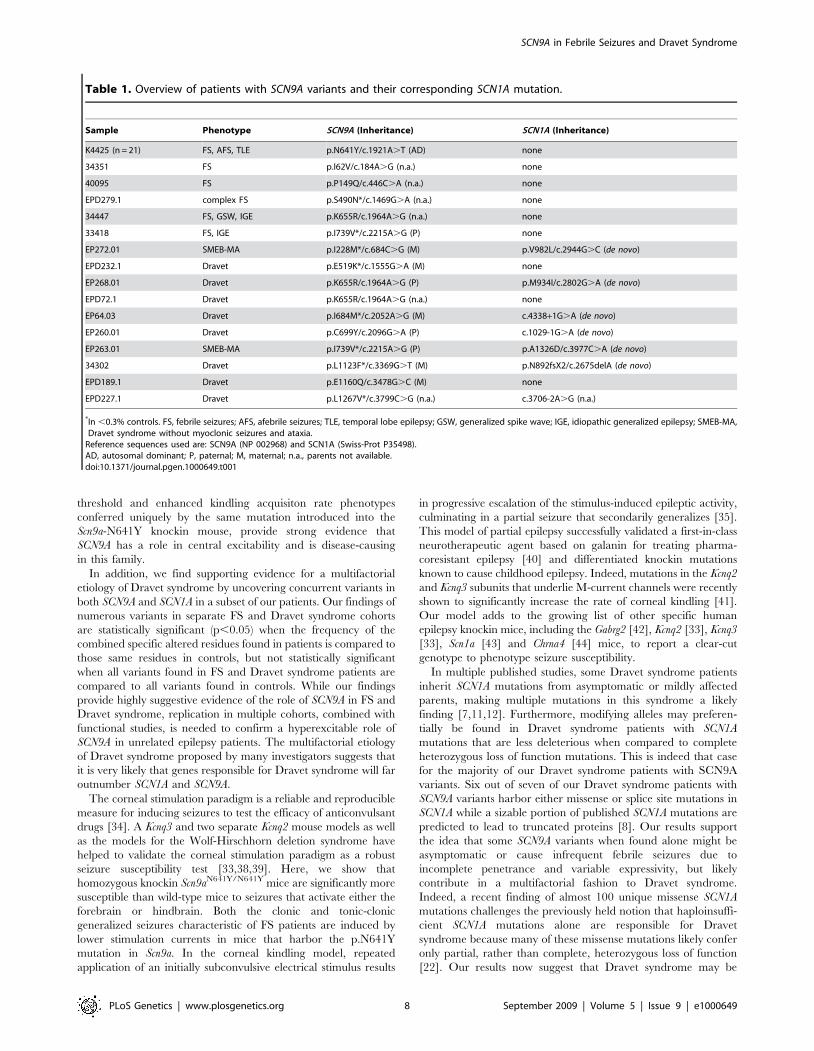
A follow-up study of a large Utah family with significant linkage to chromosome 2q24 led us to identify a new febrile seizure(FS) gene, SCN9A encoding Nav1.7. In 21 affected members, we uncovered a potential mutation in a highly conserved aminoacid, p.N641Y, in the large cytoplasmic loop between transmembrane domains I and II that was absent from 586 ethnicallymatched population control chromosomes. To establish a functional role for this mutation in seizure susceptibility, weintroduced the orthologous mutation into the murine Scn9a ortholog using targeted homologous recombination.Compared to wild-type mice, homozygous Scn9aN641Y/N641Y knockin mice exhibit significantly reduced thresholds toelectrically induced clonic and tonic-clonic seizures, and increased corneal kindling acquisition rates. Together, these datastrongly support the SCN9A p.N641Y mutation as disease-causing in this family. To confirm the role of SCN9A in FS, weanalyzed a collection of 92 unrelated FS patients and identified additional highly conserved Nav1.7 missense variants in 5%of the patients. After one of these children with FS later developed Dravet syndrome (severe myoclonic epilepsy of infancy),we sequenced the SCN1A gene, a gene known to be associated with Dravet syndrome, and identified a heterozygousframeshift mutation. Subsequent analysis of 109 Dravet syndrome patients yielded nine Nav1.7 missense variants (8% of thepatients), all in highly conserved amino acids. Six of these Dravet syndrome patients with SCN9A missense variants alsoharbored either missense or splice site SCN1A mutations and three had no SCN1A mutations. This study provides evidencefor a role of SCN9A in human epilepsies, both as a cause of FS and as a partner with SCN1A mutations.
Citation: Singh NA, Pappas C, Dahle EJ, Claes LRF, Pruess TH, et al. (2009) A Role of SCN9A in Human Epilepsies, As a Cause of Febrile Seizures and As a PotentialModifier of Dravet Syndrome. PLoS Genet 5(9): e1000649. doi:10.1371/journal.pgen.1000649
Editor: Wayne N. Frankel, The Jackson Laboratory, United States of America
Received May 14, 2009; Accepted August 14, 2009; Published September 18, 2009
Copyright: � 2009 Singh et al. This is an open-access article distributed under the terms of the Creative Commons Attribution License, which permitsunrestricted use, distribution, and reproduction in any medium, provided the original author and source are credited.
Funding: This work was supported in part by grants from the NIH (RO1 NS32666 to MFL), the Margolis Foundation (to MFL), the Keck Foundation (to MFL), andthe NCRR (UL1-RR025764). The funders had no role in study design, data collection and analysis, decision to publish, or preparation of the manuscript.
Competing Interests: The authors have declared that no competing interests exist.
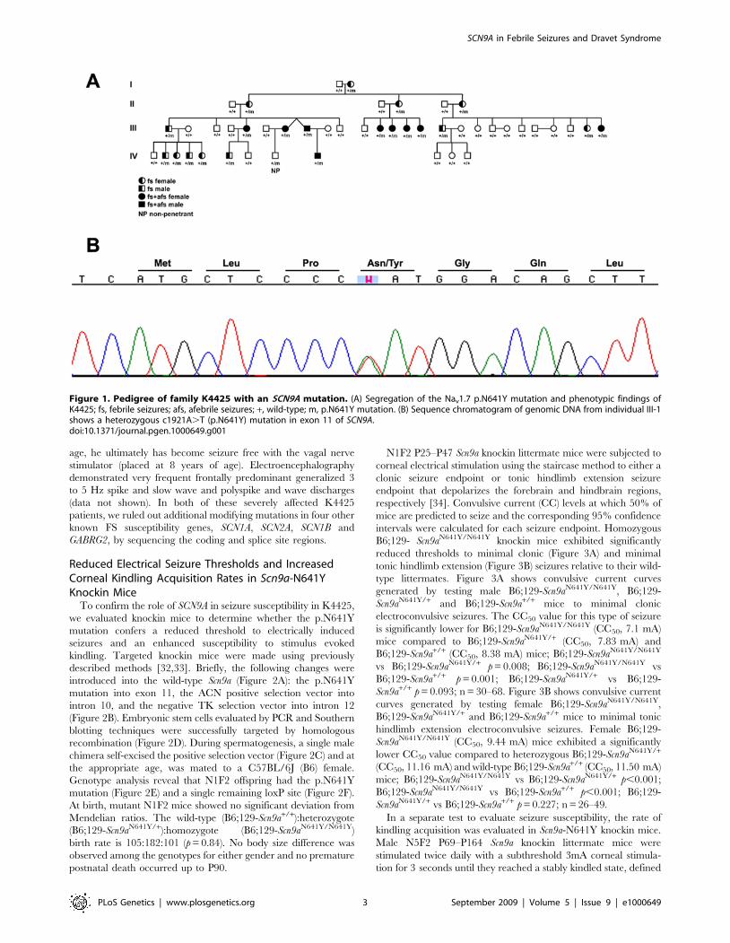
c.1921A.T) that cosegregates with all 21 affected K4425
individuals, in addition to a single non-penetrant individual (IV-
8) (Figure 1A and 1B). A non-penetrant individual is not
unexpected as they are commonly seen in autosomal dominant
diseases and are well documented in FS pedigrees. Inherited
autosomal dominant forms of FS have a reduced penetrance of
60–80% [29–31], meaning that 20–40% of individuals with
mutations who belong to FS families will not experience seizures.
The penetrance of FS in K4425 is actually rather high at
approximately 95%. The N641Y variant was absent from 586
chromosomes from an ethnically matched population of unrelated
individuals, providing supporting evidence for this nucleotide
change being the disease-causing mutation in this family.
Broad Clinical Spectrum of Seizures in K4425 Individualswith SCN9A-N641Y
A broad spectrum of seizure manifestations is observed in
K4425 family members who harbor the p.N641Y mutation [14].
Illustrating the milder end of the continuum are 11 individuals
from K4425 who experienced only FS before six years of age. The
remaining ten of the 21 affected individuals in K4425 experienced
FS before six years of age followed by later afebrile seizures. In
eight of these ten, the seizures remitted by the age of 16. Finally,
two individuals, III-14 and IV-9, developed intractable epilepsy.
Patient III-14 experienced her first simple FS at age 1.5 years
followed before age five by several non-febrile convulsions and at
least one prolonged generalized convulsive seizure lasting at least
45 minutes. After age five, she had occasional complex-partial
seizures and was diagnosed with left mesial temporal sclerosis at 22
years of age. At about one year of age, patient IV-9 began having
frequent simple FS without focal onset and never lasting more
than 2 minutes. However, he had as many as 60 such seizures until
about 4–5 years of age. Afebrile generalized convulsive seizures
began at about 6 years of age followed closely by very frequent
typical absence seizures. He has never had prolonged convulsions,
hemiclonic or secondarily generalized seizures, drop attacks,
myoclonic or astatic seizures, or ‘‘atypical absence’’ episodes,
and there has been no developmental regression. Now 11 years of
Author Summary
Febrile seizures are the most common seizure disorder ofearly childhood, and exhibit a prevalence of 2%–5% inEuropean and North American children. While the geneticbasis of febrile seizures is well-documented, efforts touncover these genes have yielded only a few genes in asmall proportion of cases. In a genomic region on humanchromosome 2 known to harbor the febrile seizure SCN1Asodium channel gene, we now report a disease-causingmutation in the adjacent gene, SCN9A (Nav1.7), in a largefamily with febrile seizures. We introduced the familymutation (N641Y) into the orthologous mouse gene tocreate a knockin mouse model, and tested seizuresusceptibility in these mice. Compared to wild-type mice,our Scn9a knockin mice have a significantly lowerthreshold to electrically induced seizures and experienceseizures at a significantly faster rate with repeatedsubthreshold stimulation. We also report novel missenseSCN9A mutations in unrelated febrile seizure patients.Furthermore, we show that a subset of patients with thecatastrophic early-onset Dravet syndrome who commonlyhave mutations in SCN1A also harbor mutations in SCN9A.This finding is important as it demonstrates for the firsttime mutational evidence for a modifying digenic mech-anism of human epilepsy. For infants with Dravetsyndrome, a genetic diagnosis will be of immediatebenefit to guide therapeutics away from the sodiumchannel blocking class of anticonvulsant drugs thatexacerbate seizures but are often the first administered.
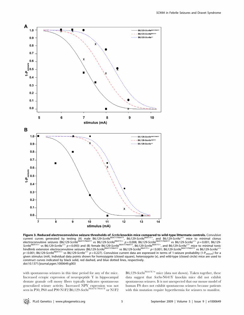
Scn9aN641Y/N641Y (CC50, 9.44 mA) mice exhibited a significantly
lower CC50 value compared to heterozygous B6;129-Scn9aN641Y/+
(CC50, 11.16 mA) and wild-type B6;129-Scn9a+/+ (CC50, 11.50 mA)
mice; B6;129-Scn9aN641Y/N641Y vs B6;129-Scn9aN641Y/+ p,0.001;
B6;129-Scn9aN641Y/N641Y vs B6;129-Scn9a+/+ p,0.001; B6;129-
Scn9aN641Y/+ vs B6;129-Scn9a+/+ p = 0.227; n = 26–49.
In a separate test to evaluate seizure susceptibility, the rate of
kindling acquisition was evaluated in Scn9a-N641Y knockin mice.
Male N5F2 P69–P164 Scn9a knockin littermate mice were
stimulated twice daily with a subthreshold 3mA corneal stimula-
tion for 3 seconds until they reached a stably kindled state, defined
Figure 1. Pedigree of family K4425 with an SCN9A mutation. (A) Segregation of the Nav1.7 p.N641Y mutation and phenotypic findings ofK4425; fs, febrile seizures; afs, afebrile seizures; +, wild-type; m, p.N641Y mutation. (B) Sequence chromatogram of genomic DNA from individual III-1shows a heterozygous c1921A.T (p.N641Y) mutation in exon 11 of SCN9A.doi:10.1371/journal.pgen.1000649.g001
seizure for B6.129-Scn9aN641Y/N641Y mice compared to B6.129-
Scn9aN641Y/+ and B6;129-Scn9a+/+ mice, respectively.
Two homozygous mutant female mice were video-monitored
continuously from P33–P47 and three homozygous mutant male
mice were continuously video-monitored from P27–P50 during
the 12-hour daylight cycle. We did not observe any behavior, such
as rearing and falling or forelimb or hindlimb clonus, consistent
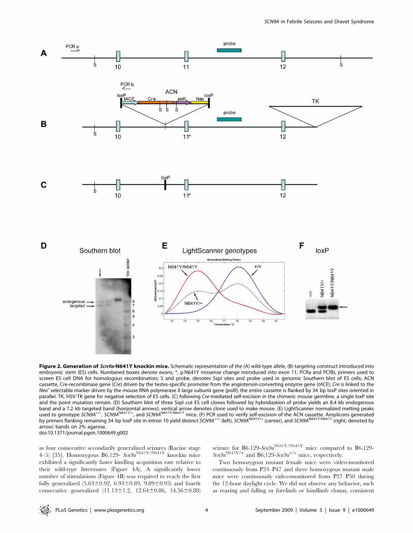
Figure 2. Generation of Scn9a-N641Y knockin mice. Schematic representation of the (A) wild-type allele, (B) targeting construct introduced intoembryonic stem (ES) cells. Numbered boxes denote exons; *, p.N641Y missense change introduced into exon 11; PCRa and PCRb, primers used toscreen ES cell DNA for homologous recombination; S and probe, denotes SspI sites and probe used in genomic Southern blot of ES cells; ACNcassette, Cre-recombinase gene (Cre) driven by the testes-specific promoter from the angiotensin-converting enzyme gene (tACE); Cre is linked to theNeor selectable marker driven by the mouse RNA polymerase II large subunit gene (polII); the entire cassette is flanked by 34 bp loxP sites oriented inparallel. TK, HSV-TK gene for negative selection of ES cells. (C) following Cre-mediated self-excision in the chimeric mouse germline, a single loxP siteand the point mutation remain. (D) Southern blot of three SspI cut ES cell clones followed by hybridization of probe yields an 8.4 kb endogenousband and a 7.2 kb targeted band (horizontal arrows); vertical arrow denotes clone used to make mouse. (E) LightScanner normalized melting peaksused to genotype SCN9A+/+, SCN9AN641Y/+, and SCN9AN641Y/N641Y mice. (F) PCR used to verify self-excision of the ACN cassette. Amplicons generatedby primers flanking remaining 34 bp loxP site in intron 10 yield distinct SCN9A+/+ (left), SCN9AN641Y/+ (center), and SCN9AN641Y/N641Y (right, denoted byarrow) bands on 2% agarose.doi:10.1371/journal.pgen.1000649.g002
with spontaneous seizures in this time period for any of the mice.
Increased ectopic expression of neuropeptide Y in hippocampal
dentate granule cell mossy fibers typically indicates spontaneous
generalized seizure activity. Increased NPY expression was not
seen in P30, P60 and P90 N1F2 B6;129-Scn9aN641Y/N641Y or N1F2
B6;129-Scn9aN641Y/+ mice (data not shown). Taken together, these
data suggest that Scn9a-N641Y knockin mice did not exhibit
spontaneous seizures. It is not unexpected that our mouse model of
human FS does not exhibit spontaneous seizures because patients
with this mutation require hyperthermia for seizures to manifest.
Figure 3. Reduced electroconvulsive seizure thresholds of Scn9a knockin mice compared to wild-type littermate controls. Convulsivecurrent curves generated by testing (A) male B6;129-Scn9aN641Y/N641Y, B6;129-Scn9aN641Y/+, and B6;129-Scn9a+/+ mice to minimal clonuselectroconvulsive seizures (B6;129-Scn9aN641Y/N641Y vs B6;129-Scn9aN641Y/+ p = 0.008; B6;129-Scn9aN641Y/N641Y vs B6;129-Scn9a+/+ p = 0.001; B6;129-Scn9aN641Y/+ vs B6;129-Scn9a+/+ p = 0.093) and (B) female B6;129-Scn9aN641Y/N641Y, B6;129-Scn9aN641Y/+, and B6;129-Scn9a+/+ mice to minimal tonichindlimb extension electroconvulsive seizures (B6;129-Scn9aN641Y/N641Y vs B6;129-Scn9aN641Y/+ p,0.001; B6;129-Scn9aN641Y/N641Y vs B6;129-Scn9a+/+
p,0.001; B6;129-Scn9aN641Y/+ vs B6;129-Scn9a+/+ p = 0.227). Convulsive current data are expressed in terms of 1-seizure probability (1-Pseizure) for agiven stimulus (mA). Individual data points shown for homozygote (closed square), heterozygote (x), and wild-type (closed circle) mice are used toconstruct curves indicated by black solid, red dashed, and blue dotted lines, respectively.doi:10.1371/journal.pgen.1000649.g003
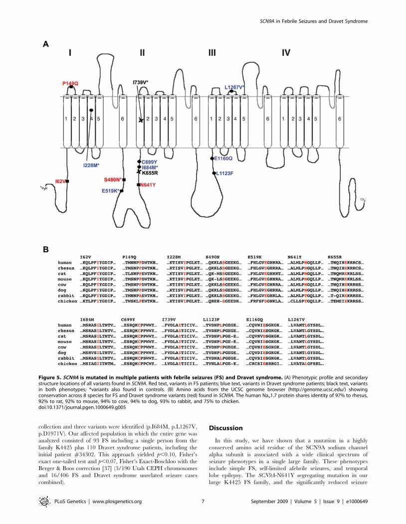
Expanding the Role of SCN9A in Unrelated Febrile SeizurePatients
To further assess the role of Nav1.7 in FS patients, we then
analyzed SCN9A in a panel of 92 unrelated patients with childhood
seizures occurring in the setting of a febrile illness, either with or
without a family history of seizures. We identified 4 additional
missense variants in our 90 Caucasian samples and 1 variant in
our 2 Hispanic samples (Figure 5A, Table 1). p.P149Q and
p.K655R were not found in at least 562 ethnically matched
Caucasian population control chromosomes, while p.S490N and
p.I739V were found only once in at least 562 ethnically matched
Caucasian population control chromosomes. For all four Cauca-
sian mutations, Fisher’s exact two-tailed test yielded p = 0.03
(4/180 unrelated FS chromosomes and 2/562 population control
chromosomes). The single Hispanic variant p.I62V was not found
in 276 ethnically matched Hispanic control chromosomes (p = 0.01
for 1/4 FS chromosomes and 0/276 population control
chromosomes, Fisher’s exact two-tailed test). All five seizure-
associated Nav1.7 variants reported here occur in codons that are
highly conserved across species (Figure 5B). To rule out the role of
SCN1A in FS susceptibility in these 5 FS patients with SCN9A
variants, we sequenced the entire coding and splice site regions of
SCN1A and did not find any potential disease-causing amino acid
variations.
A Role for SCN9A in Dravet SyndromeDuring the course of our studies on unrelated FS patients, the
diagnosis of one patient #34302 progressed from atypical FS to
Dravet syndrome. Beginning at five months of age, this Caucasian
patient experienced multiple generalized clonic seizures that were
predominantly afebrile, then progressed to frequent episodes of
status epilepticus and prolonged complex partial seizures by 16
months. Now 5 years old, this patient continues to have mixed
seizures (including myoclonic and astatic seizures) in spite of
resolute therapeutic intervention. Sequencing of SCN9A yielded a
p.L1123F missense variant found only once in 1736 ethnically
matched population control chromosomes (Fisher’s exact p-
value = 0.0023; Figure 6, Table 1). Segregation analysis showed
that the SCN9A p.L1123F variant was inherited from the
asymptomatic mother with a reported extended family history of
seizures. Subsequent sequencing of the SCN1A gene (Swiss-Prot
P35498) known to cause Dravet syndrome uncovered a hetero-
zygous frameshift mutation (c.2675delA, p.N892fsX2) in the
intracellular loop between DIIS4 and DIIS5 (Figure 6, Table 1).
The SCN1A frameshift was de novo and misinheritance was ruled
out by testing 31 polymorphic microsatellite markers (data not
shown).
The finding of variants in both SCN1A and SCN9A in a single
patient led us to investigate whether additional disease-associated
alleles in SCN9A contribute to Dravet syndrome. In an analysis of a
cohort of 109 Dravet syndrome patients, 50% of whom had
SCN1A mutations, we found 8 additional SCN9A variants within
the transmembrane domains and intracellular and extracellular
loops of Nav1.7 in 9 patients (Table 1). The missense variants
p.C699Y, K655R and p.E1160Q were not found in at least 576
control chromosomes and the remaining 5 missense variants were
found in 0.3% of at least 576 control chromosomes (p = 0.004 for
9/218 Dravet syndrome cases and 5/576 population controls,
Fisher’s exact two-tailed test). Of the 9 Dravet syndrome patients
with SCN9A variants, six harbor either splice site or missense
mutations in SCN1A (Table 1, Figure 5). Two of these SCN9A
variants (p.K655R and p.I739V) are also found in our FS patients.
In the three remaining Dravet syndrome patients without SCN1A
mutations, additional proconvulsive genes that act in concert with
SCN9A may yet be uncovered. Protein secondary structure
prediction using Consensus Data Mining [36] found coil to a-
helix (p.I684M), a-helix to coil (p.I739V, p.L1123F, p.L1267V)
and ß-sheet to coil (p.E1160Q) alterations.
An alternate statistical approach is to examine the mutational
burden of SCN9A, comparing rare (,1%) variants identified by
mutational analysis of the entire coding region in all FS and Dravet
syndrome populations combined versus the entire coding region in
population control individuals. Analysis of the coding and splice site
regions of SCN9A was performed only in a subset of our control
panel consisting of 95 healthy individuals from the Utah CEPH
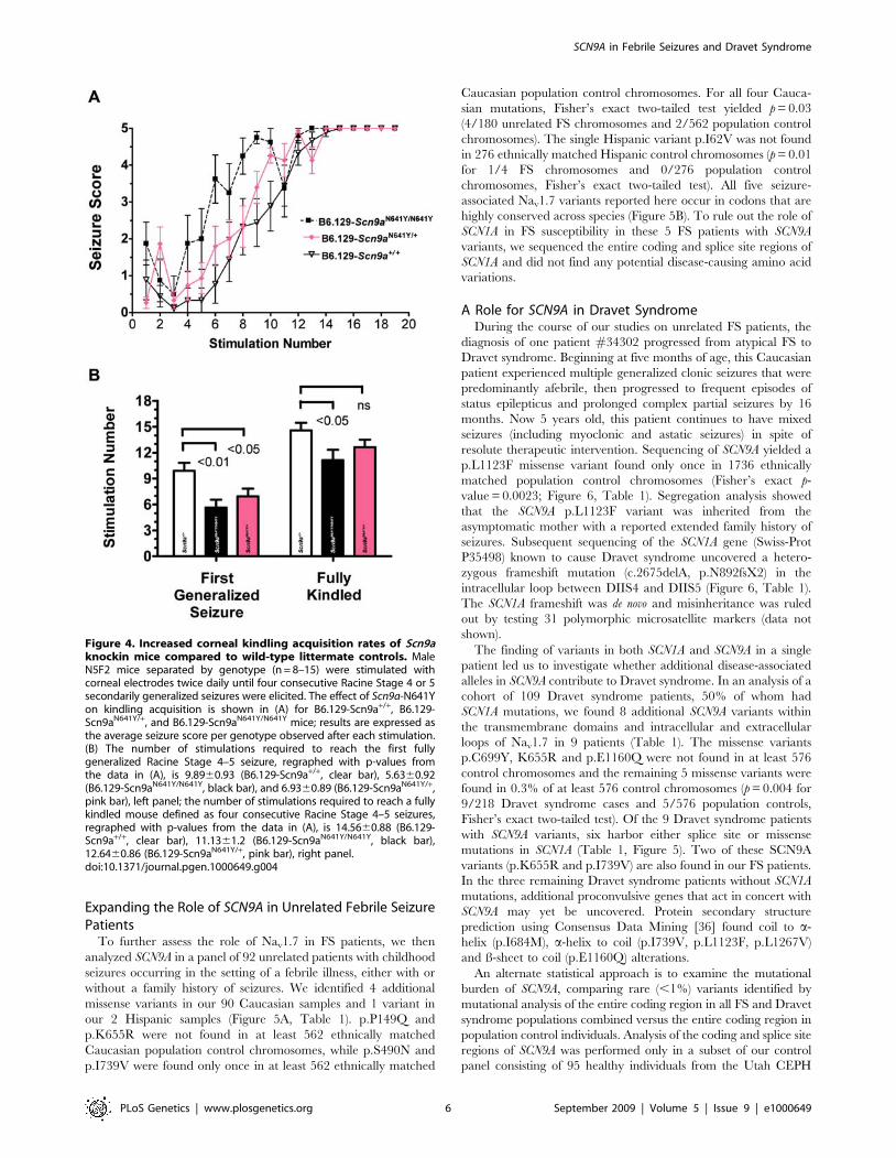
Figure 4. Increased corneal kindling acquisition rates of Scn9aknockin mice compared to wild-type littermate controls. MaleN5F2 mice separated by genotype (n = 8–15) were stimulated withcorneal electrodes twice daily until four consecutive Racine Stage 4 or 5secondarily generalized seizures were elicited. The effect of Scn9a-N641Yon kindling acquisition is shown in (A) for B6.129-Scn9a+/+, B6.129-Scn9aN641Y/+, and B6.129-Scn9aN641Y/N641Y mice; results are expressed asthe average seizure score per genotype observed after each stimulation.(B) The number of stimulations required to reach the first fullygeneralized Racine Stage 4–5 seizure, regraphed with p-values fromthe data in (A), is 9.8960.93 (B6.129-Scn9a+/+, clear bar), 5.6360.92(B6.129-Scn9aN641Y/N641Y, black bar), and 6.9360.89 (B6.129-Scn9aN641Y/+,pink bar), left panel; the number of stimulations required to reach a fullykindled mouse defined as four consecutive Racine Stage 4–5 seizures,regraphed with p-values from the data in (A), is 14.5660.88 (B6.129-Scn9a+/+, clear bar), 11.1361.2 (B6.129-Scn9aN641Y/N641Y, black bar),12.6460.86 (B6.129-Scn9aN641Y/+, pink bar), right panel.doi:10.1371/journal.pgen.1000649.g004
and 16/406 FS and Dravet syndrome unrelated seizure cases
combined).
Discussion
In this study, we have shown that a mutation in a highly
conserved amino acid residue of the SCN9A sodium channel
alpha subunit is associated with a wide clinical spectrum of
seizure phenotypes in a single large family. These phenotypes
include simple FS, self-limited afebrile seizures, and temporal
lobe epilepsy. The SCN9A-N641Y segregating mutation in our
large K4425 FS family, and the significantly reduced seizure
Figure 5. SCN9A is mutated in multiple patients with febrile seizures (FS) and Dravet syndrome. (A) Phenotypic profile and secondarystructure locations of all variants found in SCN9A. Red text, variants in FS patients; blue text, variants in Dravet syndrome patients; black text, variantsin both phenotypes; *variants also found in controls. (B) Amino acids from the UCSC genome browser (http://genome.ucsc.edu/) showingconservation across 8 species for FS and Dravet syndrome variants (red) found in SCN9A. The human Nav1.7 protein shares identity of 97% to rhesus,92% to rat, 92% to mouse, 94% to cow, 94% to dog, 93% to rabbit, and 75% to chicken.doi:10.1371/journal.pgen.1000649.g005
included in the list of disorders with ‘‘modifier’’ genes that include
Huntington’s disease and cystic fibrosis [45]. Additional new
functional data that examines the two gene mutations will be
required to test if the ‘‘two-hit’’ hypothesis is valid in certain
Dravet syndrome patients.
None of our FS or Dravet syndrome variants overlaps with the
SCN9A disease-associated changes found in the extreme pain or
insensitivity to pain disorders [24–26,46]. Furthermore, in all
published studies of PE, PEPD and CIP, an increased incidence of
seizures is not reported in patients with SCN9A mutations [24–26].
After follow-up questioning, none of the 21 affected members of
K4425 reported the easily recognized extreme pain phenotypes
associated with some SCN9A missense mutations. PEPD is often
misdiagnosed as epilepsy because tonic non-epileptic seizures are a
particular feature in infancy and early childhood. However the
‘‘slow-flat-slow’’ ictal EEG pattern associated with profound
syncope in PEPD patients is clearly not epileptiform, whereas
EEGs in K4425 patients are epileptiform [14,47]. Another
distinguishing feature is that PEPD attacks are provoked by
physical stimulation and not by hyperthermia as seen in FS.
The notion that dysfunction in the same ion channel can be
associated in distinct paroxysmal phenotypes is already known for
SCN9A. In 17 of 18 patients with SCN9A missense mutations
published to date, the rectal, ocular and mandibular pain seen in
PEPD does not overlap with the severe burning hand and foot
pain characteristic of PE [48]. We now extend the tissue specificity
of paroxysmal Nav1.7 malfunction to the central nervous system.
Additional support for discrete phenotypes resulting from the same
ion channel protein comes from the identification of SCN1A
mutations in either epilepsy or familial hemiplegic migraine [49],
and CACNA1A mutations in familial hemiplegic migraine, episodic
ataxia and spinocerebellar ataxia [50]. Experimentally, the Nav1.7
p.L858H PE mutation causes hyperexcitable sensory neurons and
hypoexcitable sympathetic neurons, and these opposing electrical
properties are a result of neuron specific physiologically coupled
proteins [51]. A unique complement of Nav1.7 interacting proteins
or second messenger pathways in the central nervous system may
also explain how the same gene previously implicated with
peripheral pain can also be associated with an epilepsy phenotype,
but this hypothesis will require further study.
Figure 6. Utah Dravet syndrome patient #34302 harbors mutations in both SCN9A and SCN1A. Sequence chromatograms of wild-type(top panel) and mutant (middle panel) clones of SCN1A exon 15 reveals a frameshift mutation p.N892fsX2 (c.2675delA); sequence chromatogram ofgenomic DNA shows a heterozygous p.L1123F (c.3369G.T) in exon 17 of SCN9A exon (bottom panel).doi:10.1371/journal.pgen.1000649.g006
favoring genetic heterogeneity for febrile convulsions. Epilepsia 41: 132–139.
4. Claes L, Ceulemans B, Audenaert D, Smets K, Lofgren A, et al. (2003) De novoSCN1A mutations are a major cause of severe myoclonic epilepsy of infancy.
Hum Mutat 21: 615–621.
5. Claes L, Del-Favero J, Ceulemans B, Lagae L, Van Broeckhoven C, et al. (2001)De novo mutations in the sodium-channel gene SCN1A cause severe myoclonic
epilepsy of infancy. Am J Hum Genet 68: 1327–1332.
6. Fujiwara T, Sugawara T, Mazaki-Miyazaki E, Takahashi Y, Fukushima K, et al.(2003) Mutations of sodium channel alpha subunit type 1 (SCN1A) in intractable
childhood epilepsies with frequent generalized tonic-clonic seizures. Brain 126:
531–546.
7. Nabbout R, Gennaro E, Dalla Bernardina B, Dulac O, Madia F, et al. (2003)Spectrum of SCN1A mutations in severe myoclonic epilepsy of infancy.
Neurology 60: 1961–1967.
8. Scheffer IE, Zhang YH, Jansen FE, Dibbens L (2009) Dravet syndrome orgenetic (generalized) epilepsy with febrile seizures plus? Brain Dev 31: 394–400.
9. Wallace RH, Hodgson BL, Grinton BE, Gardiner RM, Robinson R, et al.
(2003) Sodium channel alpha1-subunit mutations in severe myoclonic epilepsy ofinfancy and infantile spasms. Neurology 61: 765–769.
10. Gennaro E, Veggiotti P, Malacarne M, Madia F, Cecconi M, et al. (2003)
Familial severe myoclonic epilepsy of infancy: truncation of Nav1.1 and geneticheterogeneity. Epileptic Disord 5: 21–25.
11. Kanai K, Hirose S, Oguni H, Fukuma G, Shirasaka Y, et al. (2004) Effect of
localization of missense mutations in SCN1A on epilepsy phenotype severity.Neurology 63: 329–334.
SCN1A mutations and epilepsy. Hum Mutat 25: 535–542.
13. Scheffer IE, Berkovic SF (2003) The genetics of human epilepsy. TrendsPharmacol Sci 24: 428–433.
14. Peiffer A, Thompson J, Charlier C, Otterud B, Varvil T, et al. (1999) A locus for
febrile seizures (FEB3) maps to chromosome 2q23-24. Ann Neurol 46: 671–678.
15. Catterall WA (2000) From ionic currents to molecular mechanisms: the structureand function of voltage-gated sodium channels. Neuron 26: 13–25.
16. Escayg A, MacDonald BT, Meisler MH, Baulac S, Huberfeld G, et al. (2000)
Mutations of SCN1A, encoding a neuronal sodium channel, in two families with
GEFS+2. Nat Genet 24: 343–345.
17. Harkin LA, McMahon JM, Iona X, Dibbens L, Pelekanos JT, et al. (2007) The
spectrum of SCN1A-related infantile epileptic encephalopathies. Brain 130:
843–852.
18. Ito M, Shirasaka Y, Hirose S, Sugawara T, Yamakawa K (2004) Seizurephenotypes of a family with missense mutations in SCN2A. Pediatr Neurol 31:
150–152.
19. Herlenius E, Heron SE, Grinton BE, Keay D, Scheffer IE, et al. (2007) SCN2AMutations and Benign Familial Neonatal-Infantile Seizures: The Phenotypic
Spectrum. Epilepsia 48: 1138–1142.
20. Holland KD, Kearney JA, Glauser TA, Buck G, Keddache M, et al. (2008)Mutation of sodium channel SCN3A in a patient with cryptogenic pediatric
partial epilepsy. Neurosci Lett 433: 65–70.
21. Suls A, Claeys KG, Goossens D, Harding B, Van Luijk R, et al. (2006)Microdeletions involving the SCN1A gene may be common in SCN1A-
mutation-negative SMEI patients. Hum Mutat 27: 914–920.
22. Depienne C, Trouillard O, Saint-Martin C, An I, Bouteiller D, et al. (2008)Spectrum of SCN1A gene mutations associated with Dravet syndrome: analysis
of 333 patients. J Med Genet 46: 183–191.
23. Martin MS, Tang B, Ta N, Escayg A (2007) Characterization of 59 untranslated
regions of the voltage-gated sodium channels SCN1A, SCN2A, and SCN3A andidentification of cis-conserved noncoding sequences. Genomics 90: 225–235.
24. Cox JJ, Reimann F, Nicholas AK, Thornton G, Roberts E, et al. (2006) An
SCN9A channelopathy causes congenital inability to experience pain. Nature444: 894–898.
25. Fertleman CR, Baker MD, Parker KA, Moffatt S, Elmslie FV, et al. (2006)
SCN9A mutations in paroxysmal extreme pain disorder: allelic variants underliedistinct channel defects and phenotypes. Neuron 52: 767–774.
26. Yang Y, Wang Y, Li S, Xu Z, Li H, et al. (2004) Mutations in SCN9A, encoding
a sodium channel alpha subunit, in patients with primary erythermalgia. J MedGenet 41: 171–174.
27. Sangameswaran L, Fish LM, Koch BD, Rabert DK, Delgado SG, et al. (1997) Anovel tetrodotoxin-sensitive, voltage-gated sodium channel expressed in rat and
human dorsal root ganglia. J Biol Chem 272: 14805–14809.
28. Mechaly I, Scamps F, Chabbert C, Sans A, Valmier J (2005) Molecular diversityof voltage-gated sodium channel alpha subunits expressed in neuronal and non-
29. Johnson WG, Kugler SL, Stenroos ES, Meulener MC, Rangwalla I, et al. (1996)Pedigree analysis in families with febrile seizures. Am J Med Genet 61: 345–352.
30. Kugler SL, Johnson WG (1998) Genetics of the febrile seizure susceptibility trait.
Brain Dev 20: 265–274.
31. Wallace RH, Berkovic SF, Howell RA, Sutherland GR, Mulley JC (1996)Suggestion of a major gene for familial febrile convulsions mapping to 8q13-21.
J Med Genet 33: 308–312.
32. Bunting M, Bernstein KE, Greer JM, Capecchi MR, Thomas KR (1999)Targeting genes for self-excision in the germ line. Genes Dev 13: 1524–1528.
33. Singh NA, Otto JF, Dahle EJ, Pappas C, Leslie JD, et al. (2008) Mouse models of
human KCNQ2 and KCNQ3 mutations for benign familial neonatal
convulsions show seizures and neuronal plasticity without synaptic reorganiza-tion. J Physiol 586: 3405–3423.
34. Smith M, Wilcox KS, White HS (2007) Discovery of antiepileptic drugs.
Neurotherapeutics 4: 12–17.
35. Matagne A, Klitgaard H (1998) Validation of corneally kindled mice: a sensitivescreening model for partial epilepsy in man. Epilepsy Res 31: 59–71.
36. Cheng H, Sen TZ, Jernigan RL, Kloczkowski A (2007) Consensus Data Mining
(CDM) Protein Secondary Structure Prediction Server: combining GOR V andFragment Database Mining (FDM). Bioinformatics 23: 2628–2630.
37. Lydersen S, Fagerland MW, Laake P (2009) Recommended tests for association
in 262 tables. Stat Med 28: 1159–1175.
38. Naf D, Wilson LA, Bergstrom RA, Smith RS, Goodwin NC, et al. (2001) Mousemodels for the Wolf-Hirschhorn deletion syndrome. Hum Mol Genet 10: 91–98.
39. Yang Y, Beyer BJ, Otto JF, O’Brien TP, Letts VA, et al. (2003) Spontaneous
deletion of epilepsy gene orthologs in a mutant mouse with a lowelectroconvulsive threshold. Hum Mol Genet 12: 975–984.
40. White HS, Scholl EA, Klein BD, Flynn SP, Pruess TH, et al. (2009) Developing
Novel Antiepileptic Drugs: Characterization of NAX 5055, a Systemically-Active Galanin Analog, in Epilepsy Models. Neurotherapeutics 6: 372–380.
41. Otto JF, Singh NA, Dahle EJ, Leppert MF, Pappas CM, et al. (2009)
Electroconvulsive seizure thresholds and kindling acquisition rates are altered inmouse models of human Kcnq2 and Kcnq3 mutations for benign familial
neonatal convulsions. Epilepsia 50: 1752–1759.
42. Tan HO, Reid CA, Single FN, Davies PJ, Chiu C, et al. (2007) Reduced cortical
inhibition in a mouse model of familial childhood absence epilepsy. Proc NatlAcad Sci U S A 104: 17536–17541.
43. Ogiwara I, Miyamoto H, Morita N, Atapour N, Mazaki E, et al. (2007) Na(v)1.1
localizes to axons of parvalbumin-positive inhibitory interneurons: a circuit basisfor epileptic seizures in mice carrying an Scn1a gene mutation. J Neurosci 27:
5903–5914.
44. Klaassen A, Glykys J, Maguire J, Labarca C, Mody I, et al. (2006) Seizures andenhanced cortical GABAergic inhibition in two mouse models of human
autosomal dominant nocturnal frontal lobe epilepsy. Proc Natl Acad Sci U S A103: 19152–19157.
45. Gropman AL, Adams DR (2007) Atypical patterns of inheritance. Semin Pediatr
Neurol 14: 34–45.
46. Goldberg YP, MacFarlane J, MacDonald ML, Thompson J, Dube MP, et al.(2007) Loss-of-function mutations in the Nav1.7 gene underlie congenital
indifference to pain in multiple human populations. Clin Genet 71: 311–319.
47. Fertleman CR, Ferrie CD, Aicardi J, Bednarek NA, Eeg-Olofsson O, et al.
50. Gargus JJ (2009) Genetic calcium signaling abnormalities in the central nervoussystem: seizures, migraine, and autism. Ann N Y Acad Sci 1151: 133–156.
51. Rush AM, Dib-Hajj SD, Liu S, Cummins TR, Black JA, et al. (2006) A single
sodium channel mutation produces hyper- or hypoexcitability in different typesof neurons. Proc Natl Acad Sci U S A 103: 8245–8250.
52. Nagy A, Rossant J, Nagy R, Abramow-Newerly W, Roder JC (1993) Derivation
of completely cell culture-derived mice from early-passage embryonic stem cells.Proc Natl Acad Sci U S A 90: 8424–8428.