1035 Int. J. Morphol., 26(4):1035-1051, 2008. A Scanning Electron Microscopic Study on the Appendage Morphology of Astacus leptodactylus (Eschscholtz, 1823) and Pacifastacus leniusculus (Dana, 1852) (Crustacea: Decapoda: Astacoidea) Estudio en Microscopio Electrónico de Barrido sobre el Apéndice Morfológico del Astacus leptodactylus (Eschscholtz, 1823) y Pacifastacus leniusculus (Dana, 1852) (Crustacea: Decapoda: Astacoidea) Muzaffer Mustafa Harlioglu HARLIOGLU, M. M. A scanning electron microscopic study on the appendage morphology of Astacus leptodactylus (Eschscholtz, 1823) and Pacifastacus leniusculus (Dana, 1852) (Crustacea: Decapoda: Astacoidea). Int. J. Morphol., 26(4):1035-1051, 2008. SUMMARY: This study compares the morphology of rostrum, pereipods 1,2,4 and mouthparts of juvenile Astacus leptodactylus with those of Pacifastacus leniusculus. Differences in morphology were observed, in particular with regard to the mouthparts e.g. including setal armature and number of teeth on the mandible. In general, the shape of the rostra in the two species is similar in that both taper to a point with a pair of sharp spines distally. Laterally the rostrum of A. leptodactylus is bordered by a regular row of setae, which is not so well defined in P. leniusculus. The observations also showed that in addition to an increase in size, changes in morphology in the feeding apparatus between the developmental stages of the two species were present. It was concluded that both species have similar rostra, but different setal patterns and there are differences between the two species in the armature of mouthparts as development progresses. Therefore, important differences in the morphology of mouthparts between P. leniusculus and A. leptodactylus and in the different stages of the species might cause a difference in the feeding behavior and food choice of the species. KEY WORDS: Appendages; Astacus; Crayfish; Mouthparts; Pacifastacus; Scanning electron microscope. INTRODUCTION The signal crayfish, Pacifastacus leniusculus (Dana, 1852), is native to north-western North America inhabiting lakes, streams and rivers (Lowery & Holdich, 1988) as well as saline waters (Henry & Wheatly, 1988; Holdich et al., 1997). It is also tolerant to environmental extremes such as temperature and various pollutants (Firkins, 1993). Pacifastacus leniusculus is similar in many ways to Astacus astacus (Linnaeus, 1758) and consequently has proved po- pular alternative since the decline of the latter species due to the introduction crayfish plague into many Europe catchments (Holdich, 1999). The signal crayfish is identified by its red colour, and large robust chela including a white to turquoise patch on the claw gives this crayfish its common name. This North American crayfish has been introduced into many European watersheds. The narrow-clawed crayfish, Astacus leptodactylus (Eschscoltz, 1823) is a native of Turkey and near East Europe. It occupies a similar niche to that of P. leniusculus. However, in addition to streams, rivers, lakes, and ponds, it also inhabits swamps. Thus, it is sometimes called the swamp crayfish (Cherkashina, 1975; Köksal 1988, Harlioglu & Harlioglu, 2004). Astacus leptodactylus is commercially exploited either as imports or from introduced populations distributed widely across Europe, exception of the Iberian Peninsula and Nordic countries (Harlioglu & Holdich, 2001; Holdich 2002; Skurdal & Taugbol, 2002). This species is identified by its long claws (mainly typical for males) and is commonly named the narrow-clawed or Turkish crayfish (Harlioglu, 1996). Pacificastacus leniusculus and A. leptodactylus have become popular experimental animals. The Turkish crayfish has been subjected to biochemical, physiological and ecological studies where as physiology, ecology, reproduction and distribution have been subjects for P. leniusculus (Duvic & Soderhall, 1990; Firkins, 1993; Guan, This study is a part of PhD study of M.M. Harlıog˘lu supported by Fırat University Elazıg˘, TURKEY.
Transcript
1035
Int. J. Morphol.,26(4):1035-1051, 2008.
A Scanning Electron Microscopic Study on the AppendageMorphology of Astacus leptodactylus (Eschscholtz, 1823) andPacifastacus leniusculus (Dana, 1852) (Crustacea: Decapoda:
Astacoidea)
Estudio en Microscopio Electrónico de Barrido sobre el Apéndice Morfológico del Astacus leptodactylus(Eschscholtz, 1823) y Pacifastacus leniusculus (Dana, 1852) (Crustacea: Decapoda: Astacoidea)
Muzaffer Mustafa Harlioglu
HARLIOGLU, M. M. A scanning electron microscopic study on the appendage morphology of Astacus leptodactylus (Eschscholtz,1823) and Pacifastacus leniusculus (Dana, 1852) (Crustacea: Decapoda: Astacoidea). Int. J. Morphol., 26(4):1035-1051, 2008.
SUMMARY: This study compares the morphology of rostrum, pereipods 1,2,4 and mouthparts of juvenile Astacus leptodactyluswith those of Pacifastacus leniusculus. Differences in morphology were observed, in particular with regard to the mouthparts e.g. includingsetal armature and number of teeth on the mandible. In general, the shape of the rostra in the two species is similar in that both taper to apoint with a pair of sharp spines distally. Laterally the rostrum of A. leptodactylus is bordered by a regular row of setae, which is not sowell defined in P. leniusculus. The observations also showed that in addition to an increase in size, changes in morphology in the feedingapparatus between the developmental stages of the two species were present. It was concluded that both species have similar rostra, butdifferent setal patterns and there are differences between the two species in the armature of mouthparts as development progresses.Therefore, important differences in the morphology of mouthparts between P. leniusculus and A. leptodactylus and in the different stagesof the species might cause a difference in the feeding behavior and food choice of the species.
KEY WORDS: Appendages; Astacus; Crayfish; Mouthparts; Pacifastacus; Scanning electron microscope.
INTRODUCTION
The signal crayfish, Pacifastacus leniusculus (Dana,1852), is native to north-western North America inhabitinglakes, streams and rivers (Lowery & Holdich, 1988) as wellas saline waters (Henry & Wheatly, 1988; Holdich et al.,1997). It is also tolerant to environmental extremes such astemperature and various pollutants (Firkins, 1993).Pacifastacus leniusculus is similar in many ways to Astacusastacus (Linnaeus, 1758) and consequently has proved po-pular alternative since the decline of the latter species due tothe introduction crayfish plague into many Europecatchments (Holdich, 1999). The signal crayfish is identifiedby its red colour, and large robust chela including a white toturquoise patch on the claw gives this crayfish its commonname. This North American crayfish has been introducedinto many European watersheds.
The narrow-clawed crayfish, Astacus leptodactylus(Eschscoltz, 1823) is a native of Turkey and near East Europe.
It occupies a similar niche to that of P. leniusculus. However,in addition to streams, rivers, lakes, and ponds, it also inhabitsswamps. Thus, it is sometimes called the swamp crayfish(Cherkashina, 1975; Köksal 1988, Harlioglu & Harlioglu,2004). Astacus leptodactylus is commercially exploited eitheras imports or from introduced populations distributed widelyacross Europe, exception of the Iberian Peninsula and Nordiccountries (Harlioglu & Holdich, 2001; Holdich 2002; Skurdal& Taugbol, 2002). This species is identified by its long claws(mainly typical for males) and is commonly named thenarrow-clawed or Turkish crayfish (Harlioglu, 1996).
Pacificastacus leniusculus and A. leptodactylus havebecome popular experimental animals. The Turkish crayfishhas been subjected to biochemical, physiological andecological studies where as physiology, ecology,reproduction and distribution have been subjects for P.leniusculus (Duvic & Soderhall, 1990; Firkins, 1993; Guan,
This study is a part of PhD study of M.M. Harlıog˘lu supported by Fırat University Elazıg˘, TURKEY.
1036
1995; Warner & Green, 1995; Harlioglu, 1996). The mainreason for these researches is because the signal crayfishhas been introduced into most European countries foraquacultural and wild-stocking purposes (Lowery &Holdich). However, in recent years there has been an increasein the number of studies concerning aquaculture andimmunology of P. leniusculus as the species carries crayfishplague. Consequently, many investigators have concentratedon its immune system (e.g. Persson et al., 1987; Duvic &Soderhall; Aspan & Söderhall, 1991; Kopacek et al., 1993)and the implications for susceptible crayfish when thisspecies has been translocated (Holdich & Reeve, 1991).
Descriptions of P. leniusculus and A. leptodactylushave been given in a few keys (Curra, 1967; Pennack, 1978;Laurent & Forest, 1979; Brodski, 1983; Gledhill et al., 1993;Vigneux et al., 1993) and Andrews (1907) illustrated theappendage structure of the signal crayfish. Recently, P.leniusculus was chosen as a model in the description offunctional morphology of crayfish by Holdich & Reeve(1988), and Holdich (1992) also reviewed the early post-embryonic development of astacid, cambarid and parastacidcrayfish. However, to date no scanning electron microscopicstudies have highlighted the morphology and appendagestructure of these two species in any detail.
Differences in the morphology and structure ofmouthparts and pereiopods of crayfish are useful in classifyingthem for systematic purposes. For example, variations in therostra, chelae, mandibles and third maxillipeds were used byHobbs (1987) to classify species of Astacoides. Variations inchelae were also considered to determine taxonomicdifferences between Orconectes propinquus and O. obscurusby Tierney (1982). Morphological differences of the westernNorth American crayfish species of Pacifastacus have beenused to assign species into the subgenera Pacifastacus andHobbsastacus (Bouchard, 1977).
Scanning electron microscopy has been used to studythe external morphology of mouthparts, pereiopods andpleopods in a variety of decapod crustaceans. For example,the functional morphology of mouthparts and pereiopods hasbeen observed in the Norway lobster Nephrops norvegicusby Farmer (1974) and the shrimp Atya innocous byFelgenhauer & Abele (1983); in the prawn Panaeusmerguiensis by Hindley & Alexander (1978) and Alexanderet al. (1980). Moreover, observations on the morphologyand structure of feeding apparatus and distribution of setaein lobsters have mainly reported for Homarus americanusand H. gammarus (Solon & Cobb, 1980). In addition to these,there are also some studies on anostracan crustaceans. Forexample, Mura & Caldo (1993) have studied the structureof the molar surface of the mandibles in the shrimp
Branchinella spinosa. A similar study has been carried outon brine shrimp by Tyson & Sullivan (1981).
The aim of the present study is to compare thedifferences and similarities in the morphology of appendages(rostrum, carapace, first pereiopod, second pereiopod, fourthpereiopod) and mouthparts (third maxilliped, secondmaxilliped, first maxilliped, mandible, maxillule and maxilla)of A. leptodactylus and P. leniusculus under the scanningelectron microscope. In addition, the development ofmouthparts in stage 1, 2 and 3 juveniles of the two specieswas also evaluated.
MATERIAL AND METHOD
Juveniles of different stages (first, second and third)and juveniles 12 mm in carapace length (CL) of P. leniusculusand A. leptodactylus were scanned. The samples were rearedunder laboratory conditions in clean containers instead ofkeeping them in concrete tanks which have a muddy floorwould therefore make them dirty and unsuitable forphotography. After individuals had moulted, they weresacrificed when their body became hard enough to dissectand before they lost any appendages. To sacrifice the samples,they were placed in a freezer for ten minutes. Then they werepreserved in 70 percent alcohol.
Mouthparts and appendages were removed under alight microscope. After dissection, the selected body partsof 12 mm length (carapace) juveniles were air dried forapproximately 12 hours at room temperature (18 ± 1 0C).Because the stage 1, 2 and 3 juveniles were too delicate toapply air drying technique, the critical-point drying techniquewas applied for them.
Then, the selected appendages were attached toaluminum stubs with silver colloidal paint and coated with goldusing a Polaron Sputter Coating Unit E5100. Finally, to viewthe body parts the stubs were set up in a JSM-840 scanningelectron microscope operated at either 10, 15 or 25 KV.
The terminology used is taken from Holdich & Reeve(1988). The segments of the appendages are named fromattachment point as: coxa, basis, ischium, merus, carpus,propodus, and dactylus with a terminal unguis.
To show differences and similarities in the rostrum,carapace, second pereiopod, fourth pereiopod, third maxilliped,second maxilliped, first maxilliped and mandible between P.leniusculus and A. leptodactylus, 12 mm (carapace length)juveniles were compared. Third stage juveniles were also used
HARLIOGLU, M. M. A scanning electron microscopic study on the appendage morphology of Astacus leptodactylus (Eschscholtz, 1823) and Pacifastacus leniusculus (Dana, 1852) (Crustacea:Decapoda: Astacoidea). Int. J. Morphol., 26(4):1035-1051, 2008.
1037
to show differences in the first pereiopod (cheliped) betweenthe two species. The development of stage 1, 2, 3 and 12 mm(CL) juveniles was also evaluated within and between the twospecies (except the maxilla of stage 1 juveniles).
Due to the delicate nature of the mouthparts andappendages of juveniles it proved sometimes very difficultto prepare them for scanning electron microscopy.Consequently, some of the photographs exhibit charging(brightness). In the description below only the differencesand similarities which were apparent between the two speciesare described.
RESULTS
Differences and similarities in the morphology ofappendages and mouthparts: Differences and similaritiesin the morphology of appendages and mouthparts were foundbetween the species.
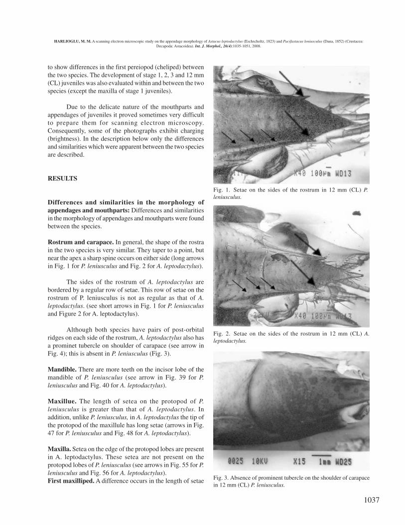
Rostrum and carapace. In general, the shape of the rostrain the two species is very similar. They taper to a point, butnear the apex a sharp spine occurs on either side (long arrowsin Fig. 1 for P. leniusculus and Fig. 2 for A. leptodactylus).
The sides of the rostrum of A. leptodactylus arebordered by a regular row of setae. This row of setae on therostrum of P. leniusculus is not as regular as that of A.leptodactylus. (see short arrows in Fig. 1 for P. leniusculusand Figure 2 for A. leptodactylus).
Although both species have pairs of post-orbitalridges on each side of the rostrum, A. leptodactylus also hasa prominet tubercle on shoulder of carapace (see arrow inFig. 4); this is absent in P. leniusculus (Fig. 3).
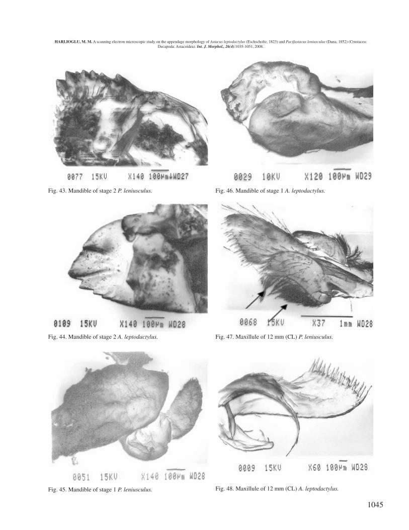
Mandible. There are more teeth on the incisor lobe of themandible of P. leniusculus (see arrow in Fig. 39 for P.leniusculus and Fig. 40 for A. leptodactylus).
Maxillue. The length of setea on the protopod of P.leniusculus is greater than that of A. leptodactylus. Inaddition, unlike P. leniusculus, in A. leptodactylus the tip ofthe protopod of the maxillule has long setae (arrows in Fig.47 for P. leniusculus and Fig. 48 for A. leptodactylus).
Maxilla. Setea on the edge of the protopod lobes are presentin A. leptodactylus. These setea are not present on theprotopod lobes of P. leniusculus (see arrows in Fig. 55 for P.leniusculus and Fig. 56 for A. leptodactylus).First maxilliped. A difference occurs in the length of setae
Fig. 1. Setae on the sides of the rostrum in 12 mm (CL) P.leniusculus.
Fig. 2. Setae on the sides of the rostrum in 12 mm (CL) A.leptodactylus.
Fig. 3. Absence of prominent tubercle on the shoulder of carapacein 12 mm (CL) P. leniusculus.
HARLIOGLU, M. M. A scanning electron microscopic study on the appendage morphology of Astacus leptodactylus (Eschscholtz, 1823) and Pacifastacus leniusculus (Dana, 1852) (Crustacea:Decapoda: Astacoidea). Int. J. Morphol., 26(4):1035-1051, 2008.
1038
on the exopod. Bigger setae are present on the exopod of P.leniusculus (Fig. 31 for P. leniusculus and Fig. 32 for A.leptodactylus).
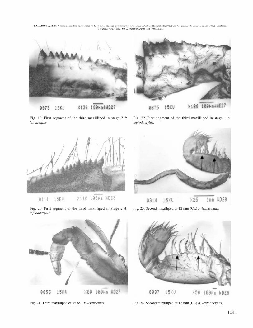
Second maxilliped. Compared to A. leptodactylus, moreabundant and very close setae are present on the endopod ofP. leniusculus (see arrows in Fig. 23 for P. leniusculus andFig. 24 for A. leptodactylus).
Third maxilliped. A spine is present on the second and thirdsegments of the third maxilliped in A. leptodactylus (seearrows in Fig. 13). These spines are not present in P.leniusculus (Fig. 14).
The number and distribution of teeth are also differenton the crista dentata (first segment) of the two species. Thereare more teeth on the crista dentata of P. leniusculus (seearrows in Fig. 15 for P. leniusculus and Fig. 16 for A.leptodactylus).
Cheliped (first pereiopod). The gap between the dactylusand propodus of the cheliped is wide in A. leptodactylus andthe margins are serrated. The gap is narrower in P. leniusculusand the inner edges of the dactylus and propodus are not soserrated (see arrows in Fig. 5 for P. leniusculus and Fig. 6for A. leptodactylus).
There is a row of setae on the propodus of the chelipedin A. leptodactylus (see long arrows in Fig. 8), but there isno a row of setae on the cheliped in P. leniusculus (Fig. 7).
The carpus of P. leniusculus has setae, but that of A.leptodactylus has not. Also, the shape of the spine on theedge of the carpus is different in the two species, that of A.leptodactylus being much larger (Fig. 7 for P. leniusculusand Fig. 8 for A. leptodactylus, see short arrow).
Second pereiopod. A difference was found regarding thepropodus and dactylus between the species. Although A.leptodactylus has an unguis at the tip of propodus anddactylus, this unguis does not appear in P. leniusculus (Fig.9 for P. leniusculus and Fig. 10 for A. leptodactylus, seearrow).
Fourth pereiopod. More abundant setae are present on theventral side of the propodus of A. leptodactylus than thoseof P. leniusculus (see arrow in Fig. 11 for P. leniusculus andFig. 12 for A. leptodactylus).
Juvenile development of Pacifastacus leniusculus andAstacus leptodactylus: In addition to an increase in size,developmental changes in morphology in the feedingapparatus of P. leniusculus and A. leptodactylus were found
Fig 4. Presence of prominent tubercle on the shoulder of carapacein 12 mm (CL) A. leptodactylus
Fig. 5. Dactylus and propodus of the first pereopod in stage 3 P.leniusculus
Fig. 6. Dactylus and propodus of the first pereopod in stage 3 A.leptodactylus.
HARLIOGLU, M. M. A scanning electron microscopic study on the appendage morphology of Astacus leptodactylus (Eschscholtz, 1823) and Pacifastacus leniusculus (Dana, 1852) (Crustacea:Decapoda: Astacoidea). Int. J. Morphol., 26(4):1035-1051, 2008.
1039
Fig. 7. Propodus and carpus of the first pereopod in stage 3 P.leniusculus.
Fig. 8. Propodus and carpus of the first pereopod in stage 3 A.leptodactylus.
Fig. 9. Propodus and dactylus of second pereopod in 12 mm (CL)P. leniusculus.
Fig. 10. Propodus and dactylus of second pereopod in 12 mm (CL)A. leptodactylus.
Fig. 11. Ventral side of propodus of fourth pereopod in 12 mm(CL) P. leniusculus.
Fig. 12. Ventral side of propodus of fourth pereopod in 12 mm(CL) A. leptodactylus.
HARLIOGLU, M. M. A scanning electron microscopic study on the appendage morphology of Astacus leptodactylus (Eschscholtz, 1823) and Pacifastacus leniusculus (Dana, 1852) (Crustacea:Decapoda: Astacoidea). Int. J. Morphol., 26(4):1035-1051, 2008.
1040
Fig. 13. Second, third, fourth and fifth segment of the thirdmaxilliped in 12 mm A. leptodactylus.
Fig. 14. Second, third, fourth and fifth segment of the thirdmaxilliped in 12 mm P. leniusculus.
Fig. 15. First segment (crista dentata) of the third maxilliped in 12mm P. leniusculus.
Fig. 16.First segment (crista dentata) of the third maxilliped in 12mm A. leptodactylus.
Fig. 17. First segment of the third maxilliped in stage 3 P.leniusculus.
Fig. 18. First segment of the third maxilliped in stage 3 A.leptodactylus.
HARLIOGLU, M. M. A scanning electron microscopic study on the appendage morphology of Astacus leptodactylus (Eschscholtz, 1823) and Pacifastacus leniusculus (Dana, 1852) (Crustacea:Decapoda: Astacoidea). Int. J. Morphol., 26(4):1035-1051, 2008.
1041
Fig. 19. First segment of the third maxilliped in stage 2 P.leniusculus.
Fig. 20. First segment of the third maxilliped in stage 2 A.leptodactylus.
Fig. 21. Third maxilliped of stage 1 P. leniusculus.
Fig. 22. First segment of the third maxilliped in stage 1 A.leptodactylus.
Fig. 23. Second maxilliped of 12 mm (CL) P. leniusculus.
Fig. 24. Second maxilliped of 12 mm (CL) A. leptodactylus.
HARLIOGLU, M. M. A scanning electron microscopic study on the appendage morphology of Astacus leptodactylus (Eschscholtz, 1823) and Pacifastacus leniusculus (Dana, 1852) (Crustacea:Decapoda: Astacoidea). Int. J. Morphol., 26(4):1035-1051, 2008.
1042
Fig. 25. Second maxilliped of stage 3 P. leniusculus.
Fig. 26. Second maxilliped of stage 3 A. leptodactylus.
Fig. 27. Second maxilliped of stage 2 P. leniusculus.
Fig. 28. Second maxilliped of stage 2 A. leptodactylus.
Fig. 29. Second maxilliped of stage 1 P. leniusculus
Fig. 30. Second maxilliped of stage 1 A. leptodactylus
HARLIOGLU, M. M. A scanning electron microscopic study on the appendage morphology of Astacus leptodactylus (Eschscholtz, 1823) and Pacifastacus leniusculus (Dana, 1852) (Crustacea:Decapoda: Astacoidea). Int. J. Morphol., 26(4):1035-1051, 2008.
1043
Fig. 31. First maxilliped of 12 mm (CL) P. leniusculus.
Fig. 32. First maxilliped of 12 mm (CL) A. leptodactylus.
Fig. 33. First maxilliped of stage 3 P. leniusculus.
Fig. 34. First maxilliped of stage 3 A. leptodactylus.
Fig. 35. First maxilliped of stage 2 P. leniusculus.
Fig. 36. First maxilliped of stage 2 A. leptodactylus.
HARLIOGLU, M. M. A scanning electron microscopic study on the appendage morphology of Astacus leptodactylus (Eschscholtz, 1823) and Pacifastacus leniusculus (Dana, 1852) (Crustacea:Decapoda: Astacoidea). Int. J. Morphol., 26(4):1035-1051, 2008.
1044
Fig. 37. First maxilliped of stage 1 P. leniusculus.
Fig. 38. First maxilliped of stage 1 A. leptodactylus.
Fig. 39. Mandible of 12 mm (CL) P. leniusculus.
Fig. 40. Mandible of 12 mm (CL) A. leptodactylus.
Fig. 41. Mandible of stage 3 P. leniusculus.
Fig. 42. Mandible of stage 3 A. leptodactylus.
HARLIOGLU, M. M. A scanning electron microscopic study on the appendage morphology of Astacus leptodactylus (Eschscholtz, 1823) and Pacifastacus leniusculus (Dana, 1852) (Crustacea:Decapoda: Astacoidea). Int. J. Morphol., 26(4):1035-1051, 2008.
1045
Fig. 43. Mandible of stage 2 P. leniusculus.
Fig. 44. Mandible of stage 2 A. leptodactylus.
Fig. 45. Mandible of stage 1 P. leniusculus.
Fig. 46. Mandible of stage 1 A. leptodactylus.
Fig. 47. Maxillule of 12 mm (CL) P. leniusculus.
Fig. 48. Maxillule of 12 mm (CL) A. leptodactylus.
HARLIOGLU, M. M. A scanning electron microscopic study on the appendage morphology of Astacus leptodactylus (Eschscholtz, 1823) and Pacifastacus leniusculus (Dana, 1852) (Crustacea:Decapoda: Astacoidea). Int. J. Morphol., 26(4):1035-1051, 2008.
1046
Fig. 49. Maxillule of stage 3 P. leniusculus.
Fig. 50. Maxillule of stage 3 A. leptodactylus.
Fig. 51. Maxillule of stage 2 P. leniusculus.
Fig. 52. Maxillule of stage 2 A. leptodactylus.
Fig. 53. Maxillule of stage 1 P. leniusculus.
Fig. 54. Maxillule of stage 1 A. leptodactylus.
HARLIOGLU, M. M. A scanning electron microscopic study on the appendage morphology of Astacus leptodactylus (Eschscholtz, 1823) and Pacifastacus leniusculus (Dana, 1852) (Crustacea:Decapoda: Astacoidea). Int. J. Morphol., 26(4):1035-1051, 2008.
1047
Fig. 55. Maxilla of 12 mm (CL) P. leniusculus.
Fig. 56. Maxilla of 12 mm (CL) A. leptodactylus.
Fig. 57. Maxilla of stage 3 P. leniusculus.
Fig. 58. Maxilla of stage 3 A. leptodactylus.
Fig. 59. Maxilla of stage 2 P. leniusculus.
Fig. 60. Maxilla of stage 2 A. leptodactylus.
HARLIOGLU, M. M. A scanning electron microscopic study on the appendage morphology of Astacus leptodactylus (Eschscholtz, 1823) and Pacifastacus leniusculus (Dana, 1852) (Crustacea:Decapoda: Astacoidea). Int. J. Morphol., 26(4):1035-1051, 2008.
1048
between the stages (stage 1, stage 2, stage 3) and 12 mm(CL) juveniles. Differences between the stages were givenin Figs. 17-22 for the third maxilliped, in Figs. 25-30 for thesecond maxilliped, in Figs. 33-38 for the first maxilliped, inFigs. 41-46 for the mandible, in Figs. 49-54 for the maxillaeand in Figs. 57-60 for the maxilla of the two species.
It was observed that differences mainly occur in thelength and abundance of setea on the feeding appendages,and in the number and dimension of teeth on the mandiblesand the crista dentata of the third maxillipeds.
The increase in the number and dimension of teethon the mandible of P. leniusculus as the crayfish moults areshown in Figs. 45, 43, 41, 39, and those of A. leptodactylusare given in Figs. 46, 44, 42, 40. Similarly, the increase inthe number and dimension of teeth on the crista dentata of P.leniusculus as the crayfish moults are shown in Figs. 21, 19,17, 15, and those of A. leptodactylus in Figs. 22, 20, 18, 16.
Presence of setae were found in the maxillipeds (first,second and third), mandible, maxillule, and maxilla of thetwo species (Figs. 37, 29, 21, 45, 53 for P. leniusculus, andFigs. 38, 30, 22, 46, 54 for A. leptodactylus respectively).Although setae were also seen in the maxilla of stage 1 P.leniusculus and A. leptodactylus, because of too muchcharging, they are not shown.
DISCUSSION
Pacificastacus leniusculus and A. leptodactylus areassigned to the Astacidae which contains only three genera:Astacus, Austropotamobius and Pacifastacus (Hobbs, 1988).Development in P. leniusculus and A. leptodactylus isepimorphic, i.e., it takes place within the egg, and whathatches out is similar to an adult (Holdich, 1992; Harlioglu,2002). Consequently, P. leniusculus and A. leptodactylus aresimilar in their morphology and share many common features(Hobbs, 1988). Indeed, in the present study, similarities wereobserved in the morphology of appendages and mouthpartsand in their setal armature between P. leniusculus and A.leptodactylus. For example, the shape of the rostra in P.leniusculus and A. leptodactylus is similar, and both specieshave two postorbital spines.
However, important differences in the morphologyof appendages and mouthparts were found between P.leniusculus and A. leptodactylus. In addition, changes in themorphology in the feeding apparatus and appendagesbetween the developmental stages of the two species wereobserved. For instance, the observations showed that A.
leptodactylus has a prominent tubercle on the shoulder ofcarapace, which is absent in P. leniusculus. According toHarlioglu (1996) this prominent tubercle is a suitablecharacter distinguish stage 2 juveniles of the two speciesunder the light microscope. This spine is also present inAustropotamobius pallipes (but not in stage 2 of A. pallipes)(NRA, 1994).
It is well known that stage 1 juveniles are not activefeeders (Thomas, 1970). Although stage 1 juveniles feed onthe yolk, setae were found on all the maxillipeds (first, secondand third), mandible, maxillule, and maxilla of the twospecies. Moreover, the differences in the setal armature ofmouthparts as observed in the present study may lead todifferences in the feeding behavior between P. leniusculusand A. leptodactylus. It seems that when the juveniles arefeed the same diet P. leniusculus might have an advantageover A. leptodactylus because of its long and abundant setaeon the second maxilliped, more teeth on the mandibles andcrista dentata, and form of dactylus and propodus of thechelipeds. Similarly, from the observations described aboveit is clear that the increase in the number and dimension ofteeth on the mandible and crista dentata of juveniles mayenable them to cope with different types of food as they getolder.
Food preference of crayfish has been subjected toseveral studies. The preference of two sizes of juvenile andan adult P. leniusculus in the consumption of aquatic weedshas been reported by Warner & Green. This preference isSpirogyra sp., Ceratophyllum demersum, Elodea canadensisor Groenlandia densa respectively for all three crayfish sizegroups. Warner et al. (1995) have also found that whendifferent sizes of snails are offerred although larger P.leniusculus (55 and 61 mm CL) show no prefence, smallerP. leniusculus (16-44 mm CL) prefer to eat some sizes ofsnails more frequently than larger and smaller sizes, andsmaller or larger snails are not eaten by the smaller P.leniusculus until prey abundance declines.
Momot (1995) is of the opinion that many crayfishspecies are primarily carnivorous. He bases this on the factthat they need animal protein to maintain their lifestyle andrelatively fast growth rates in the summer months. Hemaintains that plant food is taken in secondarily or whenanimal food is not readily available. Indeed Guan found ahigh proportion of animal food items in the guts of P.leniusculus in his study of a wild population. Others haveshown, however, that this species is effective at clearingnuisance weeds in water bodies (Blake & Laurent, 1982). Itseems likely therefore that crayfish need to possess an arrayof setal types to cope with all eventualities. As P. leniusculusand A. leptodactylus are closely related and can occupy si-
HARLIOGLU, M. M. A scanning electron microscopic study on the appendage morphology of Astacus leptodactylus (Eschscholtz, 1823) and Pacifastacus leniusculus (Dana, 1852) (Crustacea:Decapoda: Astacoidea). Int. J. Morphol., 26(4):1035-1051, 2008.
1049
milar environments it is not surprising that the setal armatureof their appendages is comparable.
Harlioglu (2000) stated that the incisor ridgemodification of mandibles may cause the difference in thefood choice of different size crayfish, because different ridgestructures of mandible may select different type of food.Harlioglu (2003) also stated that the differences in the cristadentate structure and different tooth number of the ischiumof third maxilliped, as observed in the present study, cause adifferent cutting edge and variations in the food choice ofcrayfish.
Differences in mouthparts have also been found inpenaeid prawns which fed on a wide range of food. It wasobserved that eleven species show food preferences out of31 species. Moreover, food preferences were observed inthe different stages of the same species due to the fact that
they possess different mouthpart structures and setal typesin their ontogeny (Hindley & Alexander).
In conclusion, important differences occur in themorphology of appendages and mouthparts between P.leniusculus and A. leptodactylus, and in the developmentalstages of the species. The morphological differences inmouthparts may cause a difference in the feeding behaviorand food choice of the species.
ACKNOWLEDGMENT
Many thanks are dedicated to my supervisor, Dr.David Holdich for his support and encourament during thecourse of this study. Thanks are also dedicated to Tim Smithfor helping me learn to use the scanning electron microscope,to Brain Case for developing the photographs.
HARLIOGLU, M. M. Estudio en microscópio electrónico de barrido sobre el apéndice morfológico del Astacus leptodactylus (Eschscholtz,1823) y Pacifastacus leniusculus (Dana, 1852) (Crustacea: Decapoda: Astacoidea). Int. J. Morphol., 26(4):1038-1051, 2008.
RESUMEN: Este estudio compara la morfología del rostro, pereiópodos 1,2,4 y piezas bucales de los Astacus leptodactylusjóvenes con los de Pacifastacus leniusculus. Se observaron las diferencias en la morfología, en particular, con respecto a las piezasbucales, por ejemplo incluyendo la armadura setal y el número de dientes en la mandíbula. En general, la forma del rostro en las dosespecies es similar, tanto cónicas, como en punta, con un par de espinas distalmente. Lateralmente al rostro, A. leptodactylus está bordea-da por un fila de setas, que no está tan bien definida en P. leniusculus. Las observaciones también muestran que, además de un aumentoen el tamaño, estaban presentes cambios en la morfología en el aparato masticatorio, entre las etapas de desarrollo de las dos especies. Sellegó a la conclusión que ambas especies tienen rostros similares, pero diferentes patrones setales y hay diferencias entre las dos especiesen la armadura de piezas bucales como evolución del desarrollo. Por lo tanto, importantes diferencias en la morfología de piezas bucalesentre P. leniusculus y A. leptodactylus y en las distintas etapas de la especie podrían causar una diferencia en la conducta de alimentacióny opciones de alimentación de la especie.
Alexander, C. G.; Hindley, J. P. R. & Jones, S. G. Structureand function of the third maxillipeds of the banana prawnPenaeus merguiensis. Mar. Biol., 58:245-9, 1980.
Andrews, E. A. The young of the crayfish Astacus(Pacifastacus leniusculus) and Cambarus (Orconecteslimosus). Smithsonian Contributions to Knowledge,XXXV:88, 1907.
Aspan, A. & Söderhall, K. Purification of prophenoloxidasefrom crayfish blood cells, and its activation by anendogenous serine proteinase. Insect. Biochem., 21:363-73, 1991.
Blake, G. & Laurent, P. J. Le faucardage par des ecrevisses,
resultats preliminaires. Bull. mens. Soc. linn. Lyon, 6:203-8, 1982.
Bouchard, R. W. Morphology of the mandible in holarcticcrayfishes (Decapoda: Astacidae and Cambaridae):ecological and phylogenetic implications. FreshwaterCrayfish, 3:425-52, 1977.
Brodski, S. Y. On the systematics of palaearctic crayfishes(Crustacea, Astacidae). Freshwater Crayfish, 5:464-70,1983.
Cherkashina, N. Ya. Distribution and biology of crayfish(Astacidae) along the southeastern coast of the CaspianSea. Gidrobiology ZH., 6:83-5, 1975.
HARLIOGLU, M. M. A scanning electron microscopic study on the appendage morphology of Astacus leptodactylus (Eschscholtz, 1823) and Pacifastacus leniusculus (Dana, 1852) (Crustacea:Decapoda: Astacoidea). Int. J. Morphol., 26(4):1035-1051, 2008.
1050
Curra, R. A. A key to genera, species and subspecies ofAstacinae (Nephropsidea: Astacidae). Int. Revue ges.Hydrobiol., 52:793-800, 1967.
Dana, J. D. Conspectus Crustaceorum, &c. Conspectus ofthe Crustacea of the Exploring Expedition underCaptain. C. Wilkes, U.S.N. Proceedings of the Academyof Natural Sciences of Philadelphia, 6:10-28, 1852.
Duvic, B. & Soderhall, K. Purification and characterizationof a beta 1,3 glucan binding protein from plasma of thecrayfish Pacifastacus leniusculus. J. Biol. Chem.,265:9327-32, 1990.
Eschscholtz, F. F. Descriptio novae Astacorum specieiRossicae. Memoires de la Societe Imperiale desNaturalistes du Moscu, 6:109-10, 1823.
Farmer, A. S. The functional morphology of the mouthpartsand pereiopods of Nephrops norvegicus (L) (Decapoda:Nephropidae). J. Nat. Hist., 8:121-42, 1974.
Felgenhauer, B. E. & Abele, L. G. Ultrastructure andfunctional morphology of feeding and associatedappendages in the tropical fresh-water shrimp Atyainnocous (Herbst) with notes on its ecology. J.Crustacean Biol., 3:336-63, 1983.
Firkins, I. Environmental tolerances of three species offreshwater crayfish. PhD Thesis. NottinghamUniversity, 1993. pp.288.
Gledhill, T.; Sutcliffe, D. W. & Williams, W. D. FreshwaterBiological Association, Sci. Publ., 52:1-173, 1993.
Guan, R. Z. Ecological studies on the crayfish Pacifastacusleniusculus (Dana). Unpublished PhD thesis, Universityof Buckingham, 1995. pp.186.
Harlioglu, M. M. Comparative biology of the signalcrayfish, Pacifastacus leniusculus (Dana), and thenarrow-clawed crayfish, Astacus leptodactylusEschscholtz. PhD Thesis, University of Nottingham,UK, 1996. pp.435.
Harlioglu, M. M. Incisor ridge modification of themandibles in freshwater crayfish Astacus leptodactylus.Aquaculture Int., 8:443-53, 2000.
Harlioglu, M. M. The first report on the occurrence of twinsin freshwater crayfish, Pacifastacus leniusculus(Decapoda, Astacoidea). Folia biol. (Kraków), 50:215-6, 2002.
Harlioglu, M. M. Differences in the crista dentata structure ofthe ischium of the third maxilliped in Astacus leptodactylus(Eschscholtz, 1823). Folia biol., 51:111-6, 2003.
Harlioglu, M. M. & Holdich, D. M. Meat yields in theintroduced crayfish, Pacifastacus leniusculus and Astacusleptodactylus, from British waters. Aquacult. Res., 32:411-7, 2001.
Harlioglu, M. M. & Harlioglu, A. G. The harvest of freshwatercrayfish, Astacus leptodactylus (Eschscholtz, 1823) inTurkey. Rev. Fish Biol. Fish, 14: 415-9, 2004.
Henry, R. P. & Wheatly, M. G. Dynamics of salinityadaptations in the euryhaline crayfish Pacifastacusleniusculus. Physiological Zool., 61:260-71, 1988.
Hindley, J. P. R. & Alexander, C. G. Structure and function ofthe chelate pereiopods of the banana prawn Penaeusmerguiensis. Marine Biol., 48:153-60, 1978.
Hobbs, H. H. Jr. A rewiew of the crayfish genus Astacoides(Decapoda: Parastacidae). Smithsonian Contributions toZoology, 443:1-50, 1987.
Hobbs, H. H. Crayfish distribution, adaptive radiation andevolution. In : Holdich, D. M. & Lowery, R. S. (eds).Freshwater Crayfish: Biology, Management andExploitation. London, Chapman and Hall, 1988. pp.11-51.
Holdich, D. M. Crayfish nomenclature and terminology:recommendations for uniformity. Finnish Fish Res.,14:149-55, 1992.
Holdich, D. M. The negative effects of established crayfishintroductions. In: Ghrardi, F. & Holdich, D. M. Eds.Crayfish in Europe as Alien Species. How to Make theBest of a Bad Situation? Balkema, Rotterdam/Brookfield,1999. pp.31-47.
Holdich, D. M. Present distribution of crayfish in Europe andsome adjoining countries. Bull. Fr. Pêche Piscic., 367:611-50, 2002.
Holdich, D. M. & Reeve, I. D. Functional morphology andanatomy. In: Holdich, D. M. & Lowery, R. S. Eds.Freshwater crayfish: biology, management andexploitation. London, Chapman and Hall, 1988. pp.11-51.
Holdich, D. M. & Reeve, I. D. Alien crayfish in British waters.Final report on GR3/6842 for Natural EnvironmentResearch Council, 1991. p38.
HARLIOGLU, M. M. A scanning electron microscopic study on the appendage morphology of Astacus leptodactylus (Eschscholtz, 1823) and Pacifastacus leniusculus (Dana, 1852) (Crustacea:Decapoda: Astacoidea). Int. J. Morphol., 26(4):1035-1051, 2008.
1051
Holdich, D. M.; Harlioglu, M. M. & Firkins, I. Salinityadaptations of crayfish in British waters with particularreference to Austropotamobius pallipes, Astacusleptodactylus and Pacifastacus leniusculus. Estuarine,coastal and shelf science, 44:147-54, 1997.
Laurent, P. J. & Forest, J. Donnees sur les ecrevisses qu’anpeut rencontrer en France. Pisciculture Francaise, 56:25-40, 1979.
Linnaeus, C. Systema naturae per regna tria naturae,secundum classes, ordines, genera, species, cumcharacteribus, differentiis, synonymis, locis. 10th Ed.Holmiae, Laurentii Savii, 1758.
Lowery, R. S. & Holdich, D. M. Pacifastacus leniusculus inNorth America and Europe, with details of thedistribution of introduced and native crayfish species inEurope. In: Holdich, D. M. & Lowery, R. S. Eds.Freshwater Crayfish: Biology, Management andExploitation. London, Croom Helm, 1988. pp.283-308.
Kopacek, P.; Grubhoffer, L. & Söderhall, K. Isolation andcharacterization of a hemagglutinin with affinity forlipopolysaccharides from plasma of the crayfishPacifastacus leniusculus. Dev. Comp. Immunol., 17:407-18, 1993.
Köksal, G. Astacus leptodactylus in Europe. In: Holdich, D.M. & Lowery, R. S. Eds. Freshwater Crayfish: Biology,Management and Exploitation. London, Croom Helm,1988. pp. 365-400.
Momot, W. T. Redefining the role of crayfish in aquaticecosystem. Rev. Fish Sci., 3:33-63, 1995.
Mura, G. & Caldo, L. D. A scanning electron microscopicstudy of the molar surfaces of the mandibles inBranchinella spinosa H. Milne Edwards (Branchiopoda)with some remarks on the taxonomical significance ofthis character. Crustaceana, 64:326-36, 1993.
National Rivers Authority (NRA). A Guide to IdentifyingFreshwater Crayfish in Britain and Ireland. England,NRA, 1994.
Pennack, R. W. Fresh-water invertebrates of the UnitedStates. New York, John Wiley & Sons, 1978.
Persson, M.; Cerenius, L. & Söderhall, K. The influence ofhemocyte number on the resistance of the crayfishPacifastacus leniusculus Dana to the parasitic fungusAphanomyces astaci. J. Fish Dis., 10:471-7, 1987.
Skurdal, J. & Taugbol, T. Crayfish of commercial importance-Astacus. In: Holdich, D. M. (Ed.) Biology of FreshwaterCrayfish. Oxford, Blackwell Science, 2002. pp.467-510.
Solon, M. H. & Cobb, J. S. The external morphology anddistribution of cuticular hair organs on the claws of theAmerican lobster, Homarus americanus (Milne-Edwards). J. Exp. Mar. Bio. Ecol., 48:205-15, 1980.
Thomas, W. J. The setae of Austropotamobius pallipes(Crustacea: Astacidae). J. Zool., 160:91-142, 1970.
Tierney, J. A. I˙nvidual variation and taxonomic significanceof the seminal receptacle of the crayfish, Orconectespropinquus (Girard) (Decapoda Cambaridae).Crustaceana, 43:284-95, 1982.
Tyson, G. E. & Sullivan, M. L. A scanning electronmicroscopic study of the molar surfaces of the mandiblesof the brine shrimp (Cl. Branchiopoda: O. Anostraca).J. Morphol., 170:239-51, 1981.
Vigneux, E.; Keith, P. & Noel, P. Atlas preliminaire desCrustaces Decapodes d’eau douce de France. Coll.Patrimoines Naturels, 14:55, 1993.
Warner, G. F. & Green, E. I. Choice and consumption ofaquatic weeds by signal crayfish (Pacifastacusleniusculus). Freshwater Crayfish, 8:360-3, 1995.
Warner, G. F.; Wood, J. C. & Orr-Ewing, R. H. Signal crayfish(Pacifastacus leniusculus) feeding on pond snails:optimal foraging. Freshwater Crayfish, 8:352-9, 1995.
HARLIOGLU, M. M. A scanning electron microscopic study on the appendage morphology of Astacus leptodactylus (Eschscholtz, 1823) and Pacifastacus leniusculus (Dana, 1852) (Crustacea:Decapoda: Astacoidea). Int. J. Morphol., 26(4):1035-1051, 2008.