+ All Categories
Home > Documents > A selection of photographs from the 1984 A/S/G/E Learning Corner

A selection of photographs from the 1984 A/S/G/E Learning Corner

Date post: 31-Dec-2016
Category:
Upload: dinhkien
View: 213 times
Download: 0 times
Share this document with a friend
3
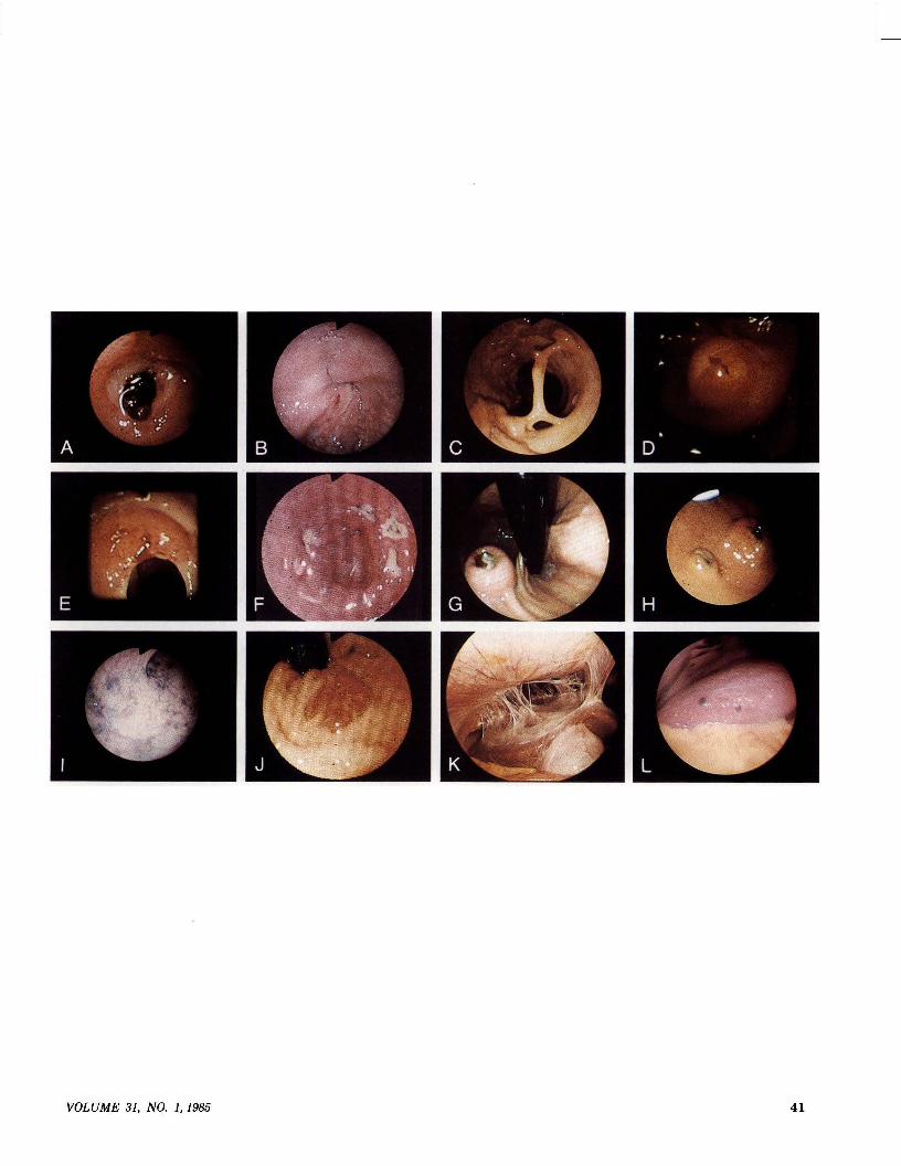
0016-6107/86/3101-0040$02.00 GASTROINTESTINAL ENDOSCOPY Copyright © 1986 by the American Society for Gastrointestinal Endoscopy Spectrum A selection of photographs from the 1984 A/S/G/E Learning Corner edited by Bennett E. Roth, MD Jay A. Noble, MD Burbank and Arcadia, California The following are true or false questions that refer to the A/S/G/E Learning Corner pfwtographs. The answers can be found on page 46. A. In regard to this finding following an acute upper gastrointestinal hemorrhage: 1. There is a high likelihood of rebleeding. 2. It is best to avoid endoscopic manipulation for fear of restart- ing bleeding. 3. Endoscopic electrocautery or laser therapy is usually recom- mended. 4. The central dark spot is the actual vessel. B. The sigmoidoscopic finding in this patient with symptoms of bowel obstruction can be attributed to: 1. Stricture of-colon. 2. Carcinoma. 3. Volvulus. 4. Ischemic colitis. C. This colonoscopy was done in a patient with longstanding ulcerative colitis. 1. The findings indicate a need for colectomy. 2. This usually occurs after many years of disease. 3. This may be seen without active symptomatology. 4. The band should be cut. D. The most likely diagnosis for this finding is: 1. Ampullary carcinoma. 2. Ulcerated duodenal polyp. 3. Lacerated papilla following spontaneous passage of stone. 4. Brunner gland hyperplasia. E. This ulcerating lesion was found in the mid esophagus. It is associated with: 1. Chronic esophagitis. 2. Increased risk of squamous cell carcinoma of esophagus. 3. Increased occurrence of colon carcinoma. 4. Esophageal stricture. F. A 26-year-old previously healthy man developed flu symptoms and medicated himself. Three days later he had the acute onset of hematemesis. Upper gastrointestinal endoscopy revealed these le- sions in the antrum. This patient most likely has: 1. Crohn's disease of the stomach. 2. Zollinger-Ellison syndrome with multiple ulcers. 3. Salicylate gastritis. 4. Gastric lymphoma. G. A 92-year-old woman was admitted with upper gastrointestinal bleeding. An upper gastrointestinal series revealed a smooth rounded mass in the fundus of the stomach which contained a central ulceration. Endoscopy showed the pictured lesion. 1. The lesion is most likely a pancreatic rest. 40 2. The lesion is most likely an ulcerated leiomyoma. 3. There is nothing to suggest the source of bleeding on this view. 4. Surgical excision is the treatment of choice in this patient. H. A 41-year-old woman with a past history of carcinoma of the breast 2years earlierpresented with upper gastrointestinal bleeding. These lesions were seen at endoscopy. 1. The multiplicity of the lesions rules out metastatic breast cancer. 2. Pinch biopsies of these lesions may not reveal the underlying histopathology. 3. These lesions should be surgically removed prior to any other form of therapy. 4. A course of sucralfate therapy would be helpful. I. A 76-year-old man with recurrent episodes of hematochezia had this appearance throughout the left colon on flexible fiberoptic sigmoidoscopy. He most likely has: 1. Angiodysplasia. 2. Cavernous hemangioma. 3. Colonic varices. 4. Peutz-Jeghers syndrome. J. A 21-year-old man admitted with chronic cough and shortness of breath underwent flexible fiberoptic sigmoidoscopy because of diarrhea. This lesion was found on retroverting the instrument inside the rectum. 1. These lesions are often asymptomatic. 2. Biopsies usually reveal interlacing bundles of spindle cells with extravasated red blood cells. 3. These lesions should be removed by hot-biopsy electrofulgur- ation. 4. Defects in the immune mechanism may be seen in association with these lesions. K. A 21-year-old woman developed right upper quadrant pain which became quite severe. This is a laparoscopic view of the area of the right lobe of the liver. The only positive finding on system review was the presence of a chronic vaginal discharge. This patient has: 1. Postsurgical adhesions. 2. Acute cholecystitis. 3. Acute hepatitis A. 4. Fitz-Hugh and Curtis syndrome. L. A 46-year-old man underwent laparoscopy as part of a staging procedure for malignant disease found elsewhere in the body. The presence of these lesions in the right lobe of the liver confirms the presence of: 1. Hemangiosarcoma. 2. Bronchogenic carcinoma. 3. Malignant melanoma. 4. Hodgkin's disease. GASTROINTESTINAL ENDOSCOPY
Transcript

0016-6107/86/3101-0040$02.00GASTROINTESTINAL ENDOSCOPYCopyright © 1986 by the American Society for Gastrointestinal Endoscopy

Spectrum

A selection of photographsfrom the 1984 A/S/G/E Learning Corner

edited byBennett E. Roth, MD

Jay A. Noble, MDBurbank and Arcadia, California

The following are true or false questions that refer to the A/S/G/ELearning Corner pfwtographs. The answers can be found on page 46.

A. In regard to this finding following an acute upper gastrointestinalhemorrhage:

1. There is a high likelihood of rebleeding.2. It is best to avoid endoscopic manipulation for fear of restart­

ing bleeding.3. Endoscopic electrocautery or laser therapy is usually recom­

mended.4. The central dark spot is the actual vessel.

B. The sigmoidoscopic finding in this patient with symptoms ofbowel obstruction can be attributed to:

1. Stricture of-colon.2. Carcinoma.3. Volvulus.4. Ischemic colitis.

C. This colonoscopy was done in a patient with longstandingulcerative colitis.

1. The findings indicate a need for colectomy.2. This usually occurs after many years of disease.3. This may be seen without active symptomatology.4. The band should be cut.

D. The most likely diagnosis for this finding is:1. Ampullary carcinoma.2. Ulcerated duodenal polyp.3. Lacerated papilla following spontaneous passage of stone.4. Brunner gland hyperplasia.

E. This ulcerating lesion was found in the mid esophagus. It isassociated with:

1. Chronic esophagitis.2. Increased risk of squamous cell carcinoma of esophagus.3. Increased occurrence of colon carcinoma.4. Esophageal stricture.

F. A 26-year-old previously healthy man developed flu symptomsand medicated himself. Three days later he had the acute onset ofhematemesis. Upper gastrointestinal endoscopy revealed these le­sions in the antrum. This patient most likely has:

1. Crohn's disease of the stomach.2. Zollinger-Ellison syndrome with multiple ulcers.3. Salicylate gastritis.4. Gastric lymphoma.

G. A 92-year-old woman was admitted with upper gastrointestinalbleeding. An upper gastrointestinal series revealed a smoothrounded mass in the fundus of the stomach which contained acentral ulceration. Endoscopy showed the pictured lesion.

1. The lesion is most likely a pancreatic rest.

40

2. The lesion is most likely an ulcerated leiomyoma.3. There is nothing to suggest the source of bleeding on this view.4. Surgical excision is the treatment of choice in this patient.

H. A 41-year-old woman with a past history of carcinoma of thebreast 2 years earlier presented with upper gastrointestinal bleeding.These lesions were seen at endoscopy.

1. The multiplicity of the lesions rules out metastatic breastcancer.

2. Pinch biopsies of these lesions may not reveal the underlyinghistopathology.

3. These lesions should be surgically removed prior to any otherform of therapy.

4. A course of sucralfate therapy would be helpful.I. A 76-year-old man with recurrent episodes of hematochezia hadthis appearance throughout the left colon on flexible fiberopticsigmoidoscopy. He most likely has:

1. Angiodysplasia.2. Cavernous hemangioma.3. Colonic varices.4. Peutz-Jeghers syndrome.

J. A 21-year-old man admitted with chronic cough and shortnessof breath underwent flexible fiberoptic sigmoidoscopy because ofdiarrhea. This lesion was found on retroverting the instrumentinside the rectum.

1. These lesions are often asymptomatic.2. Biopsies usually reveal interlacing bundles ofspindle cells with

extravasated red blood cells.3. These lesions should be removed by hot-biopsy electrofulgur­

ation.4. Defects in the immune mechanism may be seen in association

with these lesions.K. A 21-year-old woman developed right upper quadrant pain whichbecame quite severe. This is a laparoscopic view of the area of theright lobe of the liver. The only positive finding on system reviewwas the presence of a chronic vaginal discharge. This patient has:

1. Postsurgical adhesions.2. Acute cholecystitis.3. Acute hepatitis A.4. Fitz-Hugh and Curtis syndrome.

L. A 46-year-old man underwent laparoscopy as part of a stagingprocedure for malignant disease found elsewhere in the body. Thepresence of these lesions in the right lobe of the liver confirms thepresence of:

1. Hemangiosarcoma.2. Bronchogenic carcinoma.3. Malignant melanoma.4. Hodgkin's disease.

GASTROINTESTINAL ENDOSCOPY

VOLUME 31, NO.1, 1985 41

described in the research protocol. Other study sec­tions or initial review groups may review applicationsinvolving endoscopy if the proposed research projectdoes not fall within the purview of GMA-2 or SB.Outside reviewers may also be sought to complementthe expertise of review group members in review ofendoscopy-oriented applications. Information on cur­rent membership may be obtained by contacting theexecutive secretary of the appropriate study section.

The staff of the concerned NIH and ADAMHAInstitutes welcome an increased expression of interestfrom members of the American Society for Gastroin-

testinal Endoscopy and from other scientists who turnto endoscopy in pursuing their research goals.

REFERENCES1. Eaves GN. Who reads your project-grant application to the

National Institutes of Health? Fed Proc 1972;31:2-9.2. Eaves GN, Rifkin DB, Gee HH, Malone TE, Ross R, Schimke

RT. The project-grant application of the National Institutes ofHealth. Fed Proc 1973;32:1541-1550.

3. Eaves GN. A successful grant application to the National In­stitutes of Health: case history. Grants Magazine 1978;1:263­286.

4. Allen EM. Why are research grant applications disapproved?Science 1960; 132:1532-4.

5. NIH Public Advisory Groups. Activity, structure, and function.NIH Publication No. 84-11. January 1984. 438 pages.

SPECTRUM ANSWERS

A. Visible vessel1. True2. False3. True4. False

B. Volvulus1. False2. False3. True4. False

c. Mucosal bridging1. False2. True3. True4. False

D. Lacerated papilla following spontaneous passage of a stone1. False2. False3. True4. False

E. Barrett's esophagus1. True2. False3. True4. True

F. Salicylate gastritis1. False2. False3. True4. False

46

G. Gastric leiomyoma with recent bleeding1. False2. True3. False4. False

H. Metastatic carcinoma of the breast1. False2. True3. False4. False

I. Cavernous hemangioma of the colon1. False2. True3. False4. False

J. Kaposi's sarcoma of the rectum1. True2. True3. False4. True

K. Fitz-Hugh and Curtis syndrome1. False2. False3. False4. True

L. Metastatic melanoma of the liver1. False2. False3. True4. False

GASTROINTESTINAL ENDOSCOPY


Recommended