Br. J. Pharmac. Chemother. (1968), 32, 193-200. A SIMPLE METHOD FOR RECORDING THE ELECTROCARDIOGRAM AND HEART RATE FROM CONSCIOUS ANIMALS BY J. B. FARMER AND G. P. LEVY From the Department of Pharmacology, Allen and Hanburys, Ltd., Ware, Hertfordshire (Received September 28, 1967) The small bioelectrical signals of the heart detected at the body surface of animals are usually relayed to the initial amplifier stage of the electrocardiograph by the use of plate, needle or clip electrodes (Clark, Szabuniewicz & McCrady, 1966). In a series of experiments in which rapid and repeated measurements of the electrocardiogram (e.c.g.) and resting heart rate from conscious, unrestrained guinea-pigs were required none of the standard methods proved suitable and a modification was therefore developed in which the animals stand on plate electrodes. This method has been used to obtain recordings from conscious mice, rats, cats and dogs, and some applications of the method are illustrated in the experiments described in this article. METHODS A diagram of the apparatus is shown in Fig. 1. It comprised four separate copper plate electrodes mounted in either cork or blockboard, each sealed with a waterproof mastic seal (Seelastik, Expandite Ltd.) to prevent leakage. The animal was placed on the apparatus so that each foot was in contact with a separate plate electrode. The front feet and left hind foot were used for recording standard leads while the right hind foot was grounded. Contact was facilitated by the use of gauze pads damped with isotonic saline. The dimensions of the apparatus were varied to suit the different animals. The electrical signals detected by the plate electrodes were amplified by a Devices ACI and sub-unit 3 preamplifier and displayed on a Devices pen recorder. Heart rate was measured by a Neilson instantaneous ratemeter (40-250 and 80-500 beats/min scales) triggered from the QRS complex of the e.c.g. Recordings were obtained in a quiet room maintained at 200-21' C. In the experiments described the following animals of either sex were used: mice of the I.C.I. strain (20-25 g); Wistar rats (200-300 g); cats (2-3 kg) and beagle dogs (10-17 kg). Drugs The following drugs were used: isoprenaline sulphate (Burroughs Wellcome), propranolol (Imperial Chemical Industries) and strophanthin-G (Ouabain; British Drug Houses). The drugs were dissolved in normal saline and administered by subcutaneous or intravenous injection. All doses are expressed in terms of the base. RESULTS Training and suitability of conscious laboratory animals for the method of recording A preliminary training period which varied considerably in duration for different laboratory animals was required before e.c.gs of suitable quality and stable heart rate
Transcript
Br. J. Pharmac. Chemother. (1968), 32, 193-200.
A SIMPLE METHOD FOR RECORDING THEELECTROCARDIOGRAM AND HEART RATE FROM
CONSCIOUS ANIMALSBY
J. B. FARMER AND G. P. LEVY
From the Department of Pharmacology, Allen and Hanburys, Ltd., Ware, Hertfordshire
(Received September 28, 1967)
The small bioelectrical signals of the heart detected at the body surface of animalsare usually relayed to the initial amplifier stage of the electrocardiograph by the use ofplate, needle or clip electrodes (Clark, Szabuniewicz & McCrady, 1966). In a series ofexperiments in which rapid and repeated measurements of the electrocardiogram (e.c.g.)and resting heart rate from conscious, unrestrained guinea-pigs were required none ofthe standard methods proved suitable and a modification was therefore developed inwhich the animals stand on plate electrodes. This method has been used to obtainrecordings from conscious mice, rats, cats and dogs, and some applications of themethod are illustrated in the experiments described in this article.
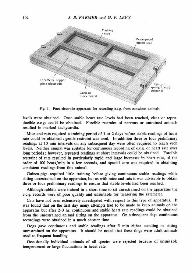
METHODSA diagram of the apparatus is shown in Fig. 1. It comprised four separate copper plate electrodes
mounted in either cork or blockboard, each sealed with a waterproof mastic seal (Seelastik, ExpanditeLtd.) to prevent leakage. The animal was placed on the apparatus so that each foot was in contactwith a separate plate electrode. The front feet and left hind foot were used for recording standardleads while the right hind foot was grounded. Contact was facilitated by the use of gauze padsdamped with isotonic saline. The dimensions of the apparatus were varied to suit the differentanimals. The electrical signals detected by the plate electrodes were amplified by a Devices ACI andsub-unit 3 preamplifier and displayed on a Devices pen recorder. Heart rate was measured by aNeilson instantaneous ratemeter (40-250 and 80-500 beats/min scales) triggered from the QRScomplex of the e.c.g. Recordings were obtained in a quiet room maintained at 200-21' C.
In the experiments described the following animals of either sex were used: mice of the I.C.I.strain (20-25 g); Wistar rats (200-300 g); cats (2-3 kg) and beagle dogs (10-17 kg).DrugsThe following drugs were used: isoprenaline sulphate (Burroughs Wellcome), propranolol (Imperial
Chemical Industries) and strophanthin-G (Ouabain; British Drug Houses). The drugs were dissolvedin normal saline and administered by subcutaneous or intravenous injection. All doses are expressedin terms of the base.
RESULTS
Training and suitability of conscious laboratory animals for the method of recordingA preliminary training period which varied considerably in duration for different
laboratory animals was required before e.c.gs of suitable quality and stable heart rate
J. B. FARMER and G. P. LEVY
Fig. 1. Foot electrode apparatus for recording e.c.g. from conscious animals.
levels were obtained. Once stable heart rate levels had been reached, clear or repro-ducible e.c.gs could be obtained. Forcible restraint of nervous or untrained animalsresulted in marked tachycardia.Mice and rats required a training period of 1 or 2 days before stable readings of heart
rate could be obtained; gentle restraint was used. In addition three or four preliminaryreadings at 10 min intervals on any subsequent day were often required to reach suchlevels. Neither animal was suitable for continuous recording of e.c.g. or heart rate overlong periods; however, repeated readings at short intervals could be obtained. Forciblerestraint of rats resulted in particularly rapid and large increases in heart rate, of theorder of 100 beats/min in a few seconds, and special care was required in obtainingconsistent readings from this animal.
Guinea-pigs required little training before giving continuous stable readings whilesitting unrestrained on the apparatus, but as with mice and rats it was advisable to obtainthree or four preliminary readings to ensure that stable levels had been reached.Although rabbits were trained in a short time to sit unrestrained on the apparatus the
e.c.g. records were of poor quality and unsuitable for triggering the ratemeter.Cats have not been extensively investigated with respect to this type of apparatus. It
was found that on the first day many attempts had to be made to keep animals on theapparatus but after 2-3 hr. continuous and stable heart rate readings could be obtainedfrom the unrestrained animal sitting on the apparatus. On subsequent days continuousrecordings were obtained in a much shorter time.Dogs gave continuous and stable readings after 5 min either standing or sitting
unrestrained on the apparatus. It should be noted that these dogs were adult animalsused to frequent handling.
Occasionally individual animals of all species were rejected because of unsuitabletemperament or large fluctuations in heart rate.
194
E.C.G. AND HEART RATE IN CONSCIOUS ANIMALS
Lead I
Mouse [In
Rat [
Guinea- rpig
Cat [
Dog [
ElectrocardiogramLead II
0.5 sec
Heart rate
Beats/min
>500
500 r4003002001000
400-300200loo0
250-200ISO100
so250200
1500'soI0t
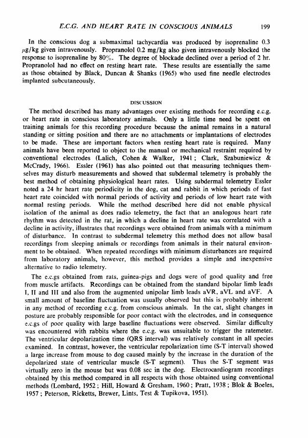
Fig. 2. Electrocardiogram and heart rate recordings obtained from conscious animals using footelectrode recording system.
Electrocardiogram and heart rate records obtained from conscious laboratory animals
Typical e.c.g. (leads I and II) and heart rate records obtained from the mouse, rat,guinea-pig, cat and dog, are shown in Fig. 2. Amplification and paper speed were variedto obtain clear records in each case. Electrocardiographic data obtained from thedifferent animals are summarized in Table 1. Although e.c.gs among individuals ofa species varied slightly they remained constant for a given animal over long periods oftime.
The heart rate of the mouse was greater than 500 beats/min and readings could not beobtained from the ratemeter; heart rate was therefore derived from the e.c.g. record. Inlead I upright P waves, QRS complexes and usually T waves were present. There wasno S-T segment; the T wave, when present, followed immediately after the QRS com-plex. In lead II the upright P waves were larger than in lead I. The QRS complex inlead II was notched ; this is a recording artifact caused by the limited frequency response
195
J. B. FARMER and G. P. LEVY
TABLE 1
ELECTROCARDIOGRAPHIC DATA OBTAINED FROM CONSCIOUS ANIMALSFigures represent mean values from stated number of animals. For each animal data were obtained from
six cardiac cyclesQRS Complex
Resting Duration Duration Amplitude Durationheart of cardiac of P-R of Q-T
No. of rate cycle interval Duration Lead I Lead II intervalSpecies animals (beats/min) (sec) (sec) (sec) (mV) (mV) (sec)
of the pen amplifier (Rappaport & Rappaport, 1943). The heart rate of the rat rangedfrom 260 to 380 beats/min. The rate was higher in the morning than in the afternoon.This gradual decline in heart rate throughout the day paralleled the decline in spontaneous
activity of the animals. In lead I upright P waves and QRS complexes were evident butT waves were absent. S waves could be seen but Q waves were rare. In lead II Pwaves, QRS complexes and T waves could be distinguished; again there was virtually no
S-T segment. The heart rate of the guinea-pig ranged from 240 to 310 beats/min. P. Rand T waves were upright in both leads. Q and S waves were evident in lead I and eithersmall or absent in lead II. In the cat, heart rate ranged from 110 to 175 beats/min. Aslight sinus arrhythmia associated with respiration was observed in some animals.Electrocardiogram records of the cat were difficult to analyse because of fluctuations inbase-line. In those sections suitable for analysis, upright P. R and T waves could bedistinguished in both lead I and II. All amplitudes were smaller in lead I. Q and Swaves were not prominent in either lead. Heart rate in the dog ranged from 70 to 120beats/min with a phasic sinus arrhythmia associated with respiration present in all dogs.P waves and R waves were upright in both leads and larger in lead II than leadI.Q waves were present in both leads but S waves were often absent in lead I. An S-Tsegment was present in both leads. T waves were variable, being either upright or
inverted in either lead I or II.
Effect of ouabain on e.c.g. and heart rate of conscious guinea-pigsOuabain 100-200jug/kg given subcutaneously or intraperitoneally to conscious
guinea-pigs caused a tachycardia of 20-40 beats/min in approximately half the animalstreated; the remainder were unaffected. Subsequent recordings revealed most of thecommon manifestations of cardiac glycoside toxicity such as prolongation of P-R interval,loss of T wave, S.A. block, A.V. block, interference dissociation and auricular andventricular dissociation. Many animals exhibited violent muscle tremors and convulsions.In this dose range ouabain proved fatal to most animals within 3 hr. Recordingsobtained from one animal are illustrated in Fig. 3. There was a progressive reduction incardiac muscle excitability with development of an A.V. block and establishment of anidioventricular rhythm. The ventricular ectopic focus caused three successive abnormalventricular beats after which normal rhythm was re-established.
196
E.C.G. AND HEART RATE IN CONSCIOUS ANIMALS
A
Ouabain ---200 gg/kg s.c. a_
B
C
I sec
Fig. 3. Electrocardiogram of guinea-pig (350 g). Panel A, control reading; panels B and C, 30 and60 min after the subcutaneous injection of ouabain 200 jig/kg.
360 F
I--
340
i 320
" 300
- 280IVx
I-
I.
I.
260L
0 30 60 90 120 150 180 210
T T ~~~~~TIsoprenaline 2.5 pg/kg 1.0 jg/kg 0.4 jg/kg
Fig. 4. Effect of graded doses of isoprenaline, given subcutaneously on the heart rate of theconscious guinea-pig.
Effect of isoprenwline and propranolol on heart rate of the guinea-pig, rat and dogThe effect of graded doses of isoprenaline on heart rate of a group of six guinea-pigs
is shown in Fig. 4. The net increase in heart rate produced by isoprenaline 0.4, 1.0 and2.5 1ug/kg was 24, 60 and 70 beats/min, respectively. The response to repeated sub-cutaneous administration of isoprenaline 1.0 pg/kg, when given at 1 hr intervals was
constant. Propranolol 10 mg/kg given subcutaneously to a group of six guinea-pigs
mV
I mV
I. mV
197
J. B. FARMER and G. P. LEVY
Propranolol10 mg/kg s.c.
Fin=6
I
Time (hr) 0 1 2 3 4 S
Fig. 5. Effect of propranolol 10 mg/kg given subcutaneously (,k) on the rcisoprenaline induced tachycardia in the conscious guinea-pig. At T isgiven subcutaneously.
6
esting heart rate and on
;oprenaline 1 ,g/kg was
340
330
.c 320-E
v 310
.0
iL 300I-
X 290
n=4n=4n=4
I-
280
270L '1/3 0 1 2 3 4 5
t Time (hr)Fig. 6. Effect of propranolol given subcutaneously (1') on resting heat rate in groups of conscious
produced a mean decrease in heart rate of 35 beats/min which was maximal at 15 minand blocked the response to isoprenaline (Fig. 5). The heart rate returned to controllevels in 3 hr but the response to isoprenaline did not recover until 4 hr.
In the rat, propranolol also produced a decrease in heart rate (Fig. 6). The netdecreases in heart rate produced by propranolol 2 and 10 mg/kg given subcutaneously togroups of four rats was 26 and 45 beats/min, respectively. The duration of action ofpropranolol was about 4-5 hr. Untreated rats also showed a decline in heart rate overthis period as described earlier.
198
320-.1C 300
Jzop260
I 240
220 F t w s s
E.C.G. AND HEART RATE IN CONSCIOUS ANIMALS
In the conscious dog a submaximal tachycardia was produced by isoprenaline 0.3ttg/kg given intravenously. Propranolol 0.2 mg/kg also given intravenously blocked theresponse to isoprenaline by 80%.. The degree of blockade declined over a period of 2 hr.Propranolol had no effect on resting heart rate. These results are essentially the sameas those obtained by Black, Duncan & Shanks (1965) who used fine needle electrodesimplanted subcutaneously.
DISCUSSION
The method described has many advantages over existing methods for recording e.c.g.or heart rate in conscious laboratory animals. Only a little time need be spent ontraining animals for this recording procedure because the animal remains in a naturalstanding or sitting position and there are no attachments or implantations of electrodesto be made. These are important factors when resting heart rate is required. Manyanimals have been reported to object to the manual or mechanical restraint required byconventional electrodes (Lalich, Cohen & Walker, 1941 ; Clark, Szabuniewicz &McCrady, 1966). Essler (1961) has also pointed out that measuring techniques them-selves may disturb measurements and showed that subdermal telemetry is probably thebest method of obtaining physiological heart rates. Using subdermal telemetry Esslernoted a 24 hr heart rate periodicity in the dog, cat and rabbit in which periods of fastheart rate coincided with normal periods of activity and periods of low heart rate withnormal resting periods. While the method described here did not enable physicalisolation of the animal as does radio telemetry, the fact that an analogous heart raterhythm was detected in the rat, in which a decline in heart rate was correlated with adecline in activity, illustrates that recordings were obtained from animals with a minimumof disturbance. In contrast to subdermal telemetry this method does not allow basalrecordings from sleeping animals or recordings from animals in their natural environ-ment to be obtained. When repeated recordings with minimum disturbances are requiredfrom laboratory animals, however, this method provides a simple and inexpensivealternative to radio telemetry.The e.c.gs obtained from rats, guinea-pigs and dogs were of good quality and free
from muscle artifacts. Recordings can be obtained from the standard bipolar limb leads1, 11 and III and also from the augmented unipolar limb leads aVR, aVL and aVF. Asmall amount of baseline fluctuation was usually observed but this is probably inherentin any method of recording e.c.g. from conscious animals. In the cat, slight changes inposture are probably responsible for poor contact with the electrodes, and in consequencee.c.gs of poor quality with large baseline fluctuations were observed. Similar difficultywas encountered with rabbits where the e.c.g. was unsuitable to trigger the ratemeter.The ventricular depolarization time (QRS interval) was relatively constant in all speciesexamined. In contrast, however, the ventricular repolarization time (S-T interval) showeda large increase from mouse to dog caused mainly by the increase in the duration of thedepolarized state of ventricular muscle (S-T segment). Thus the S-T segment wasvirtually zero in the mouse but was 0.08 sec in the dog. Electrocardiogram recordingsobtained by this method compared in all respects with those obtained using conventionalmethods (Lombard, 1952; Hill, Howard & Gresham, 1960; Pratt, 1938; Blok & Boeles,1957; Peterson, Ricketts, Brewer, Lints, Test & Tupikova, 1951).
199
J. B. FARMER and G. P. LEVY
The usefulness of the method in assessing the effect of drugs on the e.c.g. and heartrate is illustrated by the studies with ouabain and with adrenergic stimulant or blockingdrugs. The classical manifestations of cardiac glycoside toxicity after injection ofouabain were readily observed by this method. Propranolol, in addition to blockingisoprenaline tachycardia, was shown markedly to reduce the heart rate of rats andguinea-pigs in a dose-dependent fashion. The guinea-pig was a more suitable animalthan the rat for these studies because the heart rate of the guinea-pig was stable overthe experimental period (6 hr). In the dog propranolol, at the dose level used, blockedthe isoprenaline tachycardia without affecting heart rate. We have observed, however,that larger doses of propranol given orally (1 mg/kg and more) caused a fall in heart rate.
SUMMARY
1. An apparatus suitable for recording e.c.g. from conscious mice, rats, guinea-pigs,cats and dogs using foot electrodes is described.
2. Training procedures and measuring techniques for obtaining stable heart rates andreproducible e.c.gs from the different species are detailed.
3. Electrocardiogram and heart rate recordings obtained from the different speciesare described and compared.
4. The effect of ouabain on the e.c.g. of the guinea-pig and of isoprenaline andpropranolol on heart rate in the rat, guinea-pig and dog are described to illustratesome possible applications of the method.We wish to thank Mr. S. W. Smith for construction of the apparatus and Mrs. M. Tribe for
technical assistance.
REFERENCESBLACK, J. W., DUNCAN, W. A. M. & SHANKS, R. G. (1965). Comparison of some properties of pronethalol
and propranolol. Br. J. Pharmac. Chemother., 25, 577-591.BLOK, J. & BOELES, J. TH. F. (1957). The electrocardiogram of the normal cat. Acta physiol. pharmac.
neerl., 6, 95-102.CLARK, D. R., SZABUNEWICZ, M. & McCRADY, J. D. (1966). Clinical use of the electrocardiogram in
animals. Part I: Fundamentals of E.C.G. examination. Vet. Med., 61, 751-760.ESSLER, W. 0. (1961). Radio telemetry of electrocardiogram and body temperatures from surgically im-
planted transmitters. St. Univ. Stud. nat. Hist. Iowa, 20, 1-35.HILL, R., HowARD, A. N. & GRESHAM, G. A. (1960). The electrocardiographic appearances of myocardial
infarction in the rat. Br. J. exp. Path., 41, 633-637.LALICH, J., COHEN, A. M. & WALKER, G. (1941). The frequency of electrocardiographic variations in normal
unanaesthetized dogs. Am. Heart J., 22, 105-111.LoMBARD, E. A. (1952). Electrocardiograms of small mammals. Am. J. Physiol., 171, 189-193.PETERSON, E. S., RICKETTS, H. T., BREWER, N. R., LamTS, H. A., TEST, C. E. & TUPIKOVA, N. A. (1951).
Electrocardiogram of the beagle dog. Proc. Soc. exp. Biol. Med., 17, 330-332.PRATr, C. L. G. (1938). The electrocardiogram of the guinea-pig. J. Physiol., Lond., 92, 268-272.RAPPAPORT, M. B. & RAPPAPORT, I. (1943). Electrocardiographic considerations in small animal investiga-