A single-molecule digital enzyme assay usingalkaline phosphatase with a cumarin-basedfluorogenic substrate
Yusuke Obayashi,a Ryota Iinob,c and Hiroyuki Noji*a,d
Digitalization of fluorogenic enzymatic assays through the use of femtoliter chamber array technology is
an emerging approach to realizing highly quantitative bioassays with single-molecule sensitivity. However,
only a few digital fluorogenic enzyme assays have been reported, and the variations of the digital enzyme
assays are basically limited to fluorescein- and resorufin-based fluorogenic assays. This limitation
hampers the realization of a multiplex digital enzyme assay such as a digital enzyme-linked immuno-
sorbent assay (ELISA). In this study, after optimization of buffer conditions, we achieved a single-molecule
digital enzyme alkaline phosphatase (ALP) assay with a cumarin-based fluorogenic substrate, 4-methyl-
unbelliferyl phosphate (4-MUP). When ALP molecules were encapsulated in a 44-femtoliter chamber
array at a low ratio of less than 1 molecule per chamber, each chamber showed a discrete fluorescence
signal in an all-or-none manner, allowing the digital counting of the number of active enzyme molecules.
The fraction of fluorescent chambers linearly decreased with the enzyme concentration, obeying the
Poisson distribution as expected. We also demonstrated a dual-color digital enzyme assay with a ALP/
4-MUP and β-galactosidase (β-gal)/resorufin-β-D-galactopyranoside combination. The activities of single
ALP and β-gal molecules were clearly detected simultaneously. The method developed in this study will
enable us to carry out a parallelized, multiplex digital ELISA.
Introduction
Miniaturization of bioassay systems provides many benefits tobioanalysis, such as massive parallelization, reductions insample volumes, and more rapid responses due to the largesurface-to-volume ratio.1,2 Another important benefit of thedownsizing of reaction volumes is higher sensitivity. A particu-lar case is the single-molecule fluorogenic enzyme assay usingfemtoliter chambers (hereafter referred to as the digitalenzyme assay);3–6 individual enzyme molecules are stochasti-cally encapsulated with the fluorogenic assay mixture in femto-liter-sized reactor chambers, and the catalytic activity isdetected as the fluorescent signal from the reaction productmolecules accumulate in the femtoliter chambers. The meanturnover rate of enzymes is around 10 turnovers per s.7 In acube of 1 μm, which corresponds to 1 femtoliter, the concen-tration of reaction products reaches the micromolar range in a
few minutes, allowing them to be readily detectable with a con-ventional fluorescent microscope.
In a pioneering study of a single-molecule enzyme assay infemtoliter-scaled reactors,8 a diluted solution of β-galacto-sidase (β-gal) was partitioned in 7–30 μm water-in-oil (W/O)droplets with a fluorescein-based fluorogenic substrate at lessthan 1 enzyme molecule per droplet. The fluorescence signalsfrom the droplets exhibited an all-or-none manner; while mostdroplets were not fluorescent, some showed clear fluorescence.The fraction of fluorescent droplets exhibited good linearitywith the enzyme concentration, indicating that partitioning ofthe fluorogenic assay mixture into femtoliter reactors enabledthe detection of the catalytic activity of a single β-gal molecule.However, due to the large inherent heterogeneity of the W/Odroplets prepared by this technique (which was developed over50 years ago), this approach did not become widespread as ananalytical method until microfabrication technology allowedgeneration of the femtoliter chambers with identical shapesand volumes. In 2005, Rondelez et al. first reported the digitalcounting of active enzyme molecules through the use of amicrofabricated chamber system.6 They prepared a microfabri-cated PDMS sheet with identically shaped micron-sized wellson the surface and encapsulated the enzyme solution betweenthe fabricated PDMS sheet and a glass coverslip by mechanically
aDepartment of Applied Chemistry, The University of Tokyo, Japan.
E-mail: [email protected]; Fax: +81 3 5841 1872; Tel: +81 3 5841 7252bOkazaki Institute for Integrative Bioscience, Institute of Molecular Science, National
Institutes of Natural Sciences, JapancSOKENDAI (The Graduate University for Advanced Studies), JapandCREST, Japan Science and Technology Agency, Japan
pressing the PDMS sheet against the glass coverslip. When theenzyme solution was diluted to a ratio of less than 1 enzymemolecule per chamber, the individual chambers showed dis-crete fluorescence signals in the all-or-none fashion; whilemost of the chambers remained non-fluorescent, a fewchambers showed fluorescence, and only a few showed fluo-rescence signals with double intensity, indicating the encapsu-lation of zero, one, or two molecules in each chamber,respectively. This work demonstrated that a very simple micro-device allows the formation of identically shaped femtoliterchambers and enables the single-molecule detection ofenzyme molecules and quantification of the enzyme concen-trations by directly counting the number of enzyme molecules(i.e., digital counting).
In recent years, several different formats for the femtoliterchamber system have been reported. For example, researchershave reported a femtoliter chamber array system formed froma plain PDMS sheet and a chemically etched optical fiberbundle.9 In addition, Sakakihara et al. developed an arraysystem of W/O droplets that formed on micron-sized hydro-phobic patterns on glass.10 Moreover, Ge et al. integrated adroplet chamber system in a microfluidic flow channel to forma gradient of the trapping probability of target moleculesalong the microchannel.3 This system enables the automaticpreparation of a dilution series of specimens, allowing digitalcounting of enzyme molecules over a wide dynamic range. Amicrofluidic system for the generation and analysis of free-standing femtoliter droplets was also used for digital countingof enzyme molecules.11 Recently, an arrayed lipid bilayerchamber system (ALBiC) has been developed that allowsdigital counting and analysis of active transporter membraneproteins.12
Recently, the application of femtoliter chamber arrays hasalso been expanding. For example, PDMS-based femtoliterchamber arrays have been used for measurement of the chemo-mechanical coupling efficiency of a single rotary molecularmotor protein13 and for detection of individual translationevents in single bacterial cells.14 Many reports have describedother applications of femtoliter chamber array systems thatprovide high sensitivity and/or high-throughput capacity,including DNA sequencing,15 single-cell drug efflux activityanalysis,16 in vitro translation,17 and single-enzyme analysis.4,18
Among them, one of the most important applications isthe digital enzyme-linked immunosorbent assay (ELISA),3,11,19,20
in which antigen molecules recognized by enzyme-conjugatedantibodies are individually entrapped in a chamber, and thenumber of antigen molecules is counted as the number offemtoliter chambers showing enzymatically produced fluo-rescence signals. Although the first report of a digital ELISAused the PDMS-etched optical fiber plate system,20 droplet-based array systems have been frequently used in recentstudies.3,19,21 The digital ELISA has largely improved the limitof detection (LOD) down to the femto- or attomolar range, rea-lizing the ultrasensitivity of diagnostic ELISAs.
Compared with the active development of platforms andexpansion of applications of femtoliter chamber-based digital
bioassays, the variety of fluorogenic enzyme assays is stilllimited; however, researchers are hoping to develop paralle-lized digital counting assays, such as multiplex digital ELISAs,for improved analysis of multiple targets. To date, the chem-istry of fluorogenic assays has mainly been based on threemajor fluorescent dyes: fluorescein, resorufin, and cumarin.Due to its high photostability and high fluorescent intensity,fluorescein is the first choice of probes; the first digitalenzyme assay used a fluorescein derivative conjugated withgalactose.6 Subsequently, digital enzyme assays with resorufin-based fluorogenic substrates were conducted for detection ofβ-gal,22 β-glucuronidase,4 and horseradish peroxidase.18
Although resorufin and fluorescein fluorescence signals arespectrally separable, the excitation and emission spectraoverlap, causing fluorescence cross-talk. Therefore, simul-taneous, dual-color digital enzyme assays using fluorescein-and resorufin-based fluorogenic assays have not beenattempted. Since cumarin-based fluorogenic assays use exci-tation and emission wavelengths much shorter than those offluorescein and resorufin, they will be suitable for dual digitalenzyme assays with these dyes. However, digital enzyme assaysusing cumarin-based fluorogenic substrates have not beenreported. In this study, we developed a digital enzyme assaywith a cumarin-based fluorogenic substrate for detection ofEscherichia coli alkaline phosphatase (ALP), which is widelyused in diagnostic ELISAs.
Results
Before testing the digital enzyme assay with a cumarin-basedfluorogenic substrate, we explored the optimal buffer con-ditions for the catalytic reaction of ALP in solution. To obtaina high fluorescent signal, we used a mutant ALP (D101S) fromE. coli, for which the Vmax was 35-fold higher than that for thewild-type.23 We used 4-methylunbelliferyl phosphate (4-MUP)as the fluorogenic substrate for ALP; 4-MUP is a phosphory-lated cumarin derivative and hydrolyzed into inorganic phos-phate and 4-metylunbelliferon (4-MU) (Fig. 1). While 4-MUP is
Fig. 1 Fluorogenic substrates and enzymes used in this study. Sche-matic images of the fluorogenic assay of alkaline phosphatase (ALP)which hydrolyzes 4-methylunbelliferyl phosphate (4-MUP) to 4-metyl-unbelliferon (4-MU) and inorganic phosphate. While 4-MUP is non-flu-orescent, 4-MU is fluorescent (excitation peak: 372 nm, emission peak:445 nm).
non-fluorescent, 4-MU emits fluorescence (excitation peak:372 nm, emission peak: 445 nm). The enzymatic activity ofALP was monitored by determining the fluorescence of 4-MU.Fig. 2A shows typical time courses of the fluorogenic assay atdifferent initial concentrations of 4-MUP. The concentrationsof 4-MU increased linearly for 300 s, indicating the constantturnover rates at this time scale under the conditions used.Furthermore, the slope of the time course increased as the4-MUP concentration increased, indicating increased activityof ALP.
Next, we examined the effects of diethanolamine (DEA),which enhances the transphosphorylation activity of ALP as anacceptor of inorganic phosphate.24 We also tested the effectsof magnesium ions (Fig. 2B). Among the tested conditions,1 M DEA and 1 mM MgCl2 yielded the highest ALP activity.Thus, we then tested various pH conditions with 1 M DEA and1 mM MgCl2 (Fig. 2C). We found that pH 9.25 was the optimalpH showing the highest activity. Finally, we investigated theALP activity at different 4-MUP concentrations to determinethe basic kinetic parameters of ALP (Fig. 2D). By fitting
the data points with the Michaelis–Menten equation, themaximum turnover rate (kcat) and the Michaelis–Menten con-stant (KM) were determined to be 1.19 × 10³ s−1 and 183 μM,respectively. These values were consistent with a previousreport on the mutant ALP.23
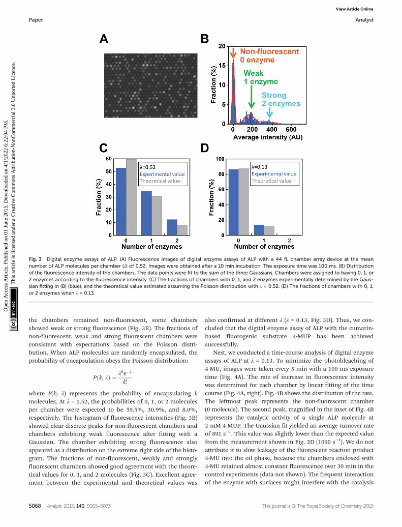
Next, digital enzyme assays for detection of the ALP activityin W/O-type femtoliter chambers were conducted at 2 mM4-MUP in the presence of 1 M DEA and 1 mM MgCl2 at pH 9.25,at which ALP hydrolyzes 4-MUP at 1090 s−1 according to theMichaelis–Menten analysis in Fig. 2D. Note that at the esti-mated consumption of 4-MUP in a 44 fL chamber containing asingle ALP molecule for 20 min reaction, is only 2.5%, so thatthe substrate consumption as well as the product inhibitionwould be negligible. After adding the enzyme solution tothe 4-MUP solution, the reaction mixture was immediatelyintroduced into the device and sealed with fluorinated oil.When the calculated mean number of ALP molecules perchamber (λ) was well below 1, the chambers showed a discretefluorescent intensity. Fig. 3A shows the fluorescence image atλ = 0.52 after a 4 min incubation. Although more than 50% of
Fig. 2 Fluorogenic assays of ALP in bulk solution. (A) Time courses of fluorogenic assays of ALP with 4-MUP at 300 μM (red), 1 mM (green), 3 mM(blue). The reaction was monitored by the fluorescence of the reaction product, 4-MU. The turnover rate was determined from the linear fitting of atime-course. (B) Effects of diethanolamine (DEA) and magnesium chloride on the turnover rate. (C) pH dependence of the turnover rate. The activitywas measured in the presence of 1 M DEA and 1 mM MgCl2. Error bars represent the standard deviations of three independent measurements. (D)Dependence of the turnover rate on 4-MUP concentration. The activity was measured in the presence of 1 M DEA and 1 mM MgCl2 at pH 9.25. Datapoints were fitted to the Michaelis–Menten equation to give a Km of 183 μM and a Vmax of 1.19 × 10³ s−1.
the chambers remained non-fluorescent, some chambersshowed weak or strong fluorescence (Fig. 3B). The fractions ofnon-fluorescent, weak and strong fluorescent chambers wereconsistent with expectations based on the Poisson distri-bution. When ALP molecules are randomly encapsulated, theprobability of encapsulation obeys the Poisson distribution:
P k; λð Þ ¼ λke�λ
k!
where P(k; λ) represents the probability of encapsulating kmolecules. At λ = 0.52, the probabilities of 0, 1, or 2 moleculesper chamber were expected to be 59.5%, 30.9%, and 8.0%,respectively. The histogram of fluorescence intensities (Fig. 3B)showed clear discrete peaks for non-fluorescent chambers andchambers exhibiting weak fluorescence after fitting with aGaussian. The chamber exhibiting strong fluorescence alsoappeared as a distribution on the extreme right side of the histo-gram. The fractions of non-fluorescent, weakly and stronglyfluorescent chambers showed good agreement with the theore-tical values for 0, 1, and 2 molecules (Fig. 3C). Excellent agree-ment between the experimental and theoretical values was
also confirmed at different λ (λ = 0.13, Fig. 3D). Thus, we con-cluded that the digital enzyme assay of ALP with the cumarin-based fluorogenic substrate 4-MUP has been achievedsuccessfully.
Next, we conducted a time-course analysis of digital enzymeassays of ALP at λ = 0.13. To minimize the photobleaching of4-MU, images were taken every 5 min with a 100 ms exposuretime (Fig. 4A). The rate of increase in fluorescence intensitywas determined for each chamber by linear fitting of the timecourse (Fig. 4A, right). Fig. 4B shows the distribution of the rate.The leftmost peak represents the non-fluorescent chamber(0 molecule). The second peak, magnified in the inset of Fig. 4Brepresents the catalytic activity of a single ALP molecule at2 mM 4-MUP. The Gaussian fit yielded an average turnover rateof 891 s−1. This value was slightly lower than the expected valuefrom the measurement shown in Fig. 2D (1090 s−1). We do notattribute it to slow leakage of the fluorescent reaction product4-MU into the oil phase, because the chambers enclosed with4-MU retained almost constant fluorescence over 30 min in thecontrol experiments (data not shown). The frequent interactionof the enzyme with surfaces might interfere with the catalysis
Fig. 3 Digital enzyme assays of ALP. (A) Fluorescence images of digital enzyme assays of ALP with a 44 fL chamber array device at the meannumber of ALP molecules per chamber (λ) of 0.52. Images were obtained after a 10 min incubation. The exposure time was 100 ms. (B) Distributionof the fluorescence intensity of the chambers. The data points were fit to the sum of the three Gaussians. Chambers were assigned to having 0, 1, or2 enzymes according to the fluorescence intensity. (C) The fractions of chambers with 0, 1, and 2 enzymes experimentally determined by the Gaus-sian fitting in (B) (blue), and the theoretical value estimated assuming the Poisson distribution with λ = 0.52. (D) The fractions of chambers with 0, 1,or 2 enzymes when λ = 0.13.
to some extent. As expected from the Poisson distribution withλ = 0.13, a few chambers (0.7%) showed double activity byencapsulation of two enzyme molecules in a chamber (Fig. 4Aand B).
In order to determine the LOD of digital counting of ALP, a10-fold dilution series of the ALP solution from λ = 0.13 to λ =0.00013 was subjected to digital enzyme assays. After a 10 minincubation, 120 fluorescence images of the different field ofview were taken for each dilution sample. Fig. 5A shows typicalfluorescence images. Each field of view contains 7600chambers, and the total number of chambers analyzed wasover 0.9 million. Because the fraction of encapsulation of twoenzyme molecules in a chamber was essentially negligible at λ≤ 0.13, each fluorescent chamber was counted as one enzymemolecule. The threshold level for fluorescent chambers was setas the mean background level plus 10 times the standard devi-ation (SD) of the background fluorescence. As shown inFig. 5B, the number of fluorescent chambers was proportionalto the λ and essentially consistent with the theoretical values,
although the experimental values were slightly higher than thetheoretical values, presumably due to the inaccuracy of proteinquantification. The background count (false-positive fluo-rescent chambers detected in the absence of ALP) was muchhigher than that for the fluorogenic digital enzyme assay ofβ-gal with fluorescein-di-β-D-galactopyranoside (about0.0001%).19 This difference could be explained by contami-nating impurities or photoresists remaining on the device (seethe Discussion section). The high background count resultedin the LOD of 7.0 fM (λ = 1.9 × 10⁻⁴) (Fig. 5C).
Finally, we tested the feasibility of dual-color digital enzymeassays of ALP and β-gal. In the fluorogenic assay for β-gal, theenzyme cleaved resorufin-β-D-galactopyranoside (RGP) to galac-tose and resorufin, yielding red fluorescence. The 4-MU andresorufin have different fluorescent emission peaks, 445 nmand 585 nm, respectively, and were spectrally separable in thefluorescence images. Because the optimal pH values for ALPand β-gal are different, the assays were conducted under nearalkaline conditions (pH 8.25), at which ALP retains 67% of the
Fig. 4 Time-course analysis of the digital enzyme assay of ALP. (A, left) Fluorescence images after 5, 10, 15, and 20 min. λ = 0.13. (A, right) Timecourse of the fluorescence intensity from (A, left). Orange, green, and light blue lines were assigned to chambers with 0, 1, and 2 enzymes, respect-ively. (B) Distribution of the rate of fluorescence increase determined by linear fitting of (A, right). The data points were fit to the sum of twoGaussians for chambers having 0 (orange) or 1 (green) enzyme molecule. Blue data points were assigned to chambers having 2 enzyme molecules.Inset indicates an enlarged distribution of chambers having 1 or 2 enzymes.
maximum activity. After mixing well-diluted enzyme solutionswith fluorogenic substrates, the reaction mixture was intro-duced into a flow cell and observed under a fluorescencemicroscope. Fig. 6 shows a pseudo-colored overlay image of4-MU and resorufin. Green and red represent the fluorescenceof 4-MU and resorufin, respectively. As expected, the green andred fluorescent chambers were distributed randomly, withsome showing both green and red fluorescence (Fig. 6, yellowarrow), indicating the simultaneous encapsulation of ALP andβ-gal. Thus, the feasibility of dual-color digital enzyme assayswith cumarin- and resorufin-based fluorogenic assays was suc-cessfully demonstrated.
Discussion
This study presented a cumarin-based fluorogenic assay thatcan be applied as a digital enzyme assay. Because manycumarin-based fluorogenic assays have been developed for
analysis and detection of enzymes, digital enzyme assays usingcumarin-based probes are expected to have a variety of appli-cations. In addition, the present study also demonstrated adual-color digital enzyme assay using cumarin and resorufinfor the first time.
Although fluorescein- and resorufin-based digital enzymeassays have been reported independently, dual-color digitalenzyme assays using these two probes have not been achieveddue to overlap of their excitation and fluorescence spectra.However, cumarin emits fluorescence signals of distinctivelyshorter wavelengths. In this study, we verified that the dual-color digital enzyme assays for ALP and β-gal were feasibleusing resorufin and cumarin. Dual-color digital enzyme assaysare expected to enable multiplex digital ELISAs; while suchmultiplex digital ELISAs have been reported using differentlycolored plastic beads to identify the captured antigen mole-cules,21 the expansion of color variations in digital enzymeassays will provide an alternative approach for multiplexdigital ELISAs or further expand the multiplicity of digitalELISAs by combination with the multi-colored bead method.
In addition to the above, multi-color digital enzyme assaysare expected to exhibit an improved background count com-pared with digital ELISAs; a high background count mayimpair the potential sensitivity of digital ELISAs. The mainfactor affecting the background count in digital ELISAs is non-specific binding of the detection antibody–enzyme conjugateto the bead surface on which the captured antibody is immobi-lized. Plastic beads are the most frequently used surface forantigen capture. When the target antigen molecule is markedwith two different detection antibody–enzyme conjugates sim-ultaneously, we can distinguish the true signal from the false-positive signal with high efficiency. This is because the simul-taneous non-specific binding of two detection conjugates on
Fig. 5 Digital counting of ALP. (A) Fluorescence images of digitalenzyme assays of ALP at λ = 0.13, 0.013, 0.0013, and 0.00013 after a10 min incubation. Typical examples of the single field of view obtainedat each λ are shown. The numbers of fluorescent chambers (n) at each λ
is also shown. (B and C) The fractions of fluorescent chambers versus λ
(B) or ALP concentration (C). Error bars are the standard deviations ofthree independent measurements. The red line represents the fractionof the background count plus 3 times the SD of the background countdetermined under the ALP-free conditions.
Fig. 6 Dual-color digital enzyme assays for ALP and β-gal. Dual-colorfluorogenic assays for ALP and β-gal were conducted by 45 min incu-bation. ALP and β-gal were encapsulated at λ = 0.033 and 0.05, respecti-vely. ALP produced 4-MU, and β-gal produced resorufin. A pseudo-colored overlay image of 4-MU (green) and resorufin (red) is shown. Theyellow color indicates a mixture of red and green signals, representingthe coexistence of ALP and β-gal.
the same bead should be much more infrequent than thesingle nonspecific binding events. Thus, by increasing thecolor variations of fluorogenic assays, the background count ofdigital ELISAs will be dramatically reduced. This strategy isexpected to be highly effective, particularly for digital ELISAstargeting multi-epitope antigens such as infectious viruses.
However, several drawbacks of this method were also foundwhen compared with a fluorescein-based digital enzyme assay.The first is the leakage of cumarin to the fluorinated oil phaseunder neutral pH conditions. Although 4-MU has anadditional hydroxyl moiety on the cumarin structure whichenhances water solubility, the hydroxyl group of 4-MU has tobe deprotonated to prevent the leakage into the fluorinated oilphase. Actually, slow leakage was found at neutral pH (pH7.0). This phenomenon is consistent with the pKa of thehydroxyl group of 4-MU (pH 7.8). Thus, 4-MU-based fluoro-genic assays are currently limited to the conditions underwhich the pH is alkaline or near alkaline. Several cumarinderivatives carrying dissociative groups with different pKa
values have been reported. To expand the cumarin-basedfluorogenic assay to the conditions under which the pH isneutral or acidic, cumarin derivatives with lower pKa, such as afluorinated 4-MU, should be tested.
Another drawback of cumarin-based digital enzyme assaysis their relatively high background count. As shown in Fig. 5B,0.03–0.04% of chambers showed apparent fluorescence signalsunder ALP-free conditions. Thus, because of this high back-ground count, the digital counting of ALP with 4-MUP has notachieved an LOD below the fM level. We tested the possiblecontamination of ALP enzymes by the bacteria grown inbuffers by using freshly prepared chemicals and buffers.However, the background count was not reduced. However,when observed with an optical setup for fluorescein imaging,such background counts were not observed. The backgroundcount was observed even when pure water was introduced intothe device. These results suggest that the background countwas attributable to the dissolution of unknown impuritiesfrom CYTOP or photoresist polymers. Thus, to reduce or elim-inate the background count, microfabrication procedures mayhave to be improved.
ExperimentalMaterials
The D101S mutant of alkali phosphatase (ALP) from E. coli wasa kind gift from Abott Japan.23 Powder of ALP was dissolved inbuffer A (20 mM Tris-HCl, pH 8.0, 1 mM MgCl2, 150 mM NaCl,0.1% sodium azide) and stored at −30 °C. The ALP stock wasdiluted in buffer A before use. Enzymatic activity wasmeasured in assay buffer containing the indicated concen-trations of diethanolamine (DEA)–HCl at pH 9.25 and mag-nesium chloride. The fluorogenic substrate for ALP,4-methylunbelliferyl phosphate (4-MUP) and the reactionproduct, 4-metylunbelliferon (4-MU) were purchased fromSigma Aldrich (St. Louis, MO, USA). Stock solutions of 4-MUP
and 4-MU were dissolved in dimethyl sulfoxide (DMSO) andstored at −30 °C.
ALP assay in bulk solution
The ALP activity in bulk solution was measured in 96-wellblack plates (Greiner, Germany). Stocks of 4-MUP and ALPsolutions were diluted in 200 μL assay buffer. The time courseof the fluorescence intensity (excitation: 372 nm, emission:445 nm) was measured at 28 °C with 30 s intervals for 5 minwith a microplate reader (Flex Station 3; Molecular Devices,USA). The turnover rate was estimated from the linear fittingof the time-course and the calibration curve between the fluo-rescence intensity and the 4-MU concentration.
Microfabrication of the femtoliter chamber array
A chamber array device was prepared as previously reported.19
A glass coverslip (24 × 32 mm) was sonicated in acetone andisopropanol, and deionized in water for 10 min. After soni-cation treatment, the coverslip was immersed in 10 M KOH forseveral hours and rinsed with deionized water. The coverslipwas then spin-coated with an amorphous fluorocarbonpolymer (CYTOP 816AP; Asahi Glass, Japan) at 3000 rpm for 30 sand baked for 1 h on a hotplate at 180 °C. The thickness ofthe CYTOP layer was 3 μm. The CYTOP-coated coverslip wasspin-coated with a positive photoresist (AZ-4903; AZ ElectronicMaterials, USA) at 4000 rpm for 60 s and baked at 55 °C for3 min and then at 110 °C for 5 min. Subsequently, photolitho-graphy was carried out with a mask structure with 3 μm holes,which were each separated by 3 μm. The resist-patterned cover-slip was dry-etched with O2 plasma in a reactive ion etchingsystem (RIE-10NR; Samco, Japan) to remove the exposedCYTOP. The substrate was then cleaned and rinsed withacetone and ethanol to remove the photoresist layer remainingon the substrate. The resulting CYTOP-on-coverslip device hadan array of exposed SiO2 patterns with a diameter of 4.3 μm,which each held a water droplet in the digital enzyme assay.The device had 120 square areas each having 28 223 (167 ×169) patterns within an area of 10 × 10 mm2.
Digital enzyme assay for ALP in the chamber array
The flow cell was constructed from a CYTOP-on-coverslipdevice and a non-fabricated coverslip, which were bound via apaper spacer with silicone grease. The ALP stock solution wasdiluted with the assay buffer containing 1.1 mg mL−1 Tween20(Sigma Aldrich) and 2 mM 4-MUP. Next, 40 μL of reactionmixture was introduced into the flow cell by manual pipetting.Then, 200 μL of fluorinated oil (Fluorinert FC-40; Sigma) wasintroduced into the flow cell to flush out an excess amount ofthe reaction mixture and form W/O droplets on the 4.3 μmwells of the device. The enzymatic activity of ALP molecules inthe chambers was measured from the fluorescence signal ofthe catalytically produced 4-MU under a fluorescence microscope.
Fluorescence image analysis
Fluorescent images were observed with a CMOS camera (NeosCMOS camera; Andor, UK) using an inverted microscope
(IX81; OLYMPUS, Japan) equipped with a 20× objective lens(UPlanSApo 20×/0.75; OLYMPUS) and a 1.6× image extenderlens (in total 32× image magnification). The 120 fluorescenceimages of the different field of view (each contains 7600chambers) in a device were taken with 100 ms exposure timefor each, and analyzed with image analysis software (Meta-Morph; Molecular Devices). The fluorescence intensity of eachchamber was determined as the averaged intensity of 7 × 7pixels (1.4 × 1.4 µm2) containing a single chamber.
Dual digital enzyme assay
The indicated amount of ALP and β-gal from E. coli (RocheApplied Science, USA) was mixed in buffer B (1 M diethanol-amine–HCl, pH 8.25, 1 M MgCl2) containing 250 μM 4-MUPand 250 μM resorufin-β-D-galactopyranoside (RGP) (LifeTechnologies, USA). After infusion into the flow cell andsealing with FC 40 oil, the chambers were imaged with a con-focal microscope (Nikon Eclipse Ti microscope; Nikon, Japan)equipped with a CMOS camera (NIKON A1R MP; Nikon). Theobjective lens used was PlanApo 60×/1.40 oil (Nikon), and401 nm and 561 nm lasers were used as the excitation lightsources for 4-MU and resorufin, respectively.
Acknowledgements
We would like to thank Dr Toru Yoshimura, Dr EisakuYoshida, and Dr Ryotaro Chiba from Abbott Japan for theexpression vector of the mutant ALP and for technical sup-ports. This research was supported by the Japan Science andTechnology Agency for Core Research for Evolutional Scienceand Technology (CREST).
References
1 T. Schneider, J. Kreutz and D. T. Chiu, The potentialimpact of droplet microfluidics in biology, Anal. Chem.,2013, 85, 3476–3482.
2 D. Witters, B. Sun, S. Begolo, J. Rodriguez-Manzano,W. Robles and R. F. Ismagilov, Digital biology and chem-istry, Lab Chip, 2014, 14, 3225–3232.
3 S. Ge, W. Liu, T. Schlappi and R. F. Ismagilov, Digital, ultra-sensitive, end-point protein measurements with largedynamic range via Brownian trapping with drift, J. Am.Chem. Soc., 2014, 136, 14662–14665.
4 R. B. Liebherr, M. Renner and H. H. Gorris, A single mole-cule perspective on the functional diversity of in vitroevolved beta-glucuronidase, J. Am. Chem. Soc., 2014, 136,5949–5955.
5 D. M. Rissin, H. H. Gorris and D. R. Walt, Distinct andlong-lived activity states of single enzyme molecules, J. Am.Chem. Soc., 2008, 130, 5349–5353.
6 Y. Rondelez, G. Tresset, K. V. Tabata, H. Arata, H. Fujita,S. Takeuchi and H. Noji, Microfabricated arrays of femtoliterchambers allow single molecule enzymology, Nat. Biotech-nol., 2005, 23, 361–365.
7 A. Bar-Even, E. Noor, Y. Savir, W. Liebermeister, D. Davidi,D. S. Tawfik and R. Milo, The moderately efficient enzyme:evolutionary and physicochemical trends shaping enzymeparameters, Biochemistry, 2011, 50, 4402–4410.
8 B. Rotman, Measurement of activity of single molecules ofbeta-D-galactosidase, Proc. Natl. Acad. Sci. U. S. A., 1961, 47,1981–1991.
9 D. M. Rissin and D. R. Walt, Digital concentration readoutof single enzyme molecules using femtoliter arrays andPoisson statistics, Nano Lett., 2006, 6, 520–523.
10 S. Sakakihara, S. Araki, R. Iino and H. Noji, A single-mole-cule enzymatic assay in a directly accessible femtoliterdroplet array, Lab Chip, 2010, 10, 3355–3362.
11 J. U. Shim, R. T. Ranasinghe, C. A. Smith, S. M. Ibrahim,F. Hollfelder, W. T. Huck, D. Klenerman and C. Abell, Ultra-rapid generation of femtoliter microfluidic droplets forsingle-molecule-counting immunoassays, ACS Nano, 2013,7, 5955–5964.
12 R. Watanabe, N. Soga, D. Fujita, K. V. Tabata, L. Yamauchi,S. Hyeon Kim, D. Asanuma, M. Kamiya, Y. Urano, H. Sugaand H. Noji, Arrayed lipid bilayer chambers allow single-molecule analysis of membrane transporter activity, Nat.Commun., 2014, 5, 4519.
13 Y. Rondelez, G. Tresset, T. Nakashima, Y. Kato-Yamada,H. Fujita, S. Takeuchi and H. Noji, Highly coupled ATP syn-thesis by F1-ATPase single molecules, Nature, 2005, 433,773–777.
14 L. Cai, N. Friedman and X. S. Xie, Stochastic proteinexpression in individual cells at the single molecule level,Nature, 2006, 440, 358–362.
15 P. A. Sims, W. J. Greenleaf, H. Duan and X. S. Xie, Fluoro-genic DNA sequencing in PDMS microreactors, Nat.Methods, 2011, 8, 575–580.
16 R. Iino, K. Hayama, H. Amezawa, S. Sakakihara, S. H. Kim,Y. Matsumono, K. Nishino, A. Yamaguchi and H. Noji, Asingle-cell drug efflux assay in bacteria by using a directlyaccessible femtoliter droplet array, Lab Chip, 2012, 12,3923–3929.
17 S. H. Kim, S. Yoshizawa, S. Takeuchi, T. Fujii andD. Fourmy, Ultra-high density protein spots achieved by onchip digitalized protein synthesis, Analyst, 2013, 138, 4663–4669.
18 B. N. Ehrl, R. B. Liebherr and H. H. Gorris, Single moleculekinetics of horseradish peroxidase exposed in large arraysof femtoliter-sized fused silica chambers, Analyst, 2013,138, 4260–4265.
19 S. H. Kim, S. Iwai, S. Araki, S. Sakakihara, R. Iino andH. Noji, Large-scale femtoliter droplet array for digitalcounting of single biomolecules, Lab Chip, 2012, 12, 4986–4991.
20 D. M. Rissin, C. W. Kan, T. G. Campbell, S. C. Howes,D. R. Fournier, L. Song, T. Piech, P. P. Patel, L. Chang,A. J. Rivnak, E. P. Ferrell, J. D. Randall, G. K. Provuncher,D. R. Walt and D. C. Duffy, Single-molecule enzyme-linkedimmunosorbent assay detects serum proteins at subfemto-molar concentrations, Nat. Biotechnol., 2010, 28, 595–599.
21 D. M. Rissin, C. W. Kan, L. Song, A. J. Rivnak,M. W. Fishburn, Q. Shao, T. Piech, E. P. Ferrell, R. E. Meyer,T. G. Campbell, D. R. Fournier and D. C. Duffy, Multiplexedsingle molecule immunoassays, Lab Chip, 2013, 13, 2902–2911.
22 H. H. Gorris, D. M. Rissin and D. R. Walt, Stochastic inhibi-tor release and binding from single-enzyme molecules,Proc. Natl. Acad. Sci. U. S. A., 2007, 104, 17680–17685.
23 W. Mandecki, M. A. Shallcross, J. Sowadski and S. Tomazic-Allen, Mutagenesis of conserved residues within the activesite of Escherichia coli alkaline phosphatase yieldsenzymes with increased kcat, Protein Eng., 1991, 4, 801–804.
24 R. B. McComb and G. N. Bowers Jr., Study of optimumbuffer conditions for measuring alkaline phosphataseactivity in human serum, Clin. Chem., 1972, 18, 97–104.