Andrés Ricardo Pérez-Riera, M.D. Ph.D. Design of Studies and Scientific Writing Laboratory in the ABC School of Medicine, Santo André, São Paulo, Brazil https://ekgvcg.wordpress.com Raimundo Barbosa-Barros, MD Chief of the Coronary Center of the Hospital de Messejana Dr. Carlos Alberto Studart Gomes. Fortaleza – CE- Brazil A small clinical-ECG/VCG detail is the diagnostic clue… Did you get it?
Transcript
Andrés Ricardo Pérez-Riera, M.D. Ph.D.Design of Studies and Scientific Writing
Laboratory in the ABC School of Medicine, Santo André, São Paulo, Brazilhttps://ekgvcg.wordpress.com
Raimundo Barbosa-Barros, MDChief of the Coronary Center of the Hospital de Messejana Dr. CarlosAlberto Studart Gomes. Fortaleza – CE- Brazil
A small clinical-ECG/VCG detail is the diagnostic clue… Did you get it?
Paciente masculino, branco, 73 anos, advogado, encaminhado por neurologista, com quem estava sendo estudado devido a déficit cognitivo, paraavaliação cardiológica. Refere ser hipertenso de longa data e concomitantemente ser portador de cardiomiopatia hipertrófica familiar (familiaresde primeiro grau – 2 irmãos - são portadores tanto de hipertensão quanto de cardiomiopatia hipertrófica). Refere também três episódios depalpitações rápidas e irregulares, revertidas espontaneamente, tendo sido registrada a última, onde se demonstra fibrilação atrial.Em uso regular do betabloqueador atenolol 50 mg/dia + olmesartana medoxomila 40 mg/dia + clortalidona 12,5 mg/dia + espironolactona 25mg/dia + rusovastatina 5 mg/dia.Realizamos ECG e VCG, assim como ecocardiograma e ressonância nuclear magnética do coração. Os dois últimos exames confirmaram acardiomiopatia hipertrófica subaórtica (espessura septal alta de 24 mm) e de parede livre 14 mm. Importante obstrução na via de saída doventrículo esquerdo, e característico movimento anterior sistólico do folheto anterior da válvula mitral. Moderado aumento do átrio esquerdo, erefluxo mitral leve a moderado.Exame físico: lúcido, corado, pressão arterial 170/95 mmHg, pulsos regulares e lentos (FC 52 bpm), ausência de congestão venosa no pescoço,ictus cordis localizado no sexto espaço intercostal esquerdo, a 1,5 cm por fora da linha hemiclavicular, intenso, e aproximadamente com 2 cm dediâmetro. Na ausculta, quarta bulha pré-sistólica e sopro sistólico regurgitante ++/4 foco mitral. Pulmões: limpos, murmúrio vesicular presente esem ruídos adventícios. O restante nada digno de nota.Perguntas:1. Qual o diagnóstico eletrocardiográfico do ECG-1?2. Qual o diagnóstico do ECG-2?3. Qual o diagnóstico clínico?
English: Case presentation
73-year-old male, Caucasian, lawyer, referred by a neurologist, with whom he was being studied due to cognitive deficit, for cardiac evaluation.He refers to be a long-standing hypertension and concomitantly carrier of familial hypertrophic cardiomyopathy (first-degree relatives - 2 siblings- are carriers of both hypertension and hypertrophic cardiomyopathy). He also refers three episodes of rapid and irregular palpitations, reversedspontaneously, and the last one was recorded, where atrial fibrillation is shown.He is in regular use of the beta-blocker atenolol 50 mg/day + olmesartan medoxomil 40 mg/day + chlortalidone 12.5 mg/day + spironolactone 25mg/day + rusovastatin 5 mg/day.We performed ECG and VCG, as well as echocardiogram and nuclear magnetic resonance of the heart. The last two exams confirmed subaortichypertrophic cardiomyopathy (high septal thickness 24 mm) and free wall 14 mm. Significant obstruction in the left ventricular outflow tract, andcharacteristic systolic anterior movement of the mitral valve anterior leaflet. Moderate enlargement of the left atrium, and mild to moderate mitralreflux.Physical examination: lucid, stained, blood pressure 170/95 mmHg, regular and slow pulses (HR 52 bpm), absence of venous congestion in theneck, ictus cordis located in the 6th left intercostal space, at 1.5 cm outside the hemiclavicular line, intense, and approximately 2 cm in diameter. Inthe auscultation, 4th pre-systolic sound and regurgitant systolic murmur ++/4 mitral focus, irradiated to axilla. Lungs: clean, Presence of vesicularmurmur, without adventitious noises. The rest is not relevant to be noted.Questions:Which is the ECG diagnosis of ECG-1?Which is the ECG diagnosis of ECG-2?Which is the clinical diagnosis?
ECG-1 at admission
ECG-2 during rapid and irregular palpitations with spontaneous reversion (2015)
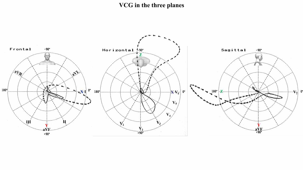
VCG in the three planes
IIIIIaVF
X I
Y
X V6
V1
V4
V5
V2
V3
Z
YaVF
V2E
0
RA
LA
E0
LA
E 0
RALA
Magnified P-loop (32x) in the three planes
Echocardiogram M-mode
Colleagues opinions
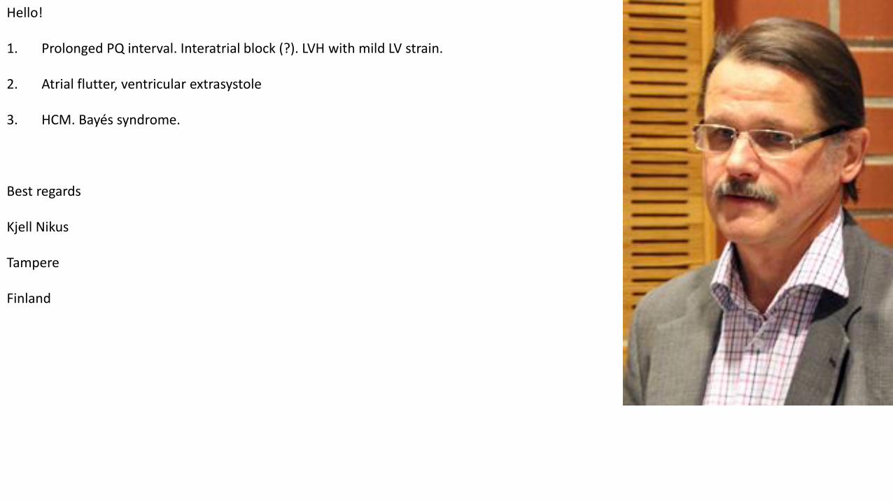
The ECG looks like LVH (Cornell criteria# ) and Left atrial abnormality. Will learn VCG abnormalities from the masters.Dr. Melvin ScheinmanProfessor of Medicine Department of Cardiac Electrophysiology, University of California San Francisco, California, USA. Electrophysiology Service 500 Parnassus AvenueSan Francisco, CA 94143-1354Telephone/FAX/E-mail: Phone: (415) 476-5706Fax: (415) 476-6260 email: [email protected]
# Cornell index (CI), Casale criteria or Cornel criteria: CI = RaVL + S V3 > than 28 mm in men or > 20 mm in women indicates LVH. Cornell product (Molloy 1992) 1985; (CorP. )*Cornell voltage-duration product It is the product of QRS voltage and QRS duration (QRSvoltage-duration product); Cornell voltage-duration product (RaVL + SV3 with 6 mm added in women x QRS duration). Values ≥2440 mm/ms arediagnostic of LVH (Positive criteria of LVH CP≥2440 mm x ms).The Cornell product is a useful ECG marker, reflecting not only left ventricular mass but also LV geometry and diastolic function in Japanesehypertensive patients (Shira 2007).Reduction in Cor P ECG LVH during antihypertensive therapy is associated with fewer hospitalizations for HF, independent of blood pressurelowering, treatment method, and other risk factors for HF. (Okin 2007).
El ECG #1: ritmo sinusal 46 lpm, bloqueo AV 1° grado, bloqueo interauricular avanzado, sobrecarga auricular izquierda (criterio de Morrispositivo en V1). Sobrecarga VI e hipertrofia VI. (Criterios de voltaje: índice de Sokolov-Lyon y de Cornell positivos), deflexión intrinsecoide >50 mseg en V5 y V6). También se observa onda Q importante en DIII < 40 mseg y de mayor voltaje que onda R y onda T positiva (pseudoinfarto)en espejo de aVL. también fQRS en cara inferior (predictor de eventos arritmicos potencialmente fatales). Presencia en QS desde V1-V2 conSupraST > 0.1 mV hasta V4 y onda T positiva Es visible también pseudo onda delta en V4. Probable onda J en V5 y V6.Signos electrocardiográficos de miocardiopatía hipertrófica obstructiva.ECG #2: Fibrilación auricular de alta respuesta ventricular.Diagnóstico clínico : Síndrome de Bayés. Miocardiopatía hipertrófica obstructiva, insuficiencia mitral.Los saludo afetuosamente
Dr Juan Carlos ManzzardoMendoza Argentina
Spanish
ECG # 1: sinus rhythm, heart ratei 46 bpm, 1st degree AV block, advanced interatrial block, left atrial enlargement (positive V1 Morris criterion). LAE+ LVH (Voltage criteria: positive Sokolov-Lyon and Cornell index), intrinsecoide deflection> 50 ms in V5 and V6). We also observed a significant Q wave in III <40 msec and higher voltage than R wave and positive T wave (pseudoinfarction) in aVL mirror. Also fQRS on the lower face (predictor of potentially fatal arrhythmic events). Presence in QS from V1-V2 with SupraST> 0.1 mV to V4 and positive T wave It is also visible pseudo delta wave in V4. Probable J wave in V5 and V6.Electrocardiographic signs of obstructive hypertrophic cardiomyopathy.
ECG # 2: Atrial fibrillation with high ventricular response.
Queridos Andrés y Raimundo,Por fin llegó un VCG de bloqueo Avanzado interauricular (BIA-A)….Bellísimo. Ya os he comentado que creo que la VCG puede facilitarel diagnóstico y sugerir grado y localización de la fibrosis…..Para ello se necesita un gold estándar de fibrosis que es la resonanciamagnética. Creo podría servir como surrogate la “speckle-tracking” ecocardiografica.Raimundo y yo ya hablamos de esto en Roma….Ánimos!!Un abrazo,AntonioProf.Antonio Bayés de LunaInvestigador Sènior- ICCCFundació d'Investigació CardiovascularHospital Sta. Creu i St. PauC/ Sant Antoni Maria Claret, 167- Pab 1108025 Barcelona, SpainTel: +34 93 556 5612Fax:+34 93 556 5563email: [email protected]
EnglishDear Andrés and Raimundo
At last came an Advanced Interatrial Block VCG (A-IAB) ... .BEAUTIFULL!!!!!. I have already commented that I believe that the VCG canfacilitate the diagnosis and suggest the degree and location of fibrosis ... .. For this you need a Gold standard fibrosis which is the MRI. I thinkit could serve as surrogate speckle-tracking echocardiography.Raimundo and I already talked about this in Rome .... Few days agoA hug,
Hola: Hermosa presentaciónEn contexto de apreciaciones referidas por Dr Nikus y Antoni el VCG muestra:1. Con un bucle de P en el plano frontal orientado en cuadrante superior izquierdo y eje máximo de P en - 40° por influencia de la despolarización de AI que se
realiza de abajo hacia arriba (y no a la inversa)por bloqueo de las vías o fibras ("autopistas" al decir de Bayes) de Bachman configurando un bloqueo inter auricular avanzado.
2. El bucle del QRS en el plano horizontal totalmente hacia atrás es sugestivo de fibrosis de segmentos anteroseptal que se correlaciona con el QRS. Además, muestra características de agrandamiento de VI.
Saludos cordialesJan José Sirena Santiago del Estero Argentina
English
Hi, beautiful presentation!In the context of the assessments referred to by Dr Nikus and Antoni the VCG shows:The P-loop axis in the frontal plane is oriented in the upper left quadrant (P- maximum axis in - 40 °) by influence of the depolarization of LA that is performed from bottom to top (and not vice versa) by blocking the tracks or fibers ("freeways" as Bayes says) by setting up an advanced inter-atrial block.(A-IAB)The QRS loop in the PH is directed fully backward and leftward. It is suggestive of anteroseptal segment fibrosis correlates with the QRS pattern. In addition, it shows features of LVH.Best RegardsJuan José Sirena Santiago del Estero Argentina
P –wave duration ≥ 120ms + biphasic - positive-negative-“plus-minus” P wave in inferior leadsII, III and aVF + caudo-cranial left atrial activation + repetitive paroxysmal atrial fibrillationepisodes: Bayes' syndrome. Advanced, “third-degree” or “complete” interatrial block (A-IAB) associated with atrial Tachyarrhythmias = Bayés´s syndrome.
+ Left Atrial Enlargement (LAE) (It is present in 90% of cases of advanced interatrial block (A-IAB). Bayés de Luna, et al perform the first consensus to separates A-IAB from LAE (Bayés deLuna 2012). The longer P-wave duration observed in HCM patients may be explained by a higherprevalence of block in one or more of the interatrial conduction routes. (Holmqvist 2007)
+LVH with strain pattern of repolarization, Cabrera LVH systolic type or VCG LVH type 1A
Interatrial block (IAB) is a ECG disturbance that entails a delayed conduction between the both atria during atrial depolarization. IAB has furtherbeen classified into partial IAB (pIAB) or second degree IAB, which is seen on the ECG as P-wave duration ≥ 120 ms, or advanced IAB (A-IAB)in which P wave duration ≥ 120ms with a biphasic positive-negative “plus-minus” P-wave in the inferior leads (II, III, and aVF). In addition theP-loop of the VCG show a characteristic P-loop pattern in the FP: the orthogonal lead “Y” lead (+90°/-90°) plus-minus with the final negativeportion ≥40 ms, consequence of retrograde caudo-cranial activation of the left atrium (LA) with final portion of P-loop delayed, notches andslurring in the last portion. (Bayes de Luna 2012) This pattern arises owing to a delay of conduction at the Bachmann region (old Bachmanbundle) resulting in retrograde, caudocranial conduction to the left atrium (LA) through the coronary sinus musculature (Cosio 2004). However, ifthe wave passes through the rim of the fossa ovalis, then retrograde activation may not necessarily occur.(Tse 2017) This abnormal caudal-cranialactivation of the LA owing to fibrosis of the Bachmann region predisposing patients to interatrial dyssynchrony. The presence of Bayés syndromein patients with atrial pacing has not been sufficiently explored. Clinicians must be cognizant of this possibility as the early identification of thispattern suggests the need for closer monitoring of AF and prophylaxis of cardioembolic events.(Britton 2017). Dr Bayés de Luna was the firstwho provided a clear description of atrial conduction block in 1979, classifying them into either inter- and intra-atrial (Bayés de Luna 1979). Inrecognition of his numerous contributions to the understanding of IAB (Bayes de Luna 1985), this syndrome was named Bayés syndrome byConde and Baranchuk, (Conde 2014). The clinical relevance of Bayés syndrome lies in the fact that is a clear arrhythmological syndrome and hasa strong association with supraventricular arrhythmias, particularly atypical atrial flutter and AF and it is independently associated with anincreased risk for non-lacunar cardioembolic stroke. Likewise, can be the cause of some cryptogenic strokes, and be related to clinically silentcerebral ischemia and vascular cognitive impairment( such as the present case), or even, vascular dementia (Arboix 2017). Likely owing todelayed, heterogeneous activation of the LA (Bayés de Luna 2012). We described and old man wit systemic hypertension and OH-HCM whoseECG/VCG shows typical A-IAB, LVH associated with repetitive episodes of paroxysmal AF which characterizes the so-called Bayes syndrome anunder-recognized clinical syndrome. Positive-negative biphasic or “plus-minus” P-wave + P-duration ≥ 120ms + AF episodes: Bayés syndromeowing to fibrosis of the Bachmann region predisposing patients to interatrial dyssynchrony and AF. Bayes’ syndrome has been identified innumerous patient populations, but its presence in patients with atrial pacing has not been sufficiently explored. Clinicians must be cognizant of thispossibility as the early identification of this pattern suggests the need for closer monitoring of AF and prophylaxis of cardioembolic events. IABmay be of 1st degree (P-wave duration >120 ms), A-IAB: P wave≥ 120ms biphasic [±] in inferior leads, and 2nd degree when these patterns appeartransiently in the same ECG recording (atrial aberrancy). These P-wave patterns are due to a block because they may (a) appear transiently, (b) bewithout associated LAE, and (c) may be reproduced experimentally.
Interatrial block and Bayés syndrome overview
Third degree block, complete or advanced interatrial block: P-duration ≥120ms+ biphasic P-wave in inferior leads
LABB
RA
A
PM
RAJ
P loop in FPP-wave in lead “Y” aVF
Third Degree, A-IAB or complete IABElectrical impulse is blocked/delayed in Bachmann’s muscular interatrial bundle or region (BB), but retrograde left atrial activation usuallyoccurs.(Ariyarajah 2005) Note the existence of an open angle between the vector of the first portion of P wave (RA) and the last portion (LA).Electrophysiological study demonstrates retrograde activation of the LA. Consequently P loop/wave in orthogonal lead “Y”, aVF and III isbiphasic plus-minus ±. LA activation occurs by an alternate route rather than proceeding from right to left via the BB.( Spodick 2007)
Complete block in Bachmann’s region
≥120ms
LA
RA
LA-60°
Y
XE
0RA
LA
aVF Leads II, III and aVFBipahisc plus-minus P wave
Biphasic P waves, in inferior leads
III II
aVF
Retrograde caudo-cranial activation of the left atrium (LA)
P-loop and inferior P-waves in the frontal Plane in the presentcase (High magnification of the P loop (0.1 mV = 3 cm)
P-loop and P-wave polarity in the frontal Planein normal cases(High magnification of the P loop (0.1 mV = 3 cm)
LA
RA
E
SÂP
Positive-negative biphasic or “plus-minus” P-wave + P-duration ≥ 120ms (A-IAB) + repetitive AF episodes: Bayés syndrome
P-axis = - 35°
P-axis = + 55°
In 1963, Thomas N. James described 3 pathways connecting the sinus node to the atrioventricular node (AVN), namely the anterior, medial, and
posterior internodal pathways (James 1963). Whether these conduction pathways were because of the presence of specialized conduction tissue or
because of the anisotropic orientation of the muscle fibers remains controversial. Nevertheless, James described the anterior pathway as leaving
the sinus node in anterior direction and giving off a secondary branch at the level of the superior vena cava to form BB.(Bachmann 1916) BB
stretches subepicardially across the interatrial groove (septal raphe). It is at the interatrial groove that the BB can be identified as a discrete bundle
(Figures next 2 slides) separated by fatty tissues from the infolded right atrial wall that is the limbus of the oval fossa. Notably, the bundle is not
surrounded by a fibrous tissue sheath. Instead, the bundle is comprised of strands of atrial myocardium that are similarly aligned in parallel
fashion. Its rightward and leftward extensions bifurcate to pass to either side of the right and left atrial appendages (LAA) (Khaja 2003). Although
they can be traced to varying extents with blunt dissection, both extensions blend into the musculature of the atrial walls. The superior arm of the
rightward extension arises in the region of the cavoatrial junction close to the site of the sinus node and in the vicinity of the sagittal bundle. The
inferior arm arises in the subepicardium of the RA vestibule. Leftward, BB buttressing part of the anterior atrial wall with its thickness (figure next
slide) is still traceable to where it encircles the neck of the LAA and blends in with the lateral atrial wall. The superior part traverses in the
infolding of the atrial wall, known to arrhythmologists as the left lateral ridge, to pass in front of the orifices of the left pulmonary veins.(Cabrera
2008) The inferior part descends toward the atrial vestibule to combine with the circumferentially aligned myocardial strands in the subepicardium
of the inferior wall. In contrast to the thinner distal extensions, BB’s body across the interatrial groove is a broader band (see next 2 figures), with
median measurements of 4 mm in thickness and 9 mm in height. It is described as trapezoidal shaped because of its short lower length (3 mm) and
longer upper length (10 mm).(Lemery 2003)
Heart viewed from the front has the tips of the right and left atrial appendages (RAA and LAA) pulled back (left). Blunt dissection revealsBachmann’s bundle (BB) crossing the interatrial groove (white arrows). The black arrows mark the rightward and leftward extensions. The blueoval marks the anticipated site of the sinus node. Longitudinal cut through the left heart (right) displays BB cut in cross-section (*). Note that itadds considerably to the thickness of the left atrial (LA) wall in this heart. LSPV indicates left superior pulmonary vein; RSPV, right superiorpulmonary vein; and SVC, superior vena cava.
Longitudinal cut through Bachman’s bundle and the atrial septum in a heart shows the bundle passing epicardial to the interatrial groove that isfilled with epicardial fat (*). IVC indicates inferior vena cava; LAA, left atrial (LA) appendage; LSPV, left superior pulmonary vein; RA, rightatrium; and SVC, superior vena cava.
* Epicardial fat
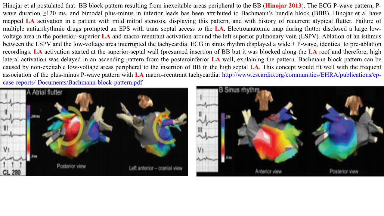
Hinojar et al postulated that BB block pattern resulting from inexcitable areas peripheral to the BB (Hinojar 2013). The ECG P-wave pattern, P-wave duration ≥120 ms, and bimodal plus-minus in inferior leads has been attributed to Bachmann’s bundle block (BBB). Hinojar et al havemapped LA activation in a patient with mild mitral stenosis, displaying this pattern, and with history of recurrent atypical flutter. Failure ofmultiple antiarrhythmic drugs prompted an EPS with trans septal access to the LA. Electroanatomic map during flutter disclosed a large low-voltage area in the posterior–superior LA and macro-reentrant activation around the left superior pulmonary vein (LSPV). Ablation of an isthmusbetween the LSPV and the low-voltage area interrupted the tachycardia. ECG in sinus rhythm displayed a wide + P-wave, identical to pre-ablationrecordings. LA activation started at the superior-septal wall (presumed insertion of BB but it was blocked along the LA roof and therefore, highlateral activation was delayed in an ascending pattern from the posteroinferior LA wall, explaining the pattern. Bachmann block pattern can becaused by non-excitable low-voltage areas peripheral to the insertion of BB in the high septal LA. This concept would fit well with the frequentassociation of the plus-minus P-wave pattern with LA macro-reentrant tachycardia: http://www.escardio.org/communities/EHRA/publications/ep-case-reports/ Documents/Bachmann-block-pattern.pdf
ECG/VC correlation in the frontal plane
IIIII
aVF
X I
Y
T
Magnified P-loop (32x)
E0
RA
LA
Electrical impulse is blocked/delayed in Bachmann’s region muscular interatrial bundle (BB), but retrograde LA activation usuallyoccurs.(Ariyarajah 2005) Note the existence of an open angle between the vector of the first portion of the P-wave (RA) and the last one(LA). Electrophysiological study demonstrates retrograde activation of the LA. Consequently, P loop/wave in orthogonal lead “Y”, aVFand III is biphasic/positive-negative plus-minus ±. LA activation occurs by an alternate route rather than proceeding from RA to LA viathe BB region.( Spodick 2007)
Y
X
aVF
IIIII
P-loop axis = - 35°
POpen angle
X V6
V1
V4
V5
V2
V3
Z
ECG/VC correlation in the horizontal plane
T
Magnified P-loop (32x)
E0
RA
LA
T
V1
z
V1
z
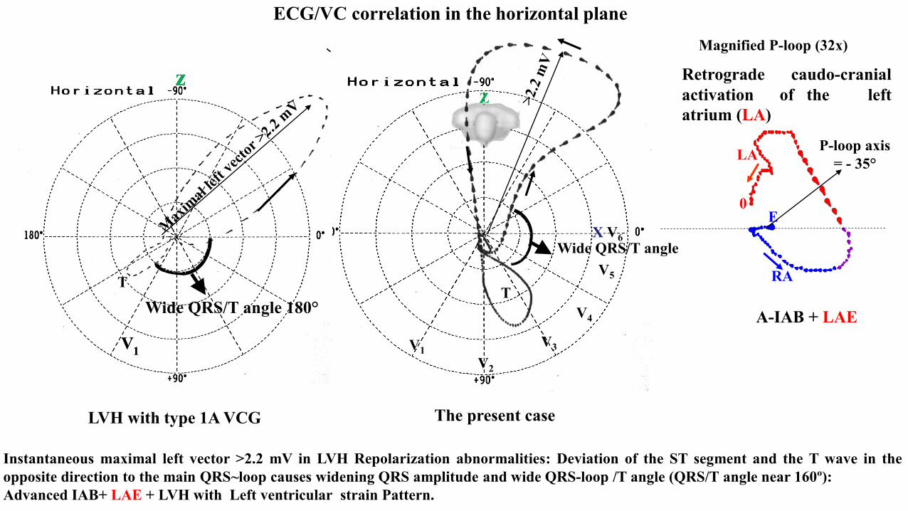
Instantaneous maximal left vector >2.2 mV in LVH Repolarization abnormalities: Deviation of the ST segment and the T wave in theopposite direction to the main QRS~loop causes widening QRS amplitude and wide QRS-loop /T angle (QRS/T angle near 160º):Advanced IAB+ LAE + LVH with Left ventricular strain Pattern.
Retrograde caudo-cranialactivation of the leftatrium (LA)
Wide QRS/T angle
Wide QRS/T angle 180°
P-loop axis = - 35°
LVH with type 1A VCG The present case
A-IAB + LAE
ECG/VC correlation in the Right Sagittal Plane (RSP)
YaVF
V2
T
Magnified P-loop (32x) in the present case
E 0
RALA
P
The normal magnified P-loop (32x) in the RSP
Z
Z
P-loop directed to back and upward (almost the entire P-loop located inposterosuperior quadrant) in RSP. In normal P wave activation P-loop is located oninferior and anterior quadrant in RSP. Plus minus P wave in aVF . The maximal leftvector >2.2 mV and directed to back. QRS-loop and T-loop with opposite directionand the angle QRS/T near 180°.
V2
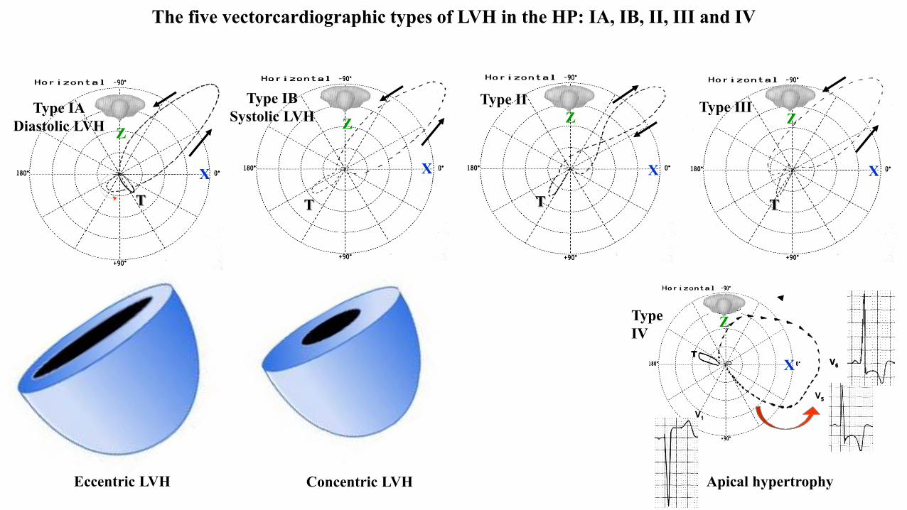
Type IADiastolic LVH
Type IBSystolic LVH Type IIIType II
T T TT
X
ZZ ZZ
XX X
V1
V5
V6TT
V1
V5
V6TT
TypeIV
Z
X
The five vectorcardiographic types of LVH in the HP: IA, IB, II, III and IV
Eccentric LVH Concentric LVH Apical hypertrophy
X V6
V1
V4
V5
V2
V3
Z
T
Wide QRS/T angleT Wide QRS/T angle
LVH Type IA vectorcardiographic The present case: LVH Type IA vectorcardiographic
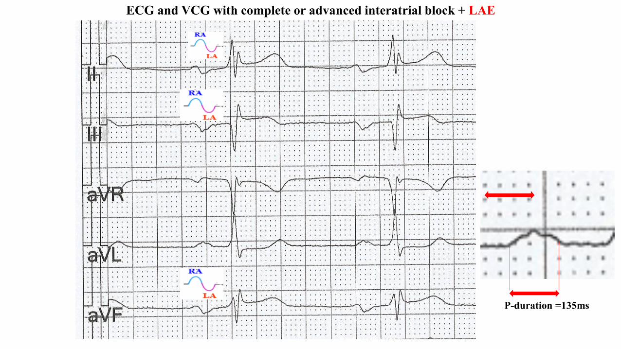
ECG and VCG with complete or advanced interatrial block + LAE
P-duration =135ms
ECG 2015
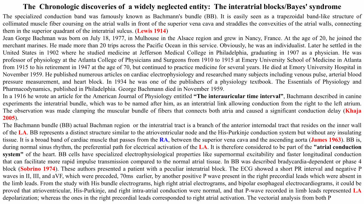
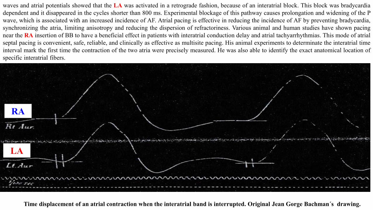
The specialized conduction band was famously known as Bachmann's bundle (BB). It is easily seen as a trapezoidal band-like structure ofcollimated muscle fiber coursing on the atrial walls in front of the superior vena cava and straddles the convexities of the atrial walls, connectingthem in the superior quadrant of the interatrial sulcus. (Lewis 1914)Jean Gorge Bachman was born on July 18, 1977, in Mulhouse in the Alsace region and grew in Nancy, France. At the age of 20, he joined themerchant marines. He made more than 20 trips across the Pacific Ocean in this service. Obviously, he was an individualist. Later he settled in theUnited States in 1902 where he studied medicine at Jefferson Medical College in Philadelphia, graduating in 1907 as a physician. He wasprofessor of physiology at the Atlanta College of Physicians and Surgeons from 1910 to 1915 at Emery University School of Medicine in Atlantafrom 1915 to his retirement in 1947 at the age of 70, but continued to practice medicine for several years. He died at Emory University Hospital inNovember 1959. He published numerous articles on cardiac electrophysiology and researched many subjects including venous pulse, arterial bloodpressure measurement, and heart block. In 1934 he was one of the publishers of a physiology textbook. The Essentials of Physiology andPharmacodynamics, published in Philadelphia. George Bachmann died in November 1959.In a 1916 he wrote an article for the American Journal of Physiology entitled “The interauricular time interval”, Bachmann described in canineexperiments the interatrial bundle, which was to be named after him, as an interatrial link allowing conduction from the right to the left atrium.The observation was made clamping the muscular bundle of fibers that connects both atria and caused a significant conduction delay (Khaja2005).The Bachmann bundle (BB) actual Bachman region or the interatrial tract is a branch of the anterior internodal tract that resides on the inner wallof the LA. BB represents a distinct structure similar to the atrioventricular node and the His-Purkinje conduction system but without any insulatingtissue. It is a broad band of cardiac muscle that passes from the RA, between the superior vena cava and the ascending aorta (James 1963). BB is,during normal sinus rhythm, the preferential path for electrical activation of the LA. It is therefore considered to be part of the "atrial conductionsystem" of the heart. BB cells have specialized electrophysiological properties like supernormal excitability and faster longitudinal conductionthat can facilitate more rapid impulse transmission compared to the normal atrial tissue. In BB was described bradycardia-dependent or phase 4block (Sobrino 1974). These authors presented a patient with a peculiar interatrial block. The ECG showed a short PR interval and negative Pwaves in II, III, and aVF, which were preceded, 70ms earlier, by another positive P wave present in the right precordial leads which were absent inthe limb leads. From the study with His bundle electrograms, high right atrial electrograms, and bipolar esophageal electrocardiograms, it could beproved that atrioventricular, His-Purkinje, and right intra-atrial conduction were normal, and that P-wave recorded in limb leads represented LAdepolarization; whereas the ones in the right precordial leads corresponded to right atrial activation. The vectorial analysis from both P
The Chronologic discoveries of a widely neglected entity: The interatrial blocks/Bayes' syndrome
waves and atrial potentials showed that the LA was activated in a retrograde fashion, because of an interatrial block. This block was bradycardiadependent and it disappeared in the cycles shorter than 800 ms. Experimental blockage of this pathway causes prolongation and widening of the Pwave, which is associated with an increased incidence of AF. Atrial pacing is effective in reducing the incidence of AF by preventing bradycardia,synchronizing the atria, limiting anisotropy and reducing the dispersion of refractoriness. Various animal and human studies have shown pacingnear the RA insertion of BB to have a beneficial effect in patients with interatrial conduction delay and atrial tachyarrhythmias. This mode of atrialseptal pacing is convenient, safe, reliable, and clinically as effective as multisite pacing. His animal experiments to determinate the interatrial timeinterval mark the first time the contraction of the two atria were precisely measured. He was also able to identify the exact anatomical location ofspecific interatrial fibers.
Time displacement of an atrial contraction when the interatrial band is interrupted. Original Jean Gorge Bachman´s drawing.
RA
LA
Jean George Bachmann 1877-1959
1. Bachmann G: The interpretation of the venous pulse. Am JM,~~ November (1908) ri Bachniann G: C Dniplcte auriculoventricular dissnciationwithout S)ncopal or epileptiform attacks. Am J Med Sci 137, 342
2. Bachmann G: Sphygmographic study of a case of complete heart. block. Arch hem Med 4, 238 (1909)3. Bachmann G: The measurement of arterial pressure in man. New Yorl Med J 93, 212 (I91)4. Bachmann G: A physiologico-pathological study of a case of heart block occurring in a dog as a result of natural causes. J,I+,, Med XVI,
I(lY12)5. Bachmann G: An automatic spinning device for the Harvard kymo. graph. JAMA LXVI, 188 (1916)6. Bachmann G: The inter-auricular time interval. Am J Physiol XLI, 309 (1916)7. Bachrnann G: The distribution of the vagus nerves to the sino-auricula, junction of the mammalian heart. Am J Phgsiol LXIII, 2 (1923)8. Bachnlann G: A Course in Erpenmenfd Physiology. Privately printed by author with illustrations by author (1924), 949. Bachmann G: The significance of splitting of the P-wave in thc &. Irocardiograin. Ann Infern Med 9, 14 (1941) ,10. Haldi J: A comparative btudy of the respiratory quotient following the ingestion of glucose and of fructose as af. fected by the lactic acid and
carbon dioxide changes in the bid J Nurri 2, 13 (1937)11. Bachmann G. Haldi J, Ensor C, Wynn W: The effects of the ingestion of glucose and of fructose on the rate or excretion of urine and various
constituents. Am J PhJsiol I, 12 (1938)12. Bachmann G, Haldi J, Ensor C. Wynn W: Creatinuria following the oral administration of caffeine. Am JPhysiol I, 138 ( 1942)13. Bachmann G, Haldi J, Wynn W, Ensor C: The respiratory quotient and carbohydate metabolism following the ingestion of glucose and of
fructose, as affected by exercise taken immediately and 30 minutes after ingestion. Am J Phgsiol 3, I20 (1937)14. Bachmann G, Haldi J, Wynn W, Ensor C: The effects produced b) decreasing the calcium and phosphorus intake on calcium and phosphorus
absorption and disposition and on various body constituents of the rat. J Nurri 2, 20 (1Y40)15. Haldi J, Bachmann G, Ensor C, Wynn W: Muscular efficiency In relation to the taking of food and to the height of the respirattlry quotient
immediately before exercise. Am J Phgsiol I, I? (1938)16. Haldi J, Bachinann G, Ensor C, Wynn W: Comparative effects of a high glucose and a high fructose diet on activity, body wei@ and various
constituents of the liver and body of the albino exercising at will. J Nurri 3, 16 (1938)17. Haldi J, Bachinann G, Ensor C. Wynn W: The effects on respiratory metabolism produced by equal amounts of caffeine in the form of coffee,
tea, and the pure alkaloid. J Nurri 4, 27 (1944)
Bachmman´s Manuscripts
➢ 1920 Papez studied the characteristic of the atria musculature (Papez1920)➢ 1946 Decherd et al described the interatrial and sinoatrial block, with an illustrative case.(Decherd 1946)➢ 1956 Bradley and Marriot described the interatrial block in Circulation( Bradley 1956)➢ 1963 Thomas described "The connecting pathways between the sinus node and A-V node and between the RA and the LA in the human heart“
in the America Heart Journal. (Thomas 1963) In the same year, Horiba published in Japan Heart Journal the stimulus conduction in atria andin the BB with intracellular microelectrodes.(Horiba 1963)
➢ 1965 Cohen and Scherf described the atrial dissociation secondary to the complete interatrial and intra-atrial block(Cohen 1965)➢ 1966 Wagner et al studied the specialized conducting fibers in the interatrial band published in Circulation Research. (Wagner 1966) The
authors concluded: 1) The interatrial band is not a homogeneous structure, but contains two fiber types. 2) In addition to ordinary atrialmuscle, specialized conducting fibers are present in the interatrial band. 3) Impulse spread in the interatrial band is not radial or uniform.Rather, it occurs through several linear paths which probably have infrequent cross-connections.
➢ 1967 De Michelis e Paparella described a case of interatrial block in Mexico.( DeMichelis 1967)➢ 1969 Childers Merideth and Moe described for the first time supernormal phenomenon in BB in an in vitro and in vivo study in dog
(Childers 1969). In the same year, Sanna et al described ECG features of complete interatrial block (Sanna 1969)➢ 1970 Waldo et al . described the P wave and PR interval and the effects of the site of origin of atrial depolarization.(Waldo 1970) The authors
studied experimentally the effects on the canine P-wave of discrete lesions in the specialized atrial tracts. In canine experiment observed thatafter the transection of anterior internodal tract, the P-wave duration increased significantly though morphology and polarity remained thesame. However, transection of the BB not only caused increased P-duration but also distorted its polarity and morphology. (Waldo 1970).
➢ 1971 Hutter and Page presented a case of atrial arrhythmias and lipomatous hypertrophy of the cardiac interatrial septum.(Hutter 1971).➢ 1972 Sangiorgi and Cannata described a partial interatrial block with duplex atriogram and with double atrial heterotopic, unstable
interferential rhythm.(Sangiorgi 1972)➢ 1973 Waldo et al described the etiology of prolongation of the PR interval in patients with an endocardial cushion defect. Further observations
on internodal conduction and the polarity of the retrograde P wave. (Waldo 1973).➢ 1974 Legato and Ferrer described for the fist time Intermittent or transient form of intra-atrial block its diagnosis, incidence and
implications. (Legato 1974a). In the same year, Legato studied the atrial ultrastructure in patients with fixed intra-atrial block. (Legato1974b).
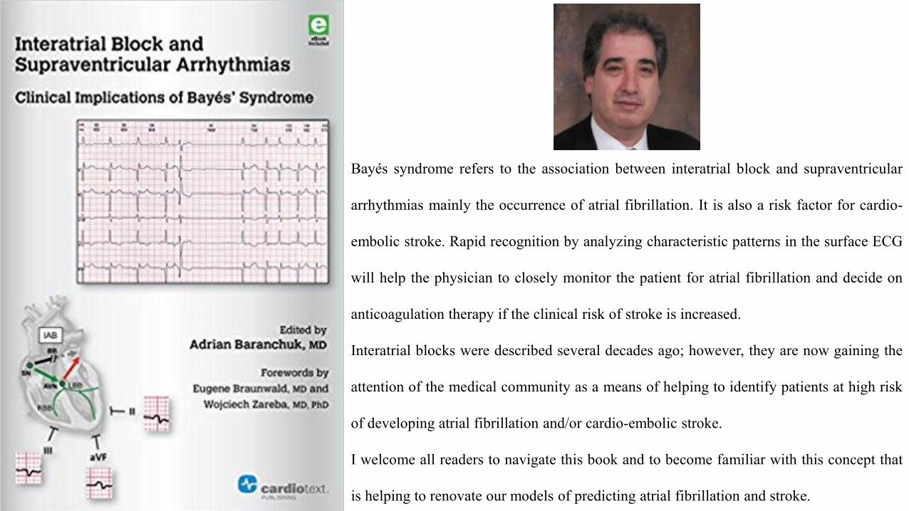
Chronology of the main discoveries related to interatrial blocks and Bayés´s syndrome
➢ 1975: Di Biase and Rizzon show a case of interatrial block with retrograde activation of the left atrium (Di Biase 1975). In this year Lee etal described "Bachmann's bundle block“ associated with P mitrale in a patient with rheumatic mitral stenosis. (Lee 1975) and Waldo et al,showed the sequence of retrograde atrial activation in the canine heart, pointing out the correlation with positive and negative retrograde Pwaves. (Waldo 1975).
➢ 1976: Zoneraich and Zoneraich studied the Intraatrial conduction disturbances and its VCGs patterns. The authors preformed the Frank Ploop VCGs in 30 normal subjects and in 40 patients who had intraatrial conduction disturbances alone or in association with cardiac disease.High magnification of the P loop (0.1 mV = 3 cm) permitted accurate measurement of the P-loop duration, magnitude and direction. High-frequency recordings allowed optimal evaluation of the notches, bites and conduction delays in the PsE loop. Four VCG patterns have beenselected as counterparts of the 4 types of enlarged P waves seen in ECGs of patients with atrial conduction disturbances. When intraatrialconduction disturbances coexisted with LA enlargement, the PsE loop was larger and smoother. The authors concluded that highmagnification, high-frequency VCG of the P-loop seems to be the best available method for determining a specific pattern of intraatrialconduction disturbance.(Zoneraich 1976)
➢ 1977 Sobrino et al presented a case of bradycardia-dependent interatrial block(phase 4 block) with retrograde LA activation.(Sobrino 1977) Inthe same year, Leier et al showed a “dissimilar atrial rhythms” in a patient with interatrial block. The patient had type A Wolff-Parkinson-White syndrome and prolonged interatrial conduction intervals developed atrial flutter during the course of an EPS. The atrial flutter blockedalong the left-to-right conduction pathways in a Wenckebach pattern. The dissimilar atrial rhythms of RA tachycardia and LA flutter evolvedas the interatrial block increased to 2:1 conduction.(Leier 1977)
➢ 1979 Bayés de Luna described the block at the auricular level.(Bayés de Luna 1979) This is the first manuscript related interatrial blockswrithed by the great Catalonian Master. In the same year, Sherf and James described the fine structure of cells and their histologicorganization within internodal pathways of the heart and its clinical and its ECG implications. (Sherf 1979) in 1 human and 2 canine heartsand correlated with histologic observations on more than 100 human and 10 canine hearts. From the electron microscopic studies 6 differentkinds of myocardial cells were classified from two locations: the Eustachian ridge (posterior internodal pathway) and the BB (anteriorinternodal pathway). 5 of the 6 kinds of cells (working myocardial cells, Purkinje-like cells, either broad or slender transitional cells andP cells, and, pleomorphic and dark in appearance, with a special intertwined relation to P cells, is newly designated as an amoeboid cell. Itwas found solely in the Eustachian ridge. In the same area a rare direct contact between a nerve and a myocardial cell was observed. Theauthors analyzed the importance of these different kinds of cells, their respective cell connections, and their topographic locations inside theinternodal pathways.
➢ 1981 Anderson et al studied the internodal atrial myocardium (Anderson 1981) The authors verified that there was nothing "special" about themyocardium between the nodes, nor was it possible to recognize tracts on the basis of either histological appearance or cellular architecture.“From the standpoint of light microscopy, there is no evidence whatsoever to support the purported concept of specialized anatomicalsubstrates for internodal conduction”.
➢ 1982 Spach et al studied experimentally in dogs the functional role of structural complexities in the propagation of depolarization in the atriumand cardiac conduction disturbances due to discontinuities of effective axial resistivity. (Spach 1982)
➢ 1983 Husson reported a case of partial interatrial block (Husson1983) involving the upper part of LA, demonstrated by esophageal recordingsat different levels. Abnormally retrograde atrial depolarization in the upper part of the LA was observed, but normal descending depolarizationin the lower part of the LA suggested that interatrial conduction was blocked on the BB but was normal on the middle interatrial pathway. Thesurface ECG showed a normal P wave duration(?); inverted P-wave in III and aVF, and flat in II an normal PR interval. The author suggests aclassification of interatrial block into three types.
➢ 1985 Bayés de Luna et al described both ECG/VCG of interatrial blocks with LA retrograde activation (Bayés de Luna 1985) from 81,000ECGs they collected 83 cases that fulfilled the criteria of interatrial conduction disturbances(IACD) with LA retrograde activation (IACD-LARA) (P +/- in II, III and VF with P duration ≥120 ms. The authors presented the detailed study of 35 cases with surface ECG/VCG and 29cases with orthogonal ECG leads. The results were then compared against two control groups: with cardiopathy (30 cases) and withoutcardiopathy (25 cases). The prevalence of IACD-LARA was nearly 1% globally, and 2% among patients with valvar heart disease. Thediagnostic criteria for IACD-LARA are:
1) ECG: P +/- in II, III and VF with P duration ≥120 ms2) Open angle (usually > 90°) between the first and the second part of the P-loop-wave.3) Orthogonal P +/- in Y lead (+90°-90°)with a negative mode of final> 40 ms.4) VCG: >50 ms. above the X( 0°/ ± 180°) and/or Z orthogonal leads of VCG axis5) Duration of the P loop > 110 ms.6) Open angle between the two parts(initial and final) of the P loop in both frontal(FP) and right sagittal planes(RSP)
(171.2° +/- 15.1), and7) Presence of notches and slurring's in the last part of the P loop.
➢ 1985 Husson et al reported the case of partial interatrial block arising in the mid zone of the LA as demonstrated by esophageal recordings.These showed that atrial depolarization descending normally in the superior part of the LA was abnormally retrograde in the inferior part.This suggests a block of the mid-interatrial pathway and conduction via BB in the upper part of the LA and via a retrograde pathway in theinferior region. The surface ECG showed a P-wave of normal duration(?) which was diphasic in III and aVF with a normal PR interval. Inaddition, endocavitary recordings in the inferior part of the RA showed the presence of preatrial potentials during three ectopic complexes.Cause? (Husson 1985)
➢ 1987 Medrano et al. produced RA and LA damage with a subepicardial infiltration of 96° alcohol in 2 groups of dogs. In 6 other dogs the leftor right portion of the interatrial band was also injured. Conventional ECG and supplementary unipolar leads were recorded usingphotographic and direct inscription polygraphs at paper speeds of 50 and 100 mm/s. Control, immediate postinjury and late tracings wereobtained. AV block was provoked to determine QTac. Bradycardia, slight P-wave duration prolongation and PR interval were observed withboth types of atrial damage. In 4 cases low RA rhythm was detected: two showed anatomohistological sinus node involvement in RA injury.Qp waves were registered over the left precordium with necrosis of both sides, but were more frequent with RA damage. Damage of the leftportion of the interatrial band delayed LA activation and split P waves in the precordial leads. Damage of the RA distorts the initial vectors,magnifying the left ones and simulating LA enlargement. The Qp registered on the RA is also detected by surface leads. Contrary distorted LAdepolarization increases the RA vector and delays the left, ones, giving rise to greater asynchronism and bimodal P waves.
➢ 1988 Bayés de Luna A et al.(Bayés de Luna 1988) studied 16 patients with ECG evidence of complete or advanced interatrial block(A-IAB)with retrograde activation of the LA: P duration ≥120 ms, and plus-minus (+/-) biphasic P-waves in inferior leads II, III, and VF. 8 patients hadvalvular heart disease, 4 had dilated cardiomyopathy and 4 had other forms of heart disease. Patients with valvular heart disease andcardiomyopathy were compared with a control group of 22 patients with similar clinical and echocardiographic characteristics, but without thistype of interatrial block. Patients with A-IAB and retrograde activation of the LA had a much higher incidence of paroxysmal supraventriculartachyarrhythmias (93.7%) during follow-up than did the control group. 11 of 16 patients (68.7%) with A-IAB and retrograde activation of LAhad atrial flutter (atypical in 7 cases, typical in 2 cases, and with two or more morphologies in 2 cases). 6 patients from the control group(27.7%) had sustained atrial tachyarrhythmias (5 AF and 1 typical atrial flutter). The atrial tachyarrhythmias were due more to A- IAB andretrograde activation of LA and frequent PACs than to LAE, because the control group with a LA of the same size, but without A-IAB andretrograde activation of LA and with less incidence of PACs, had a much lower incidence of paroxysmal tachycardia.In the same year, Vitali et al presented a case of interatrial block during acute myocardial infarction. (Vitali A 1988).
➢ 1989 Bayés de Luna et al.(Bayés de Luna 1989) demonstrated the value of preventive antiarrhythmic treatment in patients with A-IAB. In thispopulation LAE is present in 90% of cases. Using drugs (amiodarone, quinidine or verapamil) this percentage was greatly lowered (25%).Atrial tachyarrhythmias such as AF and atrial flutter in advanced IAB is observed in >90% of cases. In the same year, Dolber et al studied thestructure of canine Bachmann's bundle related to propagation of excitation. (Dolber1989)
➢ 1996 Spodick presented the relationships between interatrial block and atrial arrhythmias. (Spodick 1996) In the same year Ramsaran and,Spodick showed the electromechanical delay in the LA as a consequence of interatrial block. (Ramsaran 1996 ) RA and LAelectromechanical intervals and onsets of active right and left ventricular filling were measured in patients with interatrial block and comparedwith control patients. The authors observed that LA mechanical activity is significantly delayed by interatrial block.
➢ 1997 Spodick DH. Wrote a revision paper about interatirla block(Spodick 1997)➢ 1998 Bayes de Luna publishes a book where wrote about the interatrial blocks (Bayes de Luna 1998). In the same year Antz et al( Antz 1998)
to studied experimentally the coronary sinus (CS) musculature has electrical connections to the RA and LA and forms an RA-LA connection. 6excised dog hearts were perfused in a Langendorff preparation. A 20-electrode catheter (2-4-2-mm spacing center to center) was placed alongthe CS. Excision of the pulmonary veins provided access to the LA, and a second 20-electrode catheter was placed along the LA endocardiumopposite the CS catheter. An incision opened the CS longitudinally, and microelectrodes were inserted into the CS musculature and adjacent LAmyocardium. Continuous CS musculature was visible along a 35±9-mm length of the CS beginning at the ostium. During lateral LA pacing,CS electrodes recorded double potentials, a rounded, low-frequency potential followed by a sharp potential. The rounded initial potentialpropagated in the lateral-to-septal direction and represented "far-field" LA activation (timing coincided with adjacent LA potentials and withaction potentials recorded from microelectrodes in adjacent LA cells). The sharp potential represented CS activation (timing coincided withaction potentials recorded from CS musculature). A distal LA-CS connection (earliest sharp potential in the CS during lateral LA pacing) waslocated 26+/-7 mm from the ostium. During RA pacing posterior to the CS ostium, CS electrodes recorded septal-to-lateral activation of thehigh-frequency potential, with slightly later activation of the rounded potential (LA activation). Incisions surrounding the CS ostium isolatingthe ostium from the RA had no effect on the CS musculature andLA potentials during RA pacing within the isolated segment containing theCS ostium. RA pacing outside the isolated segment delayed activation of the CS musculature until after LA activation, confirming that the RA-CS connection was located in the region of the CS ostium as well as confirming the presence of the LA-CS connection.
➢ 2000 Parravicini et al studied the effect of M.DDD pacing and interatrial conduction block and determined the importance of optimal AVinterval setting. (Parravicini 2000). In the same year, Harrild, and Henriquez created computer model of normal conduction in the humanatria. Although considerable progress has been made in understanding the process of wavefront propagation and arrhythmogenesis in human
➢ atria, technical concerns and issues of patient safety have limited experimental investigations. The authors described a finite volume-basedcomputer model of human atrial activation. The model wad three-dimensional, incorporating both the LA and RA and the major musclebundles of the atria, including the crista terminalis, pectinate muscles, limbus of the fossa ovalis, and BB. The bundles where represented asanisotropic structures with fiber directions aligned with the bundle axes. Conductivities were assigned to the model to give realistic localconduction velocities within the bundles and bulk tissue. Results from simulations demonstrate the role of the bundles in a normal sinus rhythmand also reveal the patterns of activation in the septum. To validate the model, the simulated normal activation sequence and conductionvelocities at various locations were compared with experimental observations data. The model was also used to investigate paced activation,and a mechanism of the relative lengthening of LA versus RA stimulation. Owing to both the realistic geometry and the bundle structures, theauthors postulate that the model can be used for further analysis of the normal activation sequence and to examine abnormal conduction,including flutter.
➢ 2001 Sung et al studied the differences between conduction properties of interatrial conduits and their roles in initiation and maintenance ofsupraventricular arrhythmias. The objective was to determine details of interatrial activation in inferior atrial region and to correlate intra-atrialand interatrial activation patterns with the site of origin of atrial ectopic activation. In 9 dogs, basket-catheters carrying 64 electrodes weredeployed into both the RA and LA. A 10-electrode catheter was inserted into the coronary sinus (CS). Activation patterns of the RA, LA, andCS were compared during pacing in the CS, in RA inferoparaseptum posterior to Eustachian ridge-tendon of Todaro (TT), and in inferior RAnear the CS ostium (anterior to TT). They found that pacing in proximal and middle CS resulted in a RA breakthrough invariably at the CSostium, consistent with conduction through a CS-RA connection. Meanwhile, LA breakthrough emerged in inferoposterior region (inferior tomitral annulus), suggesting conduction through a CS- LA connection. While pacing in distal CS, LA breakthrough shifted to middleposterolateral wall. Whereas, the RA was activated by the LA directly through the septum. During pacing in RA inferoparaseptum posterior toTT, the LA was activated directly through the septum at 22±4 ms. Whereas, during pacing anterior to TT, the LA was activated through boththe CS and the septum while earliest activation was delayed by 38±5 ms. Both the interatrial septum and CS musculature form electricalconduits in inferior atrial region in canine heart. Differences in activation properties between the conduits in inferior interatrial region result inselective interatrial activation patterns during ectopic activation. ( Sung 2001). In the same year, Roithinger et al studied the effect of theatrial pacing site on the total atrial activation time. (Roithinger 2001) The effect of dual site pacing for prevention of AF may be due tosynchronization of RA and LA activation. 28 patients without structural heart disease were studied following RFCA of supraventriculararrhythmias. Pacing was performed using standard multipolar catheters from the presumed insertion site of BB, the CS ostium, the high lateralRA, and the RA appendage (n = 8 patients). Bipolar recording was performed from the distal CS, the high and low lateral RA, and the
➢ posterolateral LA (n = 13 patients). The longest conduction time from each pacing to each recording site was considered the total atrialactivation time for the respective pacing site. During high RA pacing, the total atrial activation time was determined by the conduction to thedistal CS (118 +/- 18 ms), during CS ostium pacing by the conduction to the high RA (94 +/- 18 ms), and during BB pacing by the conductionto the distal CS (74 +/- 18 ms). The total atrial activation time was significantly shorter during pacing from BB, as compared to pacing fromother RA sites. Thus, in normal atria, pacing from the insertion of BB causes a shorter total atrial activation time and less interatrial conductiondelay, as compared to pacing from other RA sites. These findings may have implications for alternative pacing sites for prevention of AF. Inthe same year James described the intermodal pathways of the human heart(James2001). In a multicenter prospective randomized study Bailinet al. compared the efficacy of Bachmann's bundle (BB) region pacing to right atrial appendage (RAA) pacing in patients with recurrentparoxysmal AF. The authors concluded that BB region pacing is safe and effective for attenuating the progression of AF.(Bailing 2001).Interatrial block is a strong correlate of LAE (enlargement) and an important predictor of supraventricular tachyarrhythmias, notably AF andflutter. It is surprising that, despite its association with arrhythmias and its effects on the electromechanical properties of the LA there iswidespread neglect of this common abnormality. Jairath and Spodic investigate the prevalence of IAB in a general hospital population. Theyprospectively evaluated the ECGs of 1,000 consecutive adult patients. analyzed for P-wave duration. The authors showed a very highprevalence of IAB (41.1% of patients in sinus rhythm and 32.8% of all patients). It was more common in patients aged > 60 years. The authorsconcluded that given this unusually high prevalence of IAB in hospital patients and its ominous portents (LAE. thrombosis and embolism,arrhythmias), physicians should be aware of its frequency and computer software should be programmed to recognize it.(Jairath 2001). In thesame year Spodick studied the of interatrial block on LA faction (Spodick 2001) and Goyal and Spodick analyzed the electromechanicaldysfunction of the left atrium associated with interatrial block.(Goyal 2001)
➢ 2002 Ho et al reviews the gross structures of RA and LA, the septum, and the connecting great veins. They observed that the RA containsprominent muscular bundles and an extensive array of pectinate muscles. The distal ramifications of the terminal crest lead to the "flutter"isthmus. By contrast, the LA has relatively smooth walls. The atrial septum is limited to the valve of the oval fossa and its immediate muscularrim. Atrial musculature extends beyond the veno-atrial junctions to the outside of the pulmonary veins. The longest sleeves are around theupper pulmonary veins, and similar sleeves are seen around the superior cava vein. They concluded that gross structure of the atriums are morethan an anatomic curiosity(Ho 2002). In the same year, Anderson et al studied the structure of the atriums. They observed that the RA isdominated by an extensive array of pectinate muscles within the extensive appendage, whereas the LA is relatively smooth-walled, with amuch smaller tubular appendage. Myoarchitecture displays parallel alignment of fibers along distinct muscle bundles, such as the terminal crestand BB. Within the smooth wall of the LA, there is a marked transmural change in the orientation of the muscular fibers.
➢ Abrupt changes in orientation, and mixed arrangements, are common between bundles. Other than BB, the muscular bridges which provideinteratrial connections, and connections between the LA and the CS and inferior cava vein, are highly variable. Inhomogeneities both in grossstructure and myoarchitecture are common in the normal heart. These should be taken into account when investigating hearts from patientsknown to have had a history of arrhythmias, in devising computer models, or when refining diagnostic and therapeutic strategies. (Anderson2002). Betts et al studied with three-dimensional mapping of RA activation during sinus rhythm and its relationship to endocardialarchitecture. Noncontact mapping of the RA was performed in 21 anesthetized swine. Isopotential and isochronal maps were superimposedupon three-dimensional reconstructions of RA geometry. Hearts were excised and endocardial dissection performed. Two patterns of RAactivation were recorded. The site of earliest endocardial activation occurred either laterally at a position consistent with the terminal crest orsuperiorly at the junction between the superior cava vein and RA appendage. The subsequent spread of depolarization followed thelongitudinal orientation of muscle fibers. Areas of conduction delay and block were seen at the junction between the terminal crest andposterior wall, the cavotricuspid isthmus, and around the margins of the triangle of Koch. Endocardial dissection at these sites demonstratedcomplex fiber orientation. A lateral site of earliest activation demonstrated a more prominent display of conduction delay or block. Theyconcluded that the spread of the sinus impulse follows endocardial myofiber orientation and is dictated by the site of earliest activation. Evenduring sinus rhythm, anisotropic conduction results in areas of conduction block or delay. These findings have implications in the developmentof reentrant arrhythmias and may influence surgical or EPS.(Betts 2002). Lemery preform cardiac mapping of atrial activation withendocardial egg-shaped multiple electrodes. This approach, provided detailed assessment of the minimum number of wavelengths requiredto sustain AF, as well as the role of interatrial connections during AF. Subsequently, several studies on bi-atrial epicardial high-density mappingin animals and humans also reported on the importance of interatrial connections, as well as the specific characteristics of the LA as comparedwith the RA during chronic AF. Endocardial bi-atrial mapping studies using electrode catheters were reported using basket-shaped catheterscarrying 64 electrodes. Animal studies suggested that septal activation was asynchronous and discordant, while a human study outlined themultiple origins of atrial ectopic beats following DC cardioversion in patients with chronic AF. The advent of non-fluoroscopic mapping systemssignificantly changed the approach to percutaneous endocardial mapping. Simultaneous bi-atrial studies using electroanatomic mapping wereperformed in sinus rhythm as well as in atrial flutter. These studies demonstrated the predominance of interatrial conduction over BB and theCS-LA connection during respectively, sinus rhythm and atrial flutter. Simultaneous bi-atrial non-contact mapping was initially performedduring porcine studies and later in humans, demonstrating asynchronous and discordant septal activation both during sinus rhythm or leftlateral atrial pacing. Preliminary studies from simultaneous bi-atrial non-contact mapping in humans in whom AF occurred spontaneously orwas induced suggests three main types of atrial activation, consisting of LA drivers causing the RA to fibrillate following conduction over
➢ interatrial connections, the RA independently sustaining AF, even after pulmonary vein disconnection, and both atria fibrillating independentlywithout activation over interatrial connections. Bi-atrial mapping has been essential for our understanding of normal and abnormal atrialactivation, and may provide new approaches for ablation of AF.(Lemery 2002)
➢ 2003 Asad and Spodick determined the prevalence of interatrial block in a general hospital population. (Asad 2003) and Agarwal et al of theSpodick group researches emphasized the association of interatrial block with development of AF. (Agarwal 2003). Farah and Spodickstudied the effect of interatrial block on coronary sinus(CS) contraction(Farah 2003) and Lemery et al studied the anatomic description of BBand its relation to the atrial septum. (Lemery 2003)
➢ 2004: Spodick categorize interatrial blocks as a poorly perceived pandemic (Spodick 2004) In the same year Lemery et al. performed ahuman study of biatrial electrical coupling: determinants of endocardial septal activation and conduction over interatrial connections. Theauthors studied 20 patients (16 men; mean age 54±11years) with a history of symptomatic AF underwent simultaneous biatrial noncontactmapping before catheter ablation of AF. The multiple electrode array catheters were positioned, respectively, in the LA; transseptally and theRA. In all but 2 patients, isopotential maps revealed that endocardial septal activations of the RA and LA were separate, independent, andasynchronous of each other. Interatrial conduction was related to the site of initial atrial depolarization, revealing conduction over BB in allpatients during sinus rhythm, high RA pacing, and pacing from the LA appendage. Pacing from the CS was associated with conduction overthe interatrial connection at the level of the CS in all patients, and conduction over BB also occurred in 5 (26%) of 19 patients. Interatrialconduction over the fossa ovalis occurred in only 2 (2%) of the 116 segments analyzed. The authors concluded that electrical coupling of theRA and LA in humans is predominantly provided by muscular connections at the level of BB and the CS. The true septum (the fossa ovalis andits limbus) of the RA and LA is asynchronous and discordant, usually without contralateral conduction during sinus rhythm or atrialpacing.(Lemery 2004).
➢ 2005 Lorbar et al. emphasized the importance of the interatrial block as a predictor of embolic stroke.(Lorbar 2005), In the same yearAriyarajah and Spodick described “a classic ECG pattern of A-IAB”. Ariyarajah et al determinate the prevalence, significance, and diagnosis of“pandemic” IAB.
➢ 2006 Frisella and Spodick confirm the pandemic frequency of IAB (present in 33% of hospitalized patients.) Interpreters of ECGs should seekIAB in all 12 leads since reliance on lead II alone resulted in only 53.3% of the total cases. The authors comments that the IAB with highprevalence and serious implications make necessary a more accurate recognition of this dromotropic disorder(Frisella 2006). In this years insuccessive papers Ariyarajah studied the potential clinical correlates and risk factors for IABs (Ariyarajah a). The progression of partial to A-IAB to atrial flutter (Ariyarajah b), and the prevalence of IAB in the Program of All-Inclusive Care for the Elderly (PACE (Ariyarajah c).
➢ Ariyarajah and Spodick. analyzed the BB and interatrial conduction. The BB, is considered one of its several accessory impulse-conductingpathways, plays a fundamental role in interatrial conduction. Delay in this pathway leads to P-wave duration prolongation (interatrial delay orblock), which in turn is a precursor for atrial tachyarrhythmias, mainly AF and significant LA electromechanical dysfunction. As such, themagnitude of its sequelae has necessitated a flurry of investigations that have been targeted toward its prevention and management. Althoughcurrent studies on the use of angiotensin-converting enzyme inhibitors and atrial pacing have indeed shown some promise, it would beshortsighted to overlook and circumvent the actual underlying lesion-BB abnormality. Thus, a thorough understanding of the CS and interatrialconduction is essential. (Ariyarajah 2006 d). In the same year Ariyarajah e al studied: 1) The optimal P-wave duration for bedside diagnosisof IAB. (e), 2) The specific ECG markers of P-wave morphology in IAB. (f) The aassociation of Duke prognostic treadmill scores withchange in P-wave duration during exercise tolerance tests in patients with IAB and CHD. (g) and 4) The reevaluation of the criterion for IAB.(h) (Ariyarajah 2006 e, f. g. h)
➢ 2007 Ariyarajah et al in 4 successive papers (Ariyarajah 2007 a;b;c;d) analyzed: 1) the frequency of IAB in patients with sinus rhythmhospitalized for stroke and comparison to those without IAB.; 2) The association of myocardial ischemia and coronary angiographic lesionswith increased LA dimension during exercise tolerance tests among patients without known CAD.; (c) The prospective evaluation of atrialtachyarrhythmias in patients with IABs and The IABs as novel risk factor for embolic stroke (e). In the same year, Holmqvist et al.demonstrated that The longer P-wave duration observed in HCM patients may be explained by a higher prevalence of block in one or more ofthe interatrial conduction routes. (Holmqvist 2007). Gialafos studied the prevalence of IAB in 1,353 young healthy men<35 years of age.1,353 young healthy men. It was found that 9.1% of healthy men aged<35 years and 5.4% of those aged<20 years had P-wave durations≥110ms. The frequent presence of IAB in leads II, V3, and V5 was also observed. Age and heart rate were independent significant determinants ofIAB. The authors concluded that IAB is a frequent phenomenon, even at young ages. Thus, the early recognition of IAB might be important,possibly contributing to the prevention of future cardiovascular complications. (Gialafos 2007) Platonov et al analyzed that pacing studiesshow promising results in IABs therapy, but further studies on larger amounts of materials are required in order to identify the population ofpatients who would benefit more effectively from this treatment as well as the optimal pacing technique. Therefore, more extensivedocumentation is required before therapeutic modalities aimed at improving interatrial conduction will become a part of the clinical routine inthe management of AF patients.(Platonov 2007)
➢ 2008 Spodick and Ariyarajah analyzed the IAB prevalence, and emphasized that the widely neglected, and portentous IAB (Spodick 2008).Loo et al observed high prevalence of widened P waves among pediatric patients in 2 separate hospitals.(Loo 2008)
➢ 2008 continuation Ariyarajah et al observed and intermittent advanced atrial depolarization abnormality? (Ariyarajah 2008a), In the sameyear, Ariyarajah et al observed exercise-induced improvement in atrial depolarization abnormality in a patient after treatment with β-blockers. (Ariyarajah 2008 b), Ariyarajah et al showed the differences in echocardiographic indices between patients with partial IAB and A-IAB. (Ariyarajah 2008 C), Platonov et al studied the substrates for intra-atrial and interatrial conduction in the atrial septum on 84 humanhearts post mortem. The atrial septum (AS), has complex structure, consequently, has been particularly difficult to study, and our knowledge ofthe muscular bundles providing routes for intra-atrial and interatrial conduction within the AS remains limited. The authors described themyocardial arrangement within the AS and adjacent parts of atrial walls for delineation of possible substrates for interatrial and intra-atrialconduction. They studied human heart specimens from 84 postmortem using conventional morphometric assessment, blunt dissection, and lightmicroscopy of serial histological sections of AS. The observed that the Interatrial muscular connections are present anteriorly, posteriorlybetween right pulmonary veins, and inferiorly between the CS and the right inferior pulmonary vein. The inferior connections can be moreprominent than the BB. Atrial musculature in the fossa ovalis consists of muscular bands isolated by fatty tissue from the endocardium of theRA and LA. They are arranged along the anterior-posterior axis and have connections with LA myocardium. Myocardial fascicles in theposterior-inferior and superior portions of the muscular rim of fossa ovalis originate on the RA side and can be traced toward the AV node.The general myocardial arrangement in the AS and adjacent regions of atrial walls are important for understanding propagation of atrialactivation for selection of the optimal treatment strategy. Raja et al studied the differences in treadmill exercise tolerance parameters betweenpatients with partial and advanced IAB. (Raja 2008). Baranchuk et al described a intermittent IAB after electrical cardioversion for AF.(Baranchuk 2008).
➢ 2009: Spodick enfatize the concept of advanced , third-degree or complete IAB (Spodick 2009). In the same year, Ho et al emphasized theimportance of knowledge of atrial structure and fibers. The general arrangement of the myofibers that make up the atrial walls was reviewedby the authors to provide a morphologic basis for atrial conduction and potential substrates of arrhythmias. The RA, dominated by itsappendage, is characterized by having an extensive array of pectinate muscles. These extend almost perpendicularly from the terminal crest.The LA has relatively smooth walls and a small tubular-shaped appendage. The myofibers show changes in orientations when traced throughthe thickness of the walls. Extensions of atrial myocardium onto the pulmonary veins and the superior cava vein are common. Apart from BB,there are other muscular bridges of variable numbers and sizes that provide interatrial connections, connections between the LA and the CS,and connections between the muscular sleeves of the right pulmonary veins and the RA.(Ho 2009). Spodick et al. correlated IAB with P-terminal forces.(Spodick 2009). Spodick et al maintains that IABs exists in pandemic proportions in unselected hospital patients. Because ofits pathologic implications it requires widespread attention which, heretofore, has been lacking. Fernandes et al. perform a preliminaryobservation of prospective assessment of cardiovascular events in patients with partial and advanced IAB.(Fernandez 2009).
➢ Havmöller et al studied 67 healthy volunteers (29 males, aged 63 ± 14 years, 48 females, 60 ± 13 years). Orthogonal lead data (X, Y, and Z)were derived from standard 12-lead ECGs (recording length 6 minutes, sampling rate 1kHz, resolution 0.625 muV) recorded at baseline (BL),and 3 years later at follow-up. P waves were then signal-averaged and analyzed regarding P-wave morphology, locations of maxima, minima,zero-crossings, and P-wave duration (PWD). They observed no differences of P-wave variables were observed at follow-up compared to BL,including PWD (127 ± 12 vs 125 ± 14 ms at BL and follow-up, respectively, n.s.). In 59 of the 67 subjects (88%), the P-wave morphologywas unaltered at follow-up . However, in the remaining 8 cases a distinctively different morphology was observed. The most common changewas from negative polarity to biphasic (-/+) in Lead Z (n=5). In one case the opposite change was observed and in two cases transition into A-IAB morphology was evident at follow-up. They concluded that In the majority of healthy subjects, P-wave morphology is stable at 3-yearfollow-up . Subtle morphological changes, observed principally in Lead Z, suggest variation of interatrial conduction. These changes could notbe detected by measuring conventional PWD that remained unchanged in the total population.(Havmöller 2009)
➢ 2011: Baranchuk studied 180 consecutive patients with obstructive sleep apnea(OSA). (Baranchuk 2011) They concluded that older ageand moderate-severe OSA are predictors of IAB. P-wave dispersion is increased in patients with moderate-severe OSA. Thismay partly explain the high prevalence of atrial arrhythmias in patients with OSA. In the same year, Spodick et al studied a casewith acute pericarditis superimposed on RBBB, LPFB , and IAB. (Spodick 2011)
➢ 2012: Proietti et al observed dynamic variations of P-wave duration in a patient with acute decompensated congestive heart failure. (Proietti2012). Zhao et al used a computer models that capture key features of the heterogeneous myofiber architecture of RA and LA and interatrialseptum(IAS) provide a means of investigating the mechanisms responsible for atrial arrhythmia.. The aims of this study were to characterizesurface geometry and myofiber architecture throughout the atrial chambers and to investigate the effects of this structure on atrial activation.Atrial surface geometry and myofiber orientations were reconstructed in 3D at 50×50×50-μm resolution from serial images acquiredthroughout the sheep atrial chambers. Myofiber orientations were determined by Eigen-analysis of the structure tensor. These data have beenincorporated into an anatomic model that provides the first quantitative representation of myofiber architecture throughout the atrial chambers.By simulating activation on this 3D structure, they have confirmed the roles of specialized myofiber tracts such as the crista terminalis,pectinate muscles, and the BB on the spread of activation from the sinus node. They also demonstrate how the complex myocyte arrangementin the posterior LA contributes to activation time dispersion adjacent to the pulmonary veins and increased vulnerability to rhythm (Zhao2012).
➢ disturbance generated by ectopic stimuli originating in the pulmonary vein sleeves. The authors developed a structurally detailed, image-basedmodel of atrial anatomy that provides deeper understanding of the role that myocyte architecture plays in normal and abnormal atrial electricfunction. Chhabra et al demonstrated that IAB is a novel risk factor for acute mesenteric ischemia. (Chhabra 2012). Bayés de Luna, et alperform the first consensus to separates A-IAB from LAE (Bayés de Luna 2012).
➢ 2013 Hinojar et al postulated that BB block pattern resulting from inexcitable areas peripheral to the BB(Hinojar 2013) The ECG P-wavepattern, P-wave duration≥120 ms, and bimodal plus-minus in inferior leads has been attributed to Bachmann’s bundle block. van Campenhoutet al have mapped LA activation in a patient with mild mitral stenosis, displaying this pattern, and with history of recurrent atypical flutter.Failure of multiple antiarrhythmic drugs prompted an EPS with trans septal access to the LA. Electroanatomic map during flutter disclosed alarge low-voltage area in the posterior–superior LA and macro-reentrant activation around the left superior pulmonary vein (LSPV). Ablationof an isthmus between the LSPV and the low-voltage area interrupted the tachycardia. ECG in sinus rhythm displayed a wide + P-wave,identical to pre-ablation recordings. LA activation started at the superior-septal wall (presumed insertion of BB but it was blocked along the LAroof and therefore, high lateral activation was delayed in an ascending pattern from the posteroinferior LA wall, explaining the pattern.Bachmann block pattern can be caused by non-excitable low-voltage areas peripheral to the insertion of BB in the high septal LA. This conceptwould fit well with the frequent association of the + - P-wave pattern with LA macro-reentrant tachycardia:http://www.escardio.org/communities/EHRA/publications/ep-case-reports/ Documents/Bachmann-block-pattern.pdf. Baranchuk et al in a letterrespond to Hinojar et al (Hinojar2013) The electroanatomical mapping (CARTO) showed by Hinojar et al. in sinus rhythm shows that the atrialactivation starts normally, but its propagation is blocked along the LA roof and the high lateral activation is delayed and directedcaudocraneally starting at the posteroinferior LA region. Therefore, this type of atrial activation may be explained by two vectors: the firstpositive component of the P-wave/loop in a craneocaudal direction, and the second negative component of the P-wave/loop due tocaudocraneal activation. This ECG pattern was reported several decades redefined the concept and also determined the ECG– VCG ydiagnostic criteria. A very frequent association with supraventricular tachyarrhythmias (atypical flutter and AF) was also reported. Thepresence of atrial tachyarrhythmias in a 2-year follow-up occurs in 94% of patients with A-IAB, being atrial flutter the most frequentpresentation (69% of cases), and only in 28% of the control group. This led different authors to consider that the association of A- IAB(prolonged P-wave duration + ECG pattern consisting in P-waves in leads II, III, and aVF with plus-minus pattern) with atrial arrhythmiasconstitutes an arrhythmological syndrome. Therefore, the case of Hinojar et al., is a clear example of the association mentioned above, andreintroduces the idea of considering antiarrhythmic treatment when this type of IAB is detected. Indeed, Baranchuck suggested this idea in asmall series presented more than two decades ago. (Baranchuck 2013)
➢ 2013 cont… Peyrou et al . Presented a case report of a Primary cardiac lymphoma in BB and involvement of interatrial connections betweenboth atria across the septum. IAB (Peyrou 2013)
➢ 2014 Mehrzad and Spodick emphasized the virtual pandemic character of IAB (Mehrzad 2014). In the same year, Chhabra et al characterizedthe IAB in the modern era.The authors highlight the value of ECG and presented their approach strategies (Chhabra 2014). Proietti et alshowed that IAB is an under-recognized ECG diagnosis with important clinical-therapeutic implications (Proietti 2014). Almehairi andBaranchuk presented an unusual cause of automatic mode switching in the absence of an atrial tachyarrhythmia (Almehairi 2014). Conde et alindicated the prevalence of IAB in patients undergoing coronary bypass graft surgery (Conde 2014a). Conde and Baranchuk used for the firsttime the eponymous Bayés syndrome for IAB as anatomical-electrical substrate for supraventricular arrhythmias (Conde 2014b). Enriquez etal showed A-IAB associated with recurrence of AF post pharmacological cardioversion (Enriquez 2014a). Huo et al showed the relationshipbetween the P-wave characteristics and histological atrial abnormalities (Huo 2014). Enriquez et al emphasized the relation of IAB to new-onset AF in patients with Chagas cardiomyopathy and ICDs (Enroquez 2014b). Baranchuk et al clarified the concepts related P-wave durationand P-wave morphology in IAB to predict AF recurrence (Baranchuk 2014). Petersson et al demonstrated that the P-wave morphology isunaffected by atrial size in healthy athletes (Peterson 2014). Chhabra et al emphasized IAB main topics in the modern era (Chhabra 2014).Enriquez et al in a letter to the Editor showed that IAB and AF could be approached with invasive and noninvasive methods. They wrote thatthe invasive measurement offers a more definitive evidence of the real interatrial conduction time. However, it is an invasive measurement thatconstitutes an important limitation for its use as a clinical risk stratification tool. They noticed that Deftereos et al did not comment on theimportance of the surface P-wave morphology, which, as we demonstrated, provides important information on how to stratify the risk ofdeveloping AF/atrial flutter in different clinical scenarios. When patients with partial IAB (P-wave duration <120 ms) and A-IAB (P-waveduration ≥120 ms plus-minus in leads II, III, and aVF) with similar clinical and LA sizes were compared, the incidence of AF/atrial flutter wasfound to be much higher in patients with A-IAB than in patients with partial IAB. These observations have been confirmed later by differentauthors in different clinical scenarios (postelectrical and pharmacological cardioversion, post–pulmonary vein isolation, and in patients withChagas disease) by different authors. It is clear now that the presence of A-IAB in ECG is a useful marker of risk to easily and noninvasivelyidentify patients who may need a closer follow-up. Conde and Baranchuk have recently coined a term in recognition of the one whoinvestigated this phenomenon—Prof. Dr. Bayés de Luna—to describe the association of A-IAB with atrial arrhythmias: Bayés syndrome.(Enriquez 2014c).
➢ 2015: Conde, Baranchuk and Bayés de Luna emphatisized tha A-IAB as a substrate of supraventricular tachyarrhythmias: a well recognizedsyndrome.(Conde 2015a). Marano et al showed that hemodialysis affects interatrial conduction (Marano 2015). Conde et al reinforced theconcept of Bayés' syndrome as the association between A-IAB and supraventricular arrhythmias (Conde 2015b). Enriquez et al showed theprogressive IAB and supraventricular arrhythmias (Enriquez 2015a). Enriquez et al demonstrated a new-onset AF after cavotricuspid isthmusablation with identification of A-IAB as clue of the diagnosis (Enriquez 2015b). Baranchuk and Bayés de Luna explained the clinicalsignificance of P-wave morphology (Baranchuk 2015). Sarrias et al wrote a letter to the Editor showing that IAB is another risk factor afterRFCA of typical atrial flutter (Sarrias 2015). van Oosten and Baranchuk demonstrated that IAB is not a predictor of post-CABG AF (vanOosten 2015). Nielsen et al showed that the P-wave duration and the risk of AF: results from the Copenhagen ECG Study (Nielsen 2015).Sadiq et al showed that A-IAB predicts new onset AF in patients with severe HF and cardiac resynchronization therapy (Sadiq 2015). Enriquezet al presented a case of intermittent second-degree IAB in hemodialysis patients (Enriquez 2015c). Marano and Baranchuk demonstrated thatLA dynamic function and IAB in hemodialysis patients (Marano 2015).
➢ 2016: Sadiq et al showed a P-wave pseudonormalization after iatrogenic coronary sinus isolation (Sadiq 2016). Baranchuk, Bayés de Luna andBreithardt wrote a letter to the Editor showing the role of A-IAB pattern as a predictor of AF (Baranchuk 2016). Martínez-Sellés et alemphasized the incidence of A-IAB in centenarians and clinical implications (Martinez-Sellés 2016a). Martínez-Sellés et al discussed if weshould anticoagulate patients at high risk of AF (Martinez-Sellés 2016b). O'Neal et al explained the ECG of A-IAB and AF risk in the generalpopulation (O’Neal 2016). Henmi et al discussed the concept of .interatrial conduction time and its capacity to predict new-onset AF afterRFCA of isolated, typical atrial flutter (Henmi 2016). Gul and Baranchuk studied A-IAB as a predictor of AF following RFCA in the LA (Gul2016). Martínez-Sellés Robledo and Baranchuk showed IAB and the risk of ischemic stroke (Martinez-Sellés 2016c). Britton, et al paced IABin Bayés' Syndrome (Britton 2016). Alexander et al discussed the impact of A-IAB on new-onset AF following TAVR procedure (Alexander2016). Martínez-Sellés, Baranchuk, Elosua and Bayés de Luna presented rationale and design of the BAYES (Interatrial Block and YearlyEvents) registry (Martinez-Sellés 2016d). Rago et al emphasized the role of the atrial electromechanical delay in predicting AF in beta-thalassemia major patients (Rago 2016). Tereshchenko presented an screening entire healthcare system ECG database observing theassociation between deep terminal negativity of P wave in lead V1 and ECG referral with mortality (Tereshchenko 2016). Lacalzada-Almeidaet al studied the value of speckle-tracking echocardiography in A-IAB (Lacalzada-Almeida 2016). Gul et al showed the IAB and interatrialseptal thickness in patients with paroxysmal AF undergoing RFCA in long-term follow-up study (Gul 2016).
➢ 2017: Barbosa-Barros et al show the first case in the literature of association between IAB and Brugada Syndrome (Barbosa-Barros 2017).Alexander et al compared the extent of CAD in patients with and without IAB and implications for new-onset AF (Alexander 2017).
➢ 2017 cont…. Zedda et al explained the meaning of changing P-wave morphology (Zedda 2017) Arboix et al showed the relationship betweenBayés syndrome and acute cardioembolic ischemic stroke (Arboix 2017). Fernández-Fernández emphasized that AF could be caused by anunderdiagnosed and easily recognizable IAB (Fernández-Fernández 2017). Morin et al in Reply to Fenández-Fernández wrote that IAB is aninteresting ECG finding that has been reported to correlate with changes in atrial structure and function, including a shift to caudocranial leftatrial activation that has been proven with VCG and electroanatomic mapping. As Dr Fernández-Fernández indicates, IAB is associated withsupraventricular tachyarrhythmias including AF and atrial flutter. One of several possible conditions linking IAB with AF may be LAE, whichis an established risk factor for AF and is found in 85% to 100% of patients with major ECG A-IAB. Thus, it is unclear whether there exists atruly causative relationship between A-IAB and AF or whether A-IAB coexists with AF simply because of their common association withLAE. This question, and others, may be answered by the longitudinal BAYES (IAB and Yearly Events) registry, which aims to follow 654patients with IAB of varying severity for 3 years. Because of the unfortunate absence of routine implantable loop recorder use, the BAYESregistry has suboptimal sensitivity for detecting incident AF, but it may offer insight into the risk of clinically evident AF as well as stroke risk.Examination of patients with IAB but structurally normal atria, admittedly an unusual finding, also would be of value (Morin 2017).
Reasons why interatrial atrial blocks are little known
1. Most of the literature studies employ only lead II or a combination of 2 or 3 leads (Stefanadis 2001).
2. Any of the 12 leads of the standard ECG may have the widest P waves, which establish the magnitude of block. Given the high prevalence of
IAB in hospital patients and its ominous portents (LA enlargement, thrombosis and embolism, arrhythmias), physicians should be aware of its
frequency and computer software should be programmed to recognize it. (Jairath 2001).
3. Much of the literature concerning IAB has loosely named it for one of its correlates in nearly every case; in IAB an abnormally wide P is seen
on ECG and IAB should be named by its precise name.
4. ECGs encoding systems do not have a code for IAB.
1. Systemic sclerosis2. Rheumatoid arthritis3. Hypertrophic Cardiomyopathy such as the present case4. Hypertensive heart disease such as the present case5. Abnormal protein deposition in atrium amyloidosis6. Diabetic cardiomyopathy7. Atrial septum aneurysm8. Atrial Septal Defects (ASDs)9. Coronary artery disease. Conde et al indicated the prevalence of IAB in patients undergoing coronary bypass graft surgery (Conde
2014). Acute myocardial infarction (Vitaly 1988)10. Neoplastic processes invading the inter-atrial conduction system, such as primary cardiac lymphoma in BB region and involvement of
interatrial connections between both atria across the septum. IAB (Peyrou 2013)11. Volume overload (transiently or intermittent (Ariyarajah. 2008)during, autonomic dysfunction (Legato 1974a) or after electrical
cardioversion for AF. (Baranchuk 2008). Intermittent or transient second degree of IAB observed in the same ECG recording (atrialaberrancy). These P-wave patterns are due to a block because they may (a) appear transiently, (b) be without associated LAE, and (c)may be reproduced experimentally. (Bayés de Luna 2012 )
12. Electrolyte disturbances (e.i from vomiting and / or diarrhea). In other patients without an obvious cause13. Autonomic dysfunction14. Chagas cardiomyopathy (Enriquez 2014b).15. After cavotricuspid isthmus ablation (Enriquez 2015b).16. Beta-thalassemia major (Rago 2016).Predisposing factors
Etiology of Bayes' Syndrome (modified from Tse 2016)
Potential molecular mechanisms leading to conduction abnormalities in Bayes' syndrome
RAS, renin-angiotensin system; Ang-II, angiotensin II; AT1R, angiotensin II receptor isoform 1; NOX, NADPH oxidase; MAPK, mitogen-activated protein kinase; MCP-1, monocyte chemoattractant protein-1; MMP, matrix metalloproteinase; MPTP, mitochondrialpermeability transition pore; NF-κB, nuclear factor kappa-light-chain-enhancer of activated B cells; RANTES, Regulated on Activation,Normal T Cell Expressed and Secreted; ROS, reactive oxygen species; RIRR, ROS-induced ROS release; TGF-β, transforming growthfactor-beta; RyR2, ryanodine receptor isoform 2.
Excess amount of reactive oxygen species (ROS) and P-wave duration and P-wave dispersion relationship (Pérez-Riera 2016)
In experimental and even clinical scenarios of AF extracellular fibrosis and inflammation as well as downregulation of several ion channels andgap junctions, nexus or macula communicans have been documented in atrial tissue. Both AF and cardiac heart failure (CHF) are associated withexcess amount of reactive oxygen species (ROS). These can cause trigger activity type arrhythmias (early after depolarizations (EADs) anddelayed after depolarizations (DADs)), reentry and potential therapeutic targets.(Sovari 2016) Due to triggered activity, arrhythmias are often dueto problems in the ion channels in the heart muscle cells. They can also occur as a side effect of certain anti-arrhythmic drugs such as digitalis.Excess amount of ROS alters multiple cardiac ion currents: Activation of CaMKII, c-Src, and PKC may mediate several important effects of ROSon ion currents resulting in arrhythmia. ROS produces Na+ current reduction (via PKC and c-Src, also via abnormal splicing mRNA) and reductionconduction velocity, abnormal splicing, activation of CaMKII, c-Src, and PKC are among emerging new antiarrhythmic therapeutic targets. ROSmay also alter intracellular Ca2+ handling in a way that generates arrhythmia.ROS may stimulate the inward L-type Ca2+ current (direct or via CaMKII activation facilitating after depolarization, which can facilitate EADs)with abnormal depolarization during phase 2 or phase 3. ROS is also responsible by inhibition of K+ channels Ito, IKr, IKs and KATP with consequentabnormal repolarization.ROS causes adversely affected splicing of mRNA of cardiac Na+ channels resulting in abnormal truncated cardiac Na+ channel proteins and areduction in normal Na+ channels.In the extracellular matrix ROS promotes cardiac fibrotic process (via TGF-β) with reduction in conduction velocity and impaired myocyte-myocyte coupling due to collagen deposition.ROS impairs gap junction affecting assembling of Cx43, resulting in reduced myocyte coupling and velocity facilitation of reentry.Activation of Ca2+/CaM-dependent kinase II, c-Src tyrosine kinase, protein kinase C, and abnormal splicing of cardiac Na+ channels are among therecently discovered molecular mechanisms of ROS-induced arrhythmia.ROS produces NCX activation, increasing inward current afterdepolarization and increasing late Na+ current (phase 2 of action potential)facilitating afterdepolarization. A new selective cardiac late Na+ current inhibitor confers concurrent protection against autonomically inducedatrial premature beats (APBs), and dual protection against vulnerability to ischemia-induced AF, and reduces atrial and ventricular repolarizationabnormalities before and during adrenergic stimulation without negative inotropic effects.(Justo 2016)In summary, ROS affects CaMKII: Ca2+/calmodulin-dependent protein kinases II; CX43: connexin 43; NCX: Na+/Ca2+ exchanger; PLB:phospholamban; RYR: ryanodine receptor; SERCA: sarco-/endoplasmic reticulum Ca2+-ATPase; TGF-β: Transforming Growth Factor-β; ZO-1:Zonula Occludens- See next figure that shows the main action points of ROS
See meaning of each number item on the next slide
1) Excess of reactive oxygen species (ROS);2) CaMKII activation or Ca2+/calmodulin-dependent protein kinases II;3) c-Src activation (SRC proto-oncogene, non-receptor tyrosine kinase);4) PKC (Protein kinase C) enzymes play important roles in several signal transduction cascades. Abnormal splicing, activation of CaMKII, c-Src,
and PKC are among emerging new antiarrhythmic;5) mRNA of Na+ current;6) Impair gap junction CX43 conduction resulting in reduced myocyte coupling;7) NCX: Na+/Ca2+ exchanger. The NCX removes a single Ca2+ ion in exchange for the import of three Na+ ions. It is considered one of the most
important cellular mechanisms for removing Ca2+;8) Phospholamban (PLB): It is a 52-amino acid integral membrane protein that regulates the Ca2+ pump in cardiac muscle and skeletal muscle
cells;9) Ryanodine receptor (RyR) participates in different signaling pathways involving Ca2+ release from intracellular organelles. It is the major
cellular mediator of Ca2+ induced Ca2+ release (CICR) in animal cells. RyR2 is primarily expressed in myocardium;10) Sarco-/endoplasmic reticulum Ca2+-ATPase (SERCA) resides in the sarcoplasmic reticulum (SR) within myocytes. It is a Ca2+ ATPase that
transfers Ca2+ from the cytosol of the cell to the lumen of the SR at the expense of ATP hydrolysis during muscle relaxation;11) Transforming Growth Factor-β (TGF-β) leads to the activation of different downstream substrates and regulatory proteins, inducing
transcription of different target genes that function in differentiation, chemotaxis, proliferation, and activation of many immune cells;12) Zonula Occludens-1 (ZO-1) or tight junction protein. It is located on a cytoplasmic membrane surface of intercellular tight junctions. The
encoded protein may be involved in signal transduction at cell–cell junctions;13) Extracellular fibroblasts activation and collagen deposition;14) Increase in L-type Ca2+ current;15) Increase in late Na+ current. Selective inhibition of cardiac late INa with eleclazine confers dual protection against vulnerability to ischemia-
induced AF and reduces atrial and ventricular repolarization abnormalities before and during adrenergic stimulation without negative inotropiceffects.
Treatment Options (Spodick 2004)Reduction of interatrial blockPrevention of atrial fibrillation an other arrhythmiasPacemaker
• Biatrial: DD pacemakers with dual atrial leads with synchronous biatrial pacing correct inter-atrial assynchrony and also preventarrhythmia recurrence.(Dubert 1994)
• Pacing: In a multicenter prospective randomized study Bailin et al. compared the efficacy of Bachmann's bundle (BB) region pacing toright atrial appendage (RAA) pacing in patients with recurrent paroxysmal AF. The authors concluded that BB region pacing is safe andeffective for attenuating the progression of AF.(Bailing 2001) Platonov et al analyzed that pacing studies show promising results in IABstherapy, but further studies on larger amounts of materials are required in order to identify the population of patients who would benefitmore effectively from this treatment as well as the optimal pacing technique. Therefore, more extensive documentation is required beforetherapeutic modalities aimed at improving interatrial conduction will become a part of the clinical routine in the management of AFpatients.(Platonov 2007).
➢ Right atrial
➢ Atrial multisite
➢ Atrial septal
➢ Bachmann Bundle (Bailing 2005) in patients undergoing coronary artery bypass surgery (CABGS). BB pacing is superior to right
atrial / no pacing in the post operative period for preventing occurrence of AF and reducing intensive care unit stay, commensurate
with a reduction in mean P wave duration on surface ECG.(Chavan 1011)
➢ Ventricular.
Nonelectric approach
• Reduction of contributory comorbidities
• Myocardial failure: LV disorders; correcting these disorders may reverse the LA abnormalities.
• Antiarrhythmic: Bayés de Luna et al (Bayés de Luna 1989) demonstrated the value of preventive antiarrhythmic treatment in patients with
advanced IAB. In this population LAE is present in 90% of cases. Using drugs (amiodarone, quinidine or verapamil) this percentage was
greatly lowered (25%)
Observation: All suggested modalities should undergo appropriately designed, prospective randomized clinical trials with sufficient statistical
power.
Main researchers in interatrial blocks
Jean George Bachmann (1877-1959), Nancy, France
Antonio J. Bayés de Luna (1936), Vich, near Barcelona, Spain
David H. Spodick (1927), Hartford, Connecticut, USA
Bayés syndrome refers to the association between interatrial block and supraventricular
arrhythmias mainly the occurrence of atrial fibrillation. It is also a risk factor for cardio-
embolic stroke. Rapid recognition by analyzing characteristic patterns in the surface ECG
will help the physician to closely monitor the patient for atrial fibrillation and decide on
anticoagulation therapy if the clinical risk of stroke is increased.
Interatrial blocks were described several decades ago; however, they are now gaining the
attention of the medical community as a means of helping to identify patients at high risk
of developing atrial fibrillation and/or cardio-embolic stroke.
I welcome all readers to navigate this book and to become familiar with this concept that
is helping to renovate our models of predicting atrial fibrillation and stroke.
Foreword by Eugene Braunwald, MD:Cardiologists are well acquainted with partial and complete blocks of conduction in the atrioventricular node, bundle of His, bundle branches, andwithin the ventricles. The prognostic implications and management of these blocks have been well described for decades. However, this has notbeen the case for blocks of interatrial conduction. While normal depolarization of the two atria and the bundle of myocardial fibers connectingthem (Bachmann s bundle) were described early in the twentieth century, the recognition of blocks in interatrial conduction leading to delayedactivation and contraction of the left atrium came much later, and until relatively recently have received little attention.Professor Antoni Bayés de Luna is a distinguished and highly respected Spanish cardiologist who is widely recognized as the leading figure incontemporary clinical electrocardiography. Among his many achievements is his work on interatrial conduction blocks, which he has pursued overalmost four decades. Professor Bayés has studied, investigated, described, and taught the cardiology community about this subject. In addition toproviding rigorous criteria for the diagnosis and classification of the severity of these conduction blocks, he has recognized and emphasized theirimportant association with supraventricular tachyarrhythmias. Quite appropriately, this syndrome of interatrial conduction blockade associatedwith these arrhythmias, most often atrial flutter or fibrillation, has been named the Bayés Syndrome.Professor Bayés has stimulated research around the world on this syndrome. It has been learned that the syndrome occurs with increasingfrequency in the elderly and may be caused by ischemia, produced most commonly by atherosclerotic obstruction of the right coronary artery, aswell as by atrial distension and/or fibrosis. Importantly, the Bayés syndrome appears to be a risk factor for cardio-embolic stroke. This has raisedthe possibility that preventive oral anticoagulant therapy, particularly with one of the newer oral anticoagulants, might be useful in patients withadvanced interatrial block, even in the absence of clinically evident atrial fibrillation.Interatrial Block and Supraventricular Arrhythmias: Clinical Implications of Bayés Syndrome, has been superbly edited by Adrian Baranchuk, animportant clinical investigator of the syndrome. Baranchuk has thoughtfully and successfully pulled together the many threads of clinical researchon the syndrome. This book will surely stimulate interest in what has been a largely neglected corner of cardiology. As a consequence of theoutstanding work of Bayés, Baranchuk, and the authors who have contributed to this book, interatrial block is no longer a stepchild.
Eugene Braunwald, MD
1. Agarwal YK, Aronow WS, Levy JA, Spodick DH. Association of interatrial block with development of atrial fibrillation. Am J Cardiol.2003;91(7):882.
2. Alexander B, Rodriguez C, de la Isla LP, et al. The impact of advanced Interatrial block on new-onset atrial fibrillation following TAVRprocedure. Int J Cardiol. 2016;223:672-73.
3. Alexander B, MacHaalany J, Lam B, et al. Comparison of the Extent of Coronary Artery Disease in Patients With Versus Without InteratrialBlock and Implications for New-Onset Atrial Fibrillation. Am J Cardiol. 2017;119(8):1162-5.
4. Almehairi M, Baranchuk A, Johri A, Caldwell J. An unusual cause of automatic mode switching in the absence of an atrial tachyarrhythmia.Pacing Clin Electrophysiol. 2014;37(6):777-80.
5. Anderson RH, Ho SY, Smith A, Becker AE. The internodal atrial myocardium. Anat Rec. 1981;201(1):75-82.6. Anderson RH, Brown NA, Webb S. Development and structure of the atrial septum. Heart. 2002;88(1):104-10.7. Antz M, Otomo K, Arruda M, et al. Electrical conduction between the right atrium and the left atrium via the musculature of the coronary
sinus. Circulation. 1998;98(17):1790-5.8. Arboix A, Martí L, Dorison S, Sánchez MJ. Bayés syndrome and acute cardioembolic ischemic stroke. World J Clin Cases. 2017;5(3):93-101.9. Asad N, Spodick DH. Prevalence of interatrial block in a general hospital population. Am J Cardiol. 2003;91(5):609-10.10. Ariyarajah V, Spodick DH. Advanced interatrial block: a classic electrocardiogram. Cardiology. 2005;104(1):33-4.11. Ariyarajah V, Asad N, Tandar A, Spodick DH. Interatrial block: pandemic prevalence, significance, and diagnosis. Chest. 2005;128(2):970-5.12. Ariyarajah V, Puri P, Spodick DH. Clinician underappreciation of interatrial block in a general hospital population. Cardiology.
2005;104(4):193-5.13. Ariyarajah V, Asad N, Spodick DH. Widely split P waves in the presence of interatrial block. J Electrocardiol. 2005;38(4):324-6.14. Apiyasawat S, Thomas AJ, Spodick DH. Interatrial block during exercise tolerance tests as an additional parameter for the diagnosis of
ischemic heart disease. J Electrocardiol. 2005;38(4 Suppl):150-3.15. Ariyarajah V, Mercado K, Apiyasawat S, Puri P, Spodick DH. Correlation of left atrial size with p-wave duration in interatrial block. Chest.
2005 Oct;128(4):2615-8.16. Ariyarajah V, Apiyasawat S, Moorthi R, Spodick DH. Potential clinical correlates and risk factors for interatrial block. Cardiology.
2006;105(4):213-8. (a)
References
17. Ariyarajah V1, Spodick DH. Progression of advanced interatrial block to atrial flutter: a prospectively-followed case. Cardiology.2006;106(3):161-3. (b)
18. Ariyarajah V, Puri P, Kranis M, Wilner DA, Spodick DH. Prevalence of interatrial block in the Program of All-Inclusive Care for the Elderly(PACE). Am J Geriatr Cardiol. 2006;15(3):174-7. (c)
19. Ariyarajah V, Spodick DH. The Bachmann Bundle and interatrial conduction. Cardiol Rev. 2006;14(4):194-9.(d)20. Ariyarajah V, Apiyasawat S, Spodick DH. Optimal P-wave duration for bedside diagnosis of interatrial block. Ann Noninvasive Electrocardiol.
2006;11(3):259-62. (e)21. Ariyarajah V, Apiyasawat S, Puri P, Spodick DH. Specific electrocardiographic markers of P-wave morphology in interatrial block. J
Electrocardiol. 2006;39(4):380-4. (f)22. Ariyarajah V, Apiyasawat S, Spodick DH. Association of Duke prognostic treadmill scores with change in P-wave duration during exercise
tolerance tests in patients with interatrial block and coronary heart disease. Am J Cardiol. 2006;98(6):786-8. (g)23. Ariyarajah V, Frisella ME, Spodick DH. Reevaluation of the criterion for interatrial block. Am J Cardiol. 2006 ;98(7):936-7. (h)24. Ariyarajah V, Spodick DH. Progression of partial to advanced interatrial block. J Electrocardiol. 2006;39(2):177-9. (i)25. Ariyarajah V, Apiyasawat S, Najjar H, Mercado K, Puri P, Spodick DH. Frequency of interatrial block in patients with sinus rhythm
hospitalized for stroke and comparison to those without interatrial block. Am J Cardiol. 2007;99(1):49-52. (a)26. Ariyarajah V, Kranis M, Apiyasawat S, Spodick DH. Association of myocardial ischemia and coronary angiographic lesions with increased
left atrial dimension during exercise tolerance tests among patients without known coronary heart disease. Am J Cardiol. 2007;99(9):1187-92.(b)
27. Ariyarajah V, Fernandes J, Kranis M, Apiyasawat S, Mercado K, Spodick DH. Prospective evaluation of atrial tachyarrhythmias in patientswith interatrial block. Int J Cardiol. 2007;118(3):332-7.(c)
28. Ariyarajah V , Puri P , Apiyasawat S, et al. Interatrial block: a novel risk factor for embolic stroke? Ann NoninvasiveElectrocardiol. 2007;12:15–20. (d)
29. Ariyarajah V, Ali M, Spodick DH. Intermittent advanced atrial depolarization abnormality? Cardiology. 2008;110(1):68-72.(a)30. Ariyarajah V, Shaikh N, Spodick DH. Exercise-induced improvement in atrial depolarization abnormality in a patient after treatment with
beta-adrenergic blockers. Cardiology. 2008;111(1):36-40. (b)31. Ariyarajah V, Fernandes J, Apiyasawat S, Spodick DH. Differences in echocardiographic indices between patients with partial and advanced
interatrial conduction delay. Am Heart Hosp J. 2008 Winter;6(1):42-7.(c)
32. Bailin SJ, Adler S, Giudici M.Prevention of chronic atrial fibrillation by pacing in the region of Bachmann's bundle: results of a multicenterrandomized trial.J Cardiovasc Electrophysiol. 2001;12(8):912-7.
33. Baranchuk A, Michael K, Syed U, Simpson CS, Redfearn DP. Intermittent interatrial block after electrical cardioversion for atrial fibrillation. JElectrocardiol. 2008;41(6):662-4.
34. Baranchuk A, Parfrey B, Lim L, Morriello F, Simpson CS, Hopman WM, Redfearn DP, Fitzpatrick M. Interatrial block in patients withobstructive sleep apnea. Cardiol J. 2011;18(2):171-5.
35. Baranchuk A, Villuendas R, Bayes-Genis A, Goldwasser D, Chiale P, Bayés de Luna A. Advanced interatrial block: a well-definedelectrocardiographic pattern with clinical arrhythmological implications. Europace. 2013;15(12):1822.
36. Baranchuk A, Conde D, Enriquez A, de Luna AB. P-wave duration or P-wave morphology? Interatrial block: seeking for the Holy Grail topredict AF recurrence. Ann Noninvasive Electrocardiol. 2014;19(4):406-8.
37. Baranchuk A, Bayés de Luna A. The P-wave morphology: what does it tell us? Herzschrittmacherther Elektrophysiol. 2015;26(3):192-9.38. Baranchuk A, de Luna AB, Breithardt G. To the Editor--The role of advanced interatrial block pattern as a predictor of atrial fibrillation. Heart
Rhythm. 2016;13(3):e87.39. Barbosa-Barros R, Alexander B, Baranchuk A. Interatrial Block in Brugada Syndrome. Rev Esp Cardiol (Engl Ed). 2017. pii: S1885-
5857(16)30451-0.40. Bayés de Luna AJ. Block at the auricular level. Rev Esp Cardiol . 1979;32:5–10.41. Bayes de Luna A, Fort de Ribot R, Trilla E, et al. Electrocardiographic and vectorcardiographic study of interatrial conduction disturbances
with left atrial retrograde activation. J Electrocardiol. 1985;18(1):1-13.42. Bayés de Luna A, Cladellas M, Oter R, et al. Interatrial conduction block and retrograde activation of the left atrium and paroxysmal
supraventricular tachyarrhythmia. Eur Heart J. 1988;9(10):1112-8.43. Bayés de Luna A, Oter MC, Guindo J. Interatrial conduction block with retrograde activation of the left atrium and paroxysmal
supraventricular tachyarrhythmias: influence of preventive antiarrhythmic treatment. Int J Cardiol. 1989;22(2):147-50.44. Bayes de Luna A, editor. Clinical electrocardiography: a textbook. 2nd edition. New York (NY): Willey-Blackwell (Futura Comany); 1998.
Electrocardiographic alterations due to atrial pathology. p. 0879936827.45. Bayés de Luna, Platonov P, Cosio FG, et al. Interatrial blocks. A separate entity from left atrial enlargement: a consensus report. J
Electrocardiol. 2012;45(5):445-51.
46. Betts TR, Ho SY, Sanchez-Quintana D, Roberts PR, Anderson RH, Morgan JM. Three-dimensional mapping of right atrial activation duringsinus rhythm and its relationship to endocardial architecture. J Cardiovasc Electrophysiol. 2002;13(11):1152-9.
47. Bradley SM, Marriott HJ. Intraatrial block. Circulation. 1956;14(6):1073-8.48. Britton S, Alexander B, Baranchuk A. Paced Interatrial Block in Bayés' Syndrome. Rev Esp Cardiol (Engl Ed). 2016. pii: S1885-
5857(16)30279-1. (in press)49. Cabrera JA, Ho SY, Climent V, Sánchez-Quintana D. The architecture of the left lateral atrial wall: a particular anatomic region with
implications for ablation of atrial fibrillation. Eur Heart J. 2008;29(3):356–62.50. Chhabra L, Srinivasan I, Sareen P, Anand C, Spodick DH. Interatrial block - a novel risk factor for acute mesenteric ischemia. Indian J
Gastroenterol. 2012;31(4):191-4.51. Chhabra L, Devadoss R, Chaubey VK, Spodick DH. Interatrial block in the modern era. Curr Cardiol Rev. 2014;10(3):181-9.52. Chhabra L, Chaubey VK, Spodick DH. Letter by Chhabra et al regarding article, "Prevalence and prognostic significance of abnormal P
terminal force in lead V1 of the electrocardiogram in the general population". Circ Arrhythm Electrophysiol. 2015;8(1):243.53. Childers RW, Merideth J, Moe GK. Supernormality in Bachmann's bundle. An in vitro and in vivo study in the dog. Circ Res. 1968
Mar;22(3):363-70.54. Cohen J, Scherf D. Complete interatrial and intra-atrial block (Atrial Dissociation). Am Heart J. 1965;70:23-3455. Conde D, Baranchuk A. Interatrial block as anatomical-electrical substrate for supraventricular arrhythmias: Bayés syndrome. Arch Cardiol
Mex. 2014;84(1):32-40.56. Conde D, van Oosten EM, Hamilton A, et al. Prevalence of interatrial block in patients undergoing coronary bypass graft surgery. Int J
Cardiol. 2014;171(3):e98-9.57. Conde D., Baranchuk A. Interatrial block as anatomical-electrical substrate for supraventricular arrhythmias: Bayes syndrome. Arch. Cardiol.
Mex. 2014; 84(1):32–40.58. Conde D, Baranchuk A, Bayés de Luna A. Advanced interatrial block as a substrate of supraventricular tachyarrhythmias: a well recognized
syndrome. J Electrocardiol. 2015;48(2):135-40.59. Conde D, Seoane L, Gysel M, Mitrione S, Bayés de Luna A, Baranchuk A. Bayés' syndrome: the association between interatrial block and
supraventricular arrhythmias. Expert Rev Cardiovasc Ther. 2015;13(5):541-50.60. De Michelis S, Paparella P. Observations in a case of interatrial block. Cardiol Prat. 1967;1(1):Suppl:169-7761. Di Biase M, Rizzon P.[Interatrial block with retrograde activation of the left atrium G Ital Cardiol. 1975;5(3):323-31.
62. Dolber PC, Spach MS. Structure of canine Bachmann's bundle related to propagation of excitation. Am J Physiol. 1989;257(5 Pt 2):H1446-57.
63. Decherd GM Jr, Ruskin A, Brindley P. Interatrial and sinoatrial block, with an illustrative case. Am Heart J. 1946;31:352-63.64. Enriquez A, Conde D, Hopman W, et al. Advanced interatrial block is associated with recurrence of atrial fibrillation post pharmacological
cardioversion. Cardiovasc Ther. 2014;32(2):52-6.65. Enriquez A, Conde D, Femenia F, et al. Relation of interatrial block to new-onset atrial fibrillation in patients with Chagas cardiomyopathy
and implantable cardioverter-defibrillators. Am J Cardiol. 2014;113(10):1740-3.66. Enriquez A, Conde D, Villuendas R, Bayes-Genis A, de Luna AB, Baranchuk A. To the Editor--interatrial block and atrial fibrillation: invasive
and noninvasive measurements may help to define the syndrome. Heart Rhythm. 2014;11(11):e197-8.67. Enriquez A, Conde D, Redfearn DP, Baranchuk A. Progressive interatrial block and supraventricular arrhythmias. Ann Noninvasive
Electrocardiol. 2015;20(4):394-6.68. Enriquez A, Sarrias A, Villuendas R, et al. New-onset atrial fibrillation after cavotricuspid isthmus ablation: identification of advanced
interatrial block is key. Europace. 2015;17(8):1289-93.69. Enriquez A, Marano M, D'Amato A, de Luna AB, Baranchuk A. Second-degree interatrial block in hemodialysis patients. Case Rep Cardiol.
2015;2015:468493.70. Farah HH, Spodick DH. Effect of interatrial block on coronary sinus contraction. Am J Cardiol. 2003;91(8):1004-6, A7-8.71. Fernandes J, Ariyarajah V, Apiyasawat S, Spodick DH. Prospective assessment of cardiovascular events in patients with partial and advanced
interatrial conduction delay: a preliminary observation. Int J Cardiol. 2009;135(1):124-5.72. Fernández-Fernández FJ. Atrial Fibrillation: Interatrial Block May Be an Underdiagnosed and Easily Recognizable Risk Factor. Mayo Clin
Proc. 2017;92(4):681-2.73. Frisella ME, Spodick DH. Confirmation of the prevalence and importance of a 12-lead investigation for diagnosis of interatrial block
[corrected]. Am J Cardiol. 2005;96(5):696-7. Erratum in: Am J Cardiol. 2006 ;97(1):154.74. Gialafos E, Psaltopoulou T, Papaioannou TG, et al Prevalence of interatrial block in young healthy men<35 years of age. Am J Cardiol.
2007;100(6):995-7.75. Goyal SB, Spodick DH. Electromechanical dysfunction of the left atrium associated with interatrial block. Am Heart J. 2001;142(5):823-7.76. Gul EE, Baranchuk A. Advanced interatrial block as a predictor of atrial fibrillation following catheter ablation in the left atrium. J Cardiol.
2016;68(3):269.
77. Gul EE, Pal R, Caldwell J, et al. Interatrial block and interatrial septal thickness in patients with paroxysmal atrial fibrillation undergoingcatheter ablation: Long-term follow-up study. Ann Noninvasive Electrocardiol. 2016. (in press) doi: 10.1111/anec.12428.
78. Harrild D, Henriquez CA computer model of normal conduction in the human atria. Circ Res. 2000;87(7):E25-3679. Havmöller R, Carlson J, Holmqvist F, Olsson B, Platonov P. Evolution of P-wave morphology in healthy individuals: a 3-year follow-up study.
Ann Noninvasive Electrocardiol. 2009;14(3):226-3380. Henmi R, Ejima K, Shoda M, Yagishita D, Hagiwara N. Interatrial Conduction Time Can Predict New-Onset Atrial Fibrillation After
Radiofrequency Ablation of Isolated, Typical Atrial Flutter. J Cardiovasc Electrophysiol. 2016. (in press) doi: 10.1111/jce.13040.81. Hinojar R, Pastor A, Cosio FG. Bachmann block pattern resulting from inexcitable areas peripheral to the Bachmann's bundle: controversial
name or concept? Europace. 2013;15(9):1272.82. Ho SY, Anderson RH, Sánchez-Quintana D. Gross structure of the atriums: more than an anatomic curiosity? Pacing Clin Electrophysiol.
2002;25(3):342-50.83. Ho SY, Sánchez-Quintana D. The importance of atrial structure and fibers. Clin Anat. 2009;22(1):52-63.84. Holmqvist F, Platonov PG, Carlson J, et al .Variable interatrial conduction illustrated in a hypertrophic cardiomyopathy population. Ann
Noninvasive Electrocardiol. 2007;12(3):227-36.85. Horiba M. Stimulus conduction in atria studied by means of intracellular microelectrode. I. That in Bachmann's bundle. Jpn Heart J.
1963;4:333-45.86. Huo Y, Mitrofanova L, Orshanskaya V, Holmberg P, Holmqvist F, Platonov PG. P-wave characteristics and histological atrial abnormality. J
Electrocardiol. 2014;47(3):275-80.87. Husson J. Partial interatrial block. Arch Mal Coeur Vaiss. 1983;76(8):959-63.88. Husson J, Froment C. Partial interatrial block due to a conduction defect of the preferential middle interatrial depolarization pathway. Arch
Mal Coeur Vaiss. 1985;78(10):1569-7489. Hutter AM Jr, Page DL. Atrial arrhythmias and lipomatous hypertrophy of the cardiac interatrial septum. Am Heart J. 1971;82(1):16-21.90. Jairath UC, Spodick DH. Exceptional prevalence of interatrial block in a general hospital population. Clin Cardiol. 2001;24(8):548-50.91. James TN. The internodal pathways of the human heart. Prog Cardiovasc Dis. 2001;43(6):495-535.92. James TN. The connecting pathways between the sinus node and a-v node and between the right and the left atrium in the human heart. Am
Heart J. 1963;66:498–508.
93. Justo F, Fuller H, Nearing BD, Rajamani S, Belardinelli L, Verrier RL. Inhibition of the cardiac late Na+ current with eleclazine protectsagainst ischemia-induced vulnerability to atrial fibrillation and reduces atrial and ventricular repolarization abnormalities in the absence andpresence of concurrent adrenergic stimulation. Heart Rhythm. 2016;13(9):1860-7.
94. Khaja A, Flaker G. Bachmann’s bundle: does it play a role in atrial fibrillation? Pacing Clin Electrophysiol. 2005;28 (8):855–63..95. Lacalzada-Almeida J, García-Niebla J, Bayés-de Luna A. Speckle-Tracking Echocardiography and Advanced Interatrial Block. Rev Esp
Cardiol (Engl Ed). 2016. pii: S1885-5857(16)30371-1. (in press) doi: 10.1016/j.rec.2016.11.014.96. Lavezzaro G, Grassi T, Minetto E. Incomplete interatrial block of an advanced stage. Report of a case with endoesophageal and right intra-
atrial recordings. Minerva Cardioangiol. 1974;22(12):871-6.97. Lee YS, Lien WP.HIS bundle electrogram in rheumatic mitral valve disease with special reference to Bachmann's bundle block in P mitrale.
Jpn Circ J. 1975;39(8):935-45.98. Legato MJ, Ferrer MI. Intermittent intra-atrial block: its diagnosis, incidence and implications. Chest. 1974 Mar;65(3):243-51.(a)99. Legato MJ, Bull MB, Ferrer MI. Atrial ultrastructure in patients with fixed intra-atrial block. Chest. 1974 Mar;65(3):252-61. (b)100.Leier CV, Schaal SF.Dissimilar atrial rhythms. A patient with interatrial block.Br Heart J. 1977;39(6):680-4.101.Lemery R.Bi-atrial mapping of atrial arrhythmias.Card Electrophysiol Rev. 2002;6(4):378-82.102.Lemery R, Guiraudon G, Veinot JP. Anatomic description of Bachmann's bundle and its relation to the atrial septum. Am J Cardiol.
2003;91(12):1482-5, A8.103.Lemery R, Soucie L, Martin B, Tang AS, Green M, Healey J.Human study of biatrial electrical coupling: determinants of endocardial septal
activation and conduction over interatrial connections. Circulation. 2004 ;110(15):2083-9.104.Lewis T, Meakins J, White PD. The excitatory process in the dog’s heart. Part 1. The auricles. Phil Trans Roy Soc Lond. 1914;205:375–426.105.Loo RM, Ariyarajah V, O'Brien D, Apiyasawat S, Silbert D, Katz S, Spodick DH. High prevalence of widened P waves among pediatric
patients in 2 separate hospitals. J Electrocardiol. 2008;41(1):63-7.106.Lorbar M, Levrault R, Phadke JG, Spodick DH. Interatrial block as a predictor of embolic stroke. Am J Cardiol. 2005;95(5):667-8.107.Marano M, D'Amato A, de Luna AB, Baranchuk A. Hemodialysis affects interatrial conduction. Ann Noninvasive Electrocardiol.
2015;20(3):299-300.108.Marano M, Baranchuk A. Left atrial dynamic function and interatrial block in hemodialysis patients: the importance of P-wave morphology
analysis. Eur Rev Med Pharmacol Sci. 2015;19(11):1941-2.
109.Martínez-Sellés M, Massó-van Roessel A, Álvarez-García J, et al. Interatrial block and atrial arrhythmias in centenarians: Prevalence,associations, and clinical implications. Heart Rhythm. 2016;13(3):645-51.
110.Martínez-Sellés M, Fernández Lozano I, Baranchuk A, Bayes-Genis A, Bayés de Luna A. Should We Anticoagulate Patients at High Risk ofAtrial Fibrillation? Rev Esp Cardiol (Engl Ed). 2016;69(4):374-6.
111.Martínez-Sellés M, Robledo LA, Baranchuk A. Interatrial Block and the Risk of Ischemic Stroke. J Atheroscler Thromb. 2016. (in press) doi:10.5551/jat.37242.
112.Martínez-Sellés M, Baranchuk A, Elosua R, de Luna AB. Rationale and design of the BAYES (Interatrial Block and Yearly Events) registry.Clin Cardiol. 2016. (in press) doi: 10.1002/clc.22647.
113.Medrano GA, de Micheli A, Osornio A.Interatrial conduction and STa in experimental atrial damage. J Electrocardiol. 1987;20(5):357-63.114.Mehrzad R, Spodick DH. Interatrial block: a virtual pandemic requiring attention. Iran J Med Sci. 2014;39(2):84-93.115.Morin DP, Bernard ML, Madias C, Rogers PA, Thihalolipavan S, Estes NA 3rd. In Reply-Atrial Fibrillation: Interatrial Block May Be an
Underdiagnosed and Easily Recognizable Risk Factor. Mayo Clin Proc. 2017;92(4):682.116.Nielsen JB , Kühl JT , Pietersen A , et al. P-wave duration and the risk of atrial fibrillation: results from the Copenhagen ECG Study. Heart
Rhythm. 2015;12:1887–95.117.O'Neal WT, Zhang ZM, Loehr LR, Chen LY, Alonso A, Soliman EZ. Electrocardiographic Advanced Interatrial Block and Atrial Fibrillation
Risk in the General Population. Am J Cardiol. 2016;117(11):1755-9.118.Papez JW. Heart musculature of the atria. Am J Anat 1920-21;27:255-77.119.Parravicini U, Mezzani A, Bielli M, Di Camillo T, Pardo NF, Iraghi G, Zenone F, Zanetta M.DDD pacing and interatrial conduction block:
importance of optimal AV interval setting. Pacing Clin Electrophysiol. 2000;23(9):1448-50.120.Peyrou J1, Park CI, Cikirikcioglu M, Shah D, Müller H. A rare cause of Bachmann interatrial block. Eur Heart J Cardiovasc Imaging.
2013;14(12):1131.121.Platonov PG, Mitrofanova L, Ivanov V, Ho SY. Substrates for intra-atrial and interatrial conduction in the atrial septum: anatomical study on
84 human hearts.Heart Rhythm. 2008;5(8):1189-95.122.Pérez-Riera AR, de Abreu LC, Barbosa-Barros R, Grindler J, Fernandes-Cardoso A, Baranchuk A. P-wave dispersion: an update. Indian
Pacing Electrophysiol J. 2016;16(4):126-33.123.Petersson R, Berge HM, Gjerdalen GF, Carlson J, Holmqvist F, Steine K, Platonov PG. P-wave morphology is unaffected by atrial size: a
study in healthy athletes. Ann Noninvasive Electrocardiol. 2014;19(4):366-73.
124.Peyrou J, Park CI, Cikirikcioglu M, Shah D, Müller H. A rare cause of Bachmann interatrial block. Eur Heart J Cardiovasc Imaging.2013;14(12):1131.
125.Platonov PG1.Interatrial conduction in the mechanisms of atrial fibrillation: from anatomy to cardiac signals and new treatment modalities.Europace. 2007 Suppl 6:vi10-6.
126.Proietti R, Mafrici A, Spodick DH. Dynamic variations of P-wave duration in a patient with acute decompensated congestive heart failure.Cardiol J. 2012;19(1):95-7.
127.Proietti R, Russo V, Sagone A, Viecca M, Spodick DH. Interatrial block: an under-recognized electrocardiographic diagnosis with importantclinical-therapeutic implications. G Ital Cardiol (Rome). 2014;15(10):561-8.
128.Rago A, Russo V, Papa AA, et al. The role of the atrial electromechanical delay in predicting atrial fibrillation in beta-thalassemia majorpatients. J Interv Card Electrophysiol. 2016. (in press)
129.Roithinger FX, Abou-Harb M, Pachinger O, Hintringer F. The effect of the atrial pacing site on the total atrial activation time. Pacing ClinElectrophysiol. 2001;24(3):316-22.
130.Sadiq Ali F, Enriquez A, Conde D, et al. Advanced Interatrial Block Predicts New Onset Atrial Fibrillation in Patients with Severe HeartFailure and Cardiac Resynchronization Therapy. Ann Noninvasive Electrocardiol. 2015;20(6):586-91.
131.Sadiq Ali F, Enriquez A, Redfearn D, Baranchuk A. P-wave pseudonormalization after iatrogenic coronary sinus isolation. J Electrocardiol.2016;49(1):13-4.
132.Sangiorgi M, Cannata D. Partial interatrial block with duplex atriogram and with double atrial heterotopic, unstable interferential rhythm. GItal Cardiol. 1972;2(5):655-64.
133.Sanna GP, Ventura L, Arcidiacono R, Vergani C. Complete interatrial block. Minerva Med. 1969;60(88):4382-5.134.Sarrias A, Villuendas R, Baranchuk A, de Luna AB, Bayes-Genis A. To the Editor--Interatrial block: Another risk to take into account after
radiofrequency ablation of typical atrial flutter. Heart Rhythm. 2015;12(9):e119-20.135.Sherf L, James TN. Fine structure of cells and their histologic organization within internodal pathways of the heart: clinical and
electrocardiographic implications. Am J Cardiol. 1979;44(2):345-69136.Sobrino JA, del Rio A, Maté I, Sobrino N. Bradycardia-dependent interatrial block with retrograde left atrial activation. Br Heart J.
1977;39(2):222-5.137.Sovari A.A. Cellular and molecular mechanisms of arrhythmia by oxidative stress. Cardiol Res Pract. 2016;2016:9656078.
138.Spach MS, Miller WT 3rd, Dolber PC, Kootsey JM, Sommer JR, Mosher CE Jr. The functional role of structural complexities in thepropagation of depolarization in the atrium of the dog. Cardiac conduction disturbances due to discontinuities of effective axial resistivity. CircRes. 1982;50(2):175-91.
139.Spodick DH. Interatrial block and atrial arrhythmias. Am J Cardiol. 1996;77(4):326.140.Spodick DH. Electrocardiology Teacher Analysis and Review - Complete Interatrial Block. Am J Geriatr Cardiol. 1997;6(6):54.141.Spodick DH. Effect of interatrial block on left atrial function. J Cardiol. 2001;38(3):169-71.142.Spodick DH. Unappreciated prevalence of interatrial block and associated consequences: a poorly perceived pandemic. Mayo Clin Proc.
2004;79(5):668-70.143.Spodick DH, Ariyarajah V, Apiyasawat S. Higher prevalence of cardiovascular events among patients with abnormal atrial depolarization and
coronary artery disease at 18 months' post-exercise tolerance testing. Am Heart Hosp J. 2007;5(4):236-40.144.Spodick DH, Ariyarajah V. Interatrial block: a prevalent, widely neglected, and portentous abnormality. J Electrocardiol. 2008;41(1):61-2.145.Spodick DH. Advanced ('third-degree' or 'complete') interatrial block. Am Heart Hosp J. 2009;7(1):63-4.146.Spodick DH, Ariyarajah V, Goldberg R. Interatrial block: correlation with P-terminal force. Clin Cardiol. 2009;32(4):181-2.147.Spodick DH, Ariyarajah V. Interatrial block: the pandemic remains poorly perceived. Pacing Clin Electrophysiol. 2009;32(5):667-72.148.Spodick DH. Acute pericarditis superimposed on right bundle branch block, posterior fascicular block, and interatrial block. Am Heart Hosp J.
2011;9(2):112-3.149.Sun H, Khoury DS. Electrical conduits within the inferior atrial region exhibit preferential roles in interatrial activation. J Electrocardiol.
2001;34(1):1-14.150.Ramsaran EK, Spodick DH. Electromechanical delay in the left atrium as a consequence of interatrial block. Am J Cardiol. 1996;77(12):1132-
4.151.Tereshchenko LG. Screening entire healthcare system ECG database: Association of deep terminal negativity of P wave in lead V1 and ECG
referral with mortality. Int J Cardiol. 2016;228:219-24.152.Thomas TN. "The connecting pathways between the sinus node and A-V node and between the right and the left atrium in the human heart".
American Heart Journal. 1963;66(4):498–508.153.Tse G, Lai ET, Yeo JM, Yan BP. Electrophysiological Mechanisms of Bayés Syndrome: Insights from Clinical and Mouse Studies. Front
Physiol. 2016;7:188.
154.van Campenhout MJ, Yaksh A, Kik C, et al. Bachmann's bundle: a key player in the development of atrial fibrillation? Circ ArrhythmElectrophysiol. 2013;6(5):1041-6.
155.van Oosten E, Baranchuk A. Interatrial Block Is Not a Predictor of Post-CABG Atrial Fibrillation. J Cardiothorac Vasc Anesth.2015;29(5):e63.
156.Vitali A, Battaglia C. Interatrial block in acute myocardial infarction. Description of a case. Minerva Cardioangiol. 1988;36(6):327-30.157.Wagner ML, Lazzara R, Weiss RM, Hoffman BF. Specialized conducting fibers in the interatrial band. Circ Res. 1966;18(5):502-18.158.Waldo AL, Vitikainen KJ, Kaiser GA, Malm JR, Hoffman BF. The P wave and P-R interval. Effects of the site of origin of atrial
depolarization. Circulation. 1970;42(4):653-71.159.Waldo AL, Vitikainen KJ, Hoffman BF. The sequence of retrograde atrial activation in the canine heart. Correlation with positive and negative
retrograde P waves. Circ Res. 1975;37(2):156-63.160.Waldo AL, Bush HL Jr, Gelband H, Zorn GL Jr, Vitikainen KJ, Hoffman BF. Effects on the canine P wave of discrete lesions in the specialized
atrial tracts. Circ Res. 1971;29(5):452-67.161.Waldo AL, Kaiser GA, Bowman FO Jr, Malm JR. Etiology of prolongation of the P-R interval in patients with an endocardial cushion defect.
Further observations on internodal conduction and the polarity of the retrograde P wave. Circulation. 1973; 48(1):19-26.162.Zedda AM, Torri F, Bertagnolli L, Arya A. Changing P-wave morphology: What is the mechanism? J Electrocardiol. 2017. pii: S0022-
0736(17)30052-3.163.Zhao J, Butters TD, Zhang H, et al. An image-based model of atrial muscular architecture: effects of structural anisotropy on electrical
activation. Circ Arrhythm Electrophysiol. 2012;5(2):361-70.164.Zoneraich O, Zoneraich S. Intraatrial conduction disturbances: vectorcardiographic patterns. Am J Cardiol. 1976;37(5):736-42.