Clinical StudyA Standardized Method for 4D Ultrasound-GuidedPeripheral Nerve Blockade and Catheter Placement
N. J. Clendenen,1 C. B. Robards,2 and S. R. Clendenen2
1 Department of Anesthesiology, Yale University School of Medicine, 333 Cedar Street, New Haven, CT 06520, USA2Department of Anesthesiology, Mayo Clinic, 4500 San Pablo Boulevard, Jacksonville, FL 32224, USA
Correspondence should be addressed to N. J. Clendenen; [email protected]
Received 1 May 2013; Revised 24 October 2013; Accepted 7 November 2013; Published 19 January 2014
We present a standardized method for using four-dimensional ultrasound (4D US) guidance for peripheral nerve blocks. 4D USallows for needle tracking inmultiple planes simultaneously and accurate measurement of the local anesthetic volume surroundingthe nerve following injection. Additionally, the morphology and proximity of local anesthetic spread around the target nerve isclearly seen with the described technique. This method provides additional spatial information in real time compared to standardtwo-dimensional ultrasound.
1. Introduction
Conventional two-dimensional (2D) ultrasound guidanceduring peripheral catheter placement is quickly becomingcommon practice due to advantages over blind or nerve stim-ulator guided techniques, namely, needle tracking and visual-ization of local anesthetic spread. However, substantial chal-lenges in ultrasound guided regional anesthesia remain suchas maintaining the needle tip in view during needle advance-ment and confirming catheter placement.
Real-time three-dimensional (4D) ultrasound offers sev-eral potential advantages compared to 2D imaging for guid-ing peripheral nerve block placement. First, 4D ultrasoundimaging allows for simultaneous visualization of multipleplanes of view, thereby permitting longitudinal, cross-sec-tional, and coronal images without probe adjustment. Sec-ond, it provides additional information about the spatial rela-tionship between anatomical structures of interest comparedto standard imaging, which could aid in preventing needlemisadventure. Additionally, 4D ultrasound also allows foraccurate volume measurements which are useful for measur-ing local anesthetic spread and tracking fluid dissipation in afascial compartment over time. Recently, a method for staticlocalization of needle and catheter placement for regionalanesthesia has been described with postacquisition volumerendering of 3D ultrasound [1] which provides additional
information but does not permit 3D needle tracking in realtime. Radiologists have employed 4D ultrasound guidednerve tracking and target localization for solid tumor biopsieswith success [2, 3]. Also, case reports have demonstrated thefeasibility of 4Dultrasound guided peripheral nerve blockade[4, 5] and thoracic paravertebral anesthesia [6]; however, astandard approach using this new technology has not beendescribed. In this report we present a standardized methodfor 4D guided peripheral nerve blocks and catheter place-ment.
2. Materials
Philips iU22 Ultrasound Imaging System
Philips X6-1 xMatrix Array Transducer
Philips Qlab Quantification Software.
3. Methods
(1) Orient the probe in a transverse position to the path ofthe nerve. Increase the field of view by widening thesector width. Optimize for the best cross-sectionalview of the nerve or vessel by adjusting the focus andgain and using XRES and harmonics.
Hindawi Publishing CorporationBioMed Research InternationalVolume 2014, Article ID 920538, 5 pageshttp://dx.doi.org/10.1155/2014/920538
2 BioMed Research International
(a) (b)
Figure 1: Image of the sciatic nerve at the popliteal fossa in short and long axis view obtained simultaneously in x-Plane mode. The yellowoutlines are added for clarity.
(2) Activate x-Plane and “right invert.” The view estab-lished in step 1 will appear in the primary window onthe left.Using lateral tilt adjust the reference line in theprimary window with the track ball so that it inter-sects the nerve. A longitudinal view of the nerve willbe visible in the secondary plane on the right.
(3) Insert the needle in line with the center of the probe atapproximately a 45- to 60-degree angle. Identify theneedle in the primary plane (short axis view) andagain use the lateral tilt to bring the needle into viewin the secondary plane (long axis view).
(4) Advance the needle to the target while keeping theneedle in view.
(5) Start 4D and select the 4 panel view showing trans-verse, longitudinal, coronal, and volume view. Withthe MPR crosshair set to partial, adjust the crosshairso that it lies over the tip of the needle in the trans-verse, longitudinal, and coronal view.
(6) Draw back on the syringe to ensure that the needle isnot intravascular and inject a test dose of local anes-thetic. Confirm that the local anesthetic spreadsappropriately by enveloping the nerve. Inject the totalvolume if a single dose nerve block is desired or 2/3 ofthe total local anesthetic dose if inserting a catheter.The spread of the local anesthetic in all four planes(transverse, longitudinal, coronal, and 3D volume)will be seen and the nerve should now be clearly vis-ible due to the fluid-nerve interface.
(7) Thread the catheter 2 to 5 cm past the needle tip.(8) Locate the catheter tip by agitating the final 1/3 of local
anesthetic and injecting it through the catheter. Theagitated local anesthetic will emerge from the cathetertip and appear as a hyper-echoic cloud.
(9) Once the total volume of local anesthetic is given,acquire a data set by pressing “save 3D volume”.
(10) Launch Q-lab to measure the approximate volume oflocal anesthetic in the perineural space.
(11) In Q-lab, adjust the threshold to better visualize thefluid/tissue interface. Press “calliper” on the touchscreen and outline the local anesthetic spreadwith theellipse tool. Use the track ball to adjust the calliper andmeasure the diameter of the fluid space. The volumecalculation appears on the screen.
The patients undergoing a nerve block demonstrated inthis work provided informed consent and received standardpre- and postoperative care for elective orthopaedic proce-dures unrelated to the study. The images are anonymized toprotect their privacy.
4. Results
Orienting the ultrasound probe perpendicular to the nerveprovides a clear view of relevant anatomical landmarks andallows for planning the needle’s path to the perineural space.Figure 1 shows the standardized view of the sciatic nerve fromthe popliteal fossa in short and long axis in biplane mode.Inserting the needle at approximately a 45- to 60-degree angleincreases the likelihood of viewing the needle in the initial2D cross-sectional scan. Advancing the needle in the biplaneview allows for simultaneous in plane and out of plane imag-ing of the needle approach. The needle is first imaged as ahyperechoic point with acoustic shadowing thatmoves in res-ponse to needlemanipulation. In x-Planemode, adjusting thetilt of the probe so that the line intersects the cross-section ofthe needle allows a longitudinal view of the needle to beclearly seen in the second x-Plane window. After switching tothe 4D mode with four windows, the MPR crosshair must beadjusted to keep the needle in view in each plane and confirmappropriate positioning prior to injection. Figure 2 is an ex-ample of the sciatic nerve from the popliteal fossa in 4Dmode. Injected local anesthetic will appear hypoechoic as itopens the perineural potential space. The catheter appears
BioMed Research International 3
(a) (b)
(c) (d)
Figure 2: Standard view of the sciatic nerve from the popliteal fossa in 4D mode. The white crosshair corresponds to the same location onthe sciatic nerve in short axis (a), long axis (b), and coronal view (c). (d) is a three-dimensional composite image that updates in real time.
(a) (b)
(c) (d)
Figure 3: Infraclavicular brachial plexus block volume calculated as 21.2 milliliters demonstrating epineural local anesthetic spread. Injectedlocal anesthetic volume was 20 milliliters.
4 BioMed Research International
(a) (b)
(c) (d)
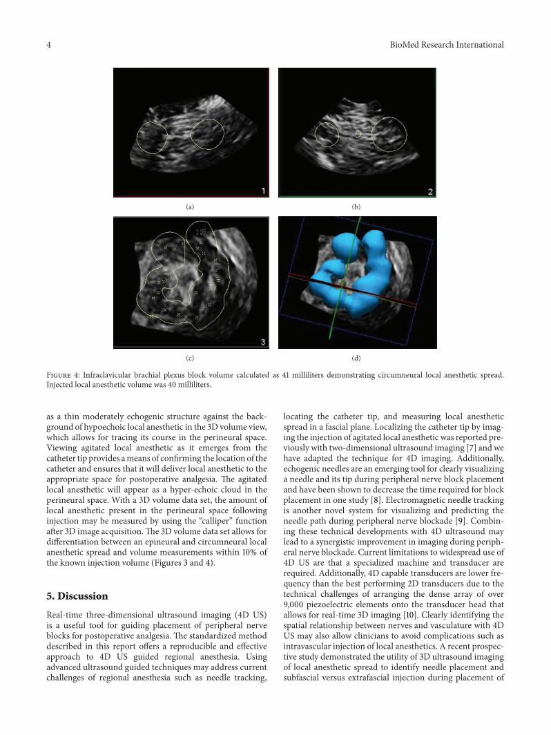
Figure 4: Infraclavicular brachial plexus block volume calculated as 41 milliliters demonstrating circumneural local anesthetic spread.Injected local anesthetic volume was 40 milliliters.
as a thin moderately echogenic structure against the back-ground of hypoechoic local anesthetic in the 3D volume view,which allows for tracing its course in the perineural space.Viewing agitated local anesthetic as it emerges from thecatheter tip provides ameans of confirming the location of thecatheter and ensures that it will deliver local anesthetic to theappropriate space for postoperative analgesia. The agitatedlocal anesthetic will appear as a hyper-echoic cloud in theperineural space. With a 3D volume data set, the amount oflocal anesthetic present in the perineural space followinginjection may be measured by using the “calliper” functionafter 3D image acquisition.The 3D volume data set allows fordifferentiation between an epineural and circumneural localanesthetic spread and volume measurements within 10% ofthe known injection volume (Figures 3 and 4).
5. Discussion
Real-time three-dimensional ultrasound imaging (4D US)is a useful tool for guiding placement of peripheral nerveblocks for postoperative analgesia. The standardized methoddescribed in this report offers a reproducible and effectiveapproach to 4D US guided regional anesthesia. Usingadvanced ultrasound guided techniques may address currentchallenges of regional anesthesia such as needle tracking,
locating the catheter tip, and measuring local anestheticspread in a fascial plane. Localizing the catheter tip by imag-ing the injection of agitated local anesthetic was reported pre-viously with two-dimensional ultrasound imaging [7] and wehave adapted the technique for 4D imaging. Additionally,echogenic needles are an emerging tool for clearly visualizinga needle and its tip during peripheral nerve block placementand have been shown to decrease the time required for blockplacement in one study [8]. Electromagnetic needle trackingis another novel system for visualizing and predicting theneedle path during peripheral nerve blockade [9]. Combin-ing these technical developments with 4D ultrasound maylead to a synergistic improvement in imaging during periph-eral nerve blockade. Current limitations to widespread use of4D US are that a specialized machine and transducer arerequired. Additionally, 4D capable transducers are lower fre-quency than the best performing 2D transducers due to thetechnical challenges of arranging the dense array of over9,000 piezoelectric elements onto the transducer head thatallows for real-time 3D imaging [10]. Clearly identifying thespatial relationship between nerves and vasculature with 4DUS may also allow clinicians to avoid complications such asintravascular injection of local anesthetics. A recent prospec-tive study demonstrated the utility of 3D ultrasound imagingof local anesthetic spread to identify needle placement andsubfascial versus extrafascial injection during placement of
BioMed Research International 5
a lateral popliteal nerve block [11]. Furtherwell-designed clin-ical trials are necessary to determine whether the additionalimaging capabilities translate into a reduction of adverseevents and improved clinical outcomes while clarifying thedifferences between techniques in regional anesthesia. Anearlier case report highlights the advantages of a multiplanarapproach during a single shot radial nerve block [12]. Wesought to formalize this approach into a standardizedmethodfor 4D ultrasound guidance of needle placement in additionto describing a method for viewing and calculating injectedlocal anesthetic volume. Future studies employing thismethod may enable additional qualitative and quantitativeassessment of techniques in regional anesthesia.
Conflict of Interests
The authors declare that there is no conflict of interestsregarding the publication of this paper.
References
[1] O. Choquet and X. Capdevila, “Case report: three-dimensionalhigh-resolution ultrasound-guided nerve blocks: a new pano-ramic vision of local anesthetic spread and perineural cathetertip location,” Anesthesia & Analgesia, vol. 116, no. 5, pp. 1176–1181, 2013.
[2] H.Albrecht, C. Stroszczynski, R. Felix, andM.Hunerbein, “Realtime 3D (4D) ultrasound-guided percutaneous biopsy of solidtumours,”Ultraschall in der Medizin, vol. 27, no. 4, pp. 324–328,2006.
[3] K. Sugimoto, F. Moriyasu, J. Shiraishi, M. Yamada, and Y. Imai,“A phantom study comparing ultrasound-guided liver tumorpuncture using new real-time 3D ultrasound and conventional2D ultrasound,”American Journal of Roentgenology, vol. 196, no.6, pp. W753–W757, 2011.
[4] S. R. Clendenen, K. Riutort, B. L. Ladlie, C. Robards, C. D.Franco, and R. A. Greengrass, “Real-time three-dimensionalultrasound-assisted axillary plexus block defines soft tissueplanes,” Anesthesia & Analgesia, vol. 108, no. 4, pp. 1347–1350,2009.
[5] N. G. Feinglass, S. R. Clendenen, K. D. Torp, R. D. Wang, R.Castello, and R. A. Greengrass, “Real-time three-dimensionalultrasound for continuous popliteal blockade: a case report andimage description,” Anesthesia & Analgesia, vol. 105, no. 1, pp.272–274, 2007.
[6] M. K. Karmakar, X. Li, J. Li, and A. Hadzic, “Volumetric three-dimensional ultrasound imaging of the anatomy relevant forthoracic paravertebral block,” Anesthesia & Analgesia, vol. 115,no. 5, pp. 1246–1250, 2012.
[7] J. D. Swenson, J. J. Davis, and J. A. Decou, “A novel approach forassessing catheter position after ultrasound-guided placementof continuous interscalene block,” Anesthesia & Analgesia, vol.106, no. 3, pp. 1015–1016, 2008.
[9] A. Kilicaslan, A. Topal, A. Tavlan, A. Erol, and S. Otelcioglu,“Differences in tip visibility and nerve block parameters bet-ween two echogenic needles during a simulation study with in-experienced anesthesia trainees,” Journal of Anesthesia, 2013.
[10] S. W.Wong, A. U. Niazi, K. J. Chin, and V. W. Chan, “Real-timeultrasound-guided spinal anesthesia using the SonixGPSneedle
tracking system: a case report,” Canadian Journal of Anesthesia,vol. 60, no. 1, pp. 50–53, 2013.
[11] G. L. Foxall, J. G. Hardman, andN.M. Bedforth, “Three-dimen-sional, multiplanar, ultrasound-guided, radial nerve block,”Regional Anesthesia and Pain Medicine, vol. 32, no. 6, pp. 516–521, 2007.
[12] A. Missair, R.Weisman, M. R. Suarez, R. Yang, and R. Gebhard,“A 3-dimensional ultrasound study of local anesthetic spreadduring lateral popliteal nerve block,” Regional Anesthesia andPain Medicine, vol. 37, no. 6, pp. 627–632, 2012.