A STUDY OF CHALARA ELEGANS IN MUCK SOILS AND DEVELOPMENT OF BLACK ROOT ROT ON CARROTS IN THE FRASER VALLEY OF BRITISH COLUMBIA Suganthi Chittaranjan B. Sc., Eastern University of Sri Lanka, 1984 THESIS SUBMITTED IN PARTIAL FULFILMENT OF THE REQUIREMENTS FOR THE DEGREE OF MASTER OF PEST MANAGEMENT in the Department of Biological Sciences O Suganthi Chittaranjan 1992 SIMON FRASER UNIVERSITY April, 1992 All rights reserved. This work may not be reproduced in whole or in part, by photocopy or other means, without permission of the author.
Transcript
A STUDY OF CHALARA ELEGANS IN MUCK SOILS AND DEVELOPMENT OF BLACK ROOT ROT ON CARROTS IN
THE FRASER VALLEY OF BRITISH COLUMBIA
Suganthi Chittaranjan
B. Sc., Eastern University of Sri Lanka, 1984
THESIS SUBMITTED IN PARTIAL FULFILMENT OF
THE REQUIREMENTS FOR THE DEGREE OF
MASTER OF PEST MANAGEMENT
in the Department
of
Biological Sciences
O Suganthi Chittaranjan 1992
SIMON FRASER UNIVERSITY
April, 1992
All rights reserved. This work may not be reproduced in whole or in part, by photocopy
or other means, without permission of the author.
Name:
Degree:
APPROVAL
SUGANTHI CHI'ITARAN JAN
Master of Pest Management
Title of Thesis:
A STUDY OF CHALARA ELEGANS IN MUCK SOILS AND DEVELOPMENT OF BLACK ROOT ROT ON CARROTS IN THE FRASER VALLEY
OF BRITISH COLUMBIA.
Examining Committee:
Chairman: Dr. P. Belton, Associate Professor
Dr. Z.K. Pun'a, Associate Profe>sor, Senibk Supervisor, JMI& -ciedces, S m Depart
. J.M. Webster, Professor, epartment of Biological Sciences, SFU
I
Mr. D.BrmI'rod, Rksearch Scientist, B.C. Ministry of Agjiculture, Cloverdale, B.C.
Ms. M. Gaye, MSc., Pqg., Project Manager, Cloverdale Soil Conservation Grou~. Surrey, B.C. Public Examiner
3 C
Date Approved
. PART I AL' COPYR I'GHT L 1 CENSE
I hereby grant to Slmon Fraser Unlverslty the rlght to lend
my thesis, project or extended essay'(th6 :ltle o f which Is shown below)
to users of the Slmon Fraser Unlversl ty LI brhy, and to maeke part la 1 or
single copies on1 y for such users or In response to a request f r,om tho
l i brary of any other unlverslty, or other educat lona l Inst i tut Ion, on
its own behalf or for one of Its users. I further agreo that permission
formultlple copylng of thls work for scholarly purposes may be granted
by me or the Ooan of Graduate Studles. It i s understood that copying
or publlcatlon of thls work for flnanclal galn shall not bo allowed
wlthout my written permlsslon.
T I tie of ~ h e s 1 s/Project/Extended Essay
4 STUDY CHaA.9 E L E W N S I;N MUCK $QTLS AND DEVELOPMENT O F 33LACK ROOT ROT ON f -.
CARROTS I N THE FRASER VALLEY O F B R I T I S H COLUMBIA.
Author: -
(signature)
Abstract
Black root rot, caused by the dematiaceous hyphomycete Chalara eleg~zns (Nag Raj and
Kendrick) [syn. Thielaviopsis basicola (Berk. and Br.) Ferr], is an important postharvest disease
on fresh market carrots in the Fraser Valley of British Columbia. Black root rot may cause
around 10% annual losses to the fresh market carrot industry, which was valued at about $2
million in 1990. To obtain a better understanding of the pathogen and factors influencing disease
development, studies were conducted to enumerate populations of Chalartr elegans, and to
determine the distribution and spatial pattern of inoculum in commercial carrot and other
vegetable production fields in the Fraser Valley. In addition, the factors influencing survival of
C. elegans were studied, and the mode of infection and disease development on carrot was
studied using light and scanning microscopy.
For quantitative studies, a semiselective medium (TBM-RBA) was developed which
contained a range of fungicides and antibiotics. This medium was effective in detecting
3 inoculum levels as low as 20 colony forming units (CFU)/cm of soil. The field sampling
studies indicated that C . elegans was present in about 80% of the fields that were selected for
sampling. The inoculum distribution in one field showed an aggregated spatial pattern within the
field. The range of inoculum densities among the fields sampled was <lo lo 560 cFU/cm3.
Studies on factors influencing survival of the phialospores (endoconidia) indicated that they
could persist in organic soil for more than 20 weeks. Soil planted to onions or- flooded and kept
at a high temperature (about 25 C) significantly reduced the population of C. elegans over time.
Survival was not affected by a carrot crop or by flooding at 4 C. The reduction in survival was
attributed to an increase of soil microorganisms which were antagonistic to C. degans.
Studies on the mode of infection of carrot roots showed that wounds were required for
pathogen growth and it's establishment, and that the periderm of carrot roots was seldom
iii
penetrated directly. Scanning electron microscopy showed rapid development of the pathogen
(within 48 hr) and fungal sporulation by 96 hr. The fungus grew both inter- and intra- celluarly.
Dedication
To my parents, who always encouraged me to pursue higher studies.
Acknowledgments
I would like to thank my senior supervisor, Dr. Zamir K. Punja, for his encouragement and
support, my supervisory committee for reviewing my thesis and making helpful suggestions,
and my lab colleagues Dee Ann Benard, Eric Urquart and others for providing a helpful and
friendly work environment and assistance.
I would like to thank Vic Bourne for his assistance with the scanning electron microscopy
and photography techniques, and Kwai Lee for help with histology techniques.
I would like to thank Dr. Andre Leveque for his help and advice and Mr. F. Bellavance for
his help with statistical analysis.
I am also very grateful to the B. C Coast Vegetable Co-operative Association, which
provided financial support for my research work, through financial contributions from the Agri-
Food Regional Development Subsidiary Agreement Project (ARDSA #11048). from the Natural
Science and Engineering Research Council of Canada (NSERC), and from the B.C. Carrot
growers research council. I thank Mr. Rick Gilmour and Ms. Mary-Margaret Gaye for providing
invaluable information during this project and for their assistance, and to various growers for
their cooperation during the field sampling studies.
Table 1. Ingredients of TBM-RBA per litre ................................................. ....................... 15
Table 2. Growth of C. elegans colonies from pure spore suspensions, when plated onto
each of four different media ......................... ...... .................... . . . . . . . . .. .. .. .. .. . . .. .. .. .. .. . 16
Table 3. Recovery of C. elegans colonies from artificially inoculated field soil. when
plated onto four different media ....................................................... . . .... .......... .... .. ... 17 3 Table 4. Recovery of C.elegans colonies (CFU/cm ) from four naturally infested field
soils located in the Cloverdale area of the Fraser Valley of British Columbia ............... 18
Table 5. Comparison of TBM-RBA medium with carrot baiting assay, when artificially
...... Figure 19. Scanning electron micrographs of the infection process of C. elegan r on carrot 70
Figure 20. Scanning electron micrographs of chlamydospore development of of C.
............................................................................................................. elegans on carrot 7 1
CHAPTER 1
INTRODUCTION:
BLACK ROOT ROT ON CARROTS - THE DISEASE:
Black root rot, caused by the dematiaceous hyphomycete Chalara elegans
(Nag Raj and Kendrick, 1975) [syn. Thielawopsis basicola (Berk. and Br.) Ferr.], is a
major postharvest disease on fresh market carrots grown in the Fraser Valley of
British Columbia (Fig 1). The pathogen is a soil-borne fungus which has a wide host
range, inclusive of cultivated and noncultivated plants (Yarwood, 1981), and it is
endemic to organic (muck) soils in the Fraser Valley . In North America, black root
rot has been reported to occur on a wide range of plants, including tobacco (Stover,
1950), cotton (King and Presley, 1942), bean (Papavizas and Davey, 1961), pea
(Lloyd and Lockwood, 1961), poinsettia (Keller and Shanks, 1955) and holly
(Lambe and Willis, 1976). On these plants, the hypocotyls and roots become
severely infected under favorable temperature and moisture regimes. Under field
conditions, C. elegans does not appear to cause any visible disease symptoms on
carrot seedlings or on mature carrot roots (Z.Punja, unpublished observatioq).
Apparently, black root rot is also not a major postharvest problem on carrots grown
in other parts of the world.
C. elegm survives over a long period of time in soil by producing resistant
chlamydospores (Tsao and Bricker, 1966). In culture, two types of spores, which are
described as endoconidia (phialospores) and chlamydospores (aleuriospores) are
formed (Nag Raj and Kendrick, 1975). Chlamydospores are thick walled, dark
brown in color and are produced in chains of 4-6 spores. Phialospores are thin
walled, hyaline and are also produced in chains.
Figure 1. Black root rot of carrots, caused b Chalara elegans. Typical blackening symptoms of this disease are ue to the production of chlamydospores on the root surface.
J' I
f
3
Wounds on the epidermis of carrots caused by harvesting, washing and
especially grading practices, become the infection sites for fungal inoculum (Punja
et al., 1992). Both chlamydospores and phialospores are capable of infecting
wounded carrots (Punja, 1990). The infections occur only in the upper 4-8 cell layers
of the carrot root (see Chapter 5). Within 2-3 days, the carrots develop grey lesions,
which expand and blacken due to the spore production on the epidermis. These
spores become secondary inoculum and infect other carrots within the polyethylene
bags.
The blackening symptoms begin to show 5-7 days after the carrots are packed
and sent to the wholesalers and retailers if stored at optimal temperatures (25-27 C)
and high moisture (98-100% RH) in the polyethylene bags. In 1989, it was
estimated that up to 40% of the carrots were returned to the B.C. Coast Vegetable
Co-operative Association during the month of August, when infections were high
personal communication). This was attributed in part to improper handling
practices at the retail end, and resulted in a substantial loss of revenue to the
growers.
CARROT HARVESTING AND GRADING:
In the Fraser Valley, fresh market carrots are produced by individual growers
on fields ranging from one to 10 ha in size. Harvesting usually takes place during the
morning, and the carrots are sent to the B.C Coast Vegetable Co-operative
Association in Richmond, B.C., where they are rinsed with water and placed in a
large holding bin (Fig 2). The carrots are then washed by roller brushes (scrubber),
hand-sorted and sent on conveyor belts through a large hydrocooler, where the
carrot surface temperature is brought to 4-6 C for 1-2 min.
Ca
rro
ts f
rom
tru
ck
s
I B
in
/
Dir
ty c
arr
ots
so
rtin
g I
I G
rad
e 1
) G
rad
e 3
/ /
Cu
lls
Gra
de
r I
Fig
ure
2.
Sch
emat
ic r
epre
sen
tati
on
of
the
pro
cess
ing
of
ca
rro
ts a
t th
e B
.C.
Co
as
t
Ve
ge
tab
le C
o-o
pe
rati
ve
Ass
oci
atio
n i
n R
ich
mo
nd
, B
.C.,
(19
91
).
Arr
ow
s in
dic
ate
dir
ecti
on
of
mo
vem
ent.
\
Gra
de
2
5
Also, chlorine is maintained at a rate of 80 pglml in the hydrocooler. Throughout
this process, muck soil can be found in the wash bins and also adhering to the
conveyer belts. After hydrocooling, the carrots are conveyed to the grader, where
they are sorted according to their diameter. Finally, the carrots are collected and
packed into 0.9-1.35 kg polyethylene bags and shipped to wholesalers and retail
stores. Bulk carrots in 22.5 kg bags are also produced.
ECONOMIC LOSSES DUE TO BLACK ROOT ROT:
Iil the lower Fraser Valley, about 350 acres are planted to carrots each year . The commercial carrot production is around 6000 Tiyear. In 1990, retail sales in the
carrot industry were valued at around $2 million. In 1979 and 1982, losses due to
black root rot in B.C were as high as $1.2 million. Therefore, a project (D.A.T.E
Project No. 135) was initiated in 1984 by the B.C. Ministry of Agriculture and
Fisheries to develop possible prevention methods to reduce this disease. It was
found that washing carrots with chlorinated water (80 pg/ml concentration of
chlorine) and cooling them to 1 C would decrease disease incidence (L. MacDonald
and DJ. Orrnrod, 1985, B.C. Ministry of Agriculture and Fisheries, Cloverdale, B.C.,
unpublished).
However, in 1988, losses due to black root rot were estimated to be about
$200,000. Studies conducted in 1989-1990 (M.M. Gaye, A.R.D.S.A Project No.
11048,) showed that even after washing with chlorinated water and hydrocooling,
89% of the carrots sampled from 339 loads developed black root rot (M.M. Gaye,
CaC03, 1 g; agar, 15 g; distilled water, 900 ml; (pH 5.3). Carrot juice and aqueous
solution of antibiotics were added to the autoclaved medium.
INOCULUM PREPARATION:
C. elegm was isolated from carrots with symptoms of black root rot by
surface sterilizing infected carrot pieces with 10% bleach [0.625% sodium
hypochlorite (NaOCl)], washing them in sterile distilled water, and incubating on V-
8 agar (V-8 juice, 150 ml; agar, 15 g; ampicillin, 100 mg; distilled water, 850 ml) at
25 C. Colonies of C. elegm were grown on V-8 agar for two weeks at room
temperature (25 C). Phialospores were harvested by flooding the colonies of C.
elegm grown on this medium with 100-200 ml of sterile distilled water and
adjusting the spore concentration with a haemocytometer.
COMPARISON OF MEDIA:
latine from Dure more - sus~ensions: - Spore suspensions were prepared
according to the above stated procedure, and up to dilutions were made in
distilled water blanks. A small volume (0.5 ml) of the suspension from each dilution
was plated onto all three media. The dishes were incubated at room temperature for
5-12 days. Observations were made on the number of colonies developing and the
size of individual colonies.
b) Soil dilution from artificiallv inoculated soil: Soil samples originating from
commercial carrot fields were screened using Yarwood's carrot root disc method
(see Chapter 3). Soil from one field which did not have any detectable C-elegm
propagules was selected for this study. To 28 cm3 of soil, two ml of a phialospre
suspension (1.32 x lo6 spores/ml) was added and the soil was thoroughly mixed.
11
Since the initial inoculum level used was high (9.5 x 16 spores/cm3), one an3 of
soil was placed in 9 ml of sterile water dilution blanks. Soil suspensions were then
diluted further (up to lo4) and 0.5 ml of the suspension from each dilution was
plated onto each of the three media. The number of colonies developing was rated
after 7-12 days of incubation at room temperature.
c) Soil dilution from natural soil: Four commercial carrot fields located in the
Cloverdale area of the Fraser Valley of B.C. that had a history of yielding carrots
with black root rot, were chosen. Each field was randomly sampled by taking 50 g of
soil from 12-15 different locations and bulking them to make a composite
sample/field. Samples were brought back to the laboratory and stored at 4 C for 1-
3 10 days prior to use. For dilution plating, a volume of 5 cm of soil was transferred
into 45 ml of sterile distilled water (10-I). The suspension was mixed thoroughly,
and further dilutions (up to lom3) were made. From each dilution, 0.5 ml of soil
suspension was evenly plated onto each medium. The plates were incubated at room
temperature (25 C) for 7-14 days.
MODIFIED TBM-RBA MEDIA:
Repeated evaluations of all of the three previously described media showed
that TBM-C had fewer fungal contaminants and, therefore, had higher recovery of
colonies of C. elegans. However, Othium, Fusarium, Penicillium, Aspergih,
Trichudema, Mucor and various bacteria were major contaminants in all of the
three media. Recovery of C. elegans was further enhanced by evaluating various
modifications of the TBM-C medium. Carrot roots (100, 200, 300 or 400 g/liter)
were either blended and filtrated through cheesecloth or cooked and mashed and
incorporated into the media before autoclaving.
12
In addition, different fungicides (per liter) were added to reduce the growth of
contaminants. The fungicides tested were metalaxyl (at 10,20,30,35 or 45 mg ai.),
dichloran (at 1,2,3,4,5 or 6 mg a.i.), benomyl (at 2 mg), rose bengal (at 25 mg) and
ampicillin (at 100 mg). Vitamin E acetate at 0.5% had been reported to induce
chlamydospore germination (Papavizas and Adams, 1969). Vitamin E acetate was
added (at 0.5%, w/v) to the media, when natural field soil was used. The pH of the
media was also adjusted (to 4,4.5,5,5.5 or 6) using 1N H2SO4 or KOH.
After determining the optimum level of all the ingredients for the new TBM-
RBA medium through several comparative studies (data not presented), the new
medium (TBM-RBA) was compared with the carrot baiting assay and the other
three media that were used in the preliminary studies. For this purpose, pure spore
suspension, artificially inoculated soil, and naturally infested soil were used in the
experiments as described above .
COMPARING TBM-RBA WITH CARROT BAITING:
A spore suspension (17 x lo6 phialospores/ml) of C. elegans was made from
a two-week-old culture grown on V-8 agar at 25 C. The spore suspension was diluted
to 170 spores/rnl. The moisture content of the field soil was determined by drying
several 20 g soil samples at 80 C for two days and re-weighing them. A 25 cm3 (15.8
g wet soil; 6.69 g dry weight;) sample of moist soil (57.6% moisture content) was
inoculated with 3 ml of the diluted spore suspension (510 spores/25 cm3). The
minimum number of spores that can be detected by TBM-RBA without a
replication plate from naturally infested soil is 20 cFU/cm3 or 74 sporeslg oven dry
weight, when the soil moisture content is 57.6 % (personal observation). Therefore
in this study, each soil sample had around 76 sporeslg dry weight.
From this inoculated soil, 5 cm3 was transferred into 45 ml of sterile distilled water.
In addition, a dilution was made. From both dilutions, 0.5 ml of soil suspension
was plated onto 8 plates of TBM-RBA media as well as onto carrot root discs (2
discs/ml). The experiment was repeated once.
COMPARISON OF MEDIA:
a) Dilution platiw from pure spore sus~ension: Colonies of C. elegans developed
within 5-6 days on TBM-C and on VDYA-PCNB media, whereas the rate of colony
growth was slower on TB-CEN2 medium. Therefore, during the evaluation of media
after 7 days, colonies on TBM-C and VDYA-PCNB were much darker and easier to
identify. The colonies formed on VDYA-PCNB were much larger (8-11 mm
diameter) than on TBM-C (5-7 mm) and were comparable to those on TB-CEN2
(7 -10 mm). The colonies on TBM-C were defined, distinct, and were easier to
count. Recovery of C. elegans was 90- 95% on TBM-C and VDYA-PCNB, whereas
the recovery rate on TB-CEN2 was lower. Also, some fungal contamination was
observed on TB-CEN2 media.
b) re cove^ of C. elepans from artificiallv inoculated soil: TB-CEN2, which
contained unautoclaved carrot juice, was highly contaminated by microorganisms
present in the organic soil type used throughout the experiment. The major
contaminants were various bacteria, Mucor and to a lesser extent, Fwarium,
Trichodenna and Pythium. Recovery on TBM-C and VDYA-PCNB were
comparable at lower dilutions but recovery was highest on TBM-C at a
dilution, when a higher initial concentration of spore suspension was used to
inoculate the soil.
14
PeniciUiUm was the major contaminant on VDYA-PCNB and Fusuiium was the
major contaminant on TBM-C. However, other contaminants such as Pythium and
Trichaiema were also found on these media
c) Recove? from naturallv infested soil: No C. elegans colonies were isolated on
VDYA-PCNB or TB-CEN2 media when naturally infested soil (presence of C.
elegans propagules was confirmed with carrot disc assay) was used in this study.
However, colonies of C. elegans developed on TBM-C media twice (from two fields)
during these experiments.
MODIFIED TBM-RBA MEDIA:
The highest recovery of C. elegans was observed in this study with the
ingredients and rates indicated in Table 1. Vitamin E acetate did not enhance the
recovery of C. elegans when added to this medium. The addition of benomyl at 2 mg
a.i/L eliminated the recovery of C. elegm.
A comparison of TBM-RBA, TBM-C, VDYA-PCNB and TB-CEN2 media
for recovery of C. elegans is shown in Tables 2-4 and in Figs. 3 and 4. In Table 2, it
can be seen that recovery from a pure spore suspension was 87.8% on TBM-RBA.
These results are also shown in Fig. 3. In Table 3, recovery from artificially
inoculated soil was 100% on TBM-RBA. These results are also shown in Fig. 4. In
these experiments, a higher initial concentration of spore suspension was used and
therefore definite colonies appeared only at lo4 dilution, when both pure spore
suspension and inoculated soil was used. TB-CEN2 had the least number of colonies
developing in these experiments. The lowest level of contamination was observed on
TBM-RBA medium by comparison. C. elegans was recovered from naturally
infested field soil only on TBM-RBA and TBM-C media (Table 4). TBM-RBA was
the only medium on which C. elegans was recovered from samples 2,3, and 4.
15
Table 1: Ingredients of TBM-RBA per liter. This medium was developed in the
:ourse of this research.
Ingrediets
Agar (Anacheim brand)
Carrot juice*
Yeast extract
oxgall
PCNB
(Pentachloronitrobenzene)
Nyatatin
Chloramphenicol
Ridomil (metalaxyl)
Botran
Ampicillin
Penicillin
Rose bengal (optional)* *
pH 5.0
Amount (a.i.)/liter
* Extract from 200 g carrot roots after blending and filtering with 350 ml of distilled
water.
* * If rose bengal is added, the medium is known as TBM-2RBA.
plated onto each of four different media.
TB-CEN2
VDYA-PCNB
TBM-C
TBM-RBA
Coloqjcs/plate at 10 dilution
% recovery
a TB-CEN: Specht, and Griffin, (1985);
VDYA-PCNB: Papavizas, (1964);
TBM-C: Maduwesi, Sneh, and Lockwood, (1976).
b Initial concentration was 1.32 x lo6.
c Rated after 7 days of incubation at 25 C.
17
Table 3: Recovery of Chalara eleganr colonies from artificially inoculated field soil
when plated onto four different media.
a Initial concentration was 0.9 x 16.
b Rated after 7 days of incubation at 25 C.
Medium
TB-CEN2
VDYA-PCNE
TBM-C
TBM-RBA
colonies/ late 3 at 10'
- I 42
44
50
CN/& a
- 8.4 x 10⁴
8.8 x 10⁴
1x10s
% recovery b
- 93
97
100
Table 4. Recovery of Chalara eleguns colonies (cFW/cm3) from four naturally
infested field soils located in the Cloverdale area of the Fraser Valley of B.C.
Medium
TB-CEN2
VDYA-PCNB
TBM-C
TBM-RBA
C F U / ~ ~ of soil *
* Average of 2 plates/ sample.
Sam~le 1
- Sample 2
- Sam~le 3
- Sample 4
-
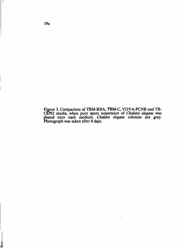
Fi re 3. Comparison of TBM-RBA, TBM-C, VDYA-PCNB and TB- ~ & 2 media, when pure spore suspension of Chalara e k g m was lated onto each medium. Chalara elegans colonies are grey.
!hotograph was taken after 8 days.
Fi e 4. Comparison of TBM-RBA, TBM-C, VDYA-PCNB, and TB- 3 N2 media, when soil that was artificially inoculated with Chalura elegans was plated. Chalara elegans colonies are grey and the F w m colonies are white; TB-CEN2 was covered with Pyyu'm. Note the low level of fungal contamination on TBM-RBA at 10-
COMPARING TBM-RBA WITH CARROT BAITING:
Carrot discs were a more sensitive assay for detecting C. elegans than TBM-RBA
(Table 5). The minimum number of spores detected by a single TBM-RBA Petri
plate would be 20 cFU/cm3 of moist soil (or 74 CFU/g oven dry soil at 57.6%
moisture content). Therefore, to detect 1 cIW/cm3 from a soil sample, at least 20
plates should be used. It should be noted that on carrot root discs, the population
level was over estimated.
DISCUSSION:
The TBM-RBA medium developed in this study was better than all of the
other media reported by previous investigators for recovery of C. elegans from soil.
When natural soil was used, recovery of C. elegans on the other media was very low
or not possible due to contamination by other fungi. This was probably due to the
high microbial activity seen in the organic (muck) soils compared to mineral soils.
Even though peeled and surface-sterilized carrots were used to prepare the
TB-CEN2 medium, some contamination was observed even when pure spore
suspensions were plated. The low recovery rate on TB-CEN2 in this assay could also
be due to the use of organic soil, which has very high populations of resident
microorganisms. Another explanation could be because etridiazol, which eliminates
Oomycetes, was unavailable , Ridomil (metalaxyl) was used for the same purpose.
Etridiazol could have eliminated contaminants in TB-CEN2 media as used by
Specht and Griffin, whereas Ridomil may not have. However, Ridomil successfully
eliminated fythium and other Oomycetes in the TBM-RBA media even at low
concentrations (35 mg/L). Because carrots have natural infections by C. elegans,
using unautoclaved carrot juice is not recommended in any medium.
Table 5. Comparison of TBM-RBA medium P t h carrot baiting assay, when artificially inoculated soil containing 20 CFU/ cm was plated.
* Average number of colonies of C. elegans from 8 replicate plates of TBM-RBA, or
from 8 carrot discs.
% recovery
87
160
Medium
TBM-RBA
Carrot baiting
colonicis, at 10-
0.87
1.6
cFU/cm3
17.4
32
23
Rapid growth of contaminants as well as larger colonies of C. elegm on
VDYA-PCNB medium could be attributed to the presence of V-8 juice and glucose
in this medium. Addition of carrot juice (which is lower in nutrients) instead of V-8
juice may be the reason why TBM-C achieved recovery of some colonies of
C.elegans from natural soil. The C. elegm colonies on TBM-RBA were very dark
and distinct and therefore easy to identify and enumerate. The colonies that
originated from artificially infested soil (phialospores) were much larger than the
colonies developing from natural soil, which probably originated from
chlamydospores. This could be due to the germination rate of phialospores being
faster than the germination rate of chlamydospores. Therefore, phialospores may
have germinated and established larger colonies on this medium before the other
contaminants could grow, whereas the colonies from chlamydospores could have
originated at the same time or later than the contaminants, giving rise to smaller
colonies.
Eliminating Fusariurn in TBM-RBA is still challenging. When benomyl was
added to eliminate Fusarium at a concentration of 2 mglliter, it also eliminated C.
elegans. Other fungicides, such as thiram or mancozeb, should be evaluated for this
purpose.
The best and consistent recovery of C. elegans was obtained when 5 cm3 of
soil was diluted in 45 ml of water and 0.5 ml was plated onto the TBM-RBA
medium, especially when naturally infested soil was used. The use of a large volume
of soil, instead of 1 an3 in 9 ml may have enhanced the probability of recovery.
If the bacterial contamination on the medium is very high, rose bengal can be
added (25 mg/L) to enhance the selectivity of the medium (Fig 5). Colonies of C.
elegans are more visible on the medium with rose bengal.
Figure 5. Corn arison of TBM-RBA (new semiselective medium developed for in this study) and TBM-2RBA media same to reduce bacterial contamination . $3 e Chalara e ans colonies are grey and are darker and more visib e on the TBM-2R % A medium.
1'
25
This medium should be incubated in the dark since rose bengal is sensitive to light
(Martin, 1950). Although TBM-RBA was less sensitive than the carrot baiting assay
(Table 5), it provided consistently better results for the quantitative estimation of C.
e legm from muck soil, which has high levels of different soil microflora. The TBM-
RBA medium was therefore used in further quantitative studies (see Chapter 3).
CHAPTER 3
DISTRIBUTION. INOCULUM DENSITY AND SPATIAL PATTERN OF
C. ELEGANS IN SOILS OF THE LOWER FRASER VALLEY OF BRITISH
COLUMBLA:
INTRODUCTION:
Determination of the distribution of a plant pathogen among commercial
fields, the inoculum density of the pathogen, and the spatial pattern of inoculum are
essential to the understanding of disease problems. Such studies can be useful for
determining whether specific control measures are applicable, or whether measures
such as pathogen avoidance (Punja et al., 1985) are advisable.
Campbell and Noe (1985) stated: " The spatial pattern of a specific organism
or set of organisms provides an opportunity to characterize organismal attributes
from a static sample in time or from a series of samples over time. The
characterization of a spatial pattern thus provides a tool for the development of
plausible biological and environmental hypotheses to account for the association
among organisms."
The distribution of Chalara elegans in nature has been intensively studied by
Yarwood (1981) using a carrot root disc assay. The fungus was found to be as
abundant in some virgin areas as in cultivated soils. It can be found associated with
over 148 plant species in different regions of the world (Yarwood, 1981).
Previous population and/or inoculum density studies of C. elegans have been
mainly conducted in tobacco fields in different areas of the U.S.A. and in Ontario,
Canada (Rittenhouse and Griffin, 1985; Specht et al., 1987; Anderson and Welacky,
1988; Meyer and Shew, 1991).
27
In some of these studies, inoculum level was shown to positively correlate with
disease development on crops such as tobacco, bean, cotton, and pea (Lucas, 1955;
Anderson and Welacky, 1988; Specht and Griffin, 1988; Meyer et al., 1989).
However, other factors such as soil pH, soil temperature, soil chemistry, host
resistance, and cultural practices (Lloyd and Lockwood, 1963; Specht et al., 1987;
Anderson and Welacky, 1988; Meyer and Shew, 1991) also were found to have a
great influence on disease development. Inoculum density of C. elegans is not
related to the field's history of black root rot. In addition, inoculum density levels
may also differ largely even between small distances (about 3.6 m) within a field
(Rittenhouse and Griffin, 1985).
Most of the previous spatial pattern studies of soilborne fungi have indicated
the aggregation or clumping of soil populations of microorganisms (Nicot et al.,
1984; Campbell and Noe, 1985; Punja et al., 1985). In addition, Rittenhouse and
Griffin (1985) showed that the distribution of C. elegans in tobacco field soil had a
clumped spatial pattern. In the Lower Fraser Valley of B.C., C. elegans was
previously isolated from eight out of twelve carrot fields sampled in 1984-1985 (L
MacDonald and D J Orrnrod, 1985).
Within the Fraser Valley of B.C., commercial fields are not continuously
planted to carrots. Fields may be planted to other vegetable crops, such as lettuce,
celery, potato, or onions, in rotation with carrots. By sampling commercial carrot
fields, as well as other vegetable fields, information can be obtained to better
understand the distribution of C. elegans in these fields. The objectives of this study
were to:
a) determine the distribution of C. elegans in carrot and other vegetable production
fields;
b) determine the inoculum density of C. elegans in commercial carrot fields; and
28
c) determine the spatial pattern of C. elegans in one carrot field which was sampled
intensively over two growing seasons (1990-1991).
MATERIALS AND METHODS:
DISTRIBUTION OF C. ELEGANS IN THE LOWER FRASER VALLEY OF
B.C.:
In each of 1990 and 1991, 30 carrot and vegetable production fields were
randomly sampled during May-July. In each field, 15-20 soil samples (each around
80-100g .of soil) were randomly collected and bulked into 15 x 20 cm polyethylene
bags. These bags were tagged with the identity of the field, brought to the laboratory
and assayed immediately or stored at 4 C in the cooler prior to examination. Assays
were conducted within one week. The presence of C. elegm was determined using
the carrot root disc assay described in Chapter 2. The areas close to the commercial
crop where weeds grew, were also tested for the presence of C. elegans.
INOCULUM DENSITY OF C. ELEGANS IN CARROT FIELDS:
Soil samples were collected from 24 commercial carrot fields in 1990 and
1991. Samples were collected in the same manner as for the distribution studies
above. The samples were analysed immediately or stored in a cooler at 4 C and
examined within 4 weeks. After thoroughly mixing each bulked soil sample, a 5 cm3
volume of soil per sample/field was placed in a 9 ml sterile distilled water blank
(10-I), and further dilutions were made. Each dilution was plated onto TBM-RBA
medium (see Chapter 2) and incubated at 25 C. The number of propagules/cm3 of
soil was calculated for each field.
29
SPATIAL PA'ITERN OF C. ELEGANS:
From the inoculum density studies above, two fields (A and B)) located in
Cloverdale, B.C. were selected to determine the spatial pattern of C. elegans. Field
A had a history of black root rot development on carrots and was planted to carrots
in 1989 and 1990. Field B had never been planted to carrots until 1990. In July
1990, samples were collected from fields A and B along two rows. Samples
comprised of 80-100 g were taken to a depth of 20 cm at 3.0 m (10 ft) intervals along
the row, placed in a polyethylene bag and tagged with the sampling distance and row
number. In field A, 20 samples/row were collected, whereas in field B, only 10
samples/row were obtained. These samples were first analysed using the carrot root
disc assay and then using TBM-RBA medium. These initial studies indicated that
the inoculum density was much higher in field B and therefore the population of C.
elegans (propagules/~3) was estimated using the TBM-RBA medium, which gives
a quantitative result over 20 propagules/cd of soil.
A portion (55 x 2.4 m2) of field B was divided into eighteen 6.1 x 1.2 m2
quadrats. About 80-100 g of soil was collected from the center of each quadrat (18
samples in total) using a hand trowel (Fig. 6). Samples were placed in polyethylene
bags and labelled with the sample location. In the laboratory, the soil samples were
analysed individually using TBM-RBA medium as described for the inoculum
density studies. Samples were obtained at four times during 1990 and three times in
1991 from approximately the same location within the quadrat. The mean number
of propagules/cm3 of soil and the variance to mean ratio (Campbell and Noe, 1985)
were calculated for each sampling date.
Figure 6. Samplin pattern used in field B. Samples were collyted from the center o f each quadrat, which measured 6.1 x 1.21 m , at various times during 1990-1991.
31
RESULT!$
DISTRIBUTION OF C. ELEGANS IN THE FRASER VALLEY OF B.C:
Among the 30 commercial fields sampled in 1990 and 1991,65% of the fields
were found to be infested with C. eleguns. Fields planted to celery, lettuce and
potato were found to be infested with C. elegm. Weeds harbored C. elegm as well.
INOCULUM DENSITY OF C. ELEGANS AMONG CARROT FIELDS:
C. elegans was found to be present in 15 out of 24 carrot fields sampled. In
most of the fields, the inoculum density was < 10 propagules/cm3 (approximately
<40 propagules g dry weight at 57% moisture content). The range of inoculum
densities among the commercial carrot fields was 6560 propagules/cm3 (Table 6).
The range of inoculum densities and the corresponding number of fields in each
category are shown in Table 6. Only two fields had extremely high inoculum
densities of C. elegm ( > 100 propagules/cm3).
SPATIAL PATTERN OF C. ELEGANS:
The highest inoculum density during the sampling years of 1990 and 1991 was
observed during the month of September, when the inoculum density ranged
between < 16410 propagules/cm3 (Table 7). The lowest inoculum level was found
during the months of June and July and the inoculum density ranged between < 10-
60 propagules/cm3 (Table 7). Mean number of propagules/cm3 of C.elegans ranged
from 18.8-68.8 from August 1990 to July 1991. The lowest and the highest mean
number of propagules/cm3 were obtained in the months of July 91 and May 91,
respectively. The variance to mean ratio was always > 1.0, which indicated the
aggregation or clumping of C. elegm inoculum in soil.
32
Table 6. Range of inoculum densities of Chalara eZegm in commercial carrot fields
sampled in 1990-1991 using TBM-RBA medium.
Inoculum densi3 (Propagules/crn
Number of fields
33 Table 7. : Inoculun levels of C. eZeguns @ropagules/crn3) in field B sampled during 1990-1991 at different sites.
The fluctuations in population levels of C. elegans during the sampling period
at two different sites are shown in Figures 7 and 8. The overall inoculum density was
higher in row I than in row II.
Dat
e of
sam
plin
g
Dis
tan
ce f
rom
th
e fi
rst
sam
plin
g p
oin
t:
Fig
ure
8.
Dis
trib
utio
n of
C
hala
ra
eleg
ans
in
Fie
ld
B
in
1990
-199
1.
Sam
ples
w
ere
coll
ecte
d at
6.1
m i
nter
v 1s
alo
ng r
ow I
I,
and
wer
e pl
ated
ont
o T
BM
-RB
A
med
ium
to
3 de
term
ine
prop
agul
es/c
m
for
each
sam
ple.
DISCUSSION:
The occurrence of C. eleguns in carrot and other vegetable production fields
was confirmed in this study as it has by other researchers (Yarwood and Levkina,
1976; Yarwood, 1981; S. MacDonald and DJ. Ormrod, 1984). Incidence of disease
was never observed on carrot or any other vegetable crop during the sampling
period. The absence of disease could be explained by the fact that firstly, C. elquns
is a weak pathogen and may cause disease only if the plants are stressed and the
environmental conditions are favorable. Mauk and Hine (1988) observed the black
cortical decay of tap roots of cotton only when the temperature was favourable for
disease. This phenomenon was also found in tobacco (Johnson and Hartman, 1919)
and in peas (Lloyd and Lockwood, 1963). The plants subsequently recovered when
conditions favoured plant growth. Secondly, even if some disease was present in the
fields, symptoms may not have been obvious. Yarwood and Karayiannis (1974) have
reported the possibility of a symbiotic association occurring between plants and C
elegm, with C. elegans promoting the growth of plants. Thirdly, disease has rarely
been observed at low soil pH (C 5.4) (Meyer and Shew, 1991), and stunting and
percentage of disease development on tobacco have been shown to increase with
increased soil pH ( > 5.6). The pH of soils in the Lower Fraser Valley tends to be on
the acidic side. Therefore, even if the pathogen is present, disease may not develop.
It is apparent from this research that C.elegans is widespread in the
Cloverdale area of the Fraser Valley of B.C., and that the inoculum density is high.
The possibility of developing approaches to reduce the population density of C.
elegm in these fields is discussed in Chapter 4.
38
During wet and cool months (September, May) the mean population density
of C. elegans was fairly high (Table 7); therefore, cool and wet weather, which is
typical for the Fraser Valley of B.C., may support the development of C. ekgans. In
contrast, the inoculum density was lower during the months of June and July, when
the weather was warm and dry. Temperature and moisture may therefore have an
influence on the populations of C. elegm in nature. It is also appeared that the
populations were increased following a lettuce crop, and reduced after a fallow
period. This observation needs to be confirmed in future studies.
Many other factors, such as cultural practices (ploughing, crop rotation), may
also influence the population levels of C. elegans. The highest inoculum density was
observed when the field was planted to lettuce (Table 7). After harvest and
ploughing, the population of C. elegans appeared to decrease. During normal
cultivation practices, these factors (temperature, moisture, ploughing, crop rotation)
cannot be separated. Therefore, it would be difficult to conclude precisely which of
these factors influenced the fluctuations in the populations of C. elegans observed in
this study. Any one of these factors or a combination of two or more factors may
influence the decrease or the increase of the populations of C. elegans in nature.
The clumped spatial pattern of C. elegans in soil is consistent with the
findings of other investigators working with soilborne fungi (Leach and Davey, 1938;
Campbell and Noe, 1985; Punja et al., 1985 ). Thus, as for most soilborne plant
pathogens, C. elegans has an aggregated or clumped spatial pattern. This conclusion
was made by examining the variance to mean ratio. This finding is similar to the
pattern observed by Rittenhouse and Griffin (1985) in tobacco fields. By mapping
inoculum levels at different sites, they obtained a visual pattern of aggregation. In
the present study, it was observed that the inoculum level is higher in some places
than others, and that it changes with time.
39
Such information is useful for predictive studies, for site selection, and for
developing control measures to reduce black root rot development. However, one
should be cautious in sampling a limited number of sites and concluding the mean
inoculum density of the field from such samples.
CHAPTER 4
FACTORS INFLUENCING SURVIVAL OF PHIALOSPORES OF CEtQLAR%
INTRODUrnON:
In most of the previous reports on survival of C. elegm in natural soil, it has
been shown that the fungus persists mainly as chlamydospores. Tsao and Bricker
(1966) plated soil that was naturally infested with C. elegm from citrus rhizopheres
on a semiselective medium. They directly observed that the colonies of C. elegm
originated mostly from chlamydospores, and colonies were never observed to
originate from endoconidia or mycelial fragments. Patrick, et al. (1965) and Tsao
and Bricker (1966) believed that the endoconidia (phialospores) of C. elegans were
short lived. However, Stover (1950) reported that a low percentage of phialospores
survived for a long period in artificial culture. In cotton soil from Tulare County,
California, the phialospores were found to be viable even after 7 months
(Linderman and Toussoun, 1967). Papavizas (1968) reported the death of a majority
of phialospores in natural soil within 1-4 weeks, but a small number could survive as
long as 10 months. Lysis and survival of the phialospores was greatly influenced by
the soil type and moisture content. When moist, sandy soil was inoculated with
phialospores, 100% were lysed after 125 days (about 18 weeks), whereas in dry
sandy soil, only 30% had lysed after 301 days (43 weeks). Likewise, lysis of
phialospores in moist and dry, clay loam was 20% and 30%, respectively, after 125
days (18 weeks) (Schippers, 1970).
4 1
Several authors have investigated survival and inoculum density changes of
C. ekans over time with different host and nonhost plants (Bateman, 1963; Lloyd
and Lockwood, 1963; Papavizas and Adams, 1969; Reddy and Patrick, 1988;), at
different soil temperatures (Lloyd and Lockwood, 1963; Rothrock, 1991) and with
different soil amendments (Papavizas and Adams, 1969; Reddy and Patrick, 1988
Kendig and Rothrock, 1991). It has been shown that planting host crops, such as
beans (Bateman, 1963; Papavizas and Adams, 1969; Reddy and Patrick, 1988) and
cotton (Rothrock, 1991) increased the population of C. elegans, mainly in the
rhizophere. In contrast, nonhost plants, such as corn, wheat (Bateman, 1963) rye
(Reddy and Patrick, 1988) and hairy vetch (Kendig and Rothrock, 1991) were found
to reduce the pathogen populations. Survival of C. elegans was significantly lower at
24 C and 28 C than at 16 C (Rothrock, 1991). When alfalfa residues were added to
soil at the time of inoculation with C. elegans, germination of both phialospores and
chlamydospores was stimulated, but the germ tubes subsequently lysed. After four
or more days of incubation, soil with alfalfa amendment had a higher level of
fungistasis than an unamended soil (Papavizas and Adams, 1969). Amending soil
with rye (Reddy and Patrick, 1988) and hairy vetch (Kendig and Rothrock, 1991)
also showed similar effects. Antagonistic bacterial populations increased when rye
was added to the soil (Reddy and Patrick, 1988).
It is clear that chlamydospores can survive longer than phialospores
regardless of the soil type. However, survival of phialospores differs with different
soil types. The factors influencing survival of phialospores in organic soil and the
length of survival are unknown. Therefore, studies were conducted to determine the
influence of soil moisture content, the presence or absence of host and nonhost
plants, and the influence of soil amendments on survival of phialospores of C.
ekgans.
42
Information on factors influencing survival may be useful in developing a strategy to
reduce populations of C. eleguns in the organic soils of the Fraser Valley of B.C.
MATERIALS AND METHODS:
SOIL CHARACT'ERISTICS:
Soil was collected during May 1990 from a field in Cloverdale, B.C. (Paul
Garvin Farm) which had black root rot history. Soil was collected from an area
where no C. eleguns propagules had been found (tested with carrot root discs and
TBM-RBA medium). The moisture content of this soil was determined by weighing
20 g of soil in duplicate, and drying them at 80 C for 24-48 hr. From this, the average
percentage moisture content of the soil was calculated.
The level of soil nutrients, such as ammonium, nitrate, phosphate, potassium,
sulphate, calcium, magnesium and aluminum were determined and the soil pH,
organic matter content and salinity were measured by Norwest Lab, Langley, B.C. A
soil moisture retension curve was developed using a pressure membrane extractor
(PME). The PME consists of a cellulose membrane supported by a fine mesh
screen. The soil samples are placed on the membrane and subjected to air pressure.
When the air pressure inside the chamber is increased above atmospheric pressure,
the higher pressure inside the chamber forces water through the minute pores in the
cellulose membrane. At equilibrium (when there is no water flow from the soil
sample through the membrane), there is an exact balance between the air pressure
in the extractor and the soil suction (and therefore the moisture content), in the soil
samples. Soil samples collected from Paul Garvins' field were brought to the
laboratory, mixed well and twelve sub-samples were placed on the membrane within
twelve small rubber rings and compacted.
43
The sub-samples were saturated with water overnight, so that all the pores in each
sub-sample were filled with water. The following day, the PME was tightly sealed
and air pressure was applied. At equilibrium, samples were taken out and oven
dried at 80 C for 24-48 hr and the moisture content of each was determined. The
level of pressure applied, which is equal to the soil matric suction, was plotted
against moisture content to obtain a moisture retension curve.
FACTORS INFLUENCING SURVIVAL OF PHIALOSPORES OF C.
ELEGANS:
a) Soil moisture and crop plant: The soil described above was used throughout all of
the experiments on spore survival. The soil was allowed to air dry for two days to
obtain an initial moisture content of 44%. The experiments were conducted at two
different soil moisture levels:
(a) Declining moisture content; no water was added throughout the experiment.
(b) Constant moisture content; sterile distilled water was added during the
experiment to keep the soil moisture content at 55k5 %. The actual moisture
content was determined by weighing the soil samples (2-3 g each) before and after
drying to 80 C.
Both moisture conditions had three treatments (Fig. 9) imposed, namely:
(i) soil planted to carrot seedlings
(ii) fallow soil
(iii) soil planted to green onion seedlings
An uninoculated control was included.
Each treatment was replicated three times.
45
In each replicate, about 200 cm3 of soil (about 126-128 g of air dried soil at a
moisture content of 44%) was placed in a 250 em3 plastic container. The soil was
left fallow or planted either to carrots or onions. The carrots and onions (1-2 plants
per pot) were planted as 3-month-old seedlings and bulbs, respectively. A 20 ml
volume of 6 x 16 phialospores/ml suspension was added to all treatments except
the uninoculated controls. The soil moisture content increased to 55%. The soil was
mixed well on a paper towel to spread the inoculum of C. eZegans and placed in the
plastic containers. Subsequently, the carrot seedlings and onion bulbs were planted
into the soil. Controls received 20 ml of sterile distilled water. A 1 cm3 sample of
soil from each treatment was immediately transferred to 9 ml of sterile distilled
water (10-l) and up to dilutions were made. From each dilution, 0.5 ml of soil
suspension was plated onto TBM-RBA medium (see Chapter 2), spread well, and
incubated at room temperature. After one week, the colonies of C. elegans that
developed on the medium were counted. Plating from each treatment and
determination of propagules/cm3 of soil per treatment was repeated at 2 week
intends for up to 20 weeks. Time (weeks) versus log propagules/cm3 of soil was
plotted for each treatment.
b) Soil flood in^ and amendments: In a second set of experiments, the influence of
five additional factors were examined, namely:
(i) flooding of soil at low temperature (4 C)
(ii) flooding of soil at high temperature (25 C)
(iii) addition of CaC* (I%, w/v)
(iv) soil planted to shallot onions
(v) fallow soil with declining moisture content (for comparison)
46
The soil was prepared as described in section (a) above. An uninoculated control
(vi) was included. For treatments (i) and (ii) distilled water was added and moisture
maintained at a level 1 cm above the soil surface. Treatments (iii) and (iv) had the
moisture content maintained at 55 25% throughout the experiment by the addition
of distilled water. Treatment (v) received no water throughout the experiment. Each
treatment had three replicates consisting of a plastic container with 200 cm3 of the
same soil as in the previous experiment. All treatments except the uninoculated
control received 20 ml of a 2.4 x 10 phialospore/ml aqueous suspension added
with a pipette, whereas the control received 20 ml of sterile distilled water. The soil
was mixed thoroughly on a paper towel to spread the inoculum. After placing the
infested soil in the plastic containers, they were subjected to the respective
treatments. A 1 cm3 sample of soil from each treatment was transferred into 9 ml of
sterile distilled water and dilutions were prepared and plated onto the TBM-2RBA
as described in the first set of experiments. After incubation of the plates for one
week, the colonies were counted and log propagules/cm3 were calculated. Dilution
plating was repeated every other week for up to 20 weeks.
STATISTICAL ANALYSIS:
For both sets of experiments, the mean, standard deviation, and standard
error were calculated for each experiment sampling date using the SAS statistical
package. The treatments were compared using a two-way ANOVA (Analysis of
Variance) with repeated measures on one factor (weeks) using the SAS program. To
compare the effect of moisture (constant moisture and declining moisture), two-way
ANOVA with repeated measures on one factor (weeks) was conducted. The F-value
was assessed at P 2 0.01% confidence level.
RESULT!$
SOIL CHARACTERISTIC:
The results from the soil analysis conducted by Norwest labs are given in
Table 8. The pH of wet soil and air dried soil (same field) were 5.2 and 5.16,
respectively, which indicates that the soil used for the survival experiment was
moderately acidic. The organic matter content was very high (80.6% for wet and
75.6% for air dried soil). The level of calcium was very high, and was the highest of
all of the mineral elements found in the soil (Table 8).
The moisture retention curve for the organic soil used is shown in Fig. 10.
Field capacity (moisture content at soil suction between 0.1-0.2 bars) for the soil was
around 57-61%. At 3 bar pressure, the moisture content was 42%, similar to that of
air-dried soil.
FACTORS INFLUENCING SURVIVAL OF PHIALOSPORES OF C.ELEGANS:
a) Soil moisture and crov vlant: Soil moisture content at the beginning of the
experiment was about 55%. After air drying the soil for two days before the
experiment, the moisture content decreased to 44%, and increased to 60% after the
addition of 20 ml of spore suspension or water. The moisture content was
maintained at 55 +. 5% throughout the experiment for the required treatments.
In the uninoculated control soil, no C. elegm colonies developed at any
time. For all treatments (planted to carrots, onions, or fallow), the number of spores
decreased significantly ( ~ ~ 0 . 0 1 ) from the initial inoculum level, and after 19 weeks,
a low percentage of viable phialospores were detected with the TBM-RBA medium.
No significant difference in survival was observed between the 'fallow soil' and
'planted to carrots' treatments at both declining and constant moisture conditions
(Figs. 11,12).
48
Table 8. Results of soil analysis conducted by the Norwest Lab, Langley, British Columbia for soil used in s u ~ v a l experiments of ChuZara elegm.
Soil components
Elements
Ammonium
Nitrate
Phosphate
Potassium
Sulphate
Calcium
Magnesium
Aluminum
Characteristics
pH
E.C (salinity)
Organic matter
Wet soila @pm)
a Soil was collected from Paul Gawins' field that
had about 55% moisture content.
b Soil was subjected to air drying for 2 days. The
moisture content was 44%.
Air dried soilb @pm)
Sam
plin
g t
ime
(wee
ks)
Figu
re
11.
Surv
ival
of
phia
losp
ores
of
Cha
lara
ele
gans
in
the
or
gani
c so
il th
at w
as
plan
ted
to
carr
ots
or
left
fa
llow
un
der
cons
tant
m
oist
ure
cond
itio
ns.
Sign
ific
ant
diff
eren
ces (P1O.O1) b
etw
een
trea
tmen
ts a
re i
ndic
ated
by
diff
eren
t le
tter
s.
Sam
plin
g ti
me
(wee
ks)
Fig
ure
12. S
urvi
val o
f ph
ialo
spor
es o
f C
hala
ra e
lega
ns i
n so
il th
at w
as p
lant
ed t
o ca
rrot
s, on
ions
or
le
ft
fallo
w
unde
r de
clin
ing
moi
stur
e co
nditi
ons.
Si
gnif
ican
t di
ffer
ence
s (P20.01) b
etw
een
trea
tmen
ts a
re i
ndic
ated
by
diff
eren
t le
tter
s.
52
However, after 17 weeks, the population of C. elegm decreased significantly
(PLO.01) in the pots planted to onions when compared with fallow soil or the pots
with carrot plants (Fig. 12).
Survival of C. elegam at constant moisture conditions (55&5%) was higher
than at declining moisture conditions at 25 C after 7 weeks. The moisture content of
'declining moisture condition' soil was 35% at the end of the experiment (Fig. 13).
b) Soil flood in^ and amendments: No colonies of C. elegans were found on any
plates from the uninoculated control throughout the experiment. The population of
C. elegam was lower than the original inoculum level for treatments 'flooding at 4
C','addition of CaC03' and 'fallow soil' only by the 2 0 ~ week and was still
detectable in the soil. However, for treatment 'flooding at 25 C' the initial
population decreased significantly (P 20.01) by the 3rd week and for treatment
'planted to onions' by the sth week compared to the initial inoculum. The highest
population of C. elegans was maintained when the soil was flooded at 4 C, and the
lowest population was maintained when the soil was flooded at higher temperature
(25 C). When a comparison between treatments was made, the population of C.
elegans in the soil that was flooded at 25 C decreased significantly (P 10.01) by the
grd week than in any other treatment. Likewise, a significant (P 20.01) reduction in
the population level was observed in the pots planted to onions on the sth week
compared with low temperature flooding, CaC03 amended soil, or fallow soil. On
the 2oth week, the population was still significantly higher in the pots flooded at 4 C
than the other treatments (Fig. 14). No significant difference in the spore population
in soil was observed between treatments that had CaC03 and the fallow soil
throughout the experiment.
Sam
plin
g ti
me
(wee
ks)
Figu
re
13.
Surv
ival
of
phia
losp
ores
of
Cha
lara
el
egan
s in
org
anic
soi
l at
co
nsta
nt
moi
stur
e (li
nes
1 an
d 3)
and
dec
linin
g m
oist
ure
cond
ition
s (li
nes
2 an
d 4)
. T
reat
men
ts
wer
e ei
ther
' p
lant
ed t
o ca
rrot
s* o
r 'fa
llow
soi
l*. S
igni
fican
t di
ffer
ence
(P1
O.O
1) b
etw
een
cond
ition
s ar
e in
dica
ted
by d
iffe
rent
lett
ers.
4 C
fallo
w
Ca
C0
3
on
ion
Sam
plin
g t
ime
(wee
ks)
Fig
ure
14.
Surv
ival
of
phia
losp
ores
of
Cha
lara
ele
gans
in
orga
nic
soil
that
was
flo
oded
(a
t 4
C,
25
C),
plan
ted
to
shal
lot
onio
ns,
had
CaC
03
or
left
fa
llow
. Si
gnif
ican
t di
ffer
ence
s (P
LO
.01)
bet
wee
n tr
eatm
ents
are
ind
icat
ed b
y di
ffer
ent
lett
ers.
DISCUSSION:
The soil analysis indicated that the soil used in these experiments has a high
organic matter and calcium content with a low soil pH. At low pH (5.0-6.0) C
eleguns grows wel in culture on V-8 medium (personal observation). Lucas (1955)
observed that the optimum growth for C. eleguns was at pH 3.9-6.2. In a recent study
(Meyer 'and Shew, 1991), analysis of soils from Western North Carolina, U.S.A,
indicated that the suppressive nature to C. elegans was found to be dependent upon
the interrelationships of soil pH, base saturation and exchangeable aluminum
(Meyer and Shew, 1991).
In the first series of experiments, the presence of carrot plants did not
enhance the survival or increase the population of C. elegans. This is not difficult to
explain since C. elegans does not infect the carrot plants in the field and therefore,
there would be no increase in the population level. When green onions were used in
the experiment, the population decreased significantly after 17 weeks. Although the
effect of onions in reducing the population level of C. elegans is gradual, onions
could be used in a crop rotation scheme to decrease C. elegans populations. It
should be noted that green onions were used in the first experiment, while in the
second experiment, shallot onions were used. When soil from the onion pots was
plated on media, an increase in the levels of antagonistic fungal populations was
observed. Three morphologically distinct Penicillium spp. and a Trichodema sp.
were isolated from the soil planted to onions. When one of the PenicilZim species
was paired in culture with C. elegans, an inhibition zone was observed, which
indicated possible in vitro production of antibiotics against C. elegans (Fig. 15a), and
the culture was over grown by Penicillium spp. When Trichodema sp. was paired in
culture with C, elegans, no inhibition zone was observed, and the culture was over
grown by Trichodem sp (Fig. 15b).
Fig. 15. Antagonism of Penicillum and Trichodema to Chalara elegans in paired culture. a) A PeniciUium sp. isolated from onion soil, inhibiting growth and
owin over Chalara elegans. Tric odenna sp. isolated from the onion soil, growing over Chalara 4 " eLans*
c) ycelium of Trichodema growing within the hyphae of C. elegans.
57
Under the microscope, the Trichodenna was observed to destroy mycelium and
conidia of C. elegans. The chlamydospores appeared unhealthy and were lightly
pigmented. The Trichodemza penetrated the mycelium (Fig. 1%) of C. elegw and
grew inside the mycelium and phialides of C. elegans. Globose, hyaline
chlamydospores of Trichodenna were found within the mycelium and phialides of C.
elegans.
An additional advantage to using onions in rotation with carrot is the
potential reduction of onion white rot, caused by Sclerotium cepivomm, which is a
major disease on onion in the Fraser Valley. When organic soil was planted to
carrots in Ontario, sclerotial populations of S. cepivonun decreased significantly
(Banks and Edgington, 1989). Therefore, rotating carrots with onions may decrease
both C. elegans and S. cepivomm populations.
Soil that was flooded and maintained at a low temperature did not reduce
the survival of C. elegans. In the Fraser Valley, commercial carrot and vegetable
fields are frequently flooded during the winter season (November-March), when the
soil temperatures are low. High populations of C. elegans in the Fraser Valley soils
might not be affected by the low temperature flooding of the fields during the winter
seasons. However, high temperature flooding was shown to decrease the population
dramatically.
While calcium was shown to increase the mycelial growth of C. elegans in
buffered potato dextrose broth (Lucas, 1955), the addition of CaC03 to the soil did
not have any significant effect on the population of C. elegans. Meyer and Shew
(1991) hypothesized that low soil calcium might be one of the mechanisms of
disease suppression in the soils they tested, because calcium is an important nutrient
for many fungi, including C. elegans.
58
Although soil suppressiveness was eliminated by raising the soil pH in their tests, it
was not nullified by raising soil calcium only, and therefore calcium deficiency was
probably not the mechanism of disease suppression in these soils. However, a high
level of calcium in the organic soils of the Fraser Valley may not be detrimental to
the survival of C. elegm.
When shallot onions were used in the experiment, there was a signifkant
decrease in the population of C. elegans and at an earlier stage than when green
onions were used. While the mechanism for reduced populations is not clear, the
increase in antagonistic fungal populations could be one of the mechanisms
responsible. It is also possible that volatile compounds produced from onion roots
may decrease the inoculum levels of C. elegans in the soil.
In the field, crop rotation with onions or flooding soil at higher temperatures
for 1-2 weeks may reduce the populations of C. elegans significantly. By reducing the
populations in the field, it may be possible to reduce the probability of black root rot
development on carrots in the future.
CHAPTER 5
MODE OF INFECTION AND DISEASE DEVEU)PMENT OF C. ELEGANS
ON CARROT:
INTRODUCTION:
Previous histopathological studies of infection of host tissue by C. eZegm
have been reported for several plant species, such as tobacco (Conant, 1927; Stover
1950), bean (Christou, 1962; Pierre and Wilkinson 1970), cotton (Mathre et al. 1966;
Mauk and Hine, 1988;), citrus (Tsao and Van Gundy, 1962) and holly (Wick and
Moore, 1983). Although the diseases are caused by the same organism, the host-
parasite interactions may or may not be similar. For example, in tobacco, hyphae
entered the roots through wounds but not by direct penetration (Conant, 1927). In
contrast, Stover (1950) reported direct penetration of tobacco roots by C. elegans.
Hyphae were shown to penetrate tobacco roots and grow intra- as well as inter-
cellularly (Stover, 1950). Conant (1927) reported that 50% of the lesions in tobacco
occurred at the origin of branch roots. In tobacco, resistance to the pathogen was
found mainly in the epidermis, at the root tips and in the zone of elongation.
Periderm formation on the roots of tobacco was the most important method of
resistance (Conant 1927). In contrast, Jewett (1938) could not observe periderm
formation as a resistance mechanism in most of the tobacco plants studied.
Direct penetration by C. elegm was observed in bean (Christou, 1962), citrus
(Tsao and Van Gundy, 1962), cotton (Mathre et al. 1966) and holly (Wick and
Moore, 1983).
60
Phidospores and chlamydospores produced germ tubes, which penetrated the host
tissue within 12 and 48 hr, respectively, at 24 C. An appresorium was produced by
both spore types and disappeared after penetration of cotton roots (Mauk and Hine,
1988). During infection of cotton roots, the pericycle was not colonized by the
fungus (Mauk and Hine, 1988). Also, numerous chlamydospores were produced
within the infected cells in cotton as well as in the other plants. Occasionally, cork
cambium tissues walled off the pathogen (Mathre et al. 1966). Bean plants can resist
penetration by the fungus by developing a thicker epidermis. Also, Pierre and
Willcinson (1970) correlated cell division in bean roots with resistance, but also
believed that a chemical by product was responsible for resistance.
In citrus, root cap and hypodermis tissue were resistant to penetration by C.
elegans (Tsao and Van Gundy, 1962). In holly plants, the pathogen did not rot the
roots. Intact nuclei in infected cells were observed in holly as well as in the other
plants. In addition, in holly, when secondary organisms were absent, cell wall and
middle lamellae were not macerated. Barrier formation below wounding was the
main resistance mechanism in holly, and in mature plants, pericycle activity resulted
in resistance of the older root tissue (Wick and Moore, 1983).
Infection by postharvest pathogens on carrots might take place in the field, as
in gray mold caused by Botlytis cinerea (Sherf and MacNab, 1986), rot caused by
Mycocentrospora acerina (Davies et al., 1981) or cottony soft rot caused by
Sclemtinia sclerotiorum (Dennis, 1983; Sherf and MacNab, 1986).
Most of the postharvest diseases on carrots appear during long-term storage.
Only a few pathogens are capable of causing diseases on carrot plants in the field as
well as in storage, as in the case of seedling disease and crown rot caused by
Rhizoctonia solani. Most of the postharvest pathogens do not cause disease on carrot
plants in the field (Sherf and MacNab, 1986).
61
Inoculum in soil or plant debris is the major source by which infection in
most postharvest diseases on carrots occur (Davies et al., 1981; Goodliffe and
Heale, 1975; Sherf and MacNab, 1986; Wall and Lewis, 1980). The pathogen can
penetrate the carrot tissue by mechanical pressure or by solvent action through
enzymes as in Sclemtinia sclerotiorum (Sherf and MacNab, 1986) or mainly through
wounds caused by harvesting practices (Dennis, 1983; Sherf and MacNab, 1986).
Disease can occur on any part of the roots, as in Botrytis cinema, S. sclerotionun and
Rhizoctonia carotae (Dennis, 1983; Goodliffe and Heale, 1975) or on certain regions
as in Mycocentrospora acerina (Davies et al., 1981).
Little is known about the mode of infection and histopathological studies
regarding black root rot on carrots. Information on how the fungus infects and the
importance of wounding can provide useful information for disease control. Studies
were conducted to determine the development and mode of infection on carrots by
Celegans. Such information could be very useful in understanding resistance
mechanisms in carrot against this disease.
MATERIALS AND METHODS:
DISEASE DEVELOPMENT IN THE FIELD:
To determine whether carrot seedlings or carrot roots become infected in the
field, 10-20 seedlings and mature roots were hand-harvested periodically during
May to October, 1990 from different commercial carrot fields. These carrot samples
were brought back to the laboratory, washed gently under tap water and incubated
on moist paper towels in plastic containers at room temperature (25 C).
Observations on the development of black root rot were made after one week.
INFLUENCE OF WOUNDING:
To determine the importance of wounding in the development of black root
rot on carrots, a field which had a high population of C. elegans was chosen. About
20 mature carrots were hand-harvested. Precautions were taken so that no
abrasions, damage or wounding occurred. Another 10 carrots were damaged by
rubbing roots against each other during the hand harvesting, and all 30 carrots were
packed with soil separately in polyethylene bags. The carrots were brought to the
laboratory and washed in tap water. Among the 20 undamaged carrots, 10 carrots
(unwounded) were incubated in plastic containers on wet paper towels for a week at
room temperature in light. The other 10 carrots were subsequently wounded during
washing. These carrots as well as the carrots which were wounded in the field were
incubated in the containers separately. Observations of symptoms of black root rot
development were made after one week of incubation at 25 C.
INFECTION DURING COMMERCIAL PROCESSING:
To determine where infections by C. elegans take place during carrot
processing at the packing plant in the B.C. Coast Vegetable Co-operative
Association, five carrots/replicate (two replicates in total) were collected from
different locations during processing (Fig. 16), packed in polyethylene bags, brought
to the laboratory and incubated at 25 C as in the previous experiment. Observations
of symptoms of black root rot development were made one week later. These
samples were obtained 8 times during July-September, 1991.
Ca
rro
ts f
rom
tru
cks
Bag
ger
I
Bin
/
I H
old
ing
I
Dir
ty c
arr
ots
'113
#*
< 1 H
ydro
coo
ler
sort
ing
H
and
-1 I
#5
#6
> Gra
de
3
Gra
der
\#
7
Gra
de
2
Gra
de
1
#9
C
u l I
s L
I Bin
/ F
igu
re 1
6. ,
Sit
es o
f c
arr
ot
sam
pli
ng
to
det
erm
ine
bla
ck r
oo
t d
evel
op
men
t in
th
e p
roce
ssin
g
pla
nt
of
the
Bri
tis
h C
olu
mb
ia V
eget
able
Co
-op
erat
ive
Ass
oci
atio
n,
Ric
hm
on
d,
B.C
., (1
991)
.
LIGHT AM) SCANNING ELECTRON MICROSCOPY:
The mode of infection of C. elegans was studied using light and scanning
electron microscopy. Carrots were hand harvested without wounding, brought to the
laboratory, and washed carefully without damaging the periderm of the roots.
C.elegans was grown on V-8 agar and the phialospores as well as chlamydospores
(Mathre and Ravenscroft, 1966) were harvested from two-week-old cultures.
Wounded and unwounded carrots were inoculated with either phialospores or
chlamydospores. After 24 hr and at 2.5, and 7 days, 3 mm2 blocks of infected tissue
were cut out from each sample and fixed for either light microscopy or scanning
electron microscopy.
For light microscopy studies, the sections were fixed in FAA (1% formalin
acetic acid) for at least 24 hr. The sections were washed well with distilled water and
then dehydrated through a 10-100% ethanol series. Dehydrated sections were left in
1:l xylene:100% ethanol mixture and moved to xylene for 2 hr. The sections were
washed in two changes of 1:l xy1ene:wax mixture and then left in the melted wax at
30 C for at least one hour before embedding in the wax. The embedded tissue was
sectioned at 10 pm thickness with a rotary hand microtome. Thin layers of sections
were placed on a drop of water on glass slides with albumin fixative. Twenty slides,
each with twelve thin sections were made from each block. The slides were left on a
slide warmer at thermostat setting 5 (Chicago Surgical and Electrical Co.), to allow
the thin sections to dry, expand and adhere to the glass. After 24 hr, the wax was
removed by washing in xylene and samples were hydrated with 100%-70% alcohol
before staining. The sections were stained with 1% fast green for 30 minutes and
dried for 5 minutes on the warmer, washed in xylene and mounted in Permount. The
slides were examined with a Zeiss compound microscope and photographs were
taken with a Contax 167 MT camera.
65 2 For scanning electron microscopy, 3 mm pieces of tissue were obtained
from all treatments as well as naturally infected carrots from retail stores. The
sections were first fixed in 3% gluteraldehyde in 0.2 M phosphate buffer solution
(monobasic sodium phosphate + dibasic sodium phosphate; pH 7.2) for 1 hr and
then fixed in 1% Osmium tetroxide. Sections were dehydrated in 10-100% ethanol
series at 10% increments, each for 15 minutes. The samples were critical point dried
with C%, and mounted on stubs with double side sticky cellotape. The specimens
were gold coated with using the NRC 3115 vacuum evaporator and examined with
an ETEC-model U scanning electron microscope.
RESULTS:
DISEASE DEVELOPMENT IN THE FIELD AND INFLUENCE OF
WOUNDING:
Undamaged, hand harvested carrots or carrot seedlings did not develop black root
rot or any related symptoms throughout the sampling period. However, when the
carrots were wounded during harvesting or washing, black root rot developed on all
20 wounded carrots in 5-7 days.
INFECTION DURING COMMERCIAL PROCESSING:
The percent disease development on carrots at different sites of sampling
(Fig. 16) in the processing plant is shown in Table 9. A low level of infection was
observed on the machine harvested carrots. After hydrocooling and chlorine
treatment, the disease incidence tended to decrease. Disease increased significantly
due to wounding from grading and packing practices.
66 Table 9. Percentane disease develovmeht on carrots at different sites of sampling in the processing plaiit.
Sample No; Location -- --
#1 Belt from holding bin
#2 Belt prior to scrubber
#3 Belt leading to cooler
#4 Hydrocooler water
#5 Belt after hydrocooler
#6 Grading belt #3
#7 Grading belt #2
#8 Grading belt #I
#9 Cull line belt
#10 Belt to large bin
#11 Belt to bin before bagger
#12 Belt to bagger
# 13 Packed bag with carrots I = 0 % infection.
+ = 1 - 25 % infection.
+ + = 25 - 50 % infection.
+ + + = 50 - 75 % infection.
+ + + + = 75 - 100 % infection.
- 67
During the grading process, carrots were observed to drop for about 1.5 m, causing
damage to the periderm, and therefore, disease development was also very high
after the carrots were graded.
LJGHT SCANNING AND MICROSCOPY:
Light microscopic observations revealed that wounding was essential for the
fungus to successfully develop. No infection or very little disease developed on
unwounded carrots where the periderm was undamaged (Fig. 17a). In those
instances when disease developed on unwounded carrots, it developed only after 7
days and at a high inoculum level (> ldl spores was required), which is not usually
found in the fields. On wounded carrots the phialospores germinated within 24 hr
and chains of phialospores were produced within 48 hours. After 7 days the infection
spread only to a depth of 4-8 cell layers of the cortex (Fig. 1%). The infected cells
were packed with intracellular hyphae (Fig. 17c). Aerial chlamydospores were
produced after 3-5 days of infection (Fig. 17d). By 7 days chlamydospores were
produced within the upper epidermal cells. No intracellular phialospores were
observed at any time. Germinating phialospores produced 1 or 2 germ tubes (Fig.
18ab, 19a) within 24 hr near the end of the spore. After infection, phialospores are
extruded out in mass (Fig. 18c) from phialides that are produced (Fig. 18d) on the
epidermis of the carrot tissue. Some germtubes branched immediately after they
were produced, and one penetrated the host cell, while the other produced the
aerial phialides, which produces the phialospores (Fig 19d). Chlamydospores were
produced as 5-10 spores in a chain (Fig. 20a,b) Occasionally, a single aerial
conidiophore branched to produce a phialide on one side and chlamydospores on
the other side (Fig. 20c,d).
Fig. 17. Observations on development of Chalara elegans on carrot, using light microscopy. a An unwounded carrot 7 days after infection. b 1 A wounded carrot 7 days after infection. c Hyphae inside the infected carrot cells. d ) Chlamydospores and phialospores produced on the epidermis of infected carrot tissue.
Fig. 18. SEM of Chalara elegans infection and develo ment on carrot.
tube (bar = 4pm). S a) Germination of phialospore at one end to pro uce germination
b) Germination tube roduced at the center of a phialospore, and 4' infection hypha (bar = pm). c) Phialos ores in mass (bar = 4pm). P d) Mass o phialides. Some phialospores are attached to the phialides (bar = 8pm).
Fig. 19. SEM of infection process of Chalara eZegm on carrot. Germinating phialospores with germination tubes (bar = 4pm). Germinating phialospore: Germination tube is penetrating the
carrot tissue (bar =4,um). c) Aerial phialide extruding a phialospore (bar = 4,um). d) Gemnation tube of phldospore branching to produce aerial mycelium and hyphae (bar = 4pm).
Fig. 20. SEM of development of chlamydospores of Chalara elegans on carrot.
Chlamydospores and phialospore (bar = 1 lpm). Enlar~ement of chain of chlamydospores showing the entaoon. Each se ent is considered an individual ser ch mydospore (bar = 1 l p m y
c) Chlamydospores (arrow indicates the same conidiophore producing chlamydospores and the phialides (bar = 1 lpm). d) Chain of chlamydospores and phialides, produced from the same conidiophore (bar = 1 lprn).
DISCUSSION:
Under field conditions, C. e @ m does not cause black root rot on carrot
seedlings or carrot plants. Yarwood and Karayiannis (1974) proposed that C. clew may be one of the few organisms which can be pathogenic to a higher plant in one
situation and symbiotic in another situation When carrot seedlings and m o t plants
were artificially wounded and inoculated with a high inoculum level (C. eleganr agar
plugs were placed close to seedlings and carrot plants were inoculated with 10'
phialospores/ml), wounded carrots showed 100% disease, whereas the unwounded
seedlings and plants showed no disease and only a few blackening symptoms. In the
field, it is possible that carrot plants can be wounded by insects, machinery or
natural breaking due to growth. However, when some carrots with growth cracks
were incubated in the laboratory, they failed to develop disease symptoms. It is not
clear why black root rot does not occur under natural conditions. It may be that
growing carrots in the field produce some kind of inhibitory chemical eg.
phytoalexins, against C. elegans or that wound healing is fast enough to exclude the
fungus.
Wounds incurred during harvesting and processing are the major means for
the pathogen to enter the carrot roots .and to become established within the tissue.
This is similar to what Conant (1927) reported on tobacco, or as seen in other
storage diseases. PhiaIospores germinated within 24 hours and penetrated the tissue,
whereas germination of chlamydospores occurred later. Following establishment
the pathogen produced numerous chains of aerial endoconidia, which could infect
other wounded sites. After 3 days, aerial chlamydospores were produced, which
could play a role in infecting healthy carrots mainly through contact. After 7 days,
chlamydospores were produced within the cells on very short conidiophores. These
chlamydospores were mostly curved.
73
The peridem of carrot roots is probably the most important resis-
mechanism against C. ekgans. Thick walled peridem cells can protect carrots from
becoming infected by C. ekgans. When this tissue was removed by wounding, carrots
became susceptible to the pathogen. Although the wounded carrots developed
disease, the mycelium and discolored cells were restricted to the upper 5-7 cell
layers. It was observed that the wounded and infected cells divided rapidly and
thickening of the cell wall occurred in order to contain the fungus. In addition, there
may be inhibitory biochemical compounds produced by carrot tissue against C.
elegans. Hampton (1962) reported that changes in phenolic and fungitoxic
compounds occur in carrot root tissue when infected by C. elegans. The resistance
mechanism of carrot to C. elegans in tissues might warrant more studies in order to
develop resistance to black root rot.
Wounding of the carrots during harvesting and processing practices is
difficult to avoid. Usually, the harvested carrots are brought to the Co-operative
with organic soil containing the C. elegans inoculum. Therefore, where possible,
organic soil should be removed by washing before the carrots are brought to the
B.C. Co-operative. Otherwise, there is always a potential for the disease to develop.
Although chlorine and hydrocooling reduced the disease, the infections occur later
in the packing plant. When the conveyor belts were tested with carrot root discs, the
highest number of colonies developed from the belt after the grading process. This
is probably due to the accumulation of spores at the later stages of the processing
line as well as the increased number of wounds on carrots at this stage. When
carrots taken from later stages of processing were stored at 4 C, however, the
disease did not develop in 7 days. If these carrots were later incubated at ideal
conditions (high humidity, 25 C), which is similar to the environment found in retail
shops, the carrots developed black root rot.
74
At present, the B.C. Coast Vegetable Co-operative Association processing
plant has implemented cleaning often and the conveyor belts are scrubbed and
sprayed with chlorine, in attempts to reduce the inoculwn. However, the plant may
require additional changes to reduce wounding of carrots and reduce black root rot
further.
75
GENERAL SUMMARY AND RECOMMENDATIONS FOR
FUTURE RESEARCH.
The TBM-RBA medium developed in this study for the quantitative
estimation of C.elegans can detect an inoculum level of 20 propagules/an3. If 20
replicate plates are used, a lower inoculum level (one propagule/cm3) can be
detected. Even though the medium is not as sensitive as carrot root discs, it is
superior to all of the other media reported in earlier studies for the quantitative
estimation of C. elegans in muck soils.
Surveys conducted in 1990 and 1991 indicated that C. elegans is prevalent in
muck soil in most of the farms sampled in the Cloverdale, B.C. area. When 12 fields
were surveyed in 1984 by S. MacDonald and DJ. Ormrod, C. elegm was isolated
from 8 fields. Population levels among the infested fields sampled in the present
research were high (C 10-560 propagules/cm3). Therefore, avoidance of infested
fields is not an effective control method.
As shown in the survival studies, the phialospores of C. elegans can sunrive
for more than 20 weeks in muck soils. Therefore, phialospores cannot be
disregarded in any survival studies, or as a potential source of inoculum.
The presence of carrot plants did not have any significant influence on the
population of C. elegans in soil. During machine harvesting, since not all the carrot
roots are pulled out, a certain percentage of the roots are left in the soil and allowed
to decompose. This practice might increase the populations of C. elegans in the soil.
Therefore, in future studies, the significance of decomposing carrots on the
populations of C. elegans in the fields should be addressed.
76
The effect of soil pH, soil chemistry, other non-host plants (eg: rye, wheat,
corn) and different soil amendments on the survival of C. elegans in organic soils
should be studied, since such studies can be valuable to develop methods to
decrease the populations of C. elegans in the fields.
Flooding the fields at higher temperatures for 1-3 weeks or rotation with
onions may reduce populations of C. elegans. If feasible, harvested carrots should be
washed immediately by the growers before they are sent to the B.C. Vegetable Co-
operative Association packing plant. Since wounding is essential for black root rot
development, care should be taken to minimize the injuries during harvesting and
packing.processes. If necessary, the packing plant should be modified to minimize
the extent of wounding, especially during grading of carrots. In addition, chlorine
should be applied at a level of 80 ,ug/rnl and the carrots should be hydrocooled as is
currently practiced. These control strategies should decrease or may even eliminate
development of black root rot.
References 7 7
Anderson, T.R. and Welacky, T.W.(1988). Populations of Thielaviopsis basicola in burley tobacco field soils and the relationship between soil inoculum concentration and the severity of disease on tobacco and soybean seedlings. Canadian Journal of Plant Pathology 10: 246-25 1.
Banks, E. and Edgington, L.V.(1989). Effect of integrated control practices on the onion white rot pathogen in organic soil. Canadian Journal of Plant Pathology 1 1 : 268- 272.
Bateman, D.F.(1963). Influence of host and non-host plants upon the populations of Thielaviopsis basicola in soil. Phytopathology 53: 1174-1 177.
Campbell, C.L. and Noe, J.P.(1985). The spatial analysis of soilborne pathogens and root diseases. Annual Review of Phytopathology 23: 129- 148.
Christou, T.(1962). Penetration and host-parasite relationships of Thielaviop is basicola in the bean plant. Phytopathology 52: 194- 198.
Conant, G.H.(1927). Histological studies of resistance in tobacco to Thielaviopsis basicola. American Journal of Botany 14: 457-480.
Davies, W.P., Lewis, B.G. and Day, J.R.(1981). Observations on infection of stored carrot roots by Mycocentrospora acerina. Transactions of the British Mycological Society 77: 139- 15 1.
Dennis, C.(1983). Post-harvest pathology of fruits and vegetables. Academic. Press, London, New York. 264pp.
Goodliffe, J.P. and Heale, J.B.(1975). Incipient infections caused by Botrytis cinerea in carrots entering storage. Annals of Applied Biology 80: 243-246.
Hampton, R.E.(1962). Changes in phenolic compounds in carrot root tissue infected with Thielaviopsis basicola. Phytopathology 52: 4 13-4 15.
Jewett, F.L. (1938). Relation of soil temperature and nutrition to the resistance of tobacco to Thielaviopsis basicola. Botanical Gazette 100: 276-297.
Johnson, J. and Hartman, R.E.(1919). Influence of soil environment on the root rot of tobacco. Journal of Agriculture Research 17: 41-86.
Kendig, S.R. and Rothrock, C.S.(1991). Suppression of Thielaviopsis basicola with hairy vetch amendments. Phytopathology 8 1 (Abstract).
King, C.J. and Presley, J.T.(1942). A root rot of cotton caused by Thielaviopsis basicola. Phytopathology 32: 752-761.
Lambe, R.C.and Willis, W.H.(1976). Thielaviopsis root-rot of Japanese hollv. (Abstract) Proceedings of American Phytopathological Society 4: 217.
Leach, L.D. and Davey, A.E.(1938). Determining the sclerotial population of ~clerot& rolfsii by soil analysis and predicting losses of sugar beets on the basis of these analyses. Joumal of Agriculture Research 56: 619-632.
Lindennan, R.G. and Toussoun, T.A.(1967). Behaviour of chlamydospores and endoconidia of Thielaviopsis basicola in nonsterilized soil. Phytopathology 57: 729-73 1.
Lloyd, A.B. and Lockwood, J.L.(1961). Pathogenicity of Thielaviopsis basicola on peas. Plant Disease Reporter 45: 422-424.
Lloyd, A.B. and Lockwood, J.L.(1963). Effect of soil temperature, host variety, and fungus strain on Thielaviopsis basicola root rot of peas. Phytopathology 53: 329- 331.
Lucas, G.B.(1955). The cardinal temperatures and pH response of Thielaviopsis basicola. Mycologia 47: 793-798.
Madeuwasi, J.N.C., Sneh, B. and Lockwood, J.L.(1976). Improved selective media for estimating populations of Thielavwpsis basicola in soil from dilution plates. Phytopathology 66: 526-530.
Martin, J.P. (1950). Use of acid, rose bengal, and streptomycin in the plate method for estimating soil fungi. Soil Science 69: 2215-232.
Mauk, P.A. and Hine, R.B.(1988). Infection, colonization, of Gossypium hirsutum and G. barbadense, and development of black root rot caused by Thielaviop~ is basicola. Phytopathology 78: 1662- 1667.
Mathre D.E., Ravenscroft, A.V. and Garber, R.H.(1966). The role of Thielaviopsis basicola as a primary cause of yield reduction in cotton in California. Phytopathology 56: 1213- 1216.
Meyer, J., Shew, H.D. and Shoemaker, P.B.(1989). Populations of Thielaviopsis basicola and the occurrence of black root rot on burley tobacco in western North Carolina. Plant Disease 73: 239-242.
Meyer, J.R. and Shew, H.D.(1991). Soils suppressive to black root rot of burley tobacco, caused by Thielaviopsis basicola. Phytopathology 8 1 : 946-954.
Nag Raj, T.R. and Kendrick, B.(1975). A Monograph of Chalara and Allied Genera. Wilfred Laurier University Press, Waterloo, Ontario, Canada, 200pp.
Nicot, P.C., Rouse, D.I. and Yandell, B.S.(1984). Comparison of statistical methods for studying spatial patterns of soilborne plant pathogens in the field. Phytopathology 74: 1399- 1402.
Papavizas, G.C. and Davey, C.B.(1961). Isolation of Thielaviopsis basicola from bean rhizosphere. Phytopathology 5 1 : 92-96.
Papavizas, G.C.(1964). New medium for the isolation of Thielaviopsis basicola on dilution plates from soil and rhizosphere. Phytopathology 54: 1475- 148 1.
Papavizas, G.C.(1968). Survival of root-infecting fungi in soil. VI. Effect of amendmkds on bean root rot caused by Thielaviopsis basicola and on inoculum density of the causal organism. Phytopathology 58: 42 1-428
Papavizas, G.C. and Adams, P.B.(1969). Survival of root- infecting fungi in soil. XII. Germination and survival of endoconidia and chlamydospores of Thielaviopsis basicola in fallow soil and in soil adjacent to germinating bean seed. Phytopathology 59: 37 1-378.
Patrick, Z.A., Toussoun, T.A. and Thorpe,H.J.(1965). Germination of chlarnydospores of Thielaviopsis basicola. Phytopathology 55: 466-467.
Pierre, R.E. and Wilkinson, R.E.(1970). Histopathological relationship of Fzrsarium and Thielaviopsis with beans. Phytopathology 60: 821-824.
Punja, Z.K.(l99 1). Development of black root rot (Thielaviopsis basicola (Berk. and Br.) Fen.) as a post-harvest disease on fresh market carrots and strategies for disease control. Phytopathology 80: 1027 (Abstract.).
Punja, Z.K., Chittaranjan, S., and Gaye, M.M.(1992). Development of black root rot caused by Chalara elegans on fresh market carrots. Submitted for publication to Canadian Journal of Plant Pathology.
Punja, Z.K., Smith, V.L., Campbell, C.L. and Jenkins, S.F.(1985). Sampling and extraction procedures to estimate numbers, spatial pattern and temporal distribution of sclerotia of Sclerotiurn rolfsii in soil. Plant Disease 69: 469-474.
Reddy, M.S. and Patrick, Z.A.(1989). Effect of host, nonhost, and fallow soil on populations of Thielaviopsis basicola and severity of black root rot. Canadian Journal of Plant Pathology 11: 68-74.
Rittenhouse, C.M. and Griffin, G.J.(1985). Pattern of Thielaviopsis basicola in tobacco field soil. Canadian Journal of Plant Pathology 7: 377-381.
Schippers, B.(1970). Survival of endoconidia of Thielaviopsis basicola in soil. Netherland Journal of Plant Pathology 76: 206-21 1.
Sherf, A.F. and MacNab, A.A.(1986). Vegetable diseases and their control. A Wiley- Interscience Publication (second edition), New York, Toronto. 728pp.
Specht, L.P. and Griffin, G.J.(1985). A selective medium for enumerating low populations of Thielaviopsis basicola in tobacco field soils. Canadian Journal of Plant Pathology 7: 438-441.
Specht, L.P. and Griffin, G.J.(1988). Relation of inoculum density of Thielaviopsis basicola to the severity of black root rot and growth of tobacco in naturally infested soil. Canadian Journal of Plant Pathology 10: 15-22.
Specht, L.P., Griffin, G.J., Reilly, J.J. and Kornrn, D.A.(1987). Inoculum densities of Thielaviopsis basicola in tobacco fields and the role of black root rot in tobacco stunting in Virginia. Plant Disease 71: 876-879.
80 Stover, R.H.(1950). The black root rot disease of tobacco. I. Studies on the causal
organism Thielaviopsis basicola. Canadian Journal of Research 28: 445-470.
Tsao, P.H. and Bricker, J.L.(1966). Chlamydospores of Thielaviopsis basicola as surviving propagules in natural soils. Phytopathology 56: 1012- 10 14.
Tsao, P.H. and Canetta, A.C.(1964). Comparative study of quantitative methods used for estimating the population of Thielaviopsis basicola in soil. Phytopathology 54: 633-635.
Tsao, P.H. and Van Gundy, S.D.(1962). Thielaviopsis basicola as a citrus root pathogen. Phytopathology 52: 78 1-786.
Wall, C.J. and Lewis, B.G.(1980). Infection of carrot plants by Mycocentrospora acerina. Transactions of the British Mycological Society 74: 587-593.
Wick, R.L. and Moore, L.D.(1983). Histopathology of root disease incited by Thielaviopsis basicola in Ilex crenata. Phytopathology 73: 561-564.
Yarwood, C.E.(1981). The occurrence of Chalara elegans. Mycologia 73: 534-530.
Yarwood, C.E. and Karayiannis, 1.(1974). Thielaviopsis may increase plant growth. Plant Disease Reporter 58: 490-492.
Yarwood, C.E. and Levkina, L. M.(1976). Crops favouring Thielaviopsis. Plant Disease Reporter 60: 347-349.