Medi�nl Journ�llJr the bl�rnic Republic of Iran Vnlunu: J Numhcr J. Payil & Zernc�t:I 1368 Fall & Wiel 191i') A STUDY OF ONYCHOMYCOSIS IN TEHRAN MAHIN MOGHADDAMI AND MOHAMMAD REZA SHIDFAR FrO/II rhe Deparrmelll of Medical Mycology. ScJwolofPllblic liealth (lIld IlmitllteofPllblic Healtll Research. Teltrllll UllitwsilY of Mediw/ Sciellces. Tehrall, h/tllllic Republic of hal/. ABSTRACT The main purpose of this original investigation is to report the results of the study on the frequency and type of the fungi that cause onychomycosis in Tehran. In a mycological study of patients with onychopathy during a period of 18months, mycotic infection of the nail plate in 268cases outof927 examined persons were noticed. The causative agents were candida (66.04%), der- matophytes (32.1 %), and mold (1.86%). Candida onychomycosis was more common in women than men. The most frequently occurring species was C. albicans (66.6%). T nguillm was predominant in the males and T rubrwl1 was the most frequently isolated fungus being present in 50% isolates. Nondermatophytic onychomycosis was seen in toe nails in all cases. The etiologic agents were Aspergillus 4 and penicillillm species 1. MJ/RI, Vo1.3, No.3 & 4, 143·149, 1989 INTRODUCTION Onychomycosis is a fungal infection of nails. ' The lhree groups of fungi involved in onychomycosis are dennatophytes. molds. and yeasts.' When fingernails are infected. the causative fungi are dermatophytes and yeasts. but in case of toenail infections dermatophytes and yeasts as well as molds may be isolated.' The clinical appearance of onychomycosis caused by olle species of fungus is indistinguishable from that caused by any other species.3 Classically, four types of onychomycosis have been described: distal subungual onychomycosis, white superficial onychomycosis. proximal subungual onychomycosis. and candida onychomycosis.3 A diagnosis of onychomycosis can not be made on clinical bases alone and also requires both microscopic- al and cultural evidence.' Tallie I. Frcqucncy distriilllliull ofcundidn nnychOlll)'C()sis in relation to sex lind uge in 177 pllticnts (Tehran 1988). �P Sex 0-5 6·11 12·17 IB·!l 24·29 30·35 36·41 p 2 TOfnl Number 27 7 3 IU 26 17 14 24 128 Fcmah: Percen! 21.U9 5.49 2.34 7.81 20,31 13.28 11l.93 IS.75 100 Number 33 r 2 I 2 I I 3 -19 Male Perccnt 67.35 12.24 4.U8 2.0� 4.08 1.04 2.0� 6.13 100 Numher 60 13 5 II 28 IS IS 27 177 Total Perccnt 33.9U 7.35 2.H2 6,22 IS,R2 10.17 8,47 15.2S IOU 143 Downloaded from mjiri.iums.ac.ir at 18:16 IRDT on Wednesday June 27th 2018
Transcript
Medi�nl Journ�llJr the
bl�rnic Republic of Iran
Vnlunu: J Numhcr J.-l
Payil & Zernc�t:lII 1368 Fall & Wintel 191i')
A STUDY OF ONYCHOMYCOSIS IN TEHRAN
MAHIN MOGHADDAMI AND MOHAMMAD REZA SHIDFAR
FrO/II rhe Deparrmelll of Medical Mycology. ScJwolofPllblic liealth (lIld IlmitllteofPllblic Healtll Research. Teltrllll UllitwsilY of Mediw/ Sciellces. Tehrall, h/tllllic Republic of hal/.
ABSTRACT
The main purpose of this original investigation is to report the results of the study on the frequency and type of the fungi that cause onychomycosis in Tehran.
In a mycological study of patients with onychopathy during a period of 18 months, mycotic infection of the nail plate in 268cases outof927 examined persons were noticed. The causative agents were candida (66.04%), dermatophytes (32.1 %), and mold (1.86%).
Candida onychomycosis was more common in women than men. The most frequently occurring species was C. albicans (66.6%). T. Llnguillm was predominant in the males and T. rubrwl1 was the most frequently isolated fungus being present in 50% isolates. Nondermatophytic onychomycosis was seen in toe nails in all cases. The etiologic agents were Aspergillus 4 and penicillillm species 1.
MJ/RI, Vo1.3, No.3 & 4, 143·149, 1989
INTRODUCTION
Onychomycosis is a fungal infection of nails. ' The lhree groups of fungi involved in onychomycosis are dennatophytes. molds. and yeasts.'
When fingernails are infected. the causative fungi are dermatophytes and yeasts. but in case of toenail infections dermatophytes and yeasts as well as molds may be isolated.' The clinical appearance of
onychomycosis caused by olle species of fungus is indistinguishable from that caused by any other species.3 Classically, four types of onychomycosis have been described: distal subungual onychomycosis, white superficial onychomycosis. proximal subungual onychomycosis. and candida onychomycosis.3
A diagnosis of onychomycosis can not be made on clinical bases alone and also requires both microscopical and cultural evidence.'
Tallie I. Frcqucncy distriilllliull ofcundidn nnychOlll)'C()sis in relation to sex lind uge in 177 pllticnts (Tehran 1988).
T:tblc II. Frequency distrihulion uf infcclcd lillj!crnails in rciuliunlu IH,TUII:lliul1.
�I Thumb Index
uccup atlon fingernail
Number 5H 10 Children
Percent 10.S(, 3.55
Nlllllbcr .9 2·\
Housewife Percent IDS S.:;:!
Numh!!f I) 5 Starr
Percent 4.6 \ I. 7K
Number 3 I Medical
group Pcrccill 1.06 0.35
r ... tiscclla Number 14 5
IlCOllS Percent ·LtJ6 1.7R
Numher 137 45
Total
Percent -tH.:';? 15.98
MATERIALS AND METHODS
From January 1987, to June 1988 (18 months), 927
palients with nail lesions resembling fungi infection were referred to the bepartmentofMedical Mycology. School of Public Health. Tehran University of Medical Sciences. Nail scrapings were examined by direct smears in 10% KOH, and cultured on Sabouraud's dextrose agar and Sabouraud's dextrose agar with cycloheximide ancl chloramphenicol and incubaled at room temperature. Cultures were identified by different techniques, including slide culture for dermatophytes and the molds; corn-meal agm. urease test and hair penetration test to differentiate TrichophylOlIlIlentagrophyres from Trichophytoll /'lIb/'lI1Il; and the corn-meal agar and API 20 c syslem were used for yeast identification.
Table III. Number of infected fillJ!,crn:lils.
One 2-3 4 fir mure
fingernail fingern ails fingernails
Number Percent Number Percent Number Percent
�7 55.42 57 32.57 21 12
144
Middle Ring lHIll' '1'111:11
fingcrmliJ fingcflmil lingcrn:lil
12 • 3 S7
·L25 1.42 1.0(, 3U.S·1
20 IH 9 12(,
�.22 6.3X 3.20 .4.10
5 5 I 2�
I.7S I.7X U.35 10.:\U
J I . S
1.1)6 0.35 l.X:!
6 • 3 32
2.12 1..f2 1.11(\ 11.)·1
52 32 I r, 2R2
IS.-I3 11.35 5.67 [00
RESULTS
268 palients wilh onychomycosis were siudied in this ohservation. In 177 cases (66.04%) the causative organism was yeast. in 86 cases (32.1 'Yo) dermatophyles. and in 51 (1.86%) molds.
Candida Onychomycosis
Details of the age and sex of the patients are shown in Table I. The youngest patient who was one month old. was born to a mother with candida vaginitis.
Candidiasis of the fingernails in 172 (97.17%).
toenails in 2 (1.13%) and combination of both sites (fingernails and toenails) in 3 (1.7%) patients were seen. Fingernails were affected with fungal paronychia in III patients (62.7%). The thumb showed a much higher frequency than expecled and housewives and children were moslly affected (Table II). The number of fingernails infecled are shown in Table III.
The predisposing factors which were seen included fingersucking.diabeles mellitus and candida vaginitis. Out of 60 children in the 0-5 year age group 18 cases had the habit of finger sucking. In aclults diabeles mellitus was seen in three patierlts and all of them were under clrug therapy. In eighl patients candida onychomycosis coexisted with candida vaginitis.
Out of 177 positive cases, 66 (37,3%) were posilive
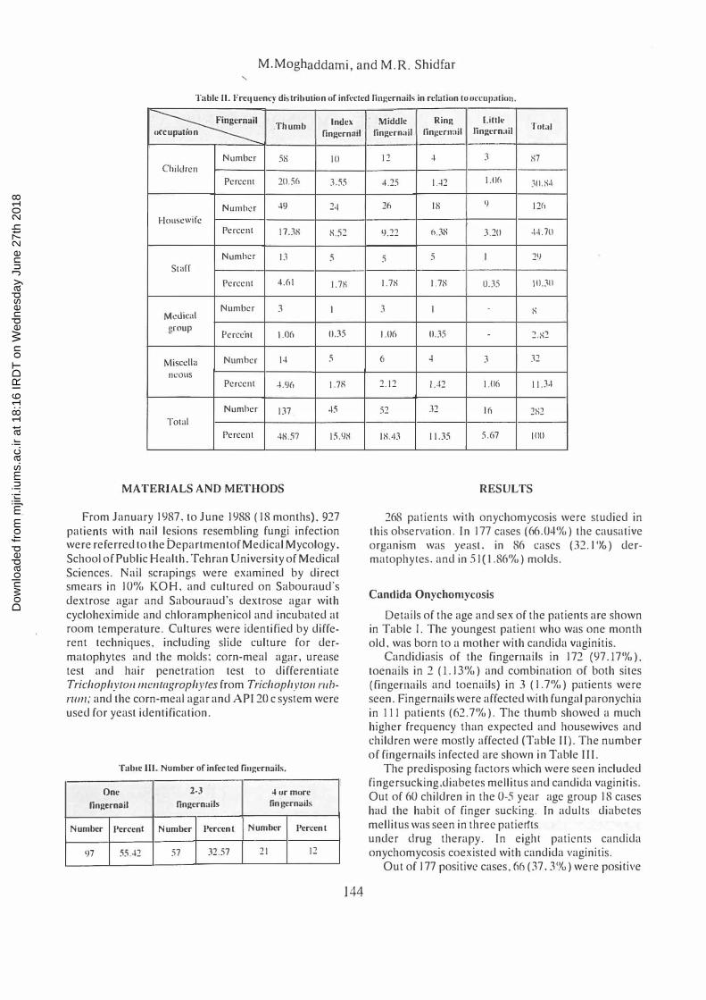
Figures 1,2, and 3. Tillea corporis and T. 1IIII;IIilllll in a p,lIiclll on corticosteroid and immunosuppressive therapy.
by the direct KOH preparation, 17 (9.6%) by culture, and 94 (53.1 %) by both methods.
The most frequently occurring species was Candida albicallS being present in 74 (66.6%) of isolates, followed by C. parapsilosis in 10 (9%), C. Iropicalis in eight (7.2%), C. gllillicrlllolldii in four (2.2%), C. sfellaroidea in two (1.8110), C. paralropicalis in two (1.8%), C. /<lIl1ala in one (0.9%), Geolrichlllll in one (0.9%), C. species in one (0.9%), and undifferentiable yeast in eight (7.2%).
Tinea llngllillnJ
Out of 86 patients with Tillea tlllguilll1l, 48 (55.8%) were male and 38 (44.2%) were female. The age and sex of the patients are shown in Table IV.
Toenails in 51 cases, fingernails in 32 cases, and a combination of both sites were seen in three cases. The 'big toenail had'a much higher rate of infection than expected (Table V and VI).
The clinical type of the lesion in all thc patients was invasive form (distal subungual onychomycosis). Nails alone were affected in 57 cases and it was associated with Tillea pedis in 21, Tillea mflllUllIll in three, Tillea cOIporis in two. both Tillea pedis and Tinea cruris in
two, Tillea capilis in one, Tinea crllris in one, and Tinea versicolor in one.
Extensive Tinea corporis was observed in two patients with both diabetes mellitus and asthma (on corticosteroid therapy) (Figures 1,2, and 3); also in a
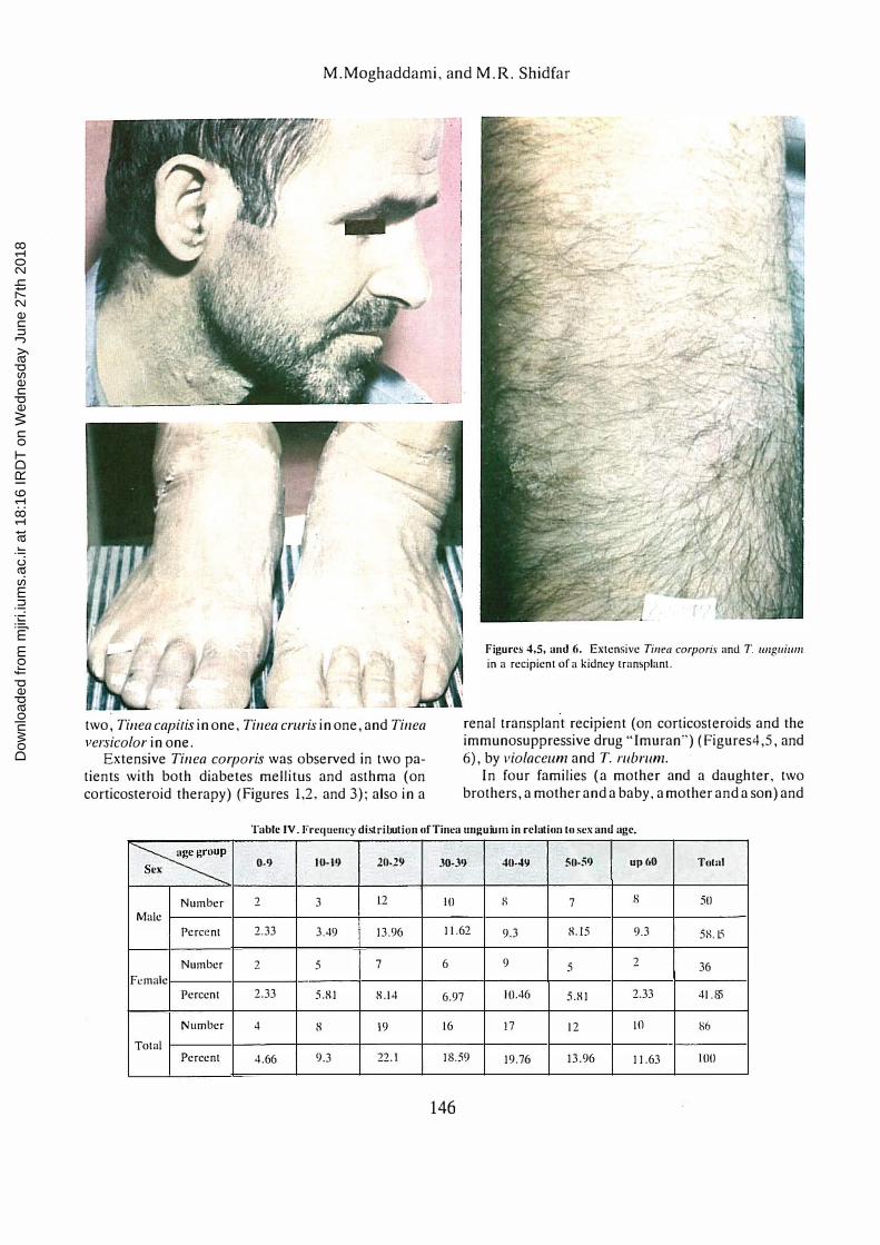
Fig ures 4,5. and 6. Extensive Tinea corporis and 1'. /lIIgllillm in a recipient of a kidney transplant.
renal transplant recipient (on corticosteroids and the immunosuppressive drug "Imuran") (Figures4,5, and 6), by violacellm and T. rubrum.
In four families (a mother and a daughter, two brothers, a mother and a baby, a mother and a son) and
Table IV Frequellt:)' distribution o f Tinca IIlIg uiulIl in relation to sex and age
� Sex 0-9 10-19 20·29 30-39 40·49 50-59 up60 Tolu)
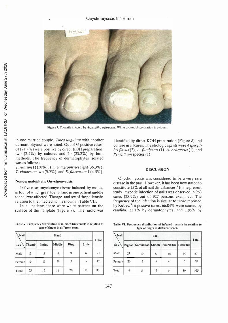
Figure 7. Toenails infected by Aspergillus ochrncells. White spotted discoloration is evident.
in one murried couple, Tinea llllguium with another dermatophytosis were noted. Out of 86 positive cases, 64 (74.4%) were positive by direct KOH preparation, two (2.4%) by culture, and 20 (23.2%) by both methods. The frequency of dermatophytes isolated was as follows: T. ,."bnlllill (50%), T. lIlelllagrophyteseight (36.3%),
T. via/acelllll two (9.2%), and E.f/accoslll1l I (4.5%).
Nondermatophylic Onychomycosis
In five cases onychomycosis was induced by molds, in four of which great toenail and in one patient middle toenail was affected. The age, and sex of the patients in relation to the infected nail is shown in Table VII.
In all patients there were white patches on the surface of the nailplate (Figure 7). The mold was
Tab)e V. FrequellCY distributioll of infected fill�crnails in relution to type uf finger in different sexes.
:s: Hand
l'tllal
Sex Thumb Index Middle Ring Little
Male 13 5 8 9 " � I
Female 10 8 S II 5 42
Total 23 13 16 20 II 83
147
identified by direct KOH preparation (Figure 8) and culture in all cases. The etiologic agents were AspergilIllS flavlls (2), A. flll1ligallls (I), A. ochracells (I), and Penicillilllll species (1).
DISCUSSION
Onychomycosis was considered to be a very rare disease in the past. However, it has been 'now stated to constitute 15% of all nail disturbances.4ln the present study, mycotic infection of nails was observed in 268 cases (28.9%) out of 927 persons examined. The frequency of the infection is similar to those reported by Kubec.5 In positive cases, 66.04% were caused by candida, 32.1 % by dermatophytes, and 1.86% by
Table VI. Frequency distrihution of infected lucnlliis in relation 10 type "ffinger in difTercn! sexes.
I� Fool
Total
Sex Big toe Second toe Middle Fourth Inc Liltle loe .-
Figure'S. Direct microscopy of nail fragments Prepared in potassium hydroxide solution (X 400).
molds, and these percentages are similar to those reported by Achten and Wanet-Rouard6As it is apparent in this study, candida infection of the nailplate is more common in women than men.]n relation to age it occurs most frequently at the age group of 0-5 (in both sexes) and at the age group 24-29 years in women. The paronychial fold is readily colonized, particularly in people whose occupation requires frequent immersion of finger in water. 7
In chronic paronychia the nail be ames invaded and only the nail may be effected. In order of frequency the thumb and the middle fingernails showed a much higher incidence than expected. These results agree with Ganor and Pumpianski's studies." In most cases involvement of only one fingernail (55.4%) was observed. Mechanical trauma which destroys the cuticle of finger is responsible for the invasion of C. albicalls into the nailfold.B In housewives the higher frequency of candida infection of the nail may be due to
Table VU. Distrihulion of nondcrrnatuphytic onychomycosis in relation tu age, sex, and the inrected nail in five cases.
Pallcnr Age Sex . Toenail
involved Len Right
24 M Middle +
2 43 F Great +
3 30 F Greal +
4 52 F Great +
5 56 F Great +
• M = Male • F = Female
148
continued immersion of fingers in water and mechanical trauma, and in children may result from a habit of finger sucking. Theoretically, the source of C. albicalls in the finger infection could be !he vagina, mouth. or bowe\. H Eight patients showed candida onychomycosis associated with vaginitis. C. albical/s is rarely found in the vagina of patients with chronic paronychia. There· fore, this site seems to have a minor epidemiological significance" The incidence of dermatophytosis or yeast infection is not significantly higher in diabetics than in normal subjectsY However. the association of onychomycosis with diabetes mellitus needs further study. The two species most frequently involved in the infection of the nails and considered to be potential pathogens, are Cal/dida albicnl/s and Cal/dida pampsilosis; although in this study C. albiCIIlIs was the predominant agent isolated.
The frequency of Tillea IIllglliul1l which was predominant in males was similar to that reported from India 10 It was most frequent in the toenails especially in big toenail in both sexes. Ringworm infection of the toenails is an exceedingly common disease condition in races accustomed to wearing shoes. and the disease has been attributed to the constant pressure applied on the toes, particularly the great toenail, caused by tight footwear.-l
The majority of Tillea llllgllilllll infections were seen at the second to fourth decades and only four cases were seen below 10 years. The rarity of Tinea wlguiul1l in children in comparison to adults is explained by a faster dynamic growth of nails in children than adults. '" In 30 cases, Tinea llflgllilllll was associated with Tinea pedis .
mamUWll, cruris, corporis, and Tillea capitis. These findings agree with that of Kamalam and Tambiah. 10
Extensive skin involvement in tWO patients with diabetes and asthma (on corticosteroid therapy) and in a renal graft recipient (on corticosteroid and immunosuppressive therapy) by T. vio/aceulII and T. rubrulll, suggests that the agents may behave opportunistically under conditions of suppressed immune response. Disseminated ringworm infections by T. vio/aceulII in two familial cases associated with a deficiency of cellular immune response and a case of multiple, subcutaneous, neutrophilic abscesses due to T. rllbrlllll in an immunosuppressed renal allograft recipient were reported by Osman et.al. and Novick et. al.,,·I' Four familial cases of dermatophytosis were observed which probably were transmilled by direct personal contact.
It is recognized that culture of dermatophytes from nails is difficult. 13 We also failed to culture 66 of 86 microscopically positive nails. Our results are similarto those reported by Walshe and English, and by Ardehali. 1 •. 15In contrast 10 the report by Ardehali, the
most frequent fungal isolates in this survey were T. rubrlllll and T. lIIe/llagrophYles. Ardehali believed that the most common dermatophytes causing T. rmguilll1l in Iran, was T.scllOell/eillii.15 As with dermatophyte infection, mould infections are much more common in toenails than in fingernails.' According to this study. in all the cases toenails were affected. In most cases the big toenail of females (four cases) were involved. The most common mold isolated was Aspergillus species. A case of onychomycosis caused by A. /Illlligatus is
reported by Rosenthal et.a1.16 and four cases of onychomycosis caused by A. lerreus are presented by Obsberg et.al. 17
ACKNOWLEDGEMENTS
We are grateful to Dr.J.Masoud and J. Gharegozlou for Iheir advice and kind cooperation.
149
REFERENCES
I. Onsbcrg P. Siahl D. Vcicn NK: Onychomycosis caused by Aspergillus l('rreIlS. Sabouraudia 16: 39-49.1978.
2. English MP: Nails and rungi. Sf J Dcnmllo194: 697-701. 1976. 3. Zaiaz N: Onychomycosis. Arch Dcnnalol105: 263- 75. 1972. 4. Ramcsh V. Reddy BSN. Singh R: Onychomycosis (Review). lnt J
Derm.10122(3): t48-52. 1983. S. Kubec K: Epidemiology of mycotic nora in the Ilnit plnlc. Ccsk.
Derm,10153 (5): 332·5. 1978. 6. Aehtcn G, Wanet Rouard J: Onychomycosis in the laboratory.
Mykoscn (t): 125-7. 197H. 7. Rippon JW: Til/ell Imgllillm, In: Medical Mycology. the
Pathogenic Fungi and the Pathogenic Actinomycetes. Philadelphia: W.B. Saunders. 190-3. 1982.
8. Ganor S. Pumpianski R:Chronic Candidll (llbic{/I/s paronychia in adult Israeli women. Br DermatoI90: 7 7 -S3. 1974.
9. Fusaro RM. Goetz FC: Common cutaneous manifestations and problem of diabetes mellitus. Posl Grad Meu 49: S4-7. 1971.
10. KamalaOl A. Thambiah AS: A study of 3 S91 cases of mycoses in (he Tropics. Sabouraudia 14: 129-48, 1976.
I I. Osman AB. Chaffai M. Aycd K, Khalfat A: Derm;JlOphytie dissemince il Trichophytoll l'iohlCt'llIIl. A propos D deux cas familliaux. Bulletin De LA Societe Francaisc Dc Mycologic Medicale XIV 2:281-4. 1985.
12. Novick NY. Tapia L. Bottone EJ: Invasive Triclwphytoll mhmlll infection in an immullocompromiseu hosl. Case report anti review of the literature. Am J Med N2(2): 321-5, 1t)�7.
13. Gentles JC: Laboratory investigatiUlls of dermatophyte inf�clions of nails. Sabouraudia 9: 149-52, 1971.
14, Walshe MM, English MP: Fungi in nails. Br J D�rmalo 7H: 11)8· 2U7.19M.
15. Ardeha!i M: DermatophYlic agentsof Tillt'a UlIglliuI1I in Iran. Int J DcrmaloI 21(5): 322-3.1973.
16. Rosenthal SA, Strilzler R. Villafane J: Onychomyt:llsiscau�eu by ..hpl'rgilll/s jilllligalll.1'. R�porl of a cas�. Arch DermaI0197:flt';5· 7. 196H.
17. Onsberg P. Stahl D. Vcicn NK: Onychomycosis caused by Aspergilll/.f (l'rl"('/O', Sabouraudia 16:39-46. 1978.


![㠢㠸㠧㠳ã å ã 㠼㠿 2007ç · (9 ÷]9]W]d] ][]/] ] ]O]2\ / ÷ ñ ® ³ .]']d] ]+]0 ÿ º 3 ò 1\» 60$57 9 + Title: 㠢㠸㠧㠳ã å ã 㠼㠿_2007ç](https://static.documents.pub/doc/80x56/60325704c2c96c22f606dc61/-2007-9-9wd-o2-.jpg)


