Rep. Lundy Field Soc. 53 A SURVEY OF MICROBIAL PARASITES IN LUNDY BROWN RATS (RATTUS NORVEGICUS) By KlM BLASDELL AND AMANDA READ Department of Veterinary Pathology, University of Liverpool, Leahurst Veterinary Station, South Wirral, CH64 7TE ABSTRACT The object of this study was to determine the parasite profile of Lundy brown rats. A combination of methods comprising culture, polymerase chain reaction (PCR) and immunofluoresence assay serology (IFA) were used to analyse past and present infection by seven micro- parasites. The results of this study show that the sampled rats were infected with two of the seven taxa of organisms tested for: Bartonella spp. and Trypanosoma spp . observed prevalence and possible reasons for presence and absence of the different organisms tested for are discussed, together with the possible implications ofthese infections at the individual and population level. Keywords: Lundy, brown rat, microbial parasites. INTRODUCTION Rodents act as reservoirs for a wide range of parasitic organisms some of which can be defined as zoonotic, meaning that they are capable of infecting humans. Rats of the genus Rattus have been well characterised as reservoirs for zoonoses such as Yersinia pestis, the causative agent of bubonic plague, and Leptospira interrogans, the agent of Weil's disease (Gerke & Rump, 2003). Whilst Yersinia pestis is no longer endemic in the UK, several other zoonotic diseases, such as cowpox and rodent- specific parasites, can be identified in the rat population. Parasites that can infect rodents may also infect other organisms, as part of their natural cycle, with rodents representing a transient host for a single stage of the life cycle. Parasites with such complex life cycles require the hosts for all stages of the cycle to be present and easily accessible. Often parasites are transmitted between hosts by arthropod vectors, such as fleas and ticks. Not all parasitic species are 86
Transcript
Rep. Lundy Field Soc. 53
A SURVEY OF MICROBIAL PARASITES IN LUNDY BROWN RATS (RATTUS NORVEGICUS)
By
KlM BLASDELL AND AMANDA READ
Department of Veterinary Pathology, University of Liverpool, Leahurst Veterinary Station, South Wirral, CH64 7TE
ABSTRACT
The object of this study was to determine the parasite profile of Lundy brown rats. A combination of methods comprising culture, polymerase chain reaction (PCR) and immunofluoresence assay serology (IFA) were used to analyse past and present infection by seven microparasites. The results of this study show that the sampled rats were infected with two of the seven taxa of organisms tested for: Bartonella spp. and Trypanosoma spp. Th·~ observed prevalence and possible reasons for presence and absence of the different organisms tested for are discussed, together with the possible implications ofthese infections at the individual and population level.
Keywords: Lundy, brown rat, microbial parasites.
INTRODUCTION
Rodents act as reservoirs for a wide range of parasitic organisms some of which can be defined as zoonotic, meaning that they are capable of infecting humans. Rats of the genus Rattus have been well characterised as reservoirs for zoonoses such as Yersinia pestis, the causative agent of bubonic plague, and Leptospira interrogans, the agent of Weil's disease (Gerke & Rump, 2003). Whilst Yersinia pestis is no longer endemic in the UK, several other zoonotic diseases, such as cowpox and rodentspecific parasites, can be identified in the rat population.
Parasites that can infect rodents may also infect other organisms, as part of their natural cycle, with rodents representing a transient host for a single stage of the life cycle. Parasites with such complex life cycles require the hosts for all stages of the cycle to be present and easily accessible. Often parasites are transmitted between hosts by arthropod vectors, such as fleas and ticks. Not all parasitic species are
86
transmitted to new hosts by this route. Some are transmitted by direct contact with the reservoir, through the respiratory tract, or by contact with an infected host 's urine or faecal material. Ectoparasites can have a significant impact on the life cycle through their specificity for either the parasite or the reservoir host(s).
Lundy is interesting to look at in terms of microbial parasites, as small islands often have considerably fewer vertebrate species than similar habitats on the mainland, therefore, fewer potential hosts for parasites and ultimately a less diverse range of parasites (MacArthur & Wilson, 1967 ; Milazzo et al., 2003). Lundy differs from many other islands as all except two mammals, the grey seal and pygmy shrew, are non-native to the island and in the case of the Sika deer, are non-native to the UK. This would almost certainly have an effect on the range of parasites capable of living in this environment. Microbial parasite load may have interesting implications on the health of the host population, particularly fecundity. A great deal of research exists examining the effect of epidemic infections but little has been published on the effects of endemic infections. Feore et al. (2004) examined the effect of cowpox infections on the reproductive success of bank voles and wood mice and concluded that fecundity was significantly reduced in infected animals. Work is ongoing in Feore's research group to examine the effects of endemic infections, which do not have clinical symptoms or generally affect survival, and will include some of the bacterial species included in this report.
Microbial parasites that are either known, or suspected, to infect species of the genus Rattus were selected for prevalence testing, these comprised viruses cowpox, MHV-68 and Lymphocytic choriomeningitis virus (LCMV), bacterial species of Bartonella and Anaplasma and the Protozoa Babesia and Trypanosoma.
All three viruses naturally infect rodents , although only Cowpox has been proven to naturally exist in rat populations. MHV-68 is a rodent-specific virus (Blasdell et al., 2003), whilst cowpox and LCMV are known to be zoonotic (Armstrong & Sweet, 1939; Baxby et al., 1994 ) , with Cowpox capable of infecting a wide range of mammalian hosts (Baxby et al., 1979). However, little work has been done on the natural host reservoirs of these viruses, particularly in regards to LCMV and MHV-68. The work reported here is part of a larger study investigating the range of natural reservoir hosts for these virus species .
Both Bartonella and Anaplasma have representatives that exist naturally in wild rodent populations. There are at least five species of Bartonella that have rodents as their reservoir host (Birtles et al. , 1994), including B. elizabethae, which is also known to be zoonotic. Of the species of Anaplasma found in the UK, A. phagocytophila is capable of infecting humans and species of Anaplasma have been isolated from rodents species (Bown et al. , 2003). Like the bacteria, both protozoan groups also contain species that reside in rodent populations. At least two species of Babesia naturally infect rodents in the UK, including B. divergens, a zoonotic pathogen (Zintl et al. , 2003) , which, along with B. microti, is known to infect rats , (Akinboade et al., 1981 ).
87
There are three species of trypanosome that infect rodents in the UK, of which T. lewisi is thought to be rat-specific and non-zoonotic (Hoare, 1972). Although both species of rats native to the UK (Rattus novegicus, brown rat; R. rattus , black rat) have been found on Lundy, this survey is restricted to the Lundy brown rat due to sample availability.
METHODS
Sample preparation
42 brown rats (Rattus norvegicus) stored at -20°C, were obtained from Lundy Island courtesy of the 'Seabird Recovery Project ' . These were dissected and a sample of blood (obtained by cardiac puncture) and the spleen were taken from each animal. The spleens were finely chopped and placed in enough Eagle's Minimum Essential Medium (EMEM), containing 1 o/o foetal calf serum (FCS), to cover the sample. Both blood and spleen were kept at -80°C until required.
DNA extraction
50f.Ll of blood and 50ml of EMEM containing spleen from each animal were used for DNA extraction. Samples were diluted in 500ml1.25% Ammonia solution and heated at 100°C for 20 minutes. Samples were vortexed and returned to the heating block for a further 25 minutes , this time with the lid of each tube open to allow evaporation and hence concentration of the DNA extract, (Bown et al. , 2003) . All samples were diluted 1:10 in double-distilled Hp. Stock DNA extracts were placed at -80°C for storage, whilst diluted samples were placed at -20°C until required.
Bacterial culture for Bartonella
A 111lloopful of rat blood was streaked onto a Columbia agar plate containing 10% defibrinated horse blood. The plates were then incubated at 3TC with 5% C0
2
saturation, in a moist environment, for between 7 and 14 days until colonies were visible. Samples were prepared ready for PCR by resuspending bacteria in water. Samples for which plates remained clear for more than 10 days were considered as being sterile.
Polymerase chain reaction
Whole-cell preparations or a small quantity of DNA extracted from blood or spleen were added to a commercially prepared PCR enzyme mix (Abgene) and primers , short nucleotide sequences complementary to known DNA sequences in the target
88
parasite, in 0.2ml thin walled tubes (Axygen). The samples were then processed through a series of heating cycles, using a thermocycler (Thermohybaid). PCR reactions were subjected to gel electrophoresis using a 1% agarose gel, containing Sf.ll of ethidium bromide at a concentration of lOmg/ml and visualised using UV light. Details of the primers used are illustrated in Table 1.
Table 1. Details of the primers used to test for the presence of the different bacterial and protozoan taxa.
TARGETED PRIMER TARGET STAGE DIRECTION SEQUENCE SPECIES NAME REGION
Trypanosom TRF 18SRNA 1" stage Forward GAAACAAGAA (Noyes et al., region ACACGGGAG 1999)
TRR Reverse CTACTGGGC AGCTTGGA
SSF Nested Forward TGGGATAACA stage AAGGAGCA
SS R Reverse CTGAGACTGTA ACCTCAAAGC
89
Immunofluorescence assay serology
A ninety-six well plate (Cos tar) was prepared so that each well contained a confluent layer ofVero (green monkey kidney) cells. These cells were infected with the relevant virus, either cowpox or MHV-68, and left to grow for three or five days respectively. Blood from each animal was placed onto two wells each at dilutions of 1:40 and I :20 (total volume 30J.Ll) . Antibodies in the blood, resulting from prior infection , bind to virus-infected cells in the wells . The plates were washed three times with Phosphate buffered saline (PBS) and a fluorescently labelled anti-rat antibody (Sigma) diluted 1:20 (total volume 30J.Ll) was added. This meant that only rats that have been infected with a virus previously and therefore had produced antibodies against it would show positive results . Fluorescent-labelled positive samples were detected using an UV microscope (Crouch et al., 1995) .
Immunofluorescence assay serology for LCMV Commercially produced slides (Charles River Laboratories) with eighteen wells were used and the blood was tested at a single dilution of 1:2 (total volume 10J.Ll). The wells of these slides contained a confluent layer of cells, both infected and uninfected (mouse cell-line), which had been fixed with acetone prior to purchase.
RESULTS
Bacterial culture
Six of the 40 blood samples yielded Bartonella-like colonies (off-white, small, smooth). These bacteria were then subjected to PCR to confirm their identity.
Polymerase chain reaction
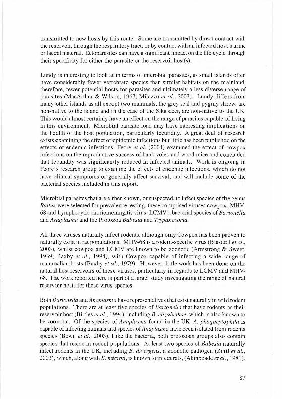
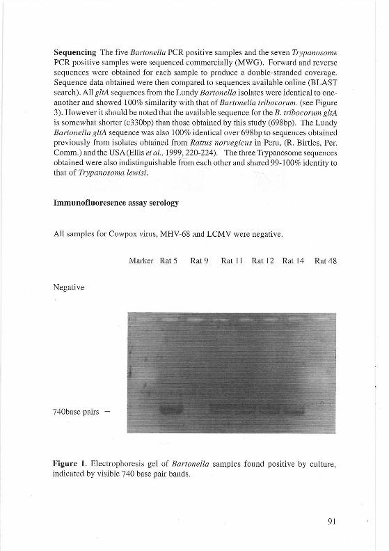
The six culture positive samples were subjected to PCR, five of the samples were confirmed to be Bartonella each yielding a PCR product of the correct size (740bp) (see Figure 1). Insufficient bacteria were available for testing the sixth sample.
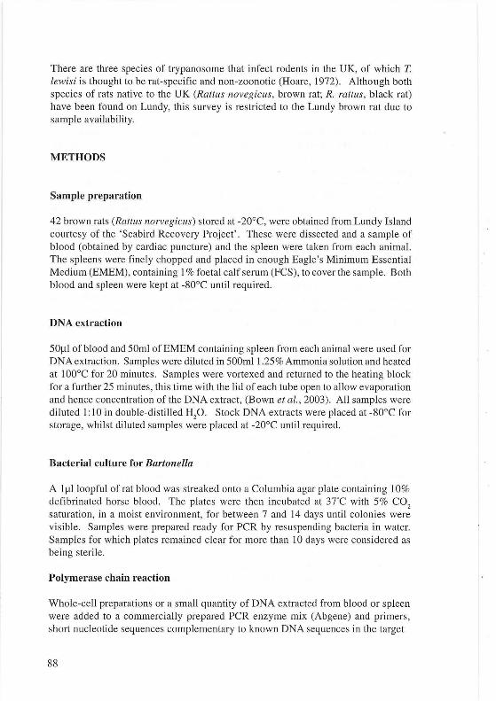
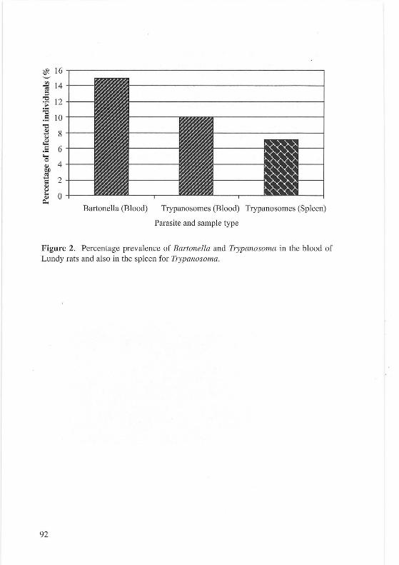
DNA extracted from the blood and spleen from each rat was tested using PCR for Anaplasma, Babesia and Trypanosoma . PCR for Anaplasma and Babesia were negative. The blood and spleen from three rats and the blood from one further rat were positive for Trypanosoma (see Figure 2). The PCR products from the samples found positive for trypanosomes were PCR purified using a DNA purification kit (Qiagen) .
90
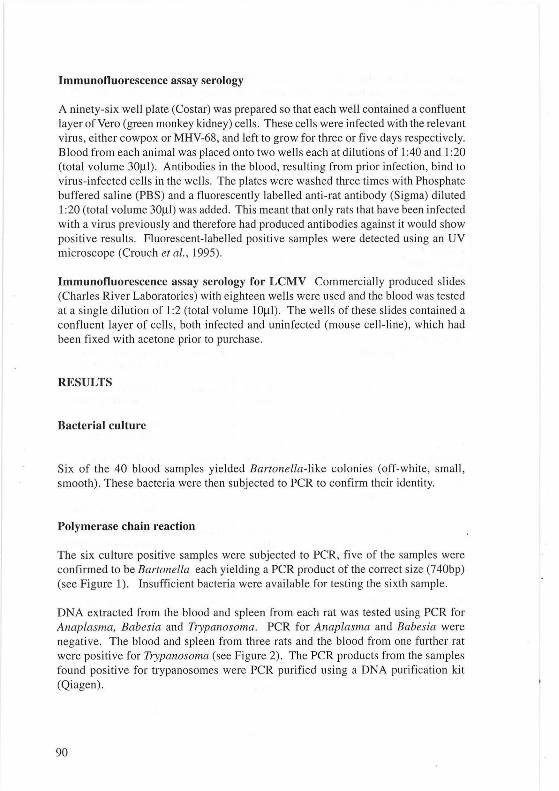
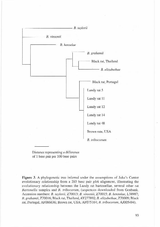
Sequencing The five Bartonella PCR positive samples and the seven Trypanosome PCR positive samples were sequenced commercially (MWG). Forward and reverse sequences were obtained for each sample to produce a double-stranded coverage. Sequence data obtained were then compared to sequences available online (BLAST search). All gltA sequences from the Lundy Bartonella isolates were identical to oneanother and showed 100% similarity with that of Bartonella tribocorum. (see Figure 3). However it should be noted that the available sequence for the B. tribocorum gltA is somewhat shorter (c330bp) than those obtained by this study (698bp). The Lundy Bartonella gltA sequence was also 100% identical over 698bp to sequences obtained previously from isolates obtained from Rattus norvegicus in Peru, (R. Birtles, Per. Comm.) and the USA (EIIis eta/ .. ; 1.999, 220-224). The three Trypanosome sequences obtained were also indistinguishable-from each other and shared 99-100% identity to that of Trypanosoma lewisi.
Immunofluoresence assay serology
All samples for Cowpox virus, MHV-68 and LCMV were negative.
Marker Rat 5 Rat9 Rat 11 Rat 12 Rat 14 Rat 48
Negative
740base pairs -
Figure 1. Electrophoresis gel of Bartonella samples found positive by culture, indicated by visible 740 base pair bands .
91
~ fl}
-; ::I -e ·;; ;a .5 -e Cl) ...... u ~ .5 ..... 0 Cl) OD ~ ...... = Cl) u ... Cl)
Figure 2. Percentage prevalence of Bartonella and Trypanosoma in the blood of Lundy rats and also in the spleen for Trypanosoma.
92
-
B l .. . tay oru
B. vinsonii
B. henselae
B. grahamii
-Black rat, Thailand
~c --=----- B. elizabethae
Distance representing a difference of 1 base pair per 1 00 base pairs
- Black rat, Portugal
Lu ndy rat 5
Lu ndy rat 11
Lu ndy rat 12 -
Lu ndy rat 14
Lu ndy rat 48
Br own rate, USA
B. tribocorum
Figure 3. A phylogenetic tree inferred under the assumptions of Juke 's Cantor evolutionary relationship from a 285 base pair gltA alignment, illustrating the evolutionary relationship between the Lundy rat bartonellae, several other rat Bartonella samples and B. tribocorum, (sequences downloaded from Genbank. Accession numbers: B. taylorii , Z70013; B. vinsonii, Z70015; B. hensalae, L38987; B. grahamii, Z70016; Black rat, Thailand, AY277892; B. elizabethae, Z70009 ; Black rat, Portugal, AF086636; Brown rat, USA; AF075164; B. tribocorum, AJ005494).
93
DISCUSSION
Bacteria of the genus Bartonella are known to infect a wide variety of ma mmalian hosts and several species are known to have rats as a natural reservoir host. B. tribocorum, B. elizabethae and B. grahamii have all been previously isola ted from rats (Ell is et al., 1999). Lundy rats were found to be infected with Bartonella, which based on avai lable sequence data is likely to be B. tribocorum. B. tribocorum is a ratspecific Bartonella species (Helier et al. , 1998). Incomplete sequence is available for the B. tribocorum gltA gene, the target of these PCR reactions, but a 100% match was obtained with the available sequence. B. tribocorum is genetically rela ted to B. elizabethae , based on analysis of several genes, which has been implicated in zoonotic infections (Helier et al., 1998; Daly et al., 1993). Prevalences found in other rat populations have ranged from approximately 0-60% in Rattus norvegicus, so the fi nding of a prevalence of approximately 15% for Lundy rats fits with this data (Birtles et al., 1994; Ellis et al., 1999).
The protozoan species Trypanosoma lewisi was identified in the Lundy samples. This parasite species has been identified in both black and brown rats, with prevalences of I I% and 22% respectively (Laakkonen et al., 2003 ; Linardi & Botelho, 2002). The observed prevalence of I 0% (blood) in this population may be slightly low, but may simply be an effect of small sample size, or possibly the poor qual ity of the samples, due to the samples being taken from animals that had been deceased and stored for an unknown length of time. Both T lewisi and B. tribocorum are transmitted by the flea, clearly demonstrating the survival of this vector on Lundy.
The three viruses, cowpox virus, LCMV and MHV-68 tested for were absent. While ev idence exists for cowpox and LCMV naturally infecting rats, these are not the primary reservoir hosts. Due to the lack of other rodent species on Lu ndy, there is less possibility of cross-infection from natural reservoir hosts , voles and mice (Marennikova et al. , 1977; M. Bennett, Pers . Comm.) . The absence of Anaplasma and Babesia can more readily be explained by considering seasonal variations in the prevalence of the arthropod vector, Ixodes ricinus (Bown et al. , 2003; Hartelt et al. , 2004). We were unable to confirm the presence or absence of ticks on Lundy, although due to the presence of sheep and deer it seems likely that ticks would be present. Alternatively, any of these parasites may have been absent from the founding population of Lundy rats, although the importance of the foundin g population would depend on whether rats from the mainland regularly infiltrated the island popu lation.
It is interesting to note that the two micro-parasite species present are both transmitted by fleas, whilst the two non-virus species that are absent are both transmitted by the same tick species, Ixodes ricinus. These data confirm that fleas are present in the Lundy animal population and are likely to be acting as vectors for infectious disease. It is unlikely that loxdes ricinus is absent from the island, this tick species is commonly found in woodland habitats, but has been shown to be capable of existing in open
94
sheep pasture (Walker et al., 2001). Due to the number of different potential host species that are present on the island, it is likely that the ticks would survive on Lundy. Tick populations fluctuate with the seasons, if the rat population was sampled in a seasonal trough period it may affect the disease prevalence. Combined with the poor quality of the original samples and the small sample size this may explain the low numbers of parasites isolated.
It would be interesting to carry out an ectoparasite survey lasting a year on Lundy, combined with a survey of the infectious agents present. Surveying the ectoparasites and micro-parasites of other mammalian species on Lundy would give a complete picture of the microbial profile of mammals on the island. Combining these studies with work determining the effects of endemic infections on the fecundity and survival of mammalian hosts , would provide interesting parallels between Lundy mammals, with an apparently limited parasite burden, and mainland mammals.
ACKNOWLEDGMENTS
Many thanks to the Lundy Warden , Ben Sampson, who stored the rats unti l they could be collected and also a great deal of thanks to all those involved in the collecting of the rats. Thanks also go to Dr Kevin Bown and Andrew Smith for the primers, Cathy Glover for technical assistance and to Professor Malcolm Bennett and Dr Richard Birtles for all of their guidance.
REFERENCES
Akinboade, O.A. , Dipeolu, 0.0. , Ogunji, F.O. & Adegoke, G.O., 1981. The Parasites Obtained and the Bacteria Isolated from House Rats (Rattus rattus, Linnaeus, 17 58) Caught in Human Habitations in Ibadan , Nigeria. International Journal of Zoonoses 8, 26-32.
Armstrong, C. & Sweet, L.K., 1939. Lymphocytic Choriomeningitis. Public Health Report 54, 673 .
Baxby, D. , Shackleton, W.B. , Wheeler, J. & Turner, A., 1979. Comparison of Cowpoxlike Viruses Isolated from European Zoos. Archives of Virology 61 , 337-340.
Baxby, D., Bennett, M. & Getty, B. 1994., Human Cowpox 1969-93: A Review Based on 54 Cases. British Journal of Dermatology 131 , 598-607.
95
Blasdell, K. , McCracken, C., Morris , A. , Nash, A.A., Begon, M. , Bennett, M. & Stewart, J.P., 2003. The Wood Mouse is a Natural Host for Murid Herpesvirus 4. Journal of General Virology 84, 111-113.
Birtles, R.J., Harrison, T.G. , & Molyneux, D.H., 1994. Grahamella in Small Woodland Mammals in the UK: Isolation Prevalence and Host Specificity. Annals of Tropical Medicine and Parasitology 88, 317-327.
Birtles , R.J . & Raoult, D. , 1996. Comparison of Partial Citrate Synthase Gene (gltA) Sequences for Phylogenetic Analysis of Bartonella Species. International Journal of Systematic Bacteriology 46, 891-897.
Bown, K.J. , Begon, M., Bennett, M. , Woldehiwet, Z. & Ogden, N.H., 2003 . Seasonal Dynamics of Anaplasma phagocytophila in a Rodent-tick (Ixodes trianguliceps) System, United Kingdom. Emerging Infectious Diseases 9, 63-70.
Crouch, A.C., Baxby, D. , McCracken, C.M. , Gaskell, R.M. & Bennett, M. , 1995. Serological Evidence for the Reservoir Hosts of Cowpox Virus in British Wildlife. Epidemiology and Infection 115, 185-191.
Daly, J.S. , Worthington , M. G. , Brenner, D.J., Moss, C.W. , Hollis , D.G. , Weyant, R.S. , Steigerwalt, A. G., Weaver, R.E., Daneshvar, M.I. & O'Connor, S.P., 1993. Rochalimaea elizabethae sp. nov. Isolated from a Patient with Endocarditis. Journal of Clincal Microbiology 31, 872-881.
Ellis, B.A. , Regnery, R.L. , Beati, L. , Bacellar, F. , Rood, M., Glass, G.G., Marston, E., Ksiazek, T.G. , Jones, D. & Childs, J.E., 1999. Rats of the Genus Rattus are Reservoir Hosts for Pathogenic Bartonella Species: An Old World Origin for a New World Disease. The Journal of Infectious Diseases 180, 220-224.
Feore, S.M. , Bennett, M. , Chantrey, J., Jones, T., Baxby, D. & Begon, M. , 2004. The Effect of Cowpox Virus Infection on Fecundity in Bank Voles and Wood Mice. Proceedings of the Royal Society of London, Series B: Biological Sciences 264, 1457-1461.
Gerke, P. & Rump, L.C., 2003. Leptospirosis - 3 Cases and a Review. Clinical Nephrology 60, 42-48.
Hartelt, K. , Oehme, R., Frank, H., Brockmann, S.O. , Hassler, D. , Kimmig, P. , 2004.
96
Pathogens and Symbionts in Ticks: Preva lence of Anaplasma phagocytophilum (Ehrlichia sp.), Wolbachia sp. , Rickettsia sp., and Babesia sp. in Southern Germany. International Journal of Medical Microbiology 293, supplement 37: 86-92.
Helier, R. , Riegel , P. , Hansmann , Y., Delacour, G., Bermond, D., Dehio, C., Lamarque, F., Monteil , H. , Chomel, B. & Piemont, Y. , 1998. Bartonella tribocorum spp. nov. , a New Bartonella Species Isolated from the Blood of Wild Rats. International Journal of Systemic Bacteriology 48, 1333-1339.
Hoare, C.A. , 1972. The Trypanosomes of Mammals. A Zoological monograph, 749. Blackwell: Oxford .
Laakkonen , J. , Lehtonen , J.T. , Ramiarinjanahary, H. & Wright, P.C. 2003 . Trypanosome Parasites in the Invading Rattus rattus and Endemic Rodents of Madagascar. AC/AR Monographs 96, 1775-1783.
Linardi , P.M. & Botelho, J.R. , 2002. Prevalence of Trypanosoma lewisi in Rattus norvegicus from Belo Horizonte, State of Minas Gerais, Brazil. Memorial Institute ofOswaldo Cruz 97 , 411-414.
MacArthur, R.H. & Wilson, E.O. , 1967. The Theory of Island Biogeography. New Jersey: Princeton University Press.
Marennikova, S.S. , Maltseva, N.N. , Korneeva, V. I. & Garanina, N.M. , 1977. Outbreak of Pox Disease Among Carnivora (Felidae) and Edentata. The Journal of Infectious Diseases 135, 358-366.
Massung, R.F. , Slater, K. , Owens, J.H. , Nicholson, W.L. , Mather, T.N. , Solberg, V.B. & Olson , J.G., 1998. Nested PCR Assay for Detection of Granulocytic Ehrlichiae. Journal of Clinical Microbiology 36, 1090-1095.
Milazzo, C., de Bellocq, J.G. , Mara, C. , Casanova, J.C. , di Bella, C. , Feliu, C. , Fons, R. , Serge, M. & Santalla, F. , 2003. Helminths and Ectoparasites of Rattus rattus and Mus musculus from Sicily, Italy. Comparative Physiology 70, 199-204.
Norman , A.F. , Regnery, R. , Jameson , P. , Greene, C. & Krause, D.C., 1995. Differentiation of Bartonella-like Isolates at the Species Level by PeRrestriction Fragment Length Polymorphism in the Citrate Synthase Gene. Journal of Clinical Microbiology 33, 1797- 1803.
Noyes, H.A., Stevens J.R. , Teixeira, M., Phelan, J. & Holz, P. , 1999. A Nested PCR for the ssrRNA Gene Detects Trypanosoma binneyi in the Platypus and Trypanosoma sp. in Wombats and Kangaroos in Australia. International Journal of Parasitology 29, 331-339.
97
Walker, A.R., Alberdi, M.P. , Urquhart, K.A. & Rose, H. , 2001. Risk Factors in Habitats of the Tick Ixodes ricinus Influencing Human Exposure to Ehrlichia phagocytophila Bacteria. Medical and Veterinary Entomology 15 , 40-49.
Zintl , A. , Mulcahy, G. , Skerrett, H.E. , Taylor, S.M. & Gray, J.S. , 2003. Babesia divergens, a Bovine Blood Parasite of Veterinary and Zoonotic Importance. Clinical Microbiology Reviews 16, 622-636.