Jessica A. Mollick, Thomas E. Hazy, Kai A. Krueger, Ananta Nair, Prescott Mackie, Seth A. Herd,and Randall C. O’Reilly
University of Colorado Boulder
We describe a neurobiologically informed computational model of phasic dopamine signaling to accountfor a wide range of findings, including many considered inconsistent with the simple reward predictionerror (RPE) formalism. The central feature of this PVLV framework is a distinction between a primaryvalue (PV) system for anticipating primary rewards (Unconditioned Stimuli [USs]), and a learned value(LV) system for learning about stimuli associated with such rewards (CSs). The LV system representsthe amygdala, which drives phasic bursting in midbrain dopamine areas, while the PV system representsthe ventral striatum, which drives shunting inhibition of dopamine for expected USs (via direct inhibitoryprojections) and phasic pausing for expected USs (via the lateral habenula). Our model accounts for datasupporting the separability of these systems, including individual differences in CS-based (sign-tracking)versus US-based learning (goal-tracking). Both systems use competing opponent-processing pathwaysrepresenting evidence for and against specific USs, which can explain data dissociating the processesinvolved in acquisition versus extinction conditioning. Further, opponent processing proved critical inaccounting for the full range of conditioned inhibition phenomena, and the closely related paradigm ofsecond-order conditioning. Finally, we show how additional separable pathways representing aversiveUSs, largely mirroring those for appetitive USs, also have important differences from the positive valencecase, allowing the model to account for several important phenomena in aversive conditioning. Overall,accounting for all of these phenomena strongly constrains the model, thus providing a well-validatedframework for understanding phasic dopamine signaling.
Phasic dopamine signaling plays a well-documented role inmany forms of learning (e.g., Wise, 2004) and understanding themechanisms involved in generating these signals is of fundamentalimportance. The temporal differences (TD) framework (Sutton &
Barto, 1981, 1990, 1998), building on the reward prediction error(RPE) theory of Rescorla and Wagner (1972), provided a majoradvance by formalizing phasic dopamine signals in terms of con-tinuously computed RPEs (Montague, Dayan, & Sejnowski, 1996;Schultz, Dayan, & Montague, 1997). To summarize this dopaminereward prediction error hypothesis (DA-RPE; Glimcher, 2011),the occurrence of better than expected reward outcomes producesbrief, short-latency increases in dopamine cell firing (phasicbursts), while worse than expected outcomes produce correspond-ing phasic decreases (pauses/dips) relative to a tonic firing base-line. These punctate error signals have been shown to function astemporally precise teaching signals for Pavlovian and instrumentallearning, and are widely believed to play an important role in theacquisition and performance of many higher cognitive functionsincluding: action selection (Frank, 2006), sequence production(Suri & Schultz, 1998), goal-directed behavior (Goto & Grace,2005), decision making (Doll & Frank, 2009; St. Onge & Floresco,2009; Takahashi et al., 2010), and working memory manipulation(O’Reilly & Frank, 2006; Rieckmann, Karlsson, Fischer, & Back-man, 2011).
Despite the well-documented explanatory power of this simpleidea, it has become increasingly clear that a more nuanced under-standing is needed, as there are many aspects of dopamine cellfiring that are hard to reconcile within a simple RPE formalism.For example, dopamine cell bursting has long been known to occurrobustly at both CS- and US-onset for a period of time early intraining (Ljungberg, Apicella, & Schultz, 1992). Moreover, recent
This article was published Online First June 11, 2020.X Jessica A. Mollick, Thomas E. Hazy, Kai A. Krueger, Ananta Nair,
Prescott Mackie, Seth A. Herd, and Randall C. O’Reilly, Department ofPsychology and Neuroscience, University of Colorado Boulder.
Jessica A. Mollick is now at the Department of Psychiatry, Yale Uni-versity. Thomas E. Hazy, Kai A. Krueger, Ananta Nair, and Seth A. Herdare now at eCortex, Inc., Boulder, Colorado.
Supported by: ONR N00014-14-1-0670, N00014-16-1-2128, N00014-13-1-0067, D00014-12-C-0638, NIH R01GM109996. Randall C. O’Reillyis CSO, Seth A. Herd is CEO, and Jessica A. Mollick, Thomas E. Hazy,Ananta Nair, and Kai A. Kruger are researchers at eCortex, Inc., Boulder,Colorado, which may derive indirect benefit from the work presented here.Some of the ideas, primarily a high-level description of the computationalmodel in this paper, were presented in a poster titled “Using a biologicallybased computational model (PVLV) to interpret BOLD signals in thedopamine system” at The Social and Affective Neuroscience Society(SANS) (2018) in Brooklyn, NY in 2018, along with an application of themodel to fMRI data.
Correspondence concerning this article should be addressed to RandallC. O’Reilly, who is now at the Department of Psychology and ComputerScience Center for Neuroscience, University of California, Davis, 1544Newton Court, Davis, CA 95618. E-mail: [email protected]
work suggests that as the delay between CS-onset and US-onsetincrease beyond a few seconds, dopamine cell bursting at the timeof the US diminishes progressively less until it is statisticallyindistinguishable from the response to randomly delivered reward,even after a task has been thoroughly learned (Fiorillo, Newsome,& Schultz, 2008; Kobayashi & Schultz, 2008). In contrast, condi-tional stimulus (CS) firing is acquired relatively robustly acrossthese same delays, albeit less so as a function of increasing delay(i.e., flatter decay slope; Fiorillo et al., 2008; Kobayashi & Schultz,2008).
More subtle anomalies include the asymmetrical pattern seen forearlier than expected versus later than expected rewards (Holler-man & Schultz, 1998); and certain aspects of the conditionedinhibition paradigm, including the lack of a RPE-like dopamineresponse at the time of omitted reward when a conditioned inhib-itor is presented alone at test (Tobler, Dickinson, & Schultz, 2003).Further, extinction learning and related reacquisition phenomenahave been shown to involve additional learning mechanisms be-yond those involved in initial acquisition, suggesting the likelihoodof additional wrinkles in the pattern of dopamine signaling in-volved. Finally, the pattern of phasic dopamine signaling seenunder aversive conditioning paradigms is not a simple mirror-image of the appetitive case, with evidence for heterogeneoussubpopulations of dopamine neurons that respond to primary aver-sive outcomes in opposite ways (Brischoux, Chakraborty, Brierley,& Ungless, 2009; Bromberg-Martin, Matsumoto, & Hikosaka,2010b; Fiorillo, 2013; Lammel, Lim, & Malenka, 2014; Lammel etal., 2012; Matsumoto & Hikosaka, 2009a). In addition, a long-standing controversy has surrounded the phasic bursting often seenfor aversive and/or high intensity stimulation (e.g., Comoli et al.,2003; Dommett et al., 2005; Fiorillo, 2013; Horvitz, 2000;Humphries, Stewart, & Gurney, 2006; Mirenowicz & Schultz,1996; Schultz, 2016), which has been interpreted as a componentof salience or novelty-coding in addition to simple RPE-coding(Kakade & Dayan, 2002).
Such departures from the simple RPE formalism should not besurprising, however, because it is an abstract, mathematical for-malism corresponding to David Marr’s (1982) algorithmic, or evencomputational, level of analysis. Thus, the present work can beseen as an attempt to bridge between the biological mechanisms atMarr’s implementational level and the higher-level RPE formal-ism, providing specific testable hypotheses about how the criticalelements of that formalism arise from interactions among distrib-uted brain systems, and the ways in which these neural systemsdiverge from the simpler high-level formalism. There is an impor-tant need for this bridging between levels of analysis, because theneuroscience literature has implicated a large and complex net-work of brain areas as involved in dopamine signaling, but under-standing the precise functional contributions of these diverse areas,and their interrelationships, is difficult without being able to seethe interacting system function as a whole. The computationalmodeling approach provides this ability, and the ability to moresystematically test and manipulate areas to determine their precisecontributions to a range of different behavioral phenomena. Fur-thermore, the considerable divergences between appetitive(reward-defined) and aversive (punishment-defined) processingare particularly challenging and informative, because the samenetworks of brain areas are involved in both to a large extent, andthe abstract RPE formalism makes no principled distinction be-
tween them. Thus, our biologically based model can help providenew principles that make sense of these discrepancies, in ways thatcould be of interest to those working at the higher abstract levels.
There have been various attempts to develop more detailedneurobiological frameworks for understanding phasic dopaminefunction (e.g., Brown, Bullock, & Grossberg, 1999; Carrere &Alexandre, 2015; Hazy, Frank, & O’Reilly, 2010; Houk, Adams,& Barto, 1995; O’Reilly, Frank, Hazy, & Watz, 2007; Redish,Jensen, Johnson, & Kurth-Nelson, 2007; Suri & Schultz, 1999,2001; Tan & Bullock, 2008; Vitay & Hamker, 2014), which webuild upon here to provide a comprehensive framework that ac-counts for the above-mentioned empirical anomalies to the simpleRPE formalism while also incorporating most of the major bio-logical elements identified to date. This framework builds on ourearlier PVLV model (primary value, learned value; pronounced“Pavlov”; Hazy et al., 2010; O’Reilly et al., 2007), and includesmechanistically explicit models of the following major brain sys-tems: the basolateral amygdalar complex (BLA); central amygdala(lateral and medial segments: CEl and CEm); pedunculopontinetegmentum (PPTg); ventral striatum (VS; including the nucleusaccumbens [NAc]); lateral habenula (LHb); and of course themidbrain dopaminergic nuclei themselves (ventral tegmental area[VTA]; and substantia nigra, pars compacta [SNc]). These areasare driven by simplified inputs representing the brain systemsencoding appetitive and aversive USs, CSs, variable contexts, andtemporally evolving working memory-like representations of US-defined goal-states mapped to ventral-medial frontal cortical areas,primarily the orbital frontal cortex (OFC).
Our overall goal is to provide a single comprehensive frame-work for understanding the full scope of phasic dopamine firingacross the biological, behavioral, and computational levels. Al-though the model is considerably more complex than the singleequation at the heart of the RPE framework, it nevertheless isbased on two core computational principles that together deter-mine much of its overall function—many more details are requiredto account for critical biological data, but these are all built uponthe foundation established by these core computational principles.The basic learning equations are consistent with the classic Re-scorla-Wagner/delta rule framework (Rescorla & Wagner, 1972),but the first core computational principle is that two separatesystems are needed to enable this form of learning to account forboth the anticipatory nature of dopamine firing (at the time of aCS, which occurs in the LV or learned-value system, associatedwith the amygdala), and the discounting of expected outcomes atthe time of the US (in the PV or primary-value system, associatedwith the ventral striatum). These two systems give the PVLVmodel its name, and have remained the central feature of theframework since its inception (Hazy et al., 2010; O’Reilly et al.,2007). The recent discovery of strong individual differences inbehavioral phenotypes, termed sign-tracking (CS-focused learningand behavior) versus goal-tracking (US-focused learning and be-havior) is suggestive of this kind of anatomical dissociation (Flagelet al., 2011; Flagel et al., 2010).
The second core computational principle, which cuts across boththe LV and PV systems in our model, is the use of opponent-processing pathways based on the reciprocal functioning of dopa-mine D1 versus D2 receptors (Collins & Frank, 2014; Frank, 2005;Frank, Loughry, & O’Reilly, 2001; Mink, 1996). The value ofopponent-processing has long been recognized, in terms of en-
Thi
sdo
cum
ent
isco
pyri
ghte
dby
the
Am
eric
anPs
ycho
logi
cal
Ass
ocia
tion
oron
eof
itsal
lied
publ
ishe
rs.
Thi
sar
ticle
isin
tend
edso
lely
for
the
pers
onal
use
ofth
ein
divi
dual
user
and
isno
tto
bedi
ssem
inat
edbr
oadl
y.
973A SYSTEMS-NEUROSCIENCE MODEL OF PHASIC DOPAMINE
abling fundamentally relative (instead of absolute) comparisons(e.g., in color vision), and allowing more flexible forms of learn-ing, for example learning a broad positive association with specificnegative exceptions. Furthermore, the dopamine modulation ofthese pathways supports both the opposite valence-orientation ofappetitive versus aversive conditioning, as well as acquisitionversus extinction learning, across both systems. The importance ofthis opponent-processing framework is particularly evident in theextinction learning case, where the context-specificity of extinc-tion can be understood as the learning of context-specific excep-tions in the opponent pathway relative to the retained initialassociation.
Thus, it is important to appreciate that we did not just addbiological mechanisms in an ad hoc manner to account for specificdata—our goal was to simplify and exploit essential computationalmechanisms, while remaining true to the known biological andbehavioral data. As the famous saying attributed to Einstein goes:“Everything should be made as simple as possible, but not sim-pler”—here we weigh heavier on the “but not simpler” part ofthings relative to the abstract RPE framework and associatedmodels, in order to account for relevant biological data. Neverthe-less, neuroscientists may still regard our models as overly abstractand computational—it is precisely this middle ground that we seekto provide, so that we can build bridges between these levels, eventhough it may not fully satisfy many on either side. As such, thismodel represents a suitable platform for generating numerousnovel, testable predictions across the spectrum from biology tobehavior, and for understanding the nature of various complexdisorders that can arise within the dynamics of these brain systems,which have been implicated in a number of major mental disor-ders.
As noted earlier, PVLV builds upon various neural-level imple-mentational models that have been proposed for the phasic dopa-mine system, integrating proposed neural mechanisms that explainthe effects of both timing (Houk et al., 1995; Vitay & Hamker,2014) and reward magnitude and probability on phasic dopamineresponses (Montague et al., 1996; Tan & Bullock, 2008), as wellas the neural mechanisms underlying inhibitory learning that con-tribute to extinction of responses to reward (Pan, Schmidt, Wick-ens, & Hyland, 2005; Redish et al., 2007). Several models alsointegrate timing and magnitude and probability signals, proposingthat separate neural pathways may be involved in each type ofcomputation (Brown et al., 1999; Contreras-Vidal & Schultz,1999).
Also relevant, although not explicitly about the phasic dopaminesignaling system, are recent neural models of fear conditioning inthe amygdala. These models have highlighted the circuitry thatcontributes to the learning and extinction of responses to negativevalence stimuli, including neural circuits implementing the effectsof context on learning and extinction (Carrere & Alexandre, 2015;Krasne, Fanselow, & Zelikowsky, 2011; Moustafa et al., 2013).Despite this wealth of neural modeling work, the PVLV modelprovides additional explanatory power beyond these prior modelsby incorporating both the positive and negative valence pathways,along with excitatory and inhibitory learning in both systems. andtheir effects on the phasic dopamine system, grounded in a widerange of neural data supporting the computations made by eachpart of the model and their effects on phasic dopamine firing.
Motivating Phenomena
Several empirical phenomena—and related neurocomputationalconsiderations—have especially guided our thinking about phasicdopamine signaling as a functioning neurobiological system.These are briefly summarized here, with additional details pro-vided later in the relevant sections.
1. The acquisition of phasic dopamine bursting for CSs, andreduction for expected USs, are dissociable phenomena.The dissociation between these two aspects of phasic do-pamine function is central to the PVLV model, as notedabove, and reviewed extensively in our earlier articles(Hazy et al., 2010; O’Reilly et al., 2007). The evidence forthis dissociation includes: (a) phasic bursting at both CSand US onset coexist for a period of time before the latteris lost (e.g., Ljungberg et al., 1992); (b) at interstimulusintervals greater than about four seconds, very little loss ofUS-triggered bursting is observed in spite of extensiveovertraining—even though substantial bursting to CS-onsetis acquired (Fiorillo et al., 2008; Kobayashi & Schultz,2008); and (c) under probabilistic reward schedules theacquired CS signals come to reflect the expected value ofthe outcomes, but US-time signals adjust to reflect therange or variance of outcomes that occur (Tobler, Fiorillo,& Schultz, 2005). Thus, CS- and US-triggered bursting areneither mutually exclusive nor conserved, in contradistinc-tion to simple TD models that predict a fixed-sumbackward-chaining of phasic signals. There now seems tobe a consensus among biologically oriented modelers thatthere are two distinct (though interdependent) subsystemswith multiple sites of plasticity (e.g., Hazy et al., 2010; Tan& Bullock, 2008; Vitay & Hamker, 2014). Under the PVLVframework, the acquisition of phasic dopamine cell burstingat CS-onset (i.e., LV learning) is mapped to the amygdala,while the loss of phasic bursting at US-onset (PV learning)is mapped to the ventral striatum (VS, including the nucleusaccumbens [NAc]). In the present version of the model, wealso include an explicit lateral habenula (LHb) componentthat is driven by the VS to cause phasic pauses in dopaminecell firing, for example, for omissions of expected rewards.
2. Rewards that occur earlier than expected produce phasicdopamine cell bursting, but no pausing at the usual time ofreward, whereas rewards that occur late produce bothsignals. While a simple RPE formalism predicts that bothearly and late rewards should exhibit both bursts andpauses, the empirically observed result (Hollerman &Schultz, 1998; Suri & Schultz, 1999) actually makes bettersense ecologically: Once an expected reward is obtained anagent should not continue to expect it. We interpret thiswithin a larger theoretical framework in which a temporallyprecise goal-state representation for a particular US devel-ops in the OFC as each CS–US association is acquired. Theoccurrence of a CS activates this OFC representation,which is then maintained via robust frontal active-maintenance mechanisms, and it is cleared when the USactually occurs (i.e., when the goal outcome is achieved). Itis the clearing of this expectation representation that pre-
Thi
sdo
cum
ent
isco
pyri
ghte
dby
the
Am
eric
anPs
ycho
logi
cal
Ass
ocia
tion
oron
eof
itsal
lied
publ
ishe
rs.
Thi
sar
ticle
isin
tend
edso
lely
for
the
pers
onal
use
ofth
ein
divi
dual
user
and
isno
tto
bedi
ssem
inat
edbr
oadl
y.
974 MOLLICK ET AL.
vents the pause from occurring after early rewards. Thisrole of OFC active maintenance in bridging between thetwo systems in PVLV (LV/CS and PV/US) replaces thetemporal chaining dynamic in the TD model, and providesan important additional functional and anatomical basis forthe specialization of these systems: The PV (VS) systemdepends critically on OFC input for learning when to expectUS outcomes, while the LV (amygdala) system is morestrongly driven by sensory inputs that then acquire CSstatus through learning. In other words, the LV/amygdalasystem is critical for sign tracking while the PV/VS systemis critical for goal tracking (Flagel et al., 2010; see GeneralDiscussion). In the present model, we do not explicitlysimulate the active maintenance dynamics of the OFC sys-tem, but other models have done so (Frank & Claus, 2006;Pauli, Atallah, & O’Reilly, 2010; Pauli, Hazy, & O’Reilly,2012).
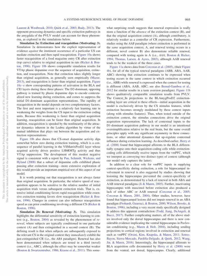
3. Extinction is not simply the unlearning of acquisition. Ex-tinction and the related phenomena of reacquisition, spon-taneous recovery, renewal, and reinstatement exhibit clearidiosyncrasies in comparison with initial acquisition. Forexample, reacquisition generally proceeds faster after ex-tinction than does original acquisition (rapid reacquisition;Pavlov, 1927; Rescorla, 2003; Ricker & Bouton, 1996), anda single unpredicted presentation of a US after extinctioncan reinstate Conditioned Responses (CRs) to near preex-tinction levels (reinstatement; Bouton, 2004; Pavlov, 1927).In addition, extinction learning has a significantly strongerdependency on context than does initial acquisition as dem-onstrated in the renewal paradigm (Bouton, 2004; Corc-oran, Desmond, Frey, & Maren, 2005; Krasne et al., 2011).The clear implication is that extinction learning is not thesymmetrical weakening of weights previously strengthenedduring acquisition, which a simple RPE formalism typicallyassumes, but instead involves the strengthening of a differ-ent set of weights that serve to counteract the effects of theacquisition weights. In support of this inference, muchempirical evidence implicates extinction-related plasticityin different neurobiological substrates from those impli-cated in initial acquisition (e.g., Bouton, 2004; Bouton,2011; Herry et al., 2008; Quirk & Mueller, 2008). Thesephenomena support the use of opposing pathways—one foracquisition and another for extinction—within both theLV-learning amygdala subsystem and the PV-learning VSsubsystem.
4. Although logically related, the loss of bursting at the time ofan expected reward and pausing when rewards are omittedare dissociable phenomena. There is evidence that themechanisms involved in the former are relatively tempo-rally imprecise, compared with the latter, which are neces-sarily more punctate since they cannot begin until it hasbeen determined that a reward has, in fact, been omitted.Rewards delivered early show progressively more burstingthe earlier they are, implying the mechanisms involved inblocking expected rewards are ramping up before the ex-pected time of reward (Fiorillo et al., 2008; Kobayashi &Schultz, 2008). Further, there is a slight, but statistically
significant, ramping decrease in tonic firing rate prior toexpected rewards (Bromberg-Martin, Matsumoto, & Hiko-saka, 2010a). On the other hand, the mechanisms impli-cated in producing pauses for omitted rewards are moretemporally precise, with an abrupt, discretized onset (Ma-tsumoto & Hikosaka, 2009b), and no apparent sign of earlyincreases in firing in the lateral habenula (LHb; Matsumoto& Hikosaka, 2009b). This dissociation, along with congru-ent anatomical data, motivates a distinction between theinhibitory shunting of phasic bursts (hypothesized to beaccomplished by known VS inhibitory projections directlyonto dopamine neurons; Joel & Weiner, 2000), and a sec-ond, probably collateral pathway through the LHb (andRMTg) that is responsible for pausing tonic firing. Thislatter pathway enables the system to make the determina-tion that a specific expected event has not in fact occurred(Brown et al., 1999; Hazy et al., 2010; O’Reilly et al., 2007;Tan & Bullock, 2008; and see Vitay & Hamker, 2014, foran excellent review and discussion of this important prob-lem space).
5. Conditioned inhibitors acquire the ability to generate pha-sic pauses in dopamine cell firing when presented alone.When a novel stimulus (conditioned inhibitor, CI, denotedX) is presented along with a previously trained CS (denotedA), and trained with the nonoccurrence of an expectedappetitive outcome (i.e., AX-), the CI takes on a negativevalence association and produces a phasic pause in dopa-mine firing (Tobler et al., 2003). This represents an impor-tant point of overlap between appetitive and aversive con-ditioning, because a CI stimulus (X-) behaves very muchlike a CS directly paired with an aversive US as reported byfor example, Mirenowicz and Schultz (1996). However, inthe CI case, there is no overt negative US involved—onlythe absence of a positive US. Thus, the conditioned inhibi-tion paradigm helps inform ideas about the role of USs indriving CS learning. In our framework, aversive CSs cometo excite the LHb via the striatum (and pallidum), to pro-duce dopamine cell pauses. Biologically, there is a pathwaythrough the striatum to the LHb, in addition to well-documented direct US inputs to LHb, and electrophysio-logical results consistent with the role of the striatal path-way in driving pauses in dopamine firing via the LHb(Hong & Hikosaka, 2013). Preliminary direct evidence fora role of the LHb in conditioned inhibition has recentlybeen reported (Laurent, Wong, & Balleine, 2017).
6. In Rescorla’s (1969) summation test of conditioned inhibi-tion, conditioned inhibitors tested with a different condi-tioned stimulus can immediately prevent both the expres-sion of acquired conditioned responses as well as phasicdopamine pauses. Specifically, this paradigm involves firsttraining A� and separately B�; then training AX- (i.e.,conditioned inhibition training), but not BX-; and then,finally, testing BX-. At the otherwise expected time of theB� US, there is no dopamine pause for the BX- case(Tobler et al., 2003), indicating that the X has acquired ageneralized ability to negate the expectation of the US andis not just specific to the AX compound. Furthermore,
Thi
sdo
cum
ent
isco
pyri
ghte
dby
the
Am
eric
anPs
ycho
logi
cal
Ass
ocia
tion
oron
eof
itsal
lied
publ
ishe
rs.
Thi
sar
ticle
isin
tend
edso
lely
for
the
pers
onal
use
ofth
ein
divi
dual
user
and
isno
tto
bedi
ssem
inat
edbr
oadl
y.
975A SYSTEMS-NEUROSCIENCE MODEL OF PHASIC DOPAMINE
presentation of the BX compound at test also prevents theexpression of acquired B� CRs (e.g., salivation, food-cupapproach; Tobler et al., 2003), implying that the acquired Xinhibitory representation has reached deep subcortical be-havioral pathways.
7. Conditioned inhibitors do not produce bursting at the ex-pected time of the US when presented alone. According toa simple RPE formalism of conditioned inhibition, the Xstimulus should acquire negative value itself and also serveto drive learning that predicts its occurrence, all trained bythe dopamine pauses. Subsequently, when the X is pre-sented by itself (without A-driven expectation of getting areward), an unopposed expectation of the negative (rewardomission) outcome should trigger a positive dopamine burstat the time when the US would have otherwise occurred.This is analogous to the modest relief bursting reportedwhen a trained CS is presented but the aversive US isomitted at test (Matsumoto, Tian, Uchida, & Watabe-Uchida, 2016; Matsumoto & Hikosaka, 2009a), or when asustained aversive US is terminated (Brischoux et al.,2009). In fact, however, no such X- relief burst was de-tected by Tobler, Dickinson, and Schultz (2003)—eventhough they explicitly looked for one.
8. Phasic dopamine responses to aversive outcomes includeboth pauses and bursts, with distinct subpopulations iden-tifiable. The nature of phasic dopamine responses to pri-mary aversive outcomes has been a topic of long-standingcontroversy with multiple studies reporting either pauses(e.g., Mirenowicz & Schultz, 1996), bursts (Horvitz, 2000;Horvitz, Stewart, & Jacobs, 1997), or a mixture of bothincluding cells exhibiting a biphasic response pattern (Ma-tsumoto & Hikosaka, 2009a). Although there is now a clearconsensus that bursting responses for aversive events dooccur, the interpretation remains controversial (e.g., Fior-illo, 2013; Schultz, 2016). All things considered, the mostparsimonious interpretation may be that different popula-tions of dopamine neurons may have different responseprofiles, with a majority (generally more laterally located)displaying a predominantly valence-congruent (RPE-consistent) response profile (i.e., pausing for aversive out-comes), while a smaller (more medial) subpopulation re-sponds with bursting for aversive outcomes. Functionally, itmay be that both forms of response make sense: for instru-mental learning based on reinforcing actions that produce“good” outcomes and punishing those leading to “bad” ones(e.g., Frank, 2005; Thorndike, 1898, 1911), valence-congruent dopamine signaling would seem essential to pre-vent confusion across both appetitive and aversive contexts;on the other hand, one or more smaller specialized sub-population(s) displaying bursting responses for aversiveoutcomes may be important for learning to suppress freez-ing and enable behavioral exploration for active avoidancelearning. In line with this latter idea, it now appears theremay be at least two small subpopulations of dopamine cellsthat respond with unequivocal bursting to aversive events:(a) a small subpopulation of posteromedial VTA neuronsexhibiting unequivocal bursting to aversive events project
narrowly to subareas of the accumbens shell and to certainventromedial prefrontal areas that may play a role in thesuppression of freezing (Lammel et al., 2012; Maier &Watkins, 2010; Moscarello & LeDoux, 2013); and (b) evenmore recently, a second subpopulation of aversive-burstingdopamine cells has been described in the posterolateralaspect of the SNc, with this population projecting only tothe caudal tail of the dorsal striatum and seemingly in-volved in simple avoidance learning (Menegas, Akiti,Uchida, & Watabe-Uchida, 2018; Menegas, Babayan,Uchida, & Watabe-Uchida, 2017; Menegas et al., 2015).Aversive-bursting dopamine cells are included in the PVLVframework as a second, distinct dopamine unit as discussedin Neurobiological Substrates and Mechanisms.
9. Dopamine pauses to aversive outcomes appear not to befully discounted through learned expectations. For the sub-set of dopamine neurons that exhibit valence-congruentpauses to aversive outcomes and CSs, these pauses seemnot to be fully predicted away (Fiorillo, 2013; Matsumoto& Hikosaka, 2009a). Behaviorally, it makes sense not tofully suppress aversive outcome signals since these out-comes remain undesirable, even potentially life-threatening, and an agent should continue to be biased tolearn to avoid them. In contrast, the discounting of expectedappetitive outcomes would seem to serve the beneficialpurpose of biasing the animal toward exploring for evenbetter opportunities. Thus, there are several fundamentalasymmetries between the appetitive and aversive cases thatsensibly ought to be incorporated into functional models.
10. Both appetitive and aversive processing involve many of thesame neurobiological substrates—in particular theamygdala and the lateral habenula. Overwhelming empir-ical evidence shows that the amygdala, ventral striatum, andlateral habenula all participate in both appetitive and aver-sive processing (Belova, Paton, Morrison, & Salzman,2007; Cole, Powell, & Petrovich, 2013; Donaire et al.,2019; Lee, Groshek, Petrovich, Cantalini, Gallagher, &Holland, 2005; Matsumoto & Hikosaka, 2009b; Paton, Be-lova, Morrison, & Salzman, 2006; Roitman, Wheeler, &Carelli, 2005; Setlow, Schoenbaum, & Gallagher, 2003;Shabel & Janak, 2009; Stopper & Floresco, 2013). Thisimplies that the processing of primary aversive events mustcoexist without disrupting the processing of appetitiveevents in these substrates, despite all the important differ-ences between these basic situations as noted above. Prop-erly integrating yet differentiating these two different va-lence contexts within a coherent overall framework presentsan important challenge for any comprehensive model of thephasic dopamine signaling system. We find that an oppo-nent processing framework—based on the opposite effectsof D1 and D2 dopamine receptors on cells in the striatumand amygdala—can go a long way toward meeting thischallenge, combined with an architecture that specificallysegregates the processing of individual USs.
11. Pavlovian conditioning generally requires a minimum 50-to 100-ms interval between CS-onset and US. Our original
Thi
sdo
cum
ent
isco
pyri
ghte
dby
the
Am
eric
anPs
ycho
logi
cal
Ass
ocia
tion
oron
eof
itsal
lied
publ
ishe
rs.
Thi
sar
ticle
isin
tend
edso
lely
for
the
pers
onal
use
ofth
ein
divi
dual
user
and
isno
tto
bedi
ssem
inat
edbr
oadl
y.
976 MOLLICK ET AL.
PVLV model emphasized the problem that a phasic dopa-mine signal generated by CS onset could create a positivefeedback loop of further learning to that CS, leading tosaturated synaptic weights (Hazy et al., 2010; O’Reilly etal., 2007). We now account for data indicating CSs mustprecede USs by a minimum of 50–100 ms to drive condi-tioned learning (Mackintosh, 1974; Schmajuk, 1997; Sch-neiderman, 1966; Smith, 1968; Smith, Coleman, & Gor-mezano, 1969). With this constraint in place, it is notpossible for CS-driven dopamine to reinforce itself, pre-venting the positive feedback problem. Incorporating thischange now allows our model to include the effects ofphasic dopamine on CS learning in the amygdala (in addi-tion to the important role that US inputs play in drivinglearning there, as captured in the prior models), supportingphenomena such as second-order conditioning in the BLA(Hatfield, Han, Conley, & Holland, 1996).
Conceptual Overview of the PVLV Model
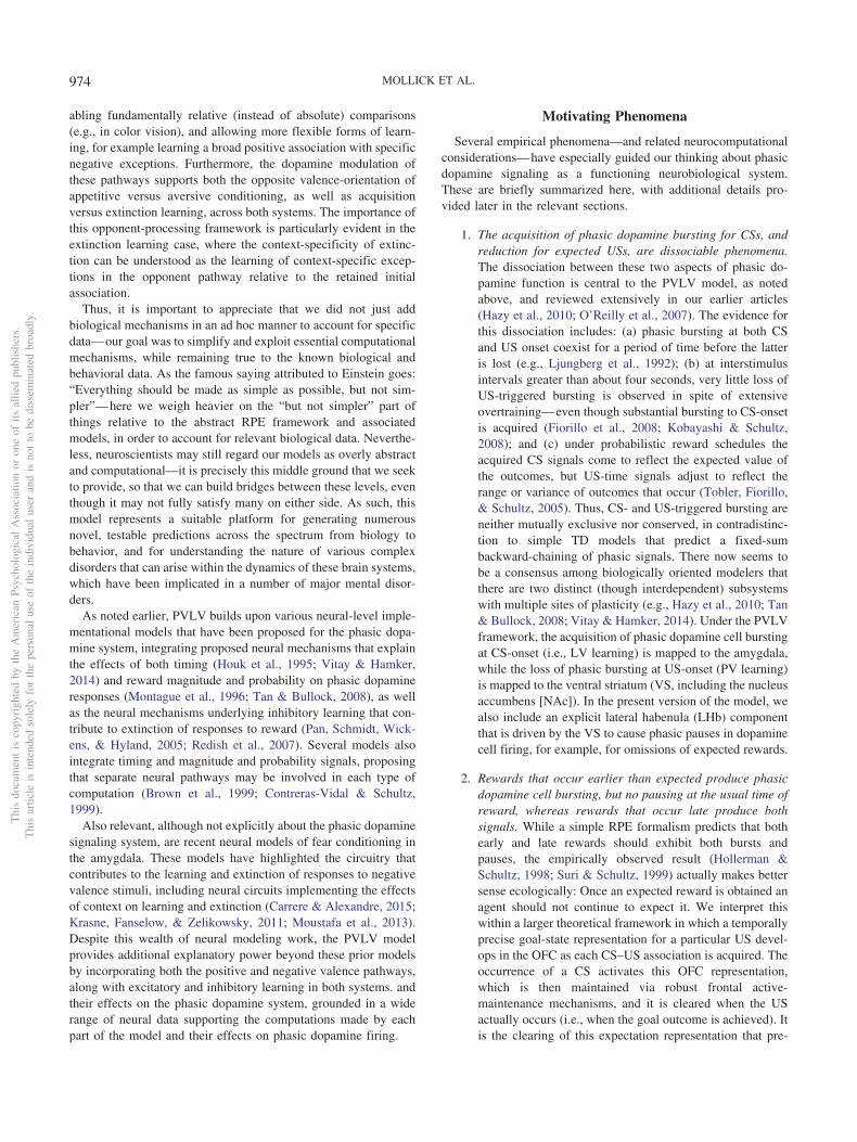
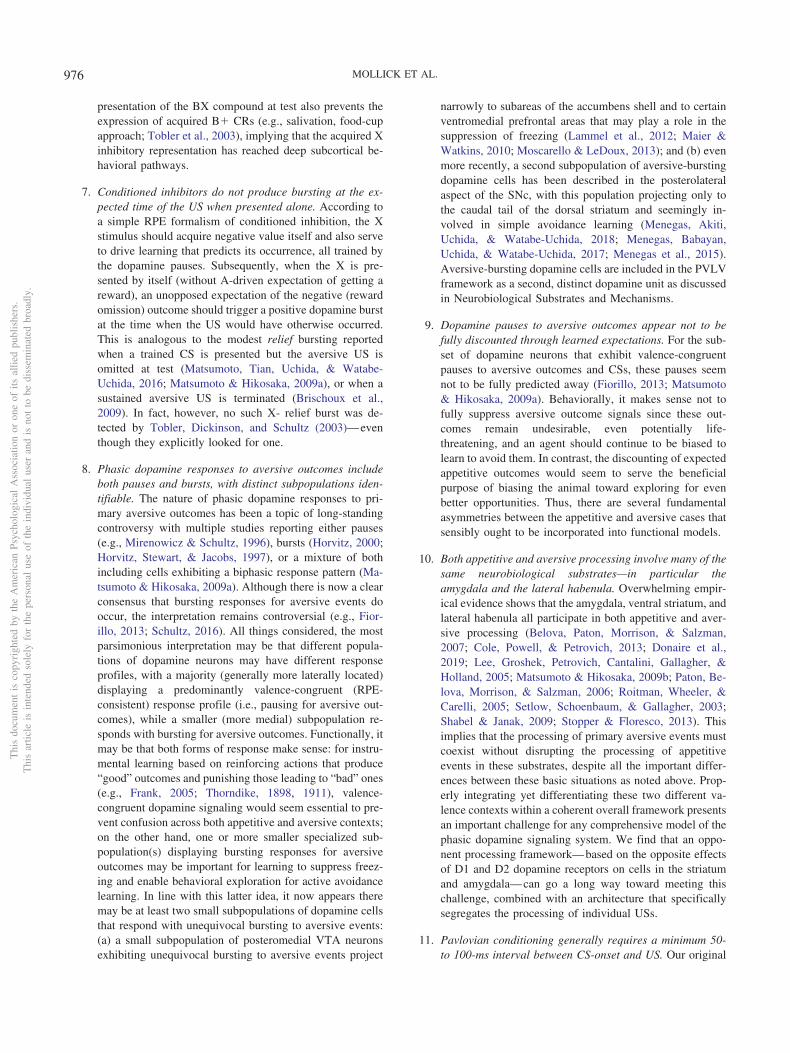
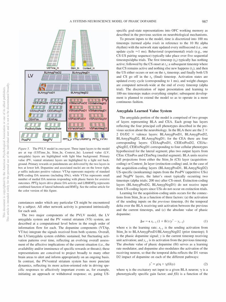
In this section we provide a high-level, conceptual overview ofthe PVLV model and how all the different parts fit together. Figure1 shows how the fundamental LV versus PV distinction cutsthrough a standard hierarchical organization of brain areas at threedifferent levels: cortex, basal ganglia (BG), and brain stem. Cortex
is generally thought to represent higher-level, more abstract, dy-namic encodings of sensory and other information, which providesa basis for learning about the US-laden value of different states ofthe world (in standard reinforcement learning terminology). Thebasolateral amygdala (BLA) is described as having a cortex-likehistology in its neural structure (e.g., Pape & Pare, 2010), but italso receives direct US inputs from various brain stem areas. Thus,it serves nicely as a critical hub/connector area that learns toassociate these cortical state representations with US outcomes,which is the core of the LV function in the PVLV framework. Incontrast, the central amygdala (CEA) has cell types and connec-tivity characteristic of the striatum of the basal ganglia (Cassell,Freedman, & Shi, 1999), and according to classic BG models (e.g.,Collins & Frank, 2014; Frank, 2005; Frank et al., 2001; Mink,1996), it should be specialized for selecting the best overall inter-pretation of the situation by separately weighing evidence-for (Go,direct pathway, CElON) versus evidence-against (NoGo, indirectpathway, CElOFF) in a competitive, opponent-process dynamic(Ciocchi et al., 2010; Li et al., 2013).
Thus, the CEA in our model takes the higher-dimensional,distributed, contextualized representations from BLA and boilsthem down to a simpler, quantitative evaluation of how likely aparticular US outcome is given the current cortical state represen-tations. When this evaluation results in an increased expectation ofpositive outcomes, it drives phasic bursting in the VTA/SNc do-pamine nuclei. This occurs via direct connections, and via thepedunculopontine tegmental nucleus (PPTg), which may help indriving bursting as a function of changes in expectations, assustained activity in BLA does not appear to drive further phasicdopamine bursting (e.g., Ono, Nishijo, & Uwano, 1995). In sum-mary, through these steps, this stack of LV areas is responsible fordriving phasic dopamine bursting in response to CS inputs.
The opponent organization scheme in the amygdala also servesto address the subtly challenging problem of learning about theabsence of an expected US outcome as occurs during extinctiontraining. This is challenging from a learning perspective becausethe absence of a US is a “nonevent,” and thus cannot drive learningin the traditional activation-based manner, and further, the issueremains of which of the indeterminate number of nonoccurringevents should direct learning. The explicit representation of ab-sence in the opponent-processing scheme solves this problem byusing selective modulatory, permissive connections fromacquisition-coding to extinction-coding units so that only USs withsome expectation of occurrence can accumulate evidence aboutnonoccurrence. Thus, only at the last step in the pathway is theUS-specific nature of the representations abstracted away to thepure value-coding nature of the effectively scalar phasic dopaminesignal, in contrast to many other computational models that onlydeal with this abstract value signal (e.g., standard TD models). Inaddition, learning constrained to separate representations for dif-ferent types of rewards (punishments) can directly account forphenomena such as unblocking by reward type, something that isotherwise challenging for value-only models like TD (e.g., Taka-hashi et al., 2017), and depends on activity of dopamine neurons(Chang, Gardner, Di Tillio, & Schoenbaum, 2017).
Bridging the CS-driven US expectations into the PV side of thesystem, the BLA also drives areas in the orbital (OFC) andventromedial prefrontal cortex (vmPFC), particularly the OFC (seeFigure 1). Projections from this cortical level to ventral striatum
Cortex
BasalGanglia
BLA
CS
hctapSV xirtamSVAEC
BrainStem VTA/
SNcLH
(US)LHb
vmPFC
(distributed, abstract, attentional)
(opponent, selection)
US, CR
LV PV(CS driven) (US expectations)
Figure 1. Overview of PVLV: The main division into LV (learned value)and PV (primary value) cuts across a hierarchy of function in cortical, basalganglia, and brain stem areas. The cortex provides high-level, abstract,dynamic state representations, and the basolateral amygdala (BLA), whichhas a cortex-like histology, links these with specific US outcomes. Thebasal-ganglia-like central amygdala (CEA) quantitatively evaluates theoverall evidence for the occurrence of reward or punishment usingopponent-processing pathways, and drives phasic dopamine bursts in themidbrain dopamine areas (VTA, SNc) if this evaluation is in favor ofexpected rewards. BLA also triggers updating of US expectations inventral/medial prefrontal cortex (vmPFC), especially the OFC (orbitofron-tal cortex), which then drives another opponent-process evaluation process,in the ventral striatum patch-like areas (VSpatch), the results of which canshunt dopamine bursts for expected US’s, and drive pauses in dopaminefiring when an expected US fails to arrive, via projections to the lateralhabenula (LHb). Various brain stem areas (e.g., the lateral hypothalamus,LH) drive US inputs into the system, and are also driven to activate CRs.See the online article for the color version of this figure.
Thi
sdo
cum
ent
isco
pyri
ghte
dby
the
Am
eric
anPs
ycho
logi
cal
Ass
ocia
tion
oron
eof
itsal
lied
publ
ishe
rs.
Thi
sar
ticle
isin
tend
edso
lely
for
the
pers
onal
use
ofth
ein
divi
dual
user
and
isno
tto
bedi
ssem
inat
edbr
oadl
y.
977A SYSTEMS-NEUROSCIENCE MODEL OF PHASIC DOPAMINE
drive a BG-like evaluation of evidence for and against the immi-nent occurrence of specific USs at particular points in time. Cellsin the patch-like compartment of the VS send direct inhibitoryprojections to the midbrain dopamine cells so as to produce ashunt-like inhibition that blocks dopamine bursts that would oth-erwise arise from an appetitive US. Furthermore, via a pallidalpathway, the VSpatch also drives a more temporally precise acti-vation (disinhibition) of the LHb that causes pausing (dips) oftonic dopamine firing if not offset by excitatory drive from anactual US occurrence. In summary, this PV stack of areas workstogether to anticipate and cancel expected US outcomes.
There is another pathway through the VS that does not fit ascleanly within the simple LV/PV distinction, which we hypothe-size is mediated by the matrix-like compartments within the VS(VSmatrix). This pathway is necessary for supporting the ability ofCS inputs to drive phasic dipping/pausing of dopamine firing,which appears to be exclusively driven by the LHb in response toVS inputs (Christoph, Leonzio, & Wilcox, 1986; Hikosaka, 2010;Hikosaka, Sesack, Lecourtier, & Shepard, 2008; Ji & Shepard,2007; Matsumoto & Hikosaka, 2007; Matsumoto & Hikosaka,2009b). We are not aware of any evidence supporting a directprojection from the amygdala to the LHb (Herkenham & Nauta,1977), which would otherwise be a more natural pathway for CSactivation of phasic dipping according to the overall PVLV frame-work. An important further motivation for this VSmatrix pathwayis that, by hypothesis, it is also responsible for gating informationthrough the thalamus so as to produce robust maintenance of USoutcome/goal state representations in OFC (Frank & Claus, 2006;Pauli et al., 2010; Pauli et al., 2012). Such working memory-likegoal state representations are hypothesized to be important forsupporting goal-directed (vs. habitual) instrumental behavior, be-havior known to depend on intact OFC (e.g., Gallagher, McMahan,& Schoenbaum, 1999). Thus, the very same plasticity eventsoccurring at corticostriatal synapses onto VSMatrix cells could beresponsible for learning to gate US information into OFC workingmemory in response to a particular CS, while acquiring an abilityto drive phasic dopamine signals (via LHb) in response to thosesame CS events.
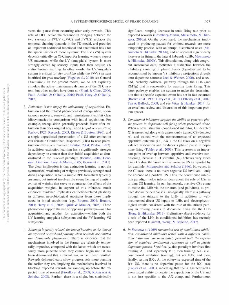
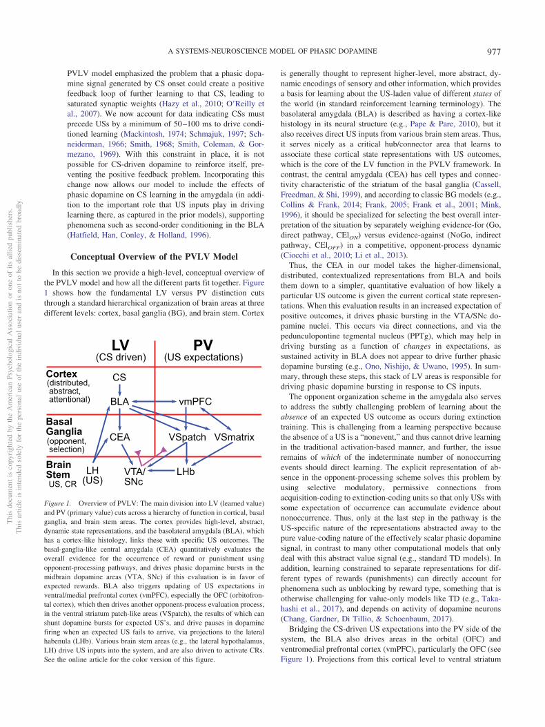
Appetitive/Aversive and Acquisition/ExtinctionPathways
The above overview is framed in terms of appetitive condition-ing, as that is the simplest and most well-established case. How-ever, a critical feature of the current model is that it incorporatespathways within the LV and PV systems for processing aversiveUSs as well, leveraging the same opponent-process dynamics, withan appropriate sign-flip, as described above. Figure 2 shows thefull set of pathways and areas in the PVLV model. As in the BG,each pathway is characterized by having a preponderance of do-pamine D1 versus D2 receptors, which then drives learning fromphasic bursts (D1) or dips (D2; e.g., Frank, 2005; Frank et al.,2001; Gerfen & Surmeier, 2011; Mink, 1996). Thus, assuming thestandard RPE form of dopamine firing, D1-dominated pathwaysare strengthened by unexpected appetitive outcomes, while D2-dominated ones are strengthened by unexpected aversive out-comes. Thus, this differential dopamine receptor expression canaccount for the differential responses of appetitive- versusaversive-coding neurons in the amygdala (LV), as shown in Figure
2. Although the BLA is not strongly topographically organized, weassume a similar opponency between subsets of neurons, as ismore clearly demonstrated in the central amygdala CElON versusCElOFF cells (Ciocchi et al., 2010; Li et al., 2013). In addition tothese lateral pathway neurons, we include a final medial outputpathway (CEm) that computes the net balance between on versusoff for each valence pathway (appetitive and aversive).
The VS (PV) system is likewise organized according to standardD1 versus D2 pathways, within the US-coding patch areas and theCS-coding matrix areas, again with separate pathways for appeti-tive versus aversive, with the sign of D1 versus D2 effects flippedas appropriate. For example, VSpatch aversive-pathway D2 neu-rons learn from unexpected aversive outcomes, and thereby learnto anticipate such outcomes. The complementary D1 pathwaythere learns from any dopamine bursts associated with the nonoc-currence of these aversive outcomes, such that the balance betweenthese pathways reflects the net expectation of the aversive out-come. Figure 2 shows how each VS pathway sends a correspond-ing net excitation or inhibition to the LHb (via a pallidal pathway),with excitation of the LHb causing inhibition of VTA/SNc tonicfiring via the RMTg (rostromedial tegmental nucleus—in ourmodel, we combine the LHb and RMTg into a single functionalunit).
In addition, the VSpatch D1 appetitive pathway sends directshunting inhibition to these midbrain dopamine areas, to blockexcitatory firing from expected US’s. Although this pathway mayseem redundant with the LHb inhibition, the differential timing ofthese two functions motivates the need for separate mechanisms.On the one hand, a complete inhibition of bursting requires aninput arriving at least slightly prior to the time of reward, or elseat least a little activity will necessarily occur on the front end. Onthe other hand, an omission-signaling input (for pausing) can onlyarrive at least slightly after the expected time of the rewardbecause an agent can determine that an expected event did notoccur only after the time it was expected, reflecting at least somefinite amount of time to compute and transmit the omission signal.Indeed, omission pauses are empirically seen to have greaterlatency than corresponding bursts.
Finally, apropos of the asymmetries between appetitive versusaversive conditioning discussed above, there are a number ofaspects where these two differ in the model. For example, appet-itive, but not aversive, pathways in the amygdala can directly drivedopamine burst firing, consistent with our overall hypothesis (andextant data) that the LHb is exclusively responsible for driving allphasic pausing in dopamine cell firing. This has some importantfunctional implications, by allowing the amygdala dopamine path-way to be positively rectified—that is, it only reports when theamygdala estimates the current situation to be better than thepreceding one. Furthermore, the extent to which VSpatch expec-tancy representations can block dopamine pauses associated withexpected aversive outcomes is significantly less than its ability toblock bursts for expected appetitive outcomes as suggested by theavailable empirical data (Matsumoto & Hikosaka, 2009a).
Differences From Previous Versions of PVLV
The present model represents a significant elaboration and re-finement of the PVLV framework since our prior publication(Hazy et al., 2010), as briefly summarized here:
Thi
sdo
cum
ent
isco
pyri
ghte
dby
the
Am
eric
anPs
ycho
logi
cal
Ass
ocia
tion
oron
eof
itsal
lied
publ
ishe
rs.
Thi
sar
ticle
isin
tend
edso
lely
for
the
pers
onal
use
ofth
ein
divi
dual
user
and
isno
tto
bedi
ssem
inat
edbr
oadl
y.
978 MOLLICK ET AL.
• Earlier versions of PVLV included only a central nucleusamygdalar component (CEA; formerly CNA). In the cur-rent version we have added a basolateral amygdalar com-plex (BLA), which serves as a primary site for CS-USpairing during acquisition (acquisition-coding cells) and,critically, for the pairing of CSs with the nonoccurrence ofexpected USs (extinction-coding cells). This is especiallyimportant in accounting for extinction-related phenomenareflecting the idea that extinction is an additional layer oflearning and not just the unlearning (weakening) of acqui-sition learning and, importantly, underlies the ability of thecurrent version to account for the differential sensitivity ofextinction to context (see Simulation 2b).
• Earlier versions of PVLV treated the inhibitory PV com-ponent as unitary with no distinction between a shuntingeffect onto dopamine cells that prevents bursting at the
time of expected rewards and the pausing effect thatoccurs when expected rewards are omitted. Since that timeit has been established that the LHb plays a critical role inthe latter phenomenon and may serve as the sole substrateresponsible for producing pauses on dopamine cell firingof any cause. Accordingly, the new version adds a LHbcomponent which receives disynaptic collaterals from thesame VSpatch cells that provide direct shunting inhibitiononto dopamine cells. These collaterals result in net excit-atory inputs onto LHb cells. Critically, the LHb alsoreceives direct (excitatory) inputs for aversive USs, aswell as net inhibitory inputs associated with both reward-ing outcomes and expectations of reward. The LHb com-ponent is important for producing the dissociation betweenshunting inhibition and overt pauses, it also enables thenew model to produce (modest) disinhibitory positive
Figure 2. Detailed components of PVLV, showing the opponent processing pathways within the PV and LVsystems, which separately encode the strength of support for and against each US, and with opposite dynamics forappetitive versus aversive valence. BLA has pathways for appetitive and aversive USs, along with distinctionsbetween acquisition and extinction learning, all of which engage in broad inhibitory competition. The BLA projectsto central amygdala (CEl, CEm) neurons that integrate the evidence for-and-against a given US, and communicate thisnet value to the VTA (and SNc, not shown). The ventral striatum (VS) has matrix and patch subsystems, where matrix(VSm) receives modulatory inputs from corresponding BLA neurons and represents CSs in a phasic manner, andpatch (VSp) anticipates and cancels USs. Both have a full complement of opposing D1- and D2-dominant pathways,which have opposing effects for appetitive versus aversive USs. LV � learned value; PV � primary value; BLA �basolateral amygdala; OFC � orbitofrontal cortex; LHb � lateral habenula; CS � conditional stimulus; VTA �ventral tegmental area; PBN � parabrachial nucleus. See the online article for the color version of this figure.
Thi
sdo
cum
ent
isco
pyri
ghte
dby
the
Am
eric
anPs
ycho
logi
cal
Ass
ocia
tion
oron
eof
itsal
lied
publ
ishe
rs.
Thi
sar
ticle
isin
tend
edso
lely
for
the
pers
onal
use
ofth
ein
divi
dual
user
and
isno
tto
bedi
ssem
inat
edbr
oadl
y.
979A SYSTEMS-NEUROSCIENCE MODEL OF PHASIC DOPAMINE
dopamine signals at the time of expected-but-omitted pun-ishment (see Simulation 4b).
• Like TD, and RPE generally, earlier versions of PVLVreally only contemplated appetitive context, that is, theoccurrence and omission of positively valenced reward; itlargely ignored learning under aversive context (e.g., fearconditioning). In the current version, additional comple-mentary channels for appetitive versus aversive processing(and associated learning) have been incorporated through-out the model, with their convergence occurring only attwo distinct sites where population coding is largely, butnot exclusively, unitary: (a) the LHb (which projects to theVTA/SNc); and (b) the dopamine cells themselves in theVTA/SNc. Incorporating aversive processing channelsalongside appetitive ones is important for demonstratingthat the core idea underlying the DA-RPE theory cansurvive the integration of all these parallel processingpathways and their significant convergence onto mostdopamine cells. This extension enabled the current PVLVversion to simulate basic aspects of aversive conditioning(see Simulation 4a, b), and provides a richer more accurateaccount of conditioned inhibition.
• Also like TD and RPE, earlier versions of PVLV treatedreward as a single scalar value throughout the modelwithout distinguishing between different kinds of reward(or punishment), for example, food versus water, or shockversus nausea. By representing different kinds of rewardseparately in both the amygdala and ventral striatum,learning in the current version of PVLV can also produceseparate expectancy representations about different re-wards. This provides a direct mechanism that can helpaccount for the phenomenon of unblocking-by-identity(e.g., see Simulation 3a).
Overview of Remainder of the Article
The next two sections examine first the neurobiology that con-strains various aspects of the PVLV framework, and then theactual computational implementation of the model. After that, theResults section describes and discusses 12 simulations coveringseveral well-established Pavlovian conditioning phenomena and,especially, serve to highlight the most important features of theoverall framework. The article concludes with a General Discus-sion in which we highlight the main contributions of the PVLVframework, compare our approach with others in the literature, andidentify several unresolved questions for future research.
Neurobiological Substrates and Mechanisms
In this section, we provide a neurobiological-level account ofthe computational model outlined above, followed in the subse-quent section by a computationally focused description. To thatend, we provide a selective review of salient biological and be-havioral data most influential in informing the overall framework,and we focus specifically on data that go beyond the foundationscovered in earlier articles (Hazy et al., 2010; O’Reilly et al., 2007).
The Amygdala: Anatomy, Connectivity, andOrganization
The amygdala is composed of a dozen or so distinct nuclei and/orsubareas (Amaral, Price, Pitkanen, & Carmichael, 1992), each ofwhich can exhibit several subdivisions (McDonald, 1992). Despitesuch anatomical complexity, however, the literature has largely con-ceptualized amygdalar function in terms of two main components: adeeper/inferior basolateral amygdalar complex (BLA) more involvedin the processing of inputs; and a more superficial/superior centralamygdalar nucleus (CEA) that has long been implicated in drivingmany of the more primitive manifestations of emotional expression(changes in heart rate, breathing, blood pressure; freezing, and so on;Figure 3a). Both BLA and CEA contain both glutamatergic andGABAergic cells (both local interneurons and projecting), with con-
a b
AB
BA
m
lITC
dl
CElm
vl
ITCd
ITCvC
CCm
IT
Sensory inputs
Subcortical outputs
dc
vHC
ACQ EXT
vmPFCCEl
ONOFF CEm
ACQ EXT
LA
Su o
CAC TXT CAC
OO
TXT
Figure 3. Basic organization, information flow, and opponent-processingin the amygdala. (a) Schematic diagram of a coronal section of unilateralamygdala with most prominent nuclei outlined according to one commonscheme. The BLA is composed of: lateral (LA), basal (BA), and accessorybasal (AB) nuclei. The central nucleus is composed of a lateral (CEl) andmedial (CEm) segments. Three collections of GABAergic cells make upthe intercalated cell masses (ITCs): the lateral paracapsular (lITC); dorsal(ITCd); and ventral (ITCv). (b) Basic information flow through theamygdala: sensory information enters via the LA predominantly flowingfrom dorsolateral (LAdl) to ventrolateral (LAvl) and medial (LAm) divi-sions. From there two parallel pathways reach the central amygdala: (1)directly from LA to CEA (via CEl; red dotted arrows); and (2) via the basal(BA) and accessory basal (AB) nuclei (blue dash arrows). (c) Opponentprocessing in the BLA following the scheme of Herry et al., 2008:acquisition-coding cells (ACQ) receive context inputs from the ventralhippocampus (vHC) and project to the ventromedial PFC, which connectsreciprocally with extinction-coding cells (EXT) in the BLA, with thevmPFC providing additional context information relevant for extinction.(d) Opponent processing in the CEl following the scheme of Pare andDuvarci (2012), with CElON � acquisition and CElOFF � extinction. Seethe online article for the color version of this figure.
Thi
sdo
cum
ent
isco
pyri
ghte
dby
the
Am
eric
anPs
ycho
logi
cal
Ass
ocia
tion
oron
eof
itsal
lied
publ
ishe
rs.
Thi
sar
ticle
isin
tend
edso
lely
for
the
pers
onal
use
ofth
ein
divi
dual
user
and
isno
tto
bedi
ssem
inat
edbr
oadl
y.
980 MOLLICK ET AL.
siderable topographic patchiness in their relative proportions; forexample, the lateral segment of the CEA (CEl) seems to be almostexclusively GABAergic. Importantly, the amygdala is richly inner-vated by all four neuromodulatory systems including a dense, heter-ogeneously distributed dopaminergic projection (Amaral et al., 1992;Fallon & Ciofi, 1992). Both main classes of dopamine receptors(D1-like, D2-like) are richly expressed, although not homogeneously(Bernal et al., 2009; de la Mora, Gallegos-Cari, Arizmendi-García,Marcellino, & Fuxe, 2010; de la Mora et al., 2012; Lee, Kim, Kwon,Lee, & Kim, 2013).
Figure 3 shows the major areas and connectivity. The BLAreceives dense afferents from much of the cerebral cortex, includ-ing the higher areas in all sensory modalities, as well as associativeand affective cortex, and from corresponding thalamic nuclei andsubcortical areas (Doyère, Schafe, Sigurdsson, & LeDoux, 2003;LeDoux, 2003; Pitkanen, 2000; Uwano, Nishijo, Ono, & Tamura,1995). The lateral nucleus (LA) receives the preponderance ofsensory input, preferentially into its dorsolateral division (Pit-kanen, 2000) and projects to CEA both directly, and indirectly viathe basal and accessary basal nuclei (Pitkanen, 2000). The basaland accessory basal nuclei exhibit extensive local and contralateralinterconnectivity, and also send feedback projections to two of thedivisions of the LA (Pitkanen, 2000), whereas the LA has rela-tively little local or contralateral interconnectivity. The BLA alsoprojects heavily to the ventral striatum and to much of the corticalmantle (Amaral et al., 1992; Pitkanen, 2000), including a strongreciprocal interconnection with the orbital frontal cortex (OFC;Ongür & Price, 2000; Schoenbaum, Chiba, & Gallagher, 1999)and parts of ventromedial prefrontal cortex including the anteriorcingulate cortex (ACC; Ongür & Price, 2000). Based on neuralrecording studies, there seems to be little discernible local topo-graphical organization of different cell responses in the BLA (i.e.,a salt-and-pepper distribution; Herry et al., 2008; Maren, 2016),with one notable exception of a recently described positive-negative valence gradient in a posterior-to-anterior direction (Kim,Pignatelli, Xu, Itohara, & Tonegawa, 2016).
The CEA can be functionally divided into medial (CEm) and lateral(CEl) segments (Figure 3a), with the CEl exerting a tonic inhibitoryinfluence on the CEm that, when released, performs a kind of gatingfunction for CEm outputs analogous to that seen in the basal ganglia.Both CEl and, especially, CEm send efferents to subcortical viscero-motor areas (autonomic processing) as well as to certain primitivemotor effector sites involved in such affective behaviors as freezing(Koo, Han, & Kim, 2004; Li et al., 2013; Veening, Swanson, &Sawchenko, 1984). Importantly, among the subcortical efferents fromCEm are projections to the VTA/SNc, both directly, and via thepedunculopontine tegmental nucleus (PPTg; Everitt, Cardinal, Hall,Parkinson, & Robbins, 2000; Fudge & Haber, 2000), and stimulationof the CEm has been shown to drive phasic dopamine cell burstingand/or dopamine release in downstream terminal fields (Ahn & Phil-lips, 2003; Fudge & Haber, 2000; Rouillard & Freeman, 1995; Stal-naker & Berridge, 2003; see Hazy et al. (2010) for detailed discus-sion). The CEA also receives broad cortical and thalamic afferentsdirectly (Amaral et al., 1992; Pitkanen, 2000); these direct inputs arepresumably responsible for the result that the CEA can supportfirst-order Pavlovian conditioning independent of the BLA (Everitt etal., 2000).
Division-of-Labor Between BLA and CEA: AnalogyWith the Cortical–Basal Ganglia System
In addition to the long-held view of basic amygdalar organiza-tion that posits the BLA as the input side and the CEA as theoutput side, we also embrace emerging ideas (e.g., Duvarci & Pare,2014; Holland & Schiffino, 2016) that posit that the two areas mayhave distinct functional roles analogous to the distinction betweenthose of the cortex (i.e., BLA) and the basal ganglia (CEA; Figure1). The BLA has long been described as cortex-like (McDonald,1992), while the CEA is more basal-ganglia like, particularly itslateral segment (CEl) whose principal cells bear a strong resem-blance with the medium spiny neurons (MSNs) of the neostriatum,with which it is contiguous laterally (Cassell et al., 1999; McDon-ald, 1992). Thus, one can think about the BLA computing com-plex, high-dimensional representations of current states of theworld (including both external and internal components) that areanchored by expectations about the imminent occurrence of spe-cific USs; in contrast, the CEA involves simpler, low-dimensionalrepresentations about particular primitive actions to be taken basedon those US-anchored anticipatory states (e.g., fear, food antici-pation). Both BLA and CEA subserve both input and output rolesand function partially in parallel as well as serially, with a majordistinction between their output projections. The BLA projects toneocortex and basal-ganglia (especially ventral striatum) and ex-erts a more modulatory effect, while CEA projects almost exclu-sively to subcortical areas (excluding the basal ganglia), and is astrong driver of subcortical visceromotor and primitive motoreffectors.
Electrophysiological recording shows that BLA neurons exhibita wide range of selectivity to different CSs, USs, and contexts(Beyeler et al., 2016; Herry et al., 2008; Johansen, Hamanaka, etal., 2010; Johansen, Tarpley, LeDoux, & Blair, 2010; Muramoto,Ono, Nishijo, & Fukuda, 1993; Ono et al., 1995; Repa et al., 2001;Roesch, Calu, Esber, & Schoenbaum, 2010; Toyomitsu, Nishijo,Uwano, Kuratsu, & Ono, 2002). By adulthood, a significant pro-portion of the principal cells in both BLA and CEA appear tostably represent specific kinds of primary rewards and punish-ments and not undergo significant change thereafter. For example,discriminative- and reversal-learning experiments have shown thatCS–US associative pairings can undergo rapid remapping whenenvironmental contingencies change, leaving the underlying US-specific representational scheme intact (Schoenbaum et al., 1999).A simple model for Pavlovian conditioning is that previouslyneutral CSs acquire the ability to activate these US-coding cells bystrengthening synapses they send to them (Muramoto et al., 1993;Ono et al., 1995; Toyomitsu et al., 2002). More recent studiesexamining larger population-level samples suggests that learningin the BLA is complex, high-dimensional, and distributed—con-sistent with a cortex-like system (Beyeler et al., 2016; Grewe et al.,2017). Nevertheless, the essential function of BLA in linking CSsand USs remains a useful overarching model.
In addition to a strong US-anchored organization for amygdalarepresentations, there are also cells in both BLA and CEA thatreflect evidence against the imminent occurrence of particular USoutcomes. For example, Herry et al. (2008) showed that a distinctset of BLA neurons progressively increased in activity in responseto CS-onset over multiple US omission trials (extinction training),in contrast with those (acquisition-coding) neurons that had ac-
Thi
sdo
cum
ent
isco
pyri
ghte
dby
the
Am
eric
anPs
ycho
logi
cal
Ass
ocia
tion
oron
eof
itsal
lied
publ
ishe
rs.
Thi
sar
ticle
isin
tend
edso
lely
for
the
pers
onal
use
ofth
ein
divi
dual
user
and
isno
tto
bedi
ssem
inat
edbr
oadl
y.
981A SYSTEMS-NEUROSCIENCE MODEL OF PHASIC DOPAMINE
quired activity in response to CS-onset during fear acquisition.Similarly, Ciocchi et al. (2010) showed opponent coding of aver-sive US presence versus absence in separate populations of CElON
versus CElOFF neurons. These CEl neurons are exclusivelyGABAergic and have mutually inhibitory connections, producinga direct opponent-processing dynamic. This pattern of opponentorganization, which is one of two core computational principles inour model, is essential for supporting extinction learning from theabsence of expected USs, and also for probabilistic learning par-adigms (Esber & Haselgrove, 2011; Fiorillo, Tobler, & Schultz,2003).
Extinction Learning and the Role of Context
Considerable behavioral data strongly supports the idea thatextinction learning is particularly sensitive to changes in bothexternal and internal context, and that areas in the vmPFC play animportant role in contextualizing extinction learning (Laurent &Westbrook, 2010; Quirk, Likhtik, Pelletier, & Paré, 2003). Further,Herry et al. (2008) looked specifically at the connectivity ofextinction-coding versus acquisition-coding cells in the BLA andfound that only the former receive connections from vmPFC. Thishas been incorporated into the PVLV framework in the form ofcontextual inputs to the model that connect exclusively to theextinction coding layers of the BLA. Somewhat surprisingly,Herry et al. (2008) also reported that hippocampal inputs to theBLA (long implicated in conditioned place preference and aver-sion) connected only with acquisition-coding cells; this ratherparadoxical situation is discussed in a section on the role andnature of context representations in the General Discussion sec-tion. In essence, it is hard to avoid the conclusion that the hip-pocampus and vmPFC must convey distinctly different forms ofcontext information to the amygdala. Simulation 2b in the Resultssection explores the differential context-sensitivity of extinctionversus acquisition learning.
There are likely differential contributions of the BLA versusCEA to extinction learning, in part due to the greater innervationof BLA by contextual inputs. For example, limited evidence sug-gests that the CEA may not be able to support extinction learningby itself and instead depends on learning in the BLA (Falls,Miserendino, & Davis, 1992; Lin, Yeh, Lu, & Gean, 2003; Lu,Walker, & Davis, 2001; Quirk & Mueller, 2008; Zimmerman &Maren, 2010). However, muscimol inactivation of BLA at differ-ent stages of extinction learning demonstrates that extinction canpersist in the absence of BLA activation (Herry et al., 2008).Although not currently implemented in PVLV, this can potentiallybe explained in terms of BLA driving learning in vmPFC whichcan in turn drive extinction via direct projections into CEA (e.g.,Anglada-Figueroa & Quirk, 2005). Finally, the intercalated cells(ITCs) have been widely discussed as suppressing fear expressionunder various circumstances (Ehrlich, Humeau, Grenier, Ciocchi,Herry, & Luthi, 2009; Likhtik, Popa, Apergis-Schoute, Fidacaro,& Paré, 2008; Maier & Watkins, 2010; Marowsky, Yanagawa,Obata, & Vogt, 2005; Pare & Duvarci, 2012; Royer, Martina, &Paré, 1999). However, some conflicting data has emerged in thisregard (Adhikari et al., 2015). Nonetheless, it seems likely thatITCs participate somehow in the opponent-processing scheme foracquisition versus extinction coding in the amygdala. Their role is
currently subsumed within the basic extinction-coding function inPVLV and not explicitly modeled.
Dopamine Modulation of Acquisition VersusExtinction Learning
Dopamine has been shown to be important for plasticity-induction in the amygdala (Andrzejewski, Spencer, & Kelley,2005; Bissière, Humeau, & Lüthi, 2003). While the other threeneuromodulatory systems (ACH, NE, 5-HT) are undoubtedly im-portant (e.g., Carrere & Alexandre, 2015), they are not currentlyincluded in the PVLV framework. There are both D1-like andD2-like receptors in in the BLA (de la Mora et al., 2010), andblocking of D2s in the BLA impaired acquisition of fear learning,reducing conditioned responses such as freezing (Guarraci, Fro-hardt, Falls, & Kapp, 2000; LaLumiere, Nguyen, & McGaugh,2004) and fear-potentiated startle (de Oliveira et al., 2011; Nader& LeDoux, 1999) to a CS. Similarly, Chang et al. (2016) reportedthat optogenetically driven pauses in DA firing produce expectedeffects consistent with aversive conditioning, while antagonism ofD1s blocked fear extinction (Hikind & Maroun, 2008). In thepositive valence domain, antagonism of D1s in the amygdalaattenuated the ability of a cue paired with cocaine to reinstateconditioned responding (Berglind, Case, Parker, Fuchs, & See,2006). Similarly consistent D1 and D2 receptor effects have beendocumented in CEl as well (De Bundel et al., 2016).
Extending the results and model of Herry et al. (2008), thePVLV framework accounts for the differential learning of acqui-sition versus extinction cells in the BLA (and acquisition only inCEl) in terms of a 2 � 2 matrix of valence X dopamine receptordominance. For example, acquisition for appetitive Pavlovian con-ditioning is trained by (appetitive) US occurrence and modulatedby phasic dopamine bursting effects on D1-expressing positiveUS-coding cells, while extinction learning is mediated by phasicdopamine pausing effects on corresponding D2-expressing cells.Conversely, aversive acquisition is trained by (aversive) US oc-currence and phasic dopamine pausing at D2-expressing, negativeUS-coding cells, and so on. Considerable circumstantial, but notyet direct, evidence supports something like this basic 2 � 2framework.
As noted earlier, the relative timing of phasic dopamine effectsis critical for our model, to prevent CS-driven bursts from rein-forcing themselves. Behaviorally, it has long been recognized thatexcitatory Pavlovian conditioning does not generally occur atCS-US interstimulus (ISIs) intervals less than approximately 50ms (Mackintosh, 1974; Schmajuk, 1997; Schneiderman, 1966;Smith, 1968; Smith et al., 1969), and becomes progressivelyweaker and more difficult at ISIs exceeding 500 ms or so, althoughthere is a great deal of variability across different CRs in theoptimal ISI, which can extend to several seconds for some CRs(Mackintosh, 1974). Importantly, virtually all of the evidencebearing on optimal ISIs appears to involve the delay conditioningparadigm in which the CS remains on until the time of US onset,which fosters stronger and/or more reliable conditioning relative totrace paradigms in which there is gap between CS-offset andUS-onset. Although not in the amygdala, recent optogenetic stud-ies have documented a temporal window of 50–2,000 ms or soafter striatal MSN activity during which phasic dopamine activitycan be effective in inducing synaptic plasticity, which serves as a
Thi
sdo
cum
ent
isco
pyri
ghte
dby
the
Am
eric
anPs
ycho
logi
cal
Ass
ocia
tion
oron
eof
itsal
lied
publ
ishe
rs.
Thi
sar
ticle
isin
tend
edso
lely
for
the
pers
onal
use
ofth
ein
divi
dual
user
and
isno
tto
bedi
ssem
inat
edbr
oadl
y.
982 MOLLICK ET AL.
kind of proof of concept (Fisher et al., 2017; Yagishita et al.,2014).
Amygdala-Driven Phasic Dopamine and the PPTg
The medial segment of the central amygdalar nucleus (CEm)has been shown to project to the midbrain dopamine nuclei bothdirectly (Fudge & Haber, 2000; Wallace, Magnuson, & Gray,1992) and indirectly via the pedunculopontine tegmental nucleus(PPTg; Fudge & Haber, 2000; Takayama & Miura, 1991; Wallaceet al., 1992), and stimulation of the CEm has been shown toproduce bursting of dopamine cells (Ahn & Phillips, 2003; Fudge& Haber, 2000; Rouillard & Freeman, 1995). It seems likely thatthe PPTg pathway (along with its functionally related neighbor thelaterodorsal tegmental nucleus, LDTg) plays a particularly impor-tant role in bursting behavior (e.g., Floresco, West, Ash, Moore, &Grace, 2003; Grace, Floresco, Goto, & Lodge, 2007; Lodge &Grace, 2006; Omelchenko & Sesack, 2005; Pan & Hyland, 2005),via direct efferents to the VTA and SNc (Watabe-Uchida, Zhu,Ogawa, Vamanrao, & Uchida, 2012). The PPTg and LDTg arelocated in the brainstem near the substantia nigra and both haveadditionally been implicated in a disparate set of functions includ-ing arousal, attention, and aspects of motor output (Redila, Kinzel,Jo, Puryear, & Mizumori, 2015). The PPTg projects preferentiallyto the SNc while the LDTg projects more to the VTA (Watabe-Uchida et al., 2012).
Both the PPTg and LDTg contain glutamatergic, GABAergic,and cholinergic cells (Wang & Morales, 2009) and all appear to beinvolved in the projection to the dopamine nuclei, although spe-cific functions assignable to each remain poorly characterized(Lodge & Grace, 2006). Recently, subpopulations of cells in PPTghave been shown to code separately for primary rewards and theirpredictors and it has been suggested that the PPTg may play thekey role in calculating RPEs (Hazy et al., 2010; Kobayashi &Okada, 2007; Okada & Kobayashi, 2013; Okada, Nakamura, &Kobayashi, 2011). The current PVLV framework implements anonlearning version of this basic idea by having the PPTg computethe positive-rectified derivative of its ongoing excitatory inputsfrom the amygdala (where the learning occurs), the positive rec-tification serving to restrict the effects of all amygdala-PPTg inputonto dopamine cells to positive-only signaling (i.e., bursting).
Homogeneity and Heterogeneity in PhasicDopamine Signaling
The midbrain dopamine system is constituted by a continuouspopulation of dopamine cells generally divided into three groupsbased on location and connectivity: retrorubral area (RRA; A8;most caudal and dorsal), substantia nigra, pars compacta (SNc;A9), and ventral tegmental area (VTA; A10; most ventromedial;Joel & Weiner, 2000). Early electrophysiological studies empha-sized the relative homogeneity of responding to reward-relatedevents, with roughly 75% of identified dopamine cells displayingthe now-iconic pattern of burst firing for unexpected rewards andreward-predicting stimuli (e.g., Schultz, 1998). However, it is nowclear that there is considerable heterogeneity in response patternsexisting within this basic homogeneity (e.g., Brischoux et al.,2009; Bromberg-Martin et al., 2010b; Lammel et al., 2014; Lam-mel et al., 2012; Menegas et al., 2018; Menegas et al., 2017;
Menegas et al., 2015). For example, it appears that a greaterproportion of the more laterally situated dopamine cells of the SNcmay exhibit a reliable, early salience-driven excitatory responseirrespective of the valence of the US. In the case of aversive USs,this results in a distinct, biphasic burst-then-pause response pattern(Matsumoto & Hikosaka, 2009a).
Furthermore, Brischoux, Chakraborty, Brierley, and Ungless(2009) has described a small subpopulation of putative dopaminecells clustered in the ventrocaudal VTA in and near the paranigralnucleus, likely not recorded from previously, that respond withrobust bursting to primary aversive events as reported byBrischoux et al. (2009). Those authors speculated that those cellsmight participate in a specialized subnetwork distinct from thepreponderance of dopamine cells, based on some older studiesreporting that cells in the paranigral nucleus project densely andselectively to the vmPFC and NAc shell (Abercrombie, Keefe,DiFrischia & Zigmond, 1989; Brischoux et al., 2009; Kalivas &Duffy, 1995). However, some caution is warranted before con-cluding that these cells are actually dopaminergic as several stud-ies have now characterized a heterogeneous population of gluta-matergic projecting cells intermingled throughout the dopaminecell population, including the VTA where they are particularlyconcentrated near the midline (see Morales & Root, 2014, forreview). Some of these cells project to the vmPFC and NAc shelland some respond with excitation to aversive stimuli (Morales &Root, 2014; Root, Estrin, & Morales, 2018; Root, Mejias-Aponte,Qi, & Morales, 2014). Thus, further studies are needed to confirmthat the cells described by Brischoux et al. (2009) are indeeddopaminergic. In any case these aversively bursting cells arelargely out of scope for the current framework, but are included inthe model largely for illustrative purposes; their efferents are notused by any downstream components for learning or otherwise(see Simulation 4a and related discussion). A possible role for suchan aversive-specific subnetwork in the learning of safety signals isdiscussed in the General Discussion.
The Ventral Striatum
The ventral striatum (VS) is a theoretical construct based onfunctional considerations. As usually defined the VS is composedof the entirety of the nucleus accumbens (NAc) as well as ventro-medial aspects of the neostriatum (caudate and putamen). The NAcis further subdivided into a core which is histologically indistin-guishable from, and continuous with, ventromedial aspects of theneostriatum (Heimer et al., 1997), and a shell which is histologi-cally distinct from the core. The shell is itself internally heteroge-neous, composed of multiple subareas participating in many dis-tinct subnetworks involving primitive processing pathways(Reynolds & Berridge, 2002). For the purposes of the currentframework, we focus only on the nonshell aspects of the ventralstriatum.
The principal and projecting cells of the striatum are known asMSNs. By hypothesis, VS MSNs can be partitioned into eightphenotypes according to a 2 � 2 � 2 cubic matrix: The first twoaxes are identical to those used to partition the principal cells of theamygdala, namely the valence of the US defining the currentsituation (positive/negative) and the dominant dopamine receptorexpressed for the MSN (D1/D2). To these are added a thirdorthogonal axis reflecting the compartment of the striatum in
Thi
sdo
cum
ent
isco
pyri
ghte
dby
the
Am
eric
anPs
ycho
logi
cal
Ass
ocia
tion
oron
eof
itsal
lied
publ
ishe
rs.
Thi
sar
ticle
isin
tend
edso
lely
for
the
pers
onal
use
ofth
ein
divi
dual
user
and
isno
tto
bedi
ssem
inat
edbr
oadl
y.
983A SYSTEMS-NEUROSCIENCE MODEL OF PHASIC DOPAMINE
which an MSN resides—patch (striosomes) versus matrix (matri-somes). The definitive work identifying this latter compartmentalpartitioning has been done in the neostriatum (e.g., Fujiyama et al.,2011; Gerfen, 1989), but these same subdivisions have been es-tablished histologically for the NAc core as well (e.g., Berendse,Groenewegen, & Lohman, 1992; Joel & Weiner, 2000)—althoughthe patch and matrix compartments are more closely intermixed inthe ventral as compared with the dorsal striatum. Both D1- andD2-expressing MSNs have been shown to reside in both compart-ments of the neostriatum (Rao, Molinoff, & Joyce, 1991), andindividual cells have been found in the VS that code selectively forappetitive or aversive USs (Roitman et al., 2005). Nonetheless,despite the considerable circumstantial evidence, our proposal forpartitioning VS MSNs into eight functional phenotypes remainsspeculative.
The positive/negative valence and D1/D2 distinctions work es-sentially the same in VS as described for the amygdala. As notedin the above model overview, we hypothesize that the patch MSNslearn to represent temporally specific expectations for when spe-cific USs should occur (based largely on external cortical inputs,not through timing mechanisms intrinsic to striatum as hypothe-sized by Brown et al., 1999). By contrast, matrix MSNs arehypothesized to learn to respond immediately based on CS inputsthat indicate the possibility of imminent specific USs, producing agating-like updating signal to OFC and vmPFC areas while simul-taneously modulating phasic dopamine via projections to the LHb.The following sections provide some key empirical data thatmotivates this basic division-of-labor.
VS Patch MSNs Learn Temporally-SpecificUS Expectations
A strong constraint distinguishing the function of patch versusmatrix subtypes comes from studies showing that at least someMSNs in the patch compartment, but not the matrix, synapsedirectly onto dopamine cells of the VTA and SNc, and this isparticularly the case for VS patch cells (Bocklisch et al., 2013;Fujiyama et al., 2011; Joel & Weiner, 2000). Further, it appearsthat the MSNs that synapse directly onto dopamine cells expressD1 receptors (Bocklisch et al., 2013; Fujiyama et al., 2011). Thus,as described in our earlier article (Hazy et al., 2010) and elsewhere(Brown et al., 1999; Houk et al., 1995; Vitay & Hamker, 2014),D1-expressing MSNs of the VS patch compartment that synapseonto dopamine cells are in a position to prevent bursting ofdopamine cells for primary appetitive events (i.e., USs) as thesebecome predictable. This produces a negative feedback loop wherephasic dopamine bursts drive learning on these D1-patch neurons,causing them to inhibit further bursting for expected rewards. Thiscorresponds directly to the classic Rescorla-Wagner learningmechanism, and the PV system in PVLV.
We extend this core model by suggesting that these sameD1-expressing VS patch MSNs also send US expectations to thelateral habenula (LHb), enabling the latter to drive pauses indopamine cell firing when expected rewards have been omitted.Complementarily, some D2-expressing VSPatch MSNs serve as anextinction-coding or evidence-against counterweight to this D1-anchored pathway, mitigating the strength of the expectation, forexample in the case of probabilistic reward schedules (see Simu-
lation 2c in Results), and conditioned inhibition training (Simula-tion 3c).
In essential symmetry with the appetitive case, a second sub-population of D2-expressing patch MSNs are hypothesized toprovide the key substate responsible for learning a temporallyexplicit expectation of aversive outcomes. Again, dopamine cellpauses provide the appropriate plasticity-inducing signals so as tostrengthen thalamo- and corticostriatal synapses at these D2-expressing MSNs. In this case, however, there is no direct shuntingof dopamine cells involved and instead it is in the LHb where thecritical cancelling out of expected punishment occurs. The inte-gration of these signals with other inputs is discussed in the sectionon the lateral habenula below.
VS Matrix MSNs Immediately Report CSs
We hypothesize that VS matrix MSNs learn to respond imme-diately to events that predict upcoming USs (i.e., CSs), with twoseparate but synergistic effects, one on phasic dopamine firing, andthe other on updating active representations in vmPFC that canencode information about potential USs with sustained firing(Frank & Claus, 2006; Pauli et al., 2012). This latter function isbased on the working memory gating model of dorsal striatum(Frank et al., 2001; Hazy, Frank, & O’Reilly, 2006, 2007; Mink,1996; O’Reilly, 2006; O’Reilly & Frank, 2006), where the director Go pathway disinhibits corticothalamic loops, and the indirector NoGo pathway is an inhibitory opponent to this process. Thesegating functions involve projections through the globus pallidusand SNr (Alexander, DeLong, & Strick, 1986; Mink, 1996), and inthe case of ventral striatum, also the ventral pallidum (VP; Kup-chik et al., 2015). One key difference from the dorsal case is thatthe D2-dominant pathway in ventral striatum would need to drivea direct-pathway-like disinhibition for aversive USs, as it serves asthe acquisition side of that pathway. Supporting this possibility,the Kupchik et al. (2015) study reported that the VS outputpathways through the VP do not seem to be as strictly segregatedas in the dorsal striatum and, more specifically, those authors alsoreported that some D2-MSNs in the NAc appear to be in a positionto disinhibit thalamic relay cells in the mediodorsal nucleus, afunction believed to be restricted to D1-MSNs in the dorsal stria-tum. Overall, this gating-like function could be much more directlytested in these VS pathways, and remains somewhat speculative. Itis also not directly included in the models reported here, althoughits effects are simulated via a controlled updating of OFC inputs tothe model.
The dopaminergic effects of VS matrix signals are hypothesizedbased on the need for VS to LHb pathways to drive phasic pausesor dips in dopamine firing—these same pathways originating inthe VS matrix could then drive pauses for aversive CSs, and we arenot aware of any other pathway for supporting this function (e.g.,there does not appear to be a direct projection from the amygdala;Herkenham & Nauta, 1977). This would require a D2-dominantpathway to produce net excitation (disinhibition) at the LHb and,according to this scheme, D1-dominant pathways would producenet inhibition in LHb. The latter could then be in a position toproduce disinhibitory bursting from dopamine cells, or at least bepermissive of such bursting. We review the relevant data on LHbnext.
Thi
sdo
cum
ent
isco
pyri
ghte
dby
the
Am
eric
anPs
ycho
logi
cal
Ass
ocia
tion
oron
eof
itsal
lied
publ
ishe
rs.
Thi
sar
ticle
isin
tend
edso
lely
for
the
pers
onal
use
ofth
ein
divi
dual
user
and
isno
tto
bedi
ssem
inat
edbr
oadl
y.
984 MOLLICK ET AL.
The Lateral Habenula and RMTg
A growing body of empirical data implicates the LHb as thecritical substrate responsible for causing tonically active (at �5Hz) dopamine cells to pause firing in response to negative out-comes (Christoph et al., 1986; Hikosaka, 2010; Hikosaka et al.,2008; Ji & Shepard, 2007; Matsumoto & Hikosaka, 2007; Matsu-moto & Hikosaka, 2009b). The LHb is composed of a largelyhomogeneous population of glutamatergic cells (Díaz, Bravo, Ro-jas, & Concha, 2011; Gonçalves, Sego, & Metzger, 2012; Zahm &Root, 2017) that have a baseline firing rate in the range of �20–30Hz (Matsumoto & Hikosaka, 2007, 2009b). Firing rates abovebaseline consistently signal negative outcomes irrespective of ap-petitive or aversive context, while rates below baseline signalpositive outcomes. Thus, primary aversive outcomes (e.g., the painof a footshock) phasically increase LHb activity via direct excit-atory inputs from the spinal cord and related structures (Coizet,Dommett, Klop, Redgrave, & Overton, 2010; Shelton, Becerra, &Borsook, 2012), and this increased LHb activity in turn producespauses in dopamine cell activity (Bromberg-Martin, Matsumoto,Hong, & Hikosaka, 2010; Christoph et al., 1986). Conversely,primary appetitive outcomes (e.g., food) produce correspondingdecreases in LHb cell activity, potentially via direct projectionsfrom the lateral hypothalamic area (Herkenham & Nauta, 1977).Unlike the other substrates described thus far, the LHb does notappear to distinguish between appetitive and aversive sources ofexcitation or inhibition, and thus represents a final common path-way where these different threads converge. Consistent with thisidea, Bernard Balleine and colleagues have recently reported thatthe LHb seems to play a critical role in conditioned inhibition(Laurent et al., 2017).
Anatomically, the primary afferents that are in a position toconvey CS and US-expectation signals to the lateral habenula(LHb) originate from a distinct set of atypical cells in the pallidum,which have been shown to convey signals from the striatum to theLHb (DeLong, 1971; Hong & Hikosaka, 2008; Parent, Lévesque,& Parent, 2001; Richardson & DeLong, 1991; Tremblay, Filion, &Be’dard, 1989; see Figure 4). These atypical, LHb-projecting cellsappear to reside in two narrow slivers of tissue at the borderbetween the GPe and GPi and between the GPi and VP (Hong &Hikosaka, 2008). Further, there appear to be LHb-projecting cellsinterspersed within the parenchyma of the VP proper as well(Hong & Hikosaka, 2013; Jhou, Fields, Baxter, Saper, & Holland,2009). As partially characterized by Hong and Hikosaka (2008),the LHb-projecting cells of the pallidum appear to be tonicallyactive in the range of 50–70 Hz and to exert a net excitatory effecton LHb cell activity, in contrast to the predominant projection cellsof the pallidum which are uniformly net inhibitory at their down-stream targets (e.g., Mink, 1996). Also relevant is the recentdemonstration that pallido-habenular axons consistently coreleaseboth glutamate and GABA (Root, Zhang, et al., 2018), which islikely important in maintaining an excitatory-inhibitory balance inthe LHb because the latter appears to have little or no localGABAergic interneurons of its own. Finally, directly stimulatingdiverse, heterogeneous regions of the striatum led to excitations,inhibitions, or neither in the lateral habenula in an indeterminate,patchy pattern (Hong & Hikosaka, 2013), although it remains to bedetermined whether those striatal cells project onto the same GPbcells that project to lateral habenula (Hong & Hikosaka, 2013), nor
has it been determined the degree to which the striatal afferents tothese cells represent collaterals of typical striatopallidal projec-tions, or arise from a distinct subpopulation.
For the various D1 versus D2 MSNs to have the appropriateeffects on the LHb, the GABA inhibitory output from the MSNsmust either be conveyed directly or the sign must be reversed, asshown in Figure 2. For example, for the appetitive VS patch D1MSNs proposed to shunt dopamine bursts, they need to have a netexcitatory effect on the LHb so that they can drive phasic pausingof dopamine firing when an anticipated reward is otherwise omit-ted. To the extent that opposing D2 VS patch MSNs act to inhibitthe LHb, they can counteract this effect, when the US expectationis reduced or extinguished. Similar logic can be carried through forall the other cases of VS MSNs.
Because the LHb neurons are predominately glutamatergic,there must be an intervening inhibitory node between those cellsand the dopamine cells in order to generate pauses. While LHb
Figure 4. Four channels may convey acquired signals from the striatumto the lateral habenula, with direct path inhibiting GPi (globus pallidusinternal segment) while indirect path via GPe (external segment) has adisinhibitory effect. The effect of GPi on LHb (lateral habenula) appears tobe net excitatory, while LHb is net inhibitory on DA (VTA, SNc) via theRMTg (rostromedial tegmental nucleus). As shown, immediate firing fromthe matrix pathway can drive appropriate phasic DA signaling (direct �positive valence; indirect � negative), while patch has more delayedtiming, with the timing becoming more precise via GP dynamics, such thatthe effect on LHb opposes the direct effect of USs (dotted lines, negativevalence for the direct pathway, positive for indirect)—if the US does notoccur, then DA responds as shown in the solid lines. See the online articlefor the color version of this figure.
Thi
sdo
cum
ent
isco
pyri
ghte
dby
the
Am
eric
anPs
ycho
logi
cal
Ass
ocia
tion
oron
eof
itsal
lied
publ
ishe
rs.
Thi
sar
ticle
isin
tend
edso
lely
for
the
pers
onal
use
ofth
ein
divi
dual
user
and
isno
tto
bedi
ssem
inat
edbr
oadl
y.
985A SYSTEMS-NEUROSCIENCE MODEL OF PHASIC DOPAMINE
cells have been shown to have a weak projection onto GABAergicinterneurons in the VTA/SNc, the main means by which LHbactivity produces pauses appears to be via a tiny, newly charac-terized GABAergic collection of cells situated between the LHband VTA called the rostromedial tegmental nucleus (RMTg;Bourdy & Barrot, 2012; Hong, Jhou, Smith, Saleem, & Hikosaka,2011; Jhou, Fields, et al., 2009; Stamatakis & Stuber, 2012).Interestingly, cells of the RMTg have also been shown to receivesome direct input from the parabrachial nucleus (PBN), whichencodes aversive USs (Jhou, Geisler, Marinelli, Degarmo, &Zahm, 2009), and thus excitation of the RMTg seems capable ofdriving dopamine cell pauses of dopamine cells via pathways otherthan the LHb.
Finally, there is evidence that a tiny subset of LHb axonssynapse directly onto a very small subpopulation of dopamine cells(Lammel et al., 2012; Watabe-Uchida et al., 2012) and a tinyminority (2/103) of dopamine cells have been reported to increasefiring in response to LHb stimulation (Ji & Shepard, 2007), pro-viding a straightforward mechanism by which aversive eventsmight drive dopamine cell bursting in that small subpopulation,which could be the same aversion-excited cells identified byBrischoux et al. (2009). Of course, as noted above, further studiesare needed to confirm that those cells are indeed dopaminergic.Also of interest, although not included in the PVLV model cur-rently, is a newly characterized population of nondopaminergiccells in the VTA that project to the LHb, coreleasing both gluta-mate and GABA just like the pallido-habenular axons noted earlier(Root, Zhang, et al., 2018). This pathway appears to be involvedaversive conditioning (Root et al., 2014).
Basolateral Amygdala to VentralStriatum Connections
Although the amygdala (LV) and VS-LHb (PV) systems func-tion largely independently, there are two important ways in whichthey interact. First, and more indirectly, VS matrix MSNs areproposed to gate US-specific working memory-like goal staterepresentations into the OFC and/or vmPFC, and these corticalareas have very strong reciprocal interconnectivity with the BLA(Holland & Gallagher, 2004; Ongür, Ferry, & Price, 2003; Ongür& Price, 2000; Pauli et al., 2012; Saddoris, Gallagher, & Schoe-nbaum, 2005; Schoenbaum, Chiba, & Gallagher, 1998, 1999;Schoenbaum, Setlow, Saddoris, & Gallagher, 2003;). More di-rectly, and in the other direction, the ventral striatum also receivesa very dense excitatory projection from the BLA originating pre-dominantly from the basal and accessory basal nuclei (Amaral etal., 1992; Ambroggi, Ishikawa, Fields, & Nicola, 2008; Stuber etal., 2011), and there is good reason to believe that these BLA-VSconnections may not function as simple driving inputs and insteadserve a more modulatory function. For example, in addition toproducing excitation of MSNs, Floresco, Yang, Phillips, and Blaha(1998) showed that BLA inputs can also cause the release ofdopamine from VTA derived terminals in the absence of axonalactivation; and changes in extracellular dopamine levels in VS canmodulate the relative influence between corticostriatal versus hip-pocampostriatal inputs in driving MSN behavior (Goto & Grace,2005). Finally, limited circumstantial evidence supports the notionof a kind of hard-wired one-to-one connectivity between cellscoding for similar USs in BLA and VS (e.g., food-responsive
cells connecting with food-responsive cells). This includes:some cells in both BLA (Ono et al., 1995; Uwano et al., 1995)and VS (Roitman et al., 2005) respond selectively to distinctUSs; and the BLA-to-VS projection is substantially topographic(McDonald, 1991).
Based on these considerations the BLA-VS projection is imple-mented in the PVLV framework as nonlearning, modulatory con-nections whose main function is to constrain learning to VS MSNs(both patch and matrix) coding for the same US representationscurrently active in the BLA as a result of CS-US pairing. Themodulatory nature of these connections also makes sense by al-lowing VS patch neurons to integrate appropriate timing signalsand fire at the expected time of US outcomes, whereas standardexcitatory inputs from BLA would tend to drive immediate ratherthan delayed firing. In the following section, we integrate all ofthese biological considerations into the explicit computationalmechanisms of the PVLV model.
Methods: PVLV Model ComputationalImplementation
This section describes the essential computational features ofthe PVLV model, including the key learning equations and generalsimulation methods. The intention is to explain the essence of howthe model achieves the functionality it does and give the reader afoundation for understanding the simulations discussed in thesubsequent Results section. However, to truly understand a modelof this complexity and scope, the reader is encouraged to downloadand explore the implemented model which is implemented in theemergent simulation software (Aisa, Mingus, & O’Reilly, 2008).See the Appendix for instructions for downloading emergent aswell as the PVLV model. The Appendix also contains additionaldetails about the computational implementation beyond that pro-vided here.
General Methods
PVLV is implemented within the general Leabra framework(O’Reilly, Munakata, Frank, Hazy, & Contributors, 2012) using arate-code version of the adapting exponential (AdEx) model ofGerstner and colleagues (Brette & Gerstner, 2005), which providesa standard ion-conductance model of individual neuron dynamics,with excitation, inhibition, and leak channels, integrated in a singleelectrical compartment. Except for the BLA layers, simple localistrepresentations of different USs are used, to facilitate analysis andvisual understanding of model behavior. Four parallel appetitiveand four aversive US-coding pathways are implemented throughboth the amygdala and VS components in order to support fourkinds of rewards (e.g., water, food; indexed 0–3) and punishments(e.g., shock, hotness; indexed 0–3) and these are easily extensibleto accommodate more, if desired.
A schematic of the overall PVLV architecture was shown inFigure 2, and the actual emergent network used for all the simu-lations is shown in Figure 5, where differing subtypes of neuronsare organized within separate layers with names as shown. USoccurrence is conveyed to the network via PosPV and NegPV(primary value) input layers, CS-type activity via a Stim_In inputlayer, and context information via a Context_In layer representingunique conjunctive information associated with the various cir-
Thi
sdo
cum
ent
isco
pyri
ghte
dby
the
Am
eric
anPs
ycho
logi
cal
Ass
ocia
tion
oron
eof
itsal
lied
publ
ishe
rs.
Thi
sar
ticle
isin
tend
edso
lely
for
the
pers
onal
use
ofth
ein
divi
dual
user
and
isno
tto
bedi
ssem
inat
edbr
oadl
y.
986 MOLLICK ET AL.
cumstances under which any particular CS might be encounteredby a subject. All other network activity is generated intrinsicallyfor each unit.
The two major components of the PVLV model, the LVamygdala system and the PV ventral striatum (VS) system, aredescribed at a computational level below in the rough order ofinformation flow for each. The dopamine components (VTAp,VTAn) integrate the signals received from both systems. Overall,the LV/amygdala system exhibits sustained, but fluctuating acti-vation patterns over time, reflecting an evolving overall assess-ment of the affective implications of the current situation (i.e., theavailability and/or imminence of specific rewards or threats); theserepresentations are conceived to project broadly to many otherbrain areas to alert and inform appropriately on an ongoing basis.In contrast, the PV/ventral striatum system has more punctatedynamics, reflecting its more action-oriented role in driving spe-cific responses to affectively important events as, for example,initiating an approach or withdrawal response; or, gating US-
specific goal-state representations into OFC working memory asdescribed in the previous section on neurobiological mechanisms.
To present inputs to the model, time is discretized into 100 mstimesteps (termed alpha trials in reference to the 10 Hz alpharhythm) with the network state updated every millisecond (i.e., oneupdate cycle �1 ms). Behavioral (experimental) trials (e.g., oneCS-US pairing sequence) typically take place over five sequentialtimesteps/alpha trials. The first timestep (t0) typically has nothingactive; followed by the CS onset at t1; a subsequent timestep wherethat CS remains active and nothing else new happens (t2), and thenthe US either occurs or not on the t3 timestep; and finally both USand CS go off in the t4 (final) timestep. Activation states areupdated every cycle (corresponding to 1 ms), and weight changesare computed network-wide at the end of every timestep (alphatrial). The discretization of input presentation and learning to100-ms timesteps makes everything simpler; subsequent develop-ment is planned to extend the model so as to operate in a morecontinuous fashion.
Amygdala Learned Value System
The amygdala portion of the model is comprised of two groupsof layers representing BLA and CEA. Each group has layersreflecting the four principal cell phenotypes described in the pre-vious section about the neurobiology. In the BLA there are the 2 �2 D1/D2 � valence layers: BLAmygPosD1, BLAmygPosD2,BLAmygNegD2, BLAmygNegD1; for the CEA there are fourcorresponding layers: CElAcqPosD1, CElExtPosD2, CElAc-qNegD2, CElExtNegD1 corresponding to four cellular phenotypeshypothesized for the lateral segment; plus two output layers fromCEm: CEmPos and CEmNeg (medial segment). BLA units receivefull projections from either the Stim_In (CS) layer (acquisition-coding) or Context_In layer (extinction-coding) and, in the case ofthe acquisition-coding layers (BLAmygPosD1, BLAmygNegD2)US-specific (nonlearning) inputs from the PosPV (appetitive USs)and NegPV layers, the latter’s onset typically occurring twotimesteps (alpha trials; 200 ms) after CS-onset. Extinction-codinglayers (BLAmygPosD2, BLAmygNegD1) do not receive inputfrom US-coding layers since USs do not occur on extinction trials.
Learning for the acquisition-coding units occurs for the connec-tions from Stim_In as a function of three factors: (a) the activationof the sending inputs on the previous timestep, (b) the temporaldelta over the BLA receiving unit activation between the previousand the current timesteps, and (c) the absolute value of phasicdopamine:
�w � � xt�1 (1 � |��) (y* � yt�1) (1)
where � is the learning rate; xt-1 is the sending activation fromStim_In to BLAAmygPosD1/BLAmygNegD2 (prior timestep); �is the phasic dopamine signal; y is the current timestep receivingunit activation; and yt-1 is its activation from the previous timestep.The absolute value of phasic dopamine (|�|) serves as a learningrate modulator, and dopamine also modulates the activation of thereceiving neuron, so that the temporal delta reflects the D1 versusD2 impact of dopamine on each of the different pathways:
y* � g( � f(�)y) (2)
where is the excitatory net input to a given BLA neuron; is aphenotypically specific gain factor; and f(�) is a function of the
Figure 5. The PVLV model in emergent. Three input layers to the modelare at top (USTime_In, Stim_In, Context_In). Learned value (LV,amygdala) layers are highlighted with light blue background. Primaryvalue (PV, ventral striatum) layers are highlighted by a light red back-ground. Primary rewards or punishments are delivered by the two layers inbox at lower left. Dopamine and associated nuclei are on the lower right,p suffix indicates positive valence: VTAp represents majority of standardRPE-coding DA neurons (including SNc), while VTAn represents smallnumber of medial DA neurons responding with phasic bursts for aversiveoutcomes. PPTg layers drive phasic DA activity and LHbRMTg representscombined function of lateral habenula and RMTg. See the online article forthe color version of this figure.
Thi
sdo
cum
ent
isco
pyri
ghte
dby
the
Am
eric
anPs
ycho
logi
cal
Ass
ocia
tion
oron
eof
itsal
lied
publ
ishe
rs.
Thi
sar
ticle
isin
tend
edso
lely
for
the
pers
onal
use
ofth
ein
divi
dual
user
and
isno
tto
bedi
ssem
inat
edbr
oadl
y.
987A SYSTEMS-NEUROSCIENCE MODEL OF PHASIC DOPAMINE
phasic dopamine signal that has a positive relationship to dopa-mine for D1-dominant neurons, and a negative one for D2-dominant neurons. The receiving unit activity y ensures that inac-tive neurons do not experience any dopamine-dependent changes.
This learning rule allows direct US-driven signals, and/or phasicdopamine, to drive the direction of learning. It resembles a stan-dard delta rule/Rescorla-Wagner (RW) learning function, and theTD learning rule, but with a few important differences. First, thedriving activation in the delta, y�, is not a simple scalar rewardoutcome (as in RW), and nor does it explicitly contain an expec-tation of future rewards (as in TD), although the dopamine mod-ulation can be considered to reflect such an expectation in somesituations. Thus, the resulting representations are not as stronglyconstrained as in RW and TD, and in general can reflect variousinfluences from other types of external inputs, along with localinhibitory dynamics reflecting the opponency relationship betweenD1 and D2, to produce a more complex distributed representation.Due to the distributed nature of these representations, there is noconstraint that the prior time-step activation learn to predict thenext time step, as in the TD algorithm. Nevertheless, the delta ruleacross time like this does drive the BLA to generalize learning atlater times to earlier times, and more generally to be sensitive tochanges in state as compared with static, unchanging elements.These features, in common with the TD and RW rules, can beconsidered essential features of RPE-driven learning, and areshared with all of the learning in PVLV (including prior versionsof the framework, which are discussed further in the Appendix).
There is one further important difference from TD: The positiverectification of the PPTg’s derivative computation prevents thegeneration of negative dopamine signals from decreases inamygdala activity (and is generally consistent with the biologicalconstraint that the LHb is exclusively responsible for phasic do-pamine dips). This prevents the negative delta driven by US offsetfrom driving a negative dopamine signal that would otherwisecounteract the positive learning occurring at US onset. Interest-ingly, the dependence of learning on at least some level of phasicdopamine (via the |�| term) is also necessary, as otherwise thenegative delta driven by the US offset itself would drive offsettinglearning in the BLA, even if it did not otherwise drive phasicdopamine dips. In TD, an absorbing reward is typically employedto achieve a similar effect as this biologically motivated positiverectification. More generally, this positive rectification means thatwhile BLA activation states accurately track both ups and downsin US expectations (due to the US drive and opponent dynamics),it is strongly biased to only learn about and report positive im-provements in these expectations over time. This likely reflects anemphasis on overall progress toward appetitive goals (O’Reilly,Hazy, Mollick, Mackie, & Herd, 2014), and represents an impor-tant asymmetry between appetitive and aversive valence.
Extinction-coding BLA units do not receive a direct US projec-tion, and instead receive modulatory, US-specific connectionsfrom corresponding acquisition-coding units that simulate an up-state type of modulation, which has the functional effect of con-straining extinction learning about USs that are actually expectedto occur. This solves the critical problem of learning from anonevent, in an expectation-appropriate manner. For simplicity, allthe units responding to a given US are grouped together intosubgroups within the BLA layers. We impose a broad layer-levelinhibitory competition within these BLA layers, reflecting typical
cortical-like inhibitory interneuron effects. In addition, theextinction-coding layers send all-to-all inhibition back to the ac-quisition layer, to induce competition between these differentlayers. It would also be possible to include similar inhibition fromacquisition to inhibition, but that would be overcome by the abovemodulatory effects, so we left this out to make that simpler.
The central nucleus, lateral segment (CEl) units are tonicallyactive, and US-specific acquisition- and extinction-coding unitsare interconnected by mutually inhibitory connections, reflectingthe on and off subtypes. The two acquisition-coding layers (CEl-AcqPosD1, CElExtNegD2) receive learning CS sensory informa-tion as full projections from Stim_In, and also nonlearning one-to-one US projections which function as a teaching signal. Bothacquisition-coding and extinction-coding units (CElAcqPosD2,CElExtNegD1) receive US one-to-one projections from corre-sponding BLA layers. All learning connections follow the samelearning rule as for the BLA (Equation 1). CEl extinction-codingunits do not receive input from the Context_In layer and do nottherefore support extinction learning on their own. Instead theyreflect learning upstream in their BLA counterparts.
Thus, although BLA and CEl share a learning rule and basicorganization in terms of representing evidence for and against agiven US, they are envisioned to do this in different ways that alignwith their status as neocortex-like (BLA) versus basal-ganglia-like(CEA): The BLA is more high-dimensional and contextualized,while the CEA is lower-dimensional, more strongly opponent-organized, and provides a more continuous, quantitative readout.
The CEm output layer computes the net evidence in favor ofeach US, in terms of the difference between acquisition versusextinction, via one-to-one, nonlearning projections from the cor-responding CEl units. The sum of all four US-coding units in theCEmPos (only) layer projects to the single-unit PPTg layer, whichcomputes the positively rectified derivative of its net input on eachalpha trial. This signal is conveyed to the VTAp unit where it isintegrated with any PosPV layer activity, and any net disinhibitoryLHbRMTg input, to produce the net dopamine cell bursting driveon each alpha trial, which is then ultimately integrated with anydirect shunting inhibition from the VSPatch layers as well as anynet pause-promoting inhibition from the LHbRMTg (addressednext).
Ventral Striatum Components
The ventral striatum can be thought of as performing twodistinct versions of the opponent-processing evidence evaluationascribed earlier to the CEl, as is evident in Figure 2. VSPatch unitslearn to expect the timing and expected value of US outcomes,while VSMatrix layers learn to report immediate signals at thetime of CS onset. VSPatch layers constitute the primary valueinhibitory (PVi) system from earlier versions of PVLV model, andthey send shunt-like inhibitory projections directly to the maindopamine cell layer (VTAp) to cancel expected dopamine bursts(typically US-coding PosPV inputs).
Among other inputs, MSNs of the VS patch receive goal-related, US-specific information from the OFC and other vmPFCareas. As these cortical areas are currently outside the scope of thePVLV framework, a specialized input layer (USTime_In) provideshypothesized temporally evolving information about the upcomingoccurrence of particular USs to the VSPatch layers. This input
Thi
sdo
cum
ent
isco
pyri
ghte
dby
the
Am
eric
anPs
ycho
logi
cal
Ass
ocia
tion
oron
eof
itsal
lied
publ
ishe
rs.
Thi
sar
ticle
isin
tend
edso
lely
for
the
pers
onal
use
ofth
ein
divi
dual
user
and
isno
tto
bedi
ssem
inat
edbr
oadl
y.
988 MOLLICK ET AL.
layer captures the idea that VS matrix MSNs learn to report theoccurrence of events predictive of specific US occurrences andalso trigger the gating of goal-expectation representations for par-ticular USs (e.g., water) into the OFC. Consistent with neural data,a component of these representations undergoes a systematic tem-poral evolution in its activation vector that can act as a reliablesubstrate for learning about the fine-grained temporal characteris-tics of any particular CS–US interstimulus interval (ISI) up to ascale of several seconds. Here we simply implemented as a localisttime representation that is unique for each particular CS-US pair(e.g., “A” predicts US1, “A” predicts US2, “B” predicts US1, andso on).
All VSPatch units receive US-specific modulatory connectionsfrom corresponding BLA acquisition-coding units and these serveto drive an up-state condition that constrains learning to appropri-ate US-coding units, and also to bootstrap initial learning beforethe weights from the USTime_In representations are sufficientlystrong to produce activation on their own.
All VSPatch afferent connections learn according to the follow-ing, standard three-factor (dopamine, sending, and receiving acti-vation) equation, as used in many basal ganglia models (Frank,2005):
�� � � f(�)x max(y, b) (3)
where like terms are as in the earlier equations and the new termb represents the up-state conveying signal from the associatedBLA units. The max�. . .� operator serves to bootstrap learningeven when VSPatch units are not themselves yet activated, butthen transitions to letting their own activation values (y) determinelearning subsequently. This latter transition is critical for facilitat-ing the learning of appropriately calibrated expected value repre-sentations.
VSMatrix layers do not receive projections from the temporallyevolving representations of the USTime_In layer, but instead re-ceive input from the same Stim_In layer as projects to theamygdala. This reflects their role in immediately reporting eventspredictive of US occurrence. They also receive modulatory pro-jections from the BLA similar to those in the VSPatch that act toconstrain learning to the specific US expected and bootstrap learn-ing until the weights from the Stim_In layer have become strongenough to produce some VSMatrix unit activity on their own.Activation in VSMatrix units is acquired for the current alpha trialwhen CS-onset occurs and the activity across all VSMatrix layersis conveyed to the LHbRMTg layer where it is interpreted asexcitatory or inhibitory depending on the particular valence rep-resentation and dopamine receptor (D1 vs. D2) expressed.
Learning for weights afferent to the VSMatrix layers follows thegeneral three-factor learning rule, but with a synaptic-tag basedtrace mechanism that is used to span the timesteps between CS-driven VSMatrix activity and subsequent US-triggered dopaminesignals. Specifically, when a given VSMatrix unit becomes active,connections with active sending input acquire a synaptic tag-liketrace value equal to the product of sending times receiving unitactivation with the trace persisting until a subsequent phasic do-paminergic outcome signal after which it is cleared. This tracemechanism is motivated by a growing body of research implicat-ing such synaptic tagging mechanisms in LTP/D generally (e.g.,Bosch & Hayashi, 2012; Redondo & Morris, 2011; Rudy, 2015)and, particularly, recent direct electrophysiological evidence for an
eligibility trace-like mechanism operating on MSN synapses in thestriatum that serves to span delays of roughly �50 but �2,000 msbetween synaptic activation and a subsequent phasic dopaminesignal (Fisher et al., 2017; Gurney, Humphries, & Redgrave, 2015;Yagishita et al., 2014).
The synaptic tag trace activation is computed as the sender-receiver activation coproduct:
tr � x y (4)
and subsequent dopamine-modulated learning is driven by this tagtimes the phasic dopamine signal:
�w � ε f(�) tr (5)
Midbrain Dopamine Mechanisms: LHb, RMTg, VTA
The LHbRMTg layer abstracts LHb and RMTg function into asingle layer. It integrates inputs from all eight ventral striatal layersand both PV (US) layers into a single bivalent activity valuebetween 1.0 and 1.0 representing phasic activity above andbelow baseline respectively. VSPatch activities produce a net inputto the LHbRMTg at the expected time of US occurrence andreflects the relative strength of D1- versus D2-dominant pathwaysfor each valence separately. For positive valence, a positive netVSPatchPosD1–VSPatchPosD2 input produces excitation thatserves to cancel any inhibitory input from a positive US and,critically, if such excitatory input is unopposed because of USomission, the LHbRMTg can produce an negative dopamine signalin the VTAp layer. Symmetrical logic applies for correspondingaversive VSPatch and NegPV inputs, with the signs flipped andone additional wrinkle: The VSPatch input is discounted instrength so that it cannot generally fully cancel out the negative USeven when fully expected (Matsumoto & Hikosaka, 2009a).
VSMatrix inputs follow a similar overall scheme where LH-bRMTg activity reflects a net balance between D1- and D2-dominant pathways within each valence, except that the signs arereversed relative to those from the VSPatch. That is, the positivevalence pathway (VSMatrixPosD1–VSMatrixPosD2) net differ-ence has an inhibitory effect on LHbRMTg, and vice versa for theaversive valence pathway. Thus, a CS associated with an aversiveoutcome will drive a net excitation of the LHbRMTg and aresulting negative dopamine signal. See the Appendix for pseudo-code of the integration computation performed.
PVLV’s main dopamine layer (VTAp) receives input fromprimary US inputs (PosPV, NegPV), the CEm via the PPTg layer,and the LHbRMTg. It also receives a direct shunt-like inhibitoryinput from both positive-valence VSPatch layers. The CEm path-way projects to the PPTg which computes a positive-rectifiedtemporal derivative of the overall CEm activation; thus phasicdopamine signaling reflects positive-only changes in a fluctuating,variably sustained amygdala signal. Positive-rectification of thisderivative is consistent with the emerging view that the LHbpathway is the sole mechanism responsible for producing pauses intonic dopamine firing. And, as noted earlier, the positive-rectification of PPTg inputs to VTAp has important computationalimplications for avoiding anomalous learning that would otherwiseresult from negative fluctuations such as reward offset.
PVLV’s VTAp layer abstracts the valence-congruent majorityof dopamine neurons, exhibiting positive dopamine signals inresponse to direct positive-valence US inputs, and increases in
Thi
sdo
cum
ent
isco
pyri
ghte
dby
the
Am
eric
anPs
ycho
logi
cal
Ass
ocia
tion
oron
eof
itsal
lied
publ
ishe
rs.
Thi
sar
ticle
isin
tend
edso
lely
for
the
pers
onal
use
ofth
ein
divi
dual
user
and
isno
tto
bedi
ssem
inat
edbr
oadl
y.
989A SYSTEMS-NEUROSCIENCE MODEL OF PHASIC DOPAMINE
CEm temporal-derivative excitation, and negative signals fromincreases in LHbRMTg activity. In addition, direct VSPatch inputsact to shunt positive signals (dopamine cell bursting) that wouldotherwise occur from positive-valence US inputs, but these shunt-like inputs cannot produce negative signals themselves, insteadrequiring integration through the LHbRMTg pathway. The posi-tive and negative (�0.0) signals computed by the VTAp aretransmitted to all relevant PVLV layers and these are used tomodulate learning as described above.
PVLV also incorporates a negative-valence complement to theVTAp, called VTAn, which corresponds biologically to thesmaller population of valence incongruent dopamine neurons de-scribed earlier. These respond with phasic bursting to aversive USsand CSs. Currently, we do not directly utilize the outputs of thissystem, and more data is needed to fully determine its appropriatebehavior for all the relevant combinations of inputs.
Results
Overview
The simulation results here address the motivating phenomenaidentified in the Introduction, and progress in complexity fromappetitive acquisition to extinction, blocking, conditioned inhibi-tion, and finally aversive conditioning. The first set of simulationsaddresses: different time courses for acquired phasic bursting atCS-onset versus loss of bursting at US-onset; a dissociation be-tween the loss of bursting at US-onset and the generation of pausesfor its omission; the asymmetry between early versus late reward;and the differential effect of increasing delays on LV versus PVlearning. The second set of simulations on extinction and relatedphenomena, highlight the utility of explicit representations thattrack evidence against the imminent occurrence of particular USs.By exerting a counteracting effect upon previously acquired rep-resentations of US expectations, such representations engenderrapid adaptability. Phenomena addressed include: rapid reacqui-sition; renewal and the increased sensitivity of extinction-relatedphenomena to context; and, probabilistic reward contingencies(accounted for by the same basic mechanisms). Spontaneous re-covery and reinstatement are discussed as well (not simulated).The third set of simulations address the related paradigms of:blocking; conditioned inhibition; and, second order conditioning.These paradigms all introduce a second informative sensory stim-ulus (CS2) after an initial CS-US pairing has been trained. Thefourth set of simulations address phasic dopamine signaling inaversive processing, illustrating how that might be integrated intothe overall system despite some important anomalies and asym-metries relative to the appetitive case. For reference the phenom-ena explicitly simulated are listed in Table 1. Later, a separatetable (see Table 2) lists related phenomena not explicitly simu-lated, but considered within the explanatory scope of the PVLVframework and RPE-based models generally. Later, in the GeneralDiscussion section we also discuss a third category of importantphenomena involving higher-level, cortical processing consideredout-of-scope for the current framework. Finally, note that we havelisted the relevant motivating phenomena from the Introduction inthe simulation headers.
Simulations 1a–d: Two Main Subsystems, MultipleSites of Plasticity
The acquisition of phasic dopamine bursting at CS-onset and itsloss at US-onset are not a zero-sum transfer process of a conservedquantity of prediction error. This first set of simulations exploresthis dissociation and how separate subsystems—and multiple sitesof plasticity—can produce the basic pattern of empirical resultsseen in appetitive conditioning.
Simulation 1a: Robust simultaneous CS, US bursting (Mo-tivating: 1). First, this simulation illustrates the basic process ofacquisition of a Pavlovian CS–US association. The unexpectedonset of the US drives a delta-activation in BLA acquisition-coding units responsive to that US, and a phasic dopamine signal.These together drive increases in weights from CS-coding Stim_Ininputs that were active in the previous timestep (alpha trial), toactive BLA and CEl units. This logic applies regardless of thevalence of the US, but is US-specific due to one-to-one projectionsfrom the PosPV or NegPV layers. As CS-driven Stim_In-to-BLAweights get stronger (and thus BLA activations) US-driven acti-vation deltas progressively decrease as does its accompanyingdopamine signal, due to learning in the VS patch (PV) system.Thus, weight changes also decrease and unit activity can naturallyapproach some proxy of the magnitude of the US-driven activation(Belova, Paton, & Salzman, 2008; Bermudez & Schultz, 2010).
This simulation captures the finding that robust phasic dopa-mine bursting occurs for both the CS and US over a relatively largeportion of the acquisition process (Figure 6; Ljungberg et al., 1992;Pan et al., 2005). In the corresponding PVLV results, dopamineactivity at the time of CS-onset tracks learning in the BLAmyg-PosD1 and CElAcqPosD1 layers, while US-onset dopamine fol-lows (inversely) learning in the VSPatchPosD1 layer. Learning ineach of these LV versus PV pathways is at least somewhat inde-pendent from each other, although the phasic dopamine signal atthe time of the US does augment learning in the LV (amygdala).This relationship means that it is important for the PV system tolearn more slowly than the LV overall, so that it does not prema-turely cutoff learning in the LV. This co-occurrence of CS and USphasic dopamine is a necessary prediction from this framework.
Many parameterizations of the TD model would not predict thisextensive co-occurrence of CS and US dopamine firing, becausethe underlying derivation of the model from the Bellman equationcauses it to learn maximally consistent expected reward estimates
over time. Specifically, the dopamine signal � in this frameworkreports deviations from temporally consistent predictions, and thusany increase in expectation at one point in time (e.g., the CS onset)typically results in a corresponding decrease in � at later points intime (e.g., the US). Nevertheless, it is possible to parameterize thestate update using a � parameter to temporally average over states,which reduces the ability of the model to have differential expec-tations at different points in time, and thus enables a longer periodof CS and US dopamine firing, while also reducing the extent towhich the dopamine burst progresses forward in time graduallyover learning, which is also not seen in recording data (Pan et al.,
2005). Further, TD models operating over belief states have alsobeen able to capture simultaneous phasic dopamine firing to theCS and US (Daw, Courville, & Touretzky, 2006).
More generally, the different time courses for acquisition ofCS-onset dopamine signaling and its loss at US-onset has impor-tant implications for the respective effects upon behavioral changedependent on each of these signals. For example, US-triggereddopamine bursts are likely important for training a specific subsetof CRs dubbed US-generated CRs by Peter Holland (e.g., food-cupbehavior; Gallagher, Graham, & Holland, 1990; Holland, 1984), aswell as for training instrumental actions. In particular, the disso-ciation in learning between the two subsystems could play a role inthe recently described distinction between so-called sign-trackersand goal-trackers (Flagel et al., 2011; Flagel et al., 2010) asaddressed below under Simulation 1d.
Simulation 1b: Two pathways from PV to DA (Motivating:2, 4). There are two pathways in the PVLV model from the VSpatch neurons that learn to anticipate US outcomes: One thatdirectly shunts dopamine burst firing, and another via the lateralhabenula (LHb) that can drive phasic dips for omitted USs. Figure7a shows that there was flat, baseline-level activity in the LHb atthe time of a predicted reward (Matsumoto & Hikosaka, 2007),meaning that the mechanism shunting dopamine bursting at thistime must not be the LHb. This then indirectly supports ourhypothesis that the direct inhibitory projections onto dopaminecells of the VTA and SNc are responsible (Gerfen, 1985; Gerfen,Herkenham, & Thibault, 1987; Joel & Weiner, 2000; Smith &Bolam, 1990). Figure 7b shows simulation results demonstratingbalanced excitatory input to the LHbRMTg from activity in theVSPatchPosD1 layer that counteracts inhibitory input from PosPVactivity at the time of a predicted reward, resulting in flat LH-bRMTg activity. Figure 7c shows unopposed VSPatchPosD1 ac-tivity at the time of reward omission, driving increased LHbRMTgactivity and, consequently, decreased VTAp activity, that is, pha-sic pausing. One functional motivation for having these two path-ways is that the VS patch neurons likely exhibit ramping activity
Table 2Pavlovian Phenomena Not Explicitly Simulated but Within the Explanatory Scope of the PVLV Framework
Phenomenon Sim Comment
Variable reward timing See 1c Drives PV (VS) firing over broader time windowAutoshaping See 1d See sign-trackingCond orienting resp (COR) See 1d See sign-trackingIncentive salience See 1d See sign-trackingExtinction (aversive) See 2a,b Largely follows appetitive pattern.Reinstatement See 2b US-reactivation of CS-specific reps in Amygdala? (not impl).Spontaneous recovery See 2b Internal context drift? (not impl).Partial reinforcement extinction effect See 2c Reliable in Pavlovian case? (not impl).Unblocking-by-identity 3aUnblocking, upward See 3a Consistent with std RPE (trivial).Unblocking, downward — Complex timing required – unclear if real (not impl).Overexpectation See 3a Same account as unblocking-by-identity in our model.Overshadowing — Strongly dependent on relative CS salience (not impl).Reversal learning — Essentially sum of 1a–c and 4a, b, also salience (not impl).Counterconditioning — Like reversal learning, pits valence reversal competitive effects
against any acquired salience effects (not impl).Latent inhibition — Habituation of novelty-triggered bursts? (not impl).Sensory preconditioning — Cortically mediated and largely associative?Variable reward magnitude — See discussion in Neurobiological Substrates and Mechanisms.
Note. not impl � not implemented.
US-onsetCS-onset
late
early
VTApCEm PosVSPatchPosD1
CS US
Activ
ity
VTApCEm PosVSPatchPosD1
CS US
-1-0.5
11.5
0.50
t0 t1 t2 t3 t4
-1-0.5
11.5
0.50
b
c
Activ
ity
a
t0 t1 t2 t3 t4
Figure 6. Simulation 1a: Dissociable time courses of learning-inducedchanges to CS- and US-onset phasic bursting. (a) Population dopamine cellactivity during early learning (top) and fully trained (bottom), adapted fromLjungberg et al.’s, (1992), Figure 13 with permission from The AmericanPhysiological Society: Journal of Neurophysiology, copyright 1992. Noterobust firing after both CS- (left vertical line) and US-onset (right verticalline) early in training (top). (b, c) Activity in key model components duringinitial early learning (b); and, after full training (c). KEY: solid black �VTAp activity (dopamine cells); dashed red � CEmPos activity (centralamygdalar nucleus, medial segment � positive coding); zipper orange �VSPatchPosD1 activity (ventral striatum patch cells). See the online articlefor the color version of this figure.
Thi
sdo
cum
ent
isco
pyri
ghte
dby
the
Am
eric
anPs
ycho
logi
cal
Ass
ocia
tion
oron
eof
itsal
lied
publ
ishe
rs.
Thi
sar
ticle
isin
tend
edso
lely
for
the
pers
onal
use
ofth
ein
divi
dual
user
and
isno
tto
bedi
ssem
inat
edbr
oadl
y.
991A SYSTEMS-NEUROSCIENCE MODEL OF PHASIC DOPAMINE
toward the peak timing of US onset—it is useful to shunt anybursts within this ramping period, but it would not be as useful tocontinuously drive dopamine dips until after it is certain that theUS is not coming. Thus, the LHb pathway is more phasic andprecisely timed. This and other timing-related implications of thesetwo pathways are developed further in the General Discussion.
Simulation 1c: Asymmetric dopamine signaling for earlyversus late reward (Motivating: 2,4). Rewards that occur ear-lier than expected produce dopamine cell bursting, but no pausingat the usual time of reward. In contrast, rewards that occur lateproduce both signals as predicted by a simple RPE formalism(Figure 8a; Hollerman & Schultz, 1998). Figure 8b,c shows cor-responding simulation results. For late rewards, a negative dopa-mine signal at the time of expected reward is driven by theunopposed VS patch activity, followed by a now unopposed pos-itive US input driving a positive burst. This same US-driven burstoccurs for early rewards, but the subsequent negative dip no longeroccurs because of the dynamics of the OFC, which we hypothesizeis activated with a temporally evolving US-specific representationat the time of CS onset (via VS matrix phasic gating), and servesas the bridge between the LV and PV systems. Once the USoccurs, we hypothesize that this OFC representation is gated backoff (i.e., the outcome has been achieved), and thus, the correspond-ing drive from OFC to VS patch US predictions is absent, and nosuch expectation is generated. In our model, we implement thisdynamic by externally driving activation of the USTime_In inputlayer as shown in Figure 8d. These dynamics can be considered avariant of the mechanism employed by Suri and Schultz (1999) inaccounting for this same phenomenon (see also Suri, 2002), buttheir model remained in a purely CS-focused space, instead offocusing on OFC as bridging between CS and US.
In contrast to the gist of earlier articles out of Wolfram Schultz’group, which tended to emphasize the relative temporal precisionof the reward timing prediction (e.g., Hollerman & Schultz, 1998),more recent results (Fiorillo et al., 2008) have reported that bothearly and late reward delivery over a range of hundreds of milli-seconds resulted in substantially suppressed dopamine signaling.
That is, early or late rewards appear to be more predicted thanunpredicted. This, of course, implies that the expectation-conveying representations responsible for suppressing dopaminefiring are temporally smeared rather substantially. Currently,PVLV uses simple localist representations for each time step thatproduces precise temporal predictions on a scale of 100 ms. Ifdesired, PVLV could reproduce this imprecision by simply usingcoarse-coded, overlapping distributed representations for eachtimestep.
Simulation 1d: Differential effect of increasing delays on LV,PV learning (Motivating: 1). As the interval between CS andUS increases beyond a few seconds both acquired CS-onset burst-ing (LV learning) and the loss of US bursting (PV learning) areattenuated, the latter to a significantly greater degree (Figure 9a;Fiorillo et al., 2008; Kobayashi & Schultz, 2008). Note that CS-onset dopamine signals are relatively preserved even at the longerdelays (Figure 9a, left panel) as compared with the pattern seen atUS-onset (right panel). As previously noted, this dissociationrepresents circumstantial evidence that separate pathways are in-volved in LV versus PV learning. Figure 9b shows correspondingsimulation results that were produced by progressively weakeningthe strength of the USTime_In representations that serve as inputto the VS patch layers. The idea is that as CS–US intervals increasethere is a corresponding deterioration in the fidelity of the tempo-rally evolving working memory-like goal-state representations thatbridge the gap. The CS representation itself is not as workingmemory-dependent because the CS stays on until reward is deliv-ered, so LV learning is relatively preserved (although attentionaleffects are undoubtedly contributory).
Considerable interest has developed in a recently describedphenotypic distinction between so-called goal-trackers, whoseCRs are dominated by conventional US-derived CRs such asfood-cup entry, versus sign-trackers, whose CRs are dominated byCS-driven CRs such as CS approach and manipulation (Flagel etal., 2011; Flagel et al., 2010; Haight, Fraser, Akil, & Flagel, 2015;Meyer, Lovic, Saunders, Yager, Flagel, Morrow, & Robinson,2012). In other words, goal-trackers preferentially develop rela-
Figure 7. Simulation 1b: Separate pathways mediate loss of bursting for reward versus pausing for omission.(a) Empirical results from Matsumoto and Hikosaka (2007), adapted from their Figure 3a with permission fromSpringer Nature: Nature, copyright 2007, showing flat activity in the LHb following a predicted reward outcome(solid red line). Omitted reward produces phasic increase in activity (dotted blue). (b) Model results showingbalanced excitatory inputs to LHbRMTg layer (dash-dot blue line) from VSPatchPosD1 activity (zipper orange)and inhibitory input from PosPV activity (dotted magenta) at the time of predicted reward. While VSPatchPosD1activity is lower than for PosPV its input to LHbRMTg has a gain factor of 1.7 resulting in an approximatebalance. (c) Unopposed input from VSPatchPosD1 activity (zipper orange) at the time of reward omission drivesincreased LHbRMTg activity (dash-dot blue) and pausing of VTAp dopamine cell firing (solid black). See theonline article for the color version of this figure.
Thi
sdo
cum
ent
isco
pyri
ghte
dby
the
Am
eric
anPs
ycho
logi
cal
Ass
ocia
tion
oron
eof
itsal
lied
publ
ishe
rs.
Thi
sar
ticle
isin
tend
edso
lely
for
the
pers
onal
use
ofth
ein
divi
dual
user
and
isno
tto
bedi
ssem
inat
edbr
oadl
y.
992 MOLLICK ET AL.
tively exclusive incentive salience, while sign-trackers develop astrong incentive salience for the CS as well. It is also worthpointing out that a sizable subpopulation falls into an intermediaterange that varies from study to study according to how categoriesare defined.
Of particular relevance to the PVLV framework and to the issueof dopamine signaling, Flagel et al. (2011) reported that animalsthey classified as sign-trackers displayed a different pattern ofdopamine signaling relative to those animals classified as goal-trackers (see Figure 9); specifically, sign-trackers showed strongerdopamine signaling (measured as extracellular dopamine levels inventral striatum) in response to CSs (top panel) and more predict-ing away of dopamine signaling to predicted USs (bottom panel).Importantly, these experiments were performed with a CS–USinterval of roughly 8 s, which is well into the range of delaysystematically characterized by Fiorillo et al. (2008). Thus, it is
tempting to speculate that individual differences in the handling ofdelay by the dopamine signaling system may underly these resultsand may account for behavioral differences between sign-trackersand goal-trackers as well. For example, there may be differentialdopamine cell responsivity per se, or there could be differentialdownstream effects (e.g., differential learning rates, relative dopa-mine receptor densities, and/or dopamine reuptake dynamics).Possible empirical support for the last of these ideas comes from arecent study by Singer et al. (2016) implicating genetic variation inthe expression of the dopamine transporter (DAT) gene betweensign-trackers versus goal-trackers, with sign-trackers havinghigher DAT expression in the VS than goal-trackers.
The basic idea of differential delay sensitivity was simulated inPVLV (Figure 9d) by varying the strength of USTime_In repre-sentations as described above (to account for the PV results) andalso varying the strength of Stim_In connections to the VS matrix
Figure 8. Simulation 1c: Asymmetric dopamine signaling for late-versus-early reward. (a) Empirical resultsadapted from Hollerman and Schultz (1998), Figure 6b with permission from Springer Nature: Nature Neuro-science, copyright 1998, showing an asymmetric pattern of firing for late (thin arrow) versus early (thick arrow)reward delivery. (b, c) Simulation results for late-versus-early reward, respectively, capturing the empiricalresults. (d) Focus on the USTime_In input layer, representing the OFC bridging between CS and US, with atemporally evolving, US-specific pattern that drives the VS patch expectations of US timing. When the USarrives early, it resets this US timing representation, thereby preventing VS patch firing. See the online articlefor the color version of this figure.
Thi
sdo
cum
ent
isco
pyri
ghte
dby
the
Am
eric
anPs
ycho
logi
cal
Ass
ocia
tion
oron
eof
itsal
lied
publ
ishe
rs.
Thi
sar
ticle
isin
tend
edso
lely
for
the
pers
onal
use
ofth
ein
divi
dual
user
and
isno
tto
bedi
ssem
inat
edbr
oadl
y.
993A SYSTEMS-NEUROSCIENCE MODEL OF PHASIC DOPAMINE
layers based on the hypothesis that VS matrix-mediated disinhi-bition of dopamine cell activity may differentially contribute todopamine cell bursting in sign-trackers versus goal-trackers. Thesetwo mechanisms may be linked according to the proposal that VSmatrix MSNs may be responsible for the gating of goal-staterepresentations into OFC in the first place. Finally we point outthat, although not explicitly discussed by the authors, it appearsthat there may indeed be significant individual differences in thetemporal delay curve for dopamine signaling based on the resultsreported by Fiorillo et al. (2008) for their two different subjects(Figure 9e).
An implication of the PVLV framework suggested by thisconstellation of ideas is that pharmacologic or other blockade ofthe DAT in the VS ought to reduce acquired sign-tracking behav-ior in animals with the sign-tracking phenotype. And, similarly,based on the CEA dependency in acquiring CS-related CRs (e.g.,COR, autoshaping; Gallagher et al., 1990) and the idea that suchCRs are trained by CS-triggered dopamine signals (see also Hazyet al., 2010), the PVLV framework predicts that CEA lesions ought
to significantly reduce the manifestations of sign-tracking CRs andthus mitigate the behavioral distinction between sign-trackers andgoal-trackers. See also the General Discussion where these pre-dictions are stated explicitly.
Simulations 2a–c: Extinction Is Mediated by New,Contextualized Learning
Extinction and the related phenomena of rapid reacquisition andrenewal exhibit clear asymmetries in comparison with initial ac-quisition. For example, reacquisition after extinction generallyproceeds faster than original acquisition (Pavlov, 1927; Rescorla,2003); and extinction exhibits a much stronger dependency oncontext than does initial acquisition as demonstrated in the renewalparadigm (e.g., Bouton, 2004). A clear implication is that extinc-tion is not simply the weakening of weights previously strength-ened during acquisition, but instead involves a component ofstrengthening of different weights that then counteract them (Bou-ton, 2002; Herry, Ciocchi, Senn, Demmou, Müller, & Lüthi, 2008;
Figure 9. Simulation 1d: Differential effect of increasing delays on LV, PV learning. (a) Empirical resultsadapted from Fiorillo et al.’s (2008), Figure 2a, c, with permission from Springer Nature: Nature Neuroscience,copyright 2008. Showing a relatively modest decrease in CS-generated dopamine cell bursting with increasingCS–US intervals and an even greater preservation of US-triggered bursting. Results are from the subject(Monkey B) that showed the greater sensitivity to temporal delay. (b) Simulation results show a qualitativelysimilar pattern due to one potential mechanism—a deterioration in the fidelity of temporally evolving USrepresentations in OFC (USTime_In) projecting to VS patch layers. (c) Empirical results from Flagel et al.’s(2011), Figure 2b, e, adapted with permission from Springer Nature: Nature, copyright 2010. Showing greaterCS-triggered extracellular dopamine signaling in the NAc and near-complete loss of US-triggered dopamine insign-trackers (top; blue) versus goal-trackers (bottom; gold). (d) Simulations results showing a qualitativelysimilar pattern based on two possible mechanisms: (1) higher representational fidelity in sign-trackers (top)versus goal-trackers (bottom) for temporally evolving goal-state representations (PV learning); and (2) a greatercontribution of VS matrix-mediated disinhibition to CS-triggered dopamine signaling (LV learning). (e) Resultsadapted from Fiorillo et al.’s (2008), Figure 2b, d, with permission from Springer Nature: Nature Neuroscience,copyright 2008. Showing different sensitivity to temporal delay in the two monkeys they recorded from: leftpanel � CS-triggered responses; right panel � US-triggered responses; note that Monkey B (gray curves in bothpanels) appears to show considerably more delay sensitivity than Monkey A (black) for both CS- andUS-triggered dopamine signaling. See the online article for the color version of this figure.
Thi
sdo
cum
ent
isco
pyri
ghte
dby
the
Am
eric
anPs
ycho
logi
cal
Ass
ocia
tion
oron
eof
itsal
lied
publ
ishe
rs.
Thi
sar
ticle
isin
tend
edso
lely
for
the
pers
onal
use
ofth
ein
divi
dual
user
and
isno
tto
bedi
ssem
inat
edbr
oadl
y.
994 MOLLICK ET AL.
Laurent & Westbrook, 2010; Quirk et al., 2003; Rudy, 2013). Theopponent-processing dynamics and specific extinction pathways inthe amygdala of the PVLV model can account for these phenom-ena, as explored in the simulations below.
Simulation 2a: Extinction and reacquisition (Motivating: 3).Simulation 2a demonstrates how the explicit representation ofevidence against the imminent occurrence of a particular US canmediate extinction and then rapid reacquisition. Figure 10a showsfaster reacquisition of a food magazine entry CR after extinction(top curve) relative to original acquisition in rats (Ricker & Bou-ton, 1996). Figure 10b shows comparable simulation results forVTAp phasic dopamine over the sequence of acquisition, extinc-tion, and reacquisition. Note that extinction takes slightly longerthan original acquisition, as generally seen empirically (Mazur,2013), and reacquisition is faster than original acquisition. Figure10c–e show corresponding patterns of activation in the BLA andCEl layers during these three phases: The D2-dominant, opposingpathway is trained by phasic dopamine dips to encode contextu-alized new learning during extinction, and comes to suppress theinitial D1-dominant acquisition representations. The rapidity ofreacquisition in the model depends on two complementary factors.The first and most important is a relatively fast learning rate inweakening the weights from the CS input to the extinction codingunits. Because this weakening is faster than original acquisitionlearning, reacquisition can be faster than original acquisition. Inaddition, reacquisition is speeded by the nonlinearity of the attrac-tor dynamics inherent in the Leabra algorithm by virtue of themutual inhibition that plays out between the acquisition and ex-tinction representations.
Figure 10b also shows that CS-onset dopamine activity dipssomewhat below zero during extinction training, which is a con-sequence of parallel learning in the VSMatrixPosD2 layer whoseacquired activity drives positive LHbRMTg activity and thusVTAp suppression. The development of this modest negativesignal is consistent with a report by Pan, Schmidt, Wickens, andHyland (2008) that a subset of dopamine cells exhibited phasicpausing after extinction training—more extensive exploration ofthis would provide an important empirical test of this aspect of ourmodel.
It is worth pointing out that reacquisition is not always fasterthan original acquisition. In particular, the relative speed of reac-quisition appears to be sensitive to the relative number of initialacquisition trials versus subsequent extinction trials. That is, ex-tensive initial conditioning favors rapid reacquisition while exten-sive extinction training favors slow reacquisition (Ricker & Bou-ton, 1996). Changes in context can also influence reacquisitionspeed as can prior conditioning involving a different CS (Ricker &Bouton, 1996).
Simulation 2b: Renewal (Motivating: 3). This simulationhighlights the differential sensitivity of extinction learning to con-text (e.g., Bouton, 2004) as revealed by the phenomenon of re-newal, where subjects are typically conditioned in one particularcontext (A) and then extinguished in a second context (B). Thedefining result is that when subjects are subsequently exposed tothe relevant CS in the original context they immediately exhibit thejust-extinguished CR (i.e., the ABA paradigm). Renewal has alsobeen demonstrated when subjects are tested in a third (novel)context (i.e., ABC), although the effect may be somewhat weaker(Bouton & Swartzentruber, 1986; Krasne et al., 2011). This some-
what surprising result suggests that renewal expression is reallymore a function of the absence of the extinction context (B), andthat the original acquisition context (A), although contributory, isrelatively weaker as a controller of CR expression. Furthermore,studies using the AAB paradigm (where extinction is performed inthe same acquisition context, A, and renewal testing occurs in adifferent, novel context B) also demonstrate reliable renewal,compared with testing again in A (i.e., AAA; Bouton & Ricker,1994; Thomas, Larsen, & Ayres, 2003), although AAB renewaltends to be the weakest of the three cases.
Figure 11a shows data from Corcoran et al. (2005), (their Figure4b), for all of the typical renewal paradigms (ABB, ABA, AAB,ABC) showing that extinction continues to be expressed whentesting occurs in the same context in which extinction occurred(i.e., ABB) while renewal is expressed when the context for testingis different (ABA, AAB, ABC; see also Bernal-Gamboa et al.,2012 for similar results in a taste aversion paradigm). Figure 11bshows qualitatively comparable simulation results from PVLV.The Context_In projections to the BLAmygPosD2 extinction-coding layer are critical to these effects—initial acquisition in themodel is exclusively driven by the CS stimulus features, whileextinction becomes strongly modulated by these context inputs(along with stimulus features). Thus, when tested outside of theextinction context, the stimulus connections drive the originalacquisition representation. The lack of contextual inputs to theD1-dominant acquisition pathway in our model is an intentionaloversimplification relative to the real brain, but the same overallprinciples apply with any significant asymmetry in these connec-tions, or other attentional dynamics that up-regulate contextualinfluence during extinction learning. As described earlier, Herry etal. (2008) found that hippocampal afferents to the BLA differen-tially synapse onto their acquisition-coding cells while extinction-coding cells differentially receive inputs from the vmPFC, whichwe interpret as conveying two distinct types of context (althoughour model only captures the latter).
In addition to a clear role for vmPFC inputs in supplyingcontext-specificity during extinction, a role for hippocampal in-volvement in renewal is also suggested by studies showing thatlesioning the hippocampus prevented the context-specificity ofextinction, as demonstrated by a lack of renewal in both ABA andAAB renewal paradigms (Ji & Maren, 2005). Further, inactivatinghippocampus with muscimol before extinction also produced alack of either ABC or AAB renewal (Corcoran et al., 2005;Corcoran & Maren, 2001, 2004). Other studies, however, havefound that hippocampal lesions did not impair renewal in an ABAparadigm (Frohardt, Guarraci, & Bouton, 2000; Wilson, Brooks, &Bouton, 1996), including a very recent study specifically designedto address this apparent contradiction (Todd, Jiang, DeAngeli, &Bucci, 2017). Further complicating matters, all of the above stud-ies involved only the dorsal hippocampus and there is now con-siderable evidence implicating the ventral hippocampus in Pavlov-ian conditioning (e.g., Maren & Holt, 2004), including sendingprojections to cortical regions involved in extinction and renewalsuch as vmPFC (Orsini, Kim, Knapska, & Maren, 2011; Sotres-Bayon, Sierra-Mercado, Pardilla-Delgado, & Quirk, 2012; Wang,Jin, & Maren, 2016). Interestingly, the hippocampal afferents toBLA acquisition cells documented by Herry et al. (2008) werefrom the ventral, not dorsal, hippocampus. Clearly, additional
Thi
sdo
cum
ent
isco
pyri
ghte
dby
the
Am
eric
anPs
ycho
logi
cal
Ass
ocia
tion
oron
eof
itsal
lied
publ
ishe
rs.
Thi
sar
ticle
isin
tend
edso
lely
for
the
pers
onal
use
ofth
ein
divi
dual
user
and
isno
tto
bedi
ssem
inat
edbr
oadl
y.
995A SYSTEMS-NEUROSCIENCE MODEL OF PHASIC DOPAMINE
Figure 10. Simulation 2a: Extinction and rapid reacquisition. (a) Empirical learning curves for initial acquisition(lower curve) and reacquisition (upper), documenting rapid reacquisition, from Ricker and Bouton (1996) withpermission from Springer Nature: Animal Learning & Behavior, copyright 1996. (b) Simulation results showing theevolution of dopamine signaling over a sequence of acquisition, extinction, and reacquisition; CS-onset dopamine �solid line; US-onset � dotted line; (c–e) Focus on network activity in the amygdalar layers after acquisition training(c), extinction (d), and reacquisition (e). Initial acquisition is mediated by BLAmygPosD1 and CElAcqPosD1D1-dominant cells, while extinction drives opponent BLAmygPosD2 and CElExtPosD2 D2-dominant cells (learningvia dopamine dips). Extinction takes longer due to the need for learning in extinction cells to out-compete theacquisition cells. Reacquisition is fast because the original acquisition weights are largely intact, and the relativebalance can be rapidly shifted. See the online article for the color version of this figure.
Thi
sdo
cum
ent
isco
pyri
ghte
dby
the
Am
eric
anPs
ycho
logi
cal
Ass
ocia
tion
oron
eof
itsal
lied
publ
ishe
rs.
Thi
sar
ticle
isin
tend
edso
lely
for
the
pers
onal
use
ofth
ein
divi
dual
user
and
isno
tto
bedi
ssem
inat
edbr
oadl
y.
996 MOLLICK ET AL.
work is needed to sort out the roles played by the dorsal versusventral hippocampus within the overall system.
Finally, to account for the relative strength of renewal thought toexist across the different paradigms (i.e., ABA � ABC � AAB)we would hypothesize that the connections from hippocampus toBLA acquisition cells are relatively slow-learning and strengthenonly modestly during initial acquisition in the presence of a spe-cific, strongly salient CS candidate. This modest strengtheningcould then produce a modest advantage for ABA renewal relativeto ABC and AAB renewal. On the other hand, in the absence ofany strongly salient CS candidates these same context-conveyingconnections could strengthen robustly to produce explicit contextconditioning such as conditioned place preference and/or aversion(e.g., Xu et al., 2016). Hippocampal contributions to acquisitioncoding in the case of fear conditioning have been extensivelysimulated previously (Rudy & O’Reilly, 2001).
Two related phenomena not simulated are spontaneous recoveryand reinstatement. The former is the observation that after behav-ior has been fully extinguished, returning the subject to the sameenvironment typically results in some partial recovery of thepreviously extinguished behavior. This effect is likely attributableto multiple factors (Bouton, 2004) including transient synapticchanges not fully stable longer-term, or perhaps to endogenouschanges to the internal context representations over time, such thatthe effective context is different later in time, that is, a change intemporal context (Bouton, 2004).
Reinstatement is the phenomenon whereby, even after extensiveextinction training (beyond the point of any spontaneous recov-ery), an unpredicted delivery of the relevant US can immediatelyreestablish extinguished CRs without benefit of further CS–USpairing. For the framework proposed here, a straightforward, ifspeculative, account might invoke the finding that the retrieval ofextinction-related context memories seems to be less robust thatacquisition-related memories (Ricker & Bouton, 1996). In thisvein, the uncued occurrence of the US itself can serve as a cue to
retrieve and maintain a working memory-like goal-state represen-tation for that US, which can be considered itself a version of“acquisition context.” Subsequently, when the relevant CS occursthe retrieval of the extinction-context may be relatively disadvan-taged, or even suppressed, and thus less likely to be activated,allowing for the reemergence of the CRs. Also relevant are resultsshowing that the context of US presentation and subsequent CStesting must match (e.g., Bouton & Peck, 1989), as well as studiesshowing the hippocampus to be important for reinstatement of fear(Frohardt et al., 2000; Todd et al., 2017; Wilson et al., 1996).Because there can be a gap of 24� hr before CS testing,context-US associations formed during US exposure might beinvolved in reactivating working memory-like US representationsat test. In particular, therefore, the projections from hippocampusto BLA acquisition neurons may be important for encodingcontext-US associations, supporting a role in reinstatement as wellas in contextual conditioning as previously noted (Xu et al., 2016).
Simulation 2c: Probabilistic reinforcement learning (Moti-vating: 3). The same opponent dynamics between acquisitionand extinction can also account for learning under probabilisticreward schedules (Fiorillo et al., 2003). Figure 12 shows thepattern of phasic dopamine signaling observed in an exampleneuron by Fiorillo et al. (2003) using various probabilistic rewardschedules, along with corresponding simulation results. Across allcases note that bursting at CS-onset corresponds roughly to theexpected value (EV) of the reward received over that trainingblock, while activity at the time of US-onset reflects the residualsurprise relative to that expectation (1 EV). In the model, therelative balance between the acquisition and extinction pathwaysreflects the relative proportion of the corresponding trial types, andthus the model accurately tracks these expected values and drivescorresponding phasic dopamine signals.
A prominent phenomenon associated with probabilistic rein-forcement, one that has played an important role in theorizingabout Pavlovian and instrumental conditioning generally, is the
Figure 11. Simulation 2b: Context dependency of renewal. (a) Example behavioral results illustrating thecomplex role of context in extinction and renewal, adapted from Corcoran et al.’s (2005), Figure 4b withpermission from Society for Neuroscience: Journal of Neuroscience, copyright 2005. After appetitive condi-tioning using a food-cup CR in Context A (all cases), extinction occurs in either Context A or B. Subjects arethen tested in a renewal phase. As shown, the ABB sequence shows continued extinction (low food-cupbehavior; white bar), while the other three sequences (ABA, AAB, ABC) all show significant renewal (highfood-cup behavior). (b) Simulation results reproducing the same basic pattern of results. AAA is equivalent toABB in that renewal occurs in the same context as did extinction. This basic pattern of results shows that it isthe context present during extinction, not original acquisition, that is critical for determining whether extinctionis expressed in testing, or not (i.e., renewal).
Thi
sdo
cum
ent
isco
pyri
ghte
dby
the
Am
eric
anPs
ycho
logi
cal
Ass
ocia
tion
oron
eof
itsal
lied
publ
ishe
rs.
Thi
sar
ticle
isin
tend
edso
lely
for
the
pers
onal
use
ofth
ein
divi
dual
user
and
isno
tto
bedi
ssem
inat
edbr
oadl
y.
997A SYSTEMS-NEUROSCIENCE MODEL OF PHASIC DOPAMINE
partial reinforcement extinction effect. The PREE is when extinc-tion is slower following acquisition training using partial (�100%)relative to continuous (100%) reinforcement, a finding that hasproven perplexing for learning theorists from the time it was firstdescribed by Humphreys (1939)—including the Rescorla-Wagnermodel. This is because it “. . .challenged the idea that the rate ofextinction might be a simple function of the amount of associative-or habit-strength that was learned during conditioning” (Bouton,Woods, & Todd, 2014, p. 30).
The pattern of results described under the PREE has turned outto be extremely complex, occurring under most circumstances(e.g., Bouton et al., 2014; Haselgrove, Aydin, & Pearce, 2004;Haselgrove & Pearce, 2003), but not always (Bouton & Sunsay,2001; Haselgrove et al., 2004; Mackintosh, 1974; Pearce, Red-head, & Aydin, 1997). In particular, it seems that the PREE maybe less readily produced when a within-subject design is used(Bouton & Sunsay, 2001; Pearce et al., 1997), although Chan and
Harris (2019) reviewed recent results that have been more suc-cessful. In addition, it appears that many other experimental ma-nipulations can influence PREE expression including: (a) the av-erage number of nonreinforced trials between USs (Bouton et al.,2014; Capaldi, 1967, 1994); (b) accumulated time between USoccurrences (Gallistel & Gibbon, 2000); although the consensus inthe literature seems to be that time per se may be a relatively minorfactor after nonreinforced trials are considered (Bouton et al.,2014; Haselgrove et al., 2004); and (c) a change in CS durationduring extinction from that used in acquisition (Haselgrove &Pearce, 2003). However, a unifying idea introduced by Redish etal. (2007) is that the experience of unexpected and/or intermittentnonreinforcement can be used by agents to infer contextual statechanges that define current contingencies. Using this frameworkRedish et al. (2007) were able to account for the long-standing andpuzzling result that a block of continuous reinforcement followinginitial partial reinforcement training does not mitigate a PREE and
Figure 12. Simulation 2c: Probabilistic reinforcement learning accounted for by extinction-related mecha-nisms. (a) Empirical results from Fiorillo et al.’s (2003), Figure 2A, with permission from The AmericanAssociation for the Advancement of Science: Science, copyright 2003. Showing dopamine cell responses undervarying probabilistic reward schedules. (b) Simulation results reproducing the same qualitative pattern of resultsin (a).
Thi
sdo
cum
ent
isco
pyri
ghte
dby
the
Am
eric
anPs
ycho
logi
cal
Ass
ocia
tion
oron
eof
itsal
lied
publ
ishe
rs.
Thi
sar
ticle
isin
tend
edso
lely
for
the
pers
onal
use
ofth
ein
divi
dual
user
and
isno
tto
bedi
ssem
inat
edbr
oadl
y.
998 MOLLICK ET AL.
can even enhance it (Domjan, 1998; Jenkins, 1962; Theios, 1962),providing an overarching explanatory framework for several ear-lier proposals (e.g., the discrimination hypothesis: Mowrer &Jones, 1945; a generalization decrement: Capaldi, 1967, 1994).Such complex context-based effects almost certainly involve cor-tically based mechanisms not strictly in-scope for the PVLV modelcurrently, but they do suggest important areas for future explora-tion.
Simulations 3a–c: Effects of a Second CS
There are multiple important phenomena that result from theintroduction of a second CS, including blocking, conditioned in-hibition, and second-order conditioning. Early electrophysiologi-cal studies demonstrated that a CS that fully predicts a later oneeventually results in phasic dopamine signals only for the earlierone, as expected from reward-prediction-error (RPE) theory (e.g.,Schultz, Apicella, & Ljungberg, 1993; Suri, 2002). There are manyfactors, however, that can determine the resulting pattern of effectswith two CS’s, including their relative timing, both within a trialand across the experiment, and their relationship with the US (e.g.,Yin, Barnet, & Miller, 1994). Simulation 3a shows how blockingarises from the simultaneous presentation of two CSs, while Sim-ulation 3b shows how conditioned inhibition results from the sameCS-level structure, but with omitted instead of delivered USs.Simulation 3c shows that just staggering the two CS’s in timecompared with conditioned inhibition results in second-order con-ditioning.
Simulation 3a: Blocking (Motivating: 11). Blocking is dem-onstrated by first training one CS (A) to predict a given USoutcome, followed by presentation of two simultaneous CSs pre-sented in compound (AX) followed by the same US outcome, andthen testing the response to X presented by itself. According toclassic RPE theory Rescorla and Wagner (1972), the fact that Aalready fully predicts the US outcome means that X provides noadditional predictive value and should not experience learning.This well-established behavioral phenomenon has been shown tobe mirrored by dopamine cell firing (Waelti, Dickinson, & Schultz,2001), albeit incompletely. Figure 13 shows these data, along withPVLV simulation results reproducing this basic pattern of results.Interestingly, the blocking of X is only partial in both the data andthe model, despite sufficient A-US pairing to the point where theUS no longer drove phasic dopamine bursting. In the model, thisoccurs because of the delta-activation in the amygdala driven byUS onset (which still occurs despite the A pretraining)—producingsome level of learning to the X stimulus. At test therefore, theblocked CS (X) has acquired some ability to activate thesespecific-US coding cells and these, in turn, drive some modestdopamine cell bursting.
Unblocking-by-identity is a variably observed (Betts, Brandon,& Wagner, 1996; Ganesan & Pearce, 1988) phenomenon such that,when it is seen, a previously established US (e.g., chocolate-flavored milk) is replaced by an equal-magnitude-but-different-US(e.g., vanilla-flavored milk) in the blocking phase, with the resultthat learning about the to-be-blocked stimulus is no longerblocked. Some have argued that this phenomenon is beyond thescope of DA-RPE theory and requires an attention-based explana-tion. However, the PVLV framework provides one potentiallyviable DA-RPE-based mechanism, which is described in the fol-
lowing paragraph. Some recent animal studies have shown thatappropriate regions in the PVLV model, including the basolateralamygdala, ventral striatum, and OFC, were crucial for the learningthat underlies unblocking-by-identity (Chang, McDannald,Wheeler, & Holland, 2012; McDannald, Lucantonio, Burke, Niv,& Schoenbaum, 2011; McDannald et al., 2012).
In the model, we obtained an unblocking-by-identity effectwithout any additional mechanisms (Figure 13c; compare responseto X� test with X test in b). This is due to the activation of both theoriginally expected US outcome (chocolate milk; driven bylearned associations from the CS), and the new unexpected USoutcome (vanilla milk) in the amygdala. Even allowing for repre-sentational overlap and/or some competitive inhibition betweenthe two active US representations in the CEm output of theamygdala, the downstream PPTg layer receives a larger increase inits net input than it otherwise would have with only the one USactive, which it will pass on to the VTAp (dopamine) layer as astronger excitatory drive. Thus, the VTAp computes a net positivedopamine signal that can be used to train the association betweenCS2 and the new US. An analogous account can be given foractivation in the lateral habenula in order to explain the phenom-enon of overexpectation where two previously conditioned CSs arethen presented together in a subsequent training phase that in-cludes the same magnitude of reward as used for each of the CSspreviously; that is, the expectation is now for two rewards, butonly one is delivered, for example. A prediction that follows fromthe current framework is that both unblocking-by-identity andoverexpectation effects should be dependent on an intact phasicdopamine signaling system. Indeed, regarding the latter case Taka-hashi et al. (2009) reported that bilateral lesions of the VTAdisrupted learning in an overexpectation paradigm.
Two other forms of unblocking are worth mentioning. Upwardunblocking is when the magnitude of reward is increasing for theblocking phase and is trivially accounted for by the DA-RPEframework. Downward unblocking is more problematic in that adecrease in reward can also produce excitatory conditioning of theto-be-blocked CS. However, it turns out that the circumstancesrequired to produce this effect are rather arcane; see the GeneralDiscussion for an explanation as to why we do not think it reallychallenges the basic DA-RPE framework.
Simulation 3b: Conditioned inhibition (Motivating: 5, 6, 7).The conditioned inhibition (CI) paradigm is essentially identical toblocking, except that the expected US is omitted when the pairedCSs are introduced in the second phase (AX-, with the initiallyconditioned A� CS). In addition, CI training requires continuedmaintenance trials (A�) to prevent extinction of the originalCS–US pairing. As reflected in the PVLV model, Bernard Balleineand colleagues have recently reported that the LHb plays a criticalrole in conditioned inhibition (Laurent et al., 2017).
Figure 14 shows results from Tobler et al. (2003) demonstratingthat phasic dopamine signaling after appetitive CI training con-forms to the basic pattern predicted by RPE theory. The accom-panying PVLV simulation results match this data, including cap-turing the biphasic response pattern to AX in terms of bothpositive CeMPos and negative LHbRMTg drivers of dopaminesignaling (the anatomical connectivity predicts that the amygdala-driven burst would precede the LHb-driven dip, but we do notresolve time at this scale in the model).
Thi
sdo
cum
ent
isco
pyri
ghte
dby
the
Am
eric
anPs
ycho
logi
cal
Ass
ocia
tion
oron
eof
itsal
lied
publ
ishe
rs.
Thi
sar
ticle
isin
tend
edso
lely
for
the
pers
onal
use
ofth
ein
divi
dual
user
and
isno
tto
bedi
ssem
inat
edbr
oadl
y.
999A SYSTEMS-NEUROSCIENCE MODEL OF PHASIC DOPAMINE
As pointed out by Tobler et al. (2003), there is an importantexception to a simple RPE account of CI: when presented alone, afully trained conditioned inhibitor (X) fails to produce a positive RPEat the expected time of the US, despite the absence of any negativeoutcome associated with the negative value signaled by this stimulus.This is consistent with the long-established finding that the negativevalence of the CI does not extinguish when presented alone (e.g.,Zimmer-Hart & Rescorla, 1974; see Miller, Barnet, & Grahame, 1995for review). PVLV reproduces this failure of extinction due to theminimal prediction error produced when the CI (X) is presented alone(not shown, but see Figure 14b for reference).
Tobler et al. (2003) further explored this issue by delivering asmall reward at the normal expected time after presentation of Xand found an enhanced dopamine response relative to the presen-tation of the same small reward unexpectedly. This small effect isshown in the simulation results for X- test trials, and its smallmagnitude reflects the idea that the LHb is only weakly capable ofdriving phasic dopamine bursting, in contrast to its dominant rolein driving inhibitory pausing. This asymmetry is further exploredbelow in the aversive conditioning simulations, and represents animportant deviation from standard RPE accounts.
An alternative account, mirroring the Redish et al. (2007) state-splitting account of extinction, might be that because the presen-tation of the CI-alone is a salient change in context compared tocompound training, the CI-alone context no longer carries theexpectation of explicit reward omission. This interpretation wouldnot be entirely straightforward, however, because the CI doesexhibit strong negative (inhibitory) valence when presented aloneand the new context might be expected to modulate the valuationof the CI as well. So there is a dissociation between the CS-timeand US-time effects of CI- presentation. Thus, this dissociationsuggests that any CI-triggered expectation of reward omission maybe dependent upon a concomitant expectation of reward delivery,as driven by the positive CS (e.g., A�) when both are presented incompound (AX-). Although out-of-scope for the PVLV model, wemight frame such a possibility in terms of working memory-like
Figure 13 (opposite). Simulation 3a: Blocking. (a) Empirical resultsadapted from Waelti et al.’s (2001), Figure 2c–e, with permission fromSpringer Nature: Nature, copyright 2001. Showing substantial, but incomplete,blocking of acquired dopamine bursting for a second CS (X-) in a blockingparadigm (arrows) as compared with a second CS (Y-) compounded with adifferent CS not previously paired with reward. Most cells showed no responseto the blocked stimulus (X-). (top) sample cell showing no response to X- butrobust response to Y- control; (middle) a minority of cells showed someresponse, or a biphasic response to X-; (bottom) population histogram showinga significantly larger response to X- versus Y- control. (b) Simulation resultsshowing similarly incomplete blocking produced by the PVLV model (arrow;X test). “A test” refers to presentation of the original blocking stimulusalone—it continues to show a robust dopamine response. (c) Simulation resultsfor identity change unblocking. Test results are shown for each CS presentedseparately—follows training with a compounded CS2 (A�X�) when adifferent-but-equal-magnitude US is substituted during the blocking trainingphase. Note robust dopamine signal in response to the would-be blocked CS2(compare X� test with X test in b). Presentation of the original blockingstimulus alone (A� test) shows that it now drives an even stronger dopaminesignal due to additional weight strengthening as a result of the unblockingeffect. See the online article for the color version of this figure.
Thi
sdo
cum
ent
isco
pyri
ghte
dby
the
Am
eric
anPs
ycho
logi
cal
Ass
ocia
tion
oron
eof
itsal
lied
publ
ishe
rs.
Thi
sar
ticle
isin
tend
edso
lely
for
the
pers
onal
use
ofth
ein
divi
dual
user
and
isno
tto
bedi
ssem
inat
edbr
oadl
y.
1000 MOLLICK ET AL.
goal-state representations. That is, the maintenance of any CI-associated working memory-like expectation of US omissioncould be dependent on a concomitant maintenance of an expecta-tion for US occurrence; the latter could be absent when there is noA�.
Another test for the inhibitory properties of the conditionedinhibitor (X) is to pair it with a novel CS that has been indepen-dently conditioned (C), where it should also generate an expecta-tion of reward omission. This was found empirically (Tobler et al.,2003) and in our model (Figure 14c–d). However, our model alsoshows that some of the inhibitory learning during the AX- trialsapplies to the A CS, so the novel CX pairing does not fully predictthe absence of a US. To the extent that this effect is not present inthe biological system, it might reflect attentional effects as wediscuss in the General Discussion. Importantly, it is noteworthythat the conditioned inhibitor blocks the behavioral CRs normallyelicited by both CSs when presented alone (Rescorla, 1969; Tobleret al., 2003), which implies that it inhibits an underlying US
expectation. This is another strong motivation for the opponentorganization of US representations in the PVLV model.
Finally, it is worth noting that the retardation test (Tobler et al.,2003) establishing that a conditioned inhibitor has acquired neg-ative valence is essentially a form of counterconditioning which,like discriminative reversal learning, pits valence reversal com-petitive effects against any acquired salience effects (see the dis-cussion regarding attentional effects in the General Discussion).
Simulation 3c: Second-order conditioning (Motivating: 11).Second-order conditioning is similar to conditioned inhibition,except that the two CSs are typically presented in temporal suc-cession (CS2 then CS1), instead of simultaneously, with the pre-viously conditioned CS1 driving conditioning of the CS2. To avoidthe confound of direct CS2-US-driven learning, the two CSs arepresented with the US omitted, just as in the CI paradigm. Fur-thermore, separate maintenance CS1� trials are typically (but notalways) interleaved with second-order trials in order to preventextinction of the CS1. Figure 15 (top) shows simulation results
Figure 14. Simulation 3b: Conditioned inhibition—learning to predict the omission of reward. (a) Empiricalresults from Tobler et al. (2003), adapted from Figure 3a, c, with permission from Society for Neuroscience:Journal of Neuroscience, copyright 2003. Showing the pattern of phasic dopamine signaling seen afterconditioned inhibition training, for the initially conditioned CS (A�), the conditioned inhibitor (X-), and theirpairing (AX-; top panels � single cell histograms; bottom � population histograms). Note that the small earlyactivation phase seen for X- in the population histogram was attributed to associative pairing with the A CSbecause it was eliminated by A- extinction training (while the depression component persisted). (b) Simulationresults showing qualitatively similar results produced by the PVLV model. For AX- there are both positive(CeMPos; dashed red line) and negative (LHbRMTg; speckled blue line) components driving dopaminesignaling (VTAp; solid black line), but the model does not have the temporal resolution to see these separatelyas in the empirical data. (c) empirical results from Tobler et al. (2003), adapted from Figure 6a, b, withpermission from Society for Neuroscience: Journal of Neuroscience, copyright 2003. Showing the results of asummation test is which the conditioned inhibitor (X-) is compounded with a different separately conditioned CS(C�; top panel � CX- test; bottom � AX- test.) (d) simulation results for the summation test showingqualitatively similar results. See the online article for the color version of this figure.
Thi
sdo
cum
ent
isco
pyri
ghte
dby
the
Am
eric
anPs
ycho
logi
cal
Ass
ocia
tion
oron
eof
itsal
lied
publ
ishe
rs.
Thi
sar
ticle
isin
tend
edso
lely
for
the
pers
onal
use
ofth
ein
divi
dual
user
and
isno
tto
bedi
ssem
inat
edbr
oadl
y.
1001A SYSTEMS-NEUROSCIENCE MODEL OF PHASIC DOPAMINE
Given the similarities with CI, especially the same negativecontingency with the US, it should not be surprising that second-order conditioning has long been recognized to be a nonmonotonicfunction of the number of CS2–CS1 pairings even with mainte-nance trials interleaved (Yin et al., 1994). That is, early in trainingsecond-order manifestations emerge, but with further CS2–CS1pairings second-order CSs become conditioned inhibitors providedthat CS1� maintenance trials are continued (Yin et al., 1994). Inthe end, the negative contingency between the CS2 and the USprevails. This may also help explain why second-order CSs cansometimes end up exhibiting both excitatory and inhibitory prop-erties (Yin et al., 1994).
To simulate the conversion of the CS2 to a conditioned inhib-itor, we modified the CS2 representation to have activity persistingup through the time when the US would otherwise be expected tooccur—in typical second-order conditioning CS2 activity termi-nates when the CS1 stimulus comes on. This temporal contiguitybetween CS2 with the time of US omission provides the substratefor learning by the extinction-coding cells of the amygdala layersthat associates the CS2 with the nonoccurrence of an expected US,and thus for the CS2 to become a conditioned inhibitor. Becausethe PVLV framework does not itself include components forworking memory or memory retrieval that are necessary for bridg-ing temporal gaps in trace-conditioning paradigms, the persistentCS2 activity manipulation employed effectively substitutes for a“memory” of the CS2 and changes it from a weak trace-like
conditioning CS for US omission into a stronger delay-like con-ditioning CS. Overall, this analysis serves to highlight the strongcommonality of the second-order conditioning paradigm with con-ditioned inhibition, and the fact that the CS2 really is a perfectpredictor of reward omission. The fact that it can obtain a positiveassociation is thus irrational from a purely predictive framework,and is suggestive that this type of second-order conditioned learn-ing is a generally beneficial heuristic that can sometimes be fooled.Interestingly, second-order conditioning has been shown to dependspecifically on an intact BLA, but not the CEA (e.g., Hatfield etal., 1996), consistent with the idea that BLA supports higher-order,cortex-like learning.
Also relevant are studies that explored second-order condition-ing using simultaneously presented CSs instead of the typicalsuccessive pattern just described. For example, Rescorla (1982)found that simultaneously presented CSs produce equivalentsecond-order conditioning to the typical successive paradigm—but with a critical difference. While typical CS2 ¡ CS1 pairingsproduce second-order CRs that are highly resistant to subsequentextinction of the CS1-US contingency (i.e., the second-order CRsare persistent to repeated CS1 trials), the CRs resulting fromsimultaneous CS2-CS1 presentations have turned out to be highlysensitive to subsequent extinction of the CS1-US contingency(Rescorla, 1982). This dissociation implies that the two forms ofsecond-order conditioning are mechanistically distinct. This isentirely consistent with the idea entailed in the PVLV frameworkthat typical (successive) second-order conditioning is dependenton plasticity in the amygdala that results in an effective associationof the CS2 and a representation of the expected US (triggered bythe CS1); on the other hand, the simultaneous (atypical) version ofsecond-order conditioning explored by Rescorla (1982) involvesan association between the CS2 and the CS1, which we hypothe-size occurs outside of the amygdala (and the whole PVLV model),instead occurring in the neocortex and/or hippocampus. Furtherdiscussion of these issues will be found as part of a more generaltreatment of complex contextual effects in the General Discussionsection.
Simulations 4a and b: Aversive Conditioning
As reviewed in the Introduction, phasic dopamine signaling inaversive contexts does not conform to a simple RPE interpretation,where it would be just the mirror image of the appetitive caseconsidered up to this point. Instead, we explore here two keydifferences: (a) a constraint that primary aversive events can neverbe completely predicted away (Fiorillo, 2013; Matsumoto & Hiko-saka, 2009a); and (b) the omission of anticipated punishmentsproduces only weak disinhibitory bursting (i.e., a relief burst), ascompared with both excitation-induced bursting and the strongpauses associated with omission of expected appetitive USs (Ma-tsumoto et al., 2016; Matsumoto & Hikosaka, 2009a). It is straight-forward to include these asymmetries within the full complementof aversive opponent processing pathways in the model that nev-ertheless do mirror those in the appetitive pathways. Thus, overall,we consider the aversive case as a combination of both symmetricand asymmetric with the appetitive case, in ways that make goodecological sense given their differential implications.
Figure 15. Simulation 3c: Second-order conditioning. Simulation resultscontrasting canonical second-order conditioning (top; 50% maintenancetrials) with a variant in which CS2 activity endures until the time of theomitted US (bottom; also 50% maintenance trials). The latter converts therelation between CS2 and US nonoccurrence from a trace-like to a delay-like conditioning relation and converts a positive dopamine response to theCS2 (top) into a negative one (bottom), that is, a conditioned inhibitor(Simulation 3b). See the online article for the color version of this figure.
Thi
sdo
cum
ent
isco
pyri
ghte
dby
the
Am
eric
anPs
ycho
logi
cal
Ass
ocia
tion
oron
eof
itsal
lied
publ
ishe
rs.
Thi
sar
ticle
isin
tend
edso
lely
for
the
pers
onal
use
ofth
ein
divi
dual
user
and
isno
tto
bedi
ssem
inat
edbr
oadl
y.
1002 MOLLICK ET AL.
Simulation 4a: Inability to fully cancel aversive dopaminesignals (Motivating: 8, 9, 10). Figure 16a shows results fromMatsumoto and Hikosaka (2009a) showing continued pausing indopamine cell firing even after extensive overtraining using a fullypredicted aversive (airpuff) US. Ecologically, this makes sense, inthat even if expected, aversive outcomes should continue to drivelearning to further avoid such outcomes. The PVLV model in-cludes a gain factor on the net inhibitory contribution to lateralhabenula activation such that excitatory inputs can never be fullycounteracted, and thus VTAp activity always reflects some resid-ual inhibitory effect (i.e., pausing). Figure 16b shows examplesimulation results after overtraining so that the aversive US is fullypredicted, with residual positive LHb activity and correspondingdopamine pausing.
Figure 16c also shows our model of the small subset of extremeposteroventromedial VTA neurons that appear to respond withphasic bursting to aversive outcomes (Bromberg-Martin et al.,2010b). We hypothesize that these are driven by a direct excitatoryconnection from the LHb, and thus they exhibit a mirror-imagepattern of firing compared with the standard VTA/SNc neurons wehave been considering to this point.
Simulation 4b: Weak relief bursting (Motivating: 8, 10).The omission of expected aversive USs can produce disinhibitoryrelief bursting in dopamine cells, at least under some circum-stances, but these signals are relatively weak (Brischoux et al.,2009; Matsumoto et al., 2016; Matsumoto & Hikosaka, 2009a). Itis not yet known whether or not these relief bursts are actuallyrobust enough to serve as an affirmative teaching signal for train-
ing safety signals or avoidance behaviors, but these are the obviouslogical applications of such a signal. To explore this in our model,we used an aversive version of the conditioned inhibition para-digm, where the conditioned inhibitor (U) instead becomes safetyor security signal. Figure 17 shows the simulation results, wherethis U stimulus drives a small but significant burst as a result ofhaving reliably predicted the absence of an aversive US. While toour knowledge there is no relevant electrophysiological data forthe response of dopamine neurons in this paradigm, data in relatedparadigms indicates that safety signals can act as positive reinforc-ers, as can the omission or cessation of punishment generally(Rogan, Leon, Perez, & Kandel, 2005), although the mechanismsunderlying these effects remains obscure. Nonetheless, we suspectthat phasic dopamine signaling will ultimately end up being acritical factor signaling successful avoidance in some variant of thesimplified model demonstrated here. Further, evidence for the roleof dopamine in safety learning comes from recent studies showingthat dopamine release in ventral striatum predicts successful avoid-ance (Oleson, Gentry, Chioma, & Cheer, 2012), and stimulation ofVTA neurons during successful avoidance enhanced avoidancelearning, while habenula stimulation impaired this learning (Shu-make, Ilango, Scheich, Wetzel, & Ohl, 2010).
Summary and Other Paradigms
The foregoing simulations demonstrate some of the critical ways inwhich the PVLV model can account for data that is incompatible witha simple RPE theory. In addition, there are, of course, many other
Figure 16. Simulation 4a: Inability to fully cancel aversive dopamine signals. (a) Empirical results adaptedfrom Matsumoto and Hikosaka’s (2009a), Figure 3a, with permission from Springer Nature: Nature, copyright2009. Showing persistent pausing in dopamine cell firing even after extensive overtraining using a fullypredicted aversive (airpuff) US (black arrow; 100% airpuff � 100% expectation of airpuff). (b) Correspondingsimulation results with fully predicted aversive US showing residual positive LHbRMTg (dash-dot blue line) andnegative VTAp activity (solid black). (c) Simulation results with fully predicted aversive US showing positiveactivity in the VTAn layer (dash-dot black line) that mirrors the negative VTAp activity (solid black). See theonline article for the color version of this figure.
Thi
sdo
cum
ent
isco
pyri
ghte
dby
the
Am
eric
anPs
ycho
logi
cal
Ass
ocia
tion
oron
eof
itsal
lied
publ
ishe
rs.
Thi
sar
ticle
isin
tend
edso
lely
for
the
pers
onal
use
ofth
ein
divi
dual
user
and
isno
tto
bedi
ssem
inat
edbr
oadl
y.
1003A SYSTEMS-NEUROSCIENCE MODEL OF PHASIC DOPAMINE
phenomena generally consistent with RPE-based models; these arealso within the explanatory scope of the PVLV framework. These arelisted in Table 2 with a brief commentary.
General Discussion
This article describes a neurobiologically informed computa-tional model of the phasic dopamine signaling system that helps tobridge between the large and rapidly expanding neuroscienceliterature, and the more abstract computational models based onthe reward prediction error (RPE) framework. This PVLV frame-work is founded on the distinction between a PV system foranticipating the onset of primary rewards (USs), and an LV systemfor learning about stimuli associated with such rewards (CSs). TheLV system corresponds to the amygdala and its ability to drivephasic dopamine bursting in the VTA and SNc, while the PVsystem represents the ventral striatum and its projections directlyand via the lateral habenula (LHb) to these same midbrain dopa-mine nuclei, driving shunting inhibition and phasic pausing ofdopamine firing for expected USs and omitted USs, respectively.We showed how our model can account for a range of datasupporting the separability of these systems. A critical feature ofboth systems is the use of opponent-processing pathways thatrepresent the competing strengths of the evidence in favor andopposed to specific USs, a fundamental idea going back to Konor-ski (1967) and Pearce and Hall (1980) who both proposed thelearning of CS-no-US (inhibitory) associations to account forextinction and related phenomena.
Using simulations we showed how these opponent-processingpathways can explain a range of important data dissociating the
processes involved in acquisition versus extinction conditioning,including rapid reacquisition, reinstatement, and renewal. Further-more, this opponent structure is critical for being able to accountfor the full range of conditioned inhibition phenomena, and thesurprisingly closely related paradigm of second-order condition-ing. Finally, we showed how additional separable pathways rep-resenting aversive USs, which largely mirror those for appetitiveUSs, also have some important differences from the positivevalence case, which allow the model to account for several im-portant phenomena in aversive conditioning.
Overall, we found that the attempt to account for this wide range ofempirical data at a detailed level imposed many convergent con-straints on the model—we are left with the impression that there arenot many residual degrees of freedom remaining in terms of majorfeatures of the model, particularly when the relevant anatomical andphysiological data is included. This is consistent with the convergenceof multiple different neurobiologically oriented models of reinforce-ment learning on many of the same major features as the presentframework (Brown et al., 1999; Carrere & Alexandre, 2015; Kutlu &Schmajuk, 2012; Vitay & Hamker, 2014).
In the following sections, we provide a more detailed discussionof the similarities and differences of the most comparable models,a number of testable predictions of the framework and implicationsfor other related phenomena, followed by a discussion of some ofthe most pressing remaining challenges for future work.
Comparison With Other Relevant Models
As a systems-neuroscience model of phasic dopamine signalingthe PVLV framework has been informed and constrained by a verybroad body of research, meaning that there are also many differentcategories of models relevant for comparison. We will brieflydiscuss the most informative of these ranging from those withexplicit neurobiological implications to those that are largely ab-stract. The latter includes important recent developments in the TDframework, as well as recent models based on a fundamentallyBayesian framework. Finally, we will also touch on purely psy-chological models of Pavlovian conditioning.
The relationship between PVLV and important early models withneurobiological implications has been covered in prior articles, andmuch of those points of comparison are still relevant (Hazy et al.,2010; O’Reilly et al., 2007). For example Houk et al. (1995) proposeda similar mechanism as our VSpatch (PVi) pathway, involving directinhibition of dopamine blocking phasic bursts for predicted USs, butthey also had this same striatal population performing the CS-drivenbursting via a subthalamic sideloop, virtually ignoring all of theempirical data implicating the amygdala in Pavlovian conditioninggenerally as well as in driving phasic dopamine cell bursting. Simi-larly, Brown, Bullock, and Grossberg (1999) and Tan and Bullock(2008) also ignored the amygdala’s role completely and had bothfunctions located in the striatum.
The Brown et al. (1999) and Tan and Bullock (2008) models alsoutilized the intracellular spectral timing mechanism (Grossberg &Schmajuk, 1989) for anticipating the expected US onset—localizedentirely within the striatum itself. In contrast, PVLV proposes adistributed scheme between the cortex, specifically OFC, which pro-vides CS and US specific representations of evolving time, andVSpatch which receives these corticostriatal inputs that are the sub-strate for dopamine-dependent learning. More recently, Vitay and
Figure 17. Simulation 4b: Punishment omission signals and avoidancelearning. (a) Data adapted from Matsumoto et al.’s (2016), Figure 3e, withpermission from eLife Sciences Publications, Ltd: eLife, copyright 2016.Showing a modest positive dopamine signal at the time of expected-but-omitted aversive US. (b) Simulation results showing a test trial immedi-ately following aversive conditioning showing a positive dopamine signalat the time of omitted aversive US. (c) Simulation results showing test trialsfollowing safety signal training (i.e., aversive conditioned inhibition); notethat a positive dopamine signal in response to the safety signal CS has beenacquired (U–). See the online article for the color version of this figure.
Thi
sdo
cum
ent
isco
pyri
ghte
dby
the
Am
eric
anPs
ycho
logi
cal
Ass
ocia
tion
oron
eof
itsal
lied
publ
ishe
rs.
Thi
sar
ticle
isin
tend
edso
lely
for
the
pers
onal
use
ofth
ein
divi
dual
user
and
isno
tto
bedi
ssem
inat
edbr
oadl
y.
1004 MOLLICK ET AL.
Hamker (2014), using a model with essentially the same overallfunctional anatomy as PVLV, focused specifically on the timingproblem and proposed a neurobiologically specific mechanism basedon the striatal-beat frequency model first proposed by Matell andMeck (2000) that uses a bank of cortical oscillations across a range offrequencies as the source of timing information. Interestingly, in thesimulation results described by Vitay and Hamker (2014), their mo-del’s temporal predictions were exquisitely precise, even presumablyout to several seconds (see, e.g., their Figure 8); thus, it is not clearhow well a mechanism dependent on the superposition of severaloscillations of varying frequencies to produce “beats” could producethe temporally smeared expectations described by Fiorillo et al.(2008). Finally, and in contrast with PVLV, the Vitay and Hamker(2014) model addressed only a small number of strictly appetitivephenomena; nonetheless, it provided a significant contribution to thefield.
Further, relative to the Vitay and Hamker (2014) model, as wellas to earlier PVLV versions, the current PVLV model has a moreelaborated representation of the amygdala circuitry, with separateBLA and CEA components, and opponent dynamics within each.Also relevant here are several recent models focused on in-traamygdalar circuitry and, specifically, its role in fear condition-ing (e.g., Li, Nair, & Quirk, 2009; Pape & Pare, 2010; Pare &Duvarci, 2012; Paré, Quirk, & Ledoux, 2004). In particular, amodel by Carrere and Alexandre (2015) has a functional anatomyof the amygdala very similar to PVLV’s, including opponentdynamics within both BLA and CEA, and also includes a criticalrole for acetylcholine (ACh) modulation of amygdala learning infear conditioning and extinction paradigms. The overall role ofthese opponent pathways during acquisition and extinction, and thecritical role of vmPFC (pre- and infralimbic cortex in rodents) inproviding contextual inputs during extinction, are similar to ourmodel, except that their model uses Pearce-Hall style absolutevalue of prediction errors to modulate ACh signals for the level ofknown uncertainty, whereas we focus more on US-specific con-nectivity to support extinction learning restricted to expected USs.These are not mutually exclusive and likely both mechanisms areat work. Overall, these models paint a largely convergent func-tional picture, compatible with the data and theory of Herry et al.(2008). Other recent models of fear learning have emphasizedcortical inputs to inhibitory interneurons (ITCs) in the amygdala(Moustafa et al., 2013), or interactions between the opioid systemand extinction neurons in the amygdala, which inhibit fear outputneurons in CeM (Krasne et al., 2011); however, we consider suchadditional mechanisms to be compatible with the basic dopamine-focused framework described by PVLV.
We consider next some important developments at the purelyalgorithmic level of analysis. Throughout the article we havehighlighted many ways in which our model converges and di-verges with simple RPE-based models such as basic TD—moti-vated by the phenomena relevant to dopamine signaling that areanomalous with a simple RPE account. Although modificationsand/or extensions to TD have been shown to address various ofthese anomalies, one important distinction remaining betweenthese RPE-based models and the more biologically informedPVLV is in the use of specific US representations as comparedwith abstracted scalar value signals. In PVLV, US-specific repre-sentations are critical for opponent-process learning in ventralstriatum and the amygdala, and only in their projections down to
midbrain-level dopamine and related nuclei (including PPTg,RMTg, LHb) does this US-specificity get abstracted into a globalmodulatory “pure value” signal. As noted below, the translation ofthese “apples and oranges” into a common denominator withlimited dynamic range (i.e., the phasic dopamine signal) entails anumber of important outstanding questions regarding the contex-tualized renormalization of these value signals.
Two specific modifications to basic TD have been particularlyseminal. First is the state-splitting mechanism utilized by Redish et al.(2007) to account for the context dependency of extinction learning.Original Rescorla-Wagner and early TD models accounted for ex-tinction effects by simply reversing reward prediction value. As aresult they could not account for characteristic context-dependentextinction-related phenomena, most notably renewal. In contrast, Re-dish et al. (2007) proposed extending TD with a mechanism for“splitting” the current state into a second duplicate version triggeredby the repeated absence of expected reward. This allows the new“extinction-context” state to be differentially associated with theomission of reward, while preserving the reward associations of theoriginal (acquisition) state. This enabled their model to reproducerenewal and other context-dependent effects. PVLV’s explicit sepa-ration of different inputs to acquisition-coding versus extinction-coding units in the BLA can be seen as a neurobiologically informedversion of the basic state-splitting idea.
A second important modification of basic TD has been theintroduction of more nuanced and robust representations of time,in particular, the construct of microstimuli introduced by Ludvig,Sutton, and Kehoe (2008). This time model proposes that eachstimulus is associated with a temporally evolving, multidimen-sional memory trace, defined by a set of basic functions withtime-varying peak magnitude and temporal resolution (Ludvig etal., 2008, 2012). This framework has proven particularly applica-ble in accounting for multiple effects associated temporal delay.PVLV’s conception of CS and US specific temporally evolvingtime representations in the OFC (USTime_In layer in the model) isessentially congruent with the microstimuli idea.
Another approach for time representation was proposed by Dawet al. (2006). These authors incorporated partial observability andsemi-Markov dynamics to capture timing effects on the dopaminesignal, such as the Hollerman and Schultz (1998) data showingasymmetrical effects on prediction errors for early and late re-wards. Recent data seem to support some of the predictions of thebelief state model. For example, Starkweather, Babayan, Uchida,and Gershman (2017) showed that the temporal modulation ofprediction errors varied depending on the probability of rewardand Lak, Nomoto, Keramati, Sakagami, and Kepecs (2017)showed that dopamine signals reflected decision confidence on aperceptual decision-making task. When a cue follows a rewardwith uncertain durations, drawn from a Gaussian distribution, theypredict that prediction errors increase depending on time in thepartially observable case (90% reward), as the model predicts astronger belief in the occurrence of the nonrewarded state overtime. However, an important difference between PVLV and theCourville, Daw, and Touretzky (2006) model is that all negativereward prediction errors in the latter model are positively rectified,and thus the model relies on another error system to providenegative prediction error information. In contrast, the PVLVmodel uses both positive and negative reward prediction errorinformation. Further, when considering partially observable situ-
Thi
sdo
cum
ent
isco
pyri
ghte
dby
the
Am
eric
anPs
ycho
logi
cal
Ass
ocia
tion
oron
eof
itsal
lied
publ
ishe
rs.
Thi
sar
ticle
isin
tend
edso
lely
for
the
pers
onal
use
ofth
ein
divi
dual
user
and
isno
tto
bedi
ssem
inat
edbr
oadl
y.
1005A SYSTEMS-NEUROSCIENCE MODEL OF PHASIC DOPAMINE
ations, they assume that dopamine computes a vector error signal,containing an error for each state’s value.
The above described extensions to the basic TD frameworkshare an important emphasis on characterizing a more complexand dynamic differentiation of the state space serving as input tothe basic underlying algorithm. This emphasis on a differentiatedand dynamic state space has naturally led to the application ofBayesian network models to problems of Pavlovian and instru-mental conditioning, including the latent causes theory by Gersh-man and Niv (2012) which generalized the basic state-splittingidea of Redish et al. (2007) (specific to extinction) to the moregeneral problem of latent or hidden state inference. The core ideais that the system is attempting to infer whether some new (non-observable) latent state may be operating in the environment, toexplain otherwise inconsistent outcomes (see also Gershman, Blei,& Niv, 2010). Such inferred latent state representations, called“belief states,” constitute a posterior probability distribution overstates at a particular time, given past observations. Bayesian beliefstate models have proven fruitful in highlighting, and in providingan avenue for addressing, complex phenomena that seem to defystrictly concrete-experience based explanations, or at least simpleones. These effects are almost certainly cortically mediated andtherefore out-of-scope for PVLV, although they would drive path-ways within the PVLV model. Thus, the biologically based ap-proach taken here can provide an important bridge between higher-level, more abstract models and the more detailed and diffuseneuroscience literature.
Testable Neurobiological and Behavioral Predictions
In this section, we list several specific neurobiological andbehavioral predictions implied by the PVLV framework. Appro-priate empirical tests that follow from these predictions wouldserve to help evaluate and inform the model. Furthermore, allmanner of Pavlovian paradigms can be run in the model and manyadditional predictions generated in that way. See the Appendix forhow to download and run the model.
• During learning the emergence of increases in phasic CSbursting should precede decreases in expected US burst-ing, because acquired BLA activation for the CS onsetprovides a permissive-like input to the US-specific VSpatch MSNs hypothesized to be responsible for the shunt-ing of US-bursting. At a behavioral level, this implies thatphenomena dependent on CS-onset dopamine signals suchas second-order conditioning and the ability to supportsecondary reinforcement ought to emerge relatively earlierduring acquisition training relative to those dependent onUS-omission dopamine signals such as extinction.
• The projection from BLA to VS exhibits strong US-specific one-to-one connectivity by adulthood; for exam-ple, food-coding cells in BLA connect with food-codingcells in VS, and so on for water-coding cells, shock-codingcells, and so forth. By hypothesis, it is this US-specificconnectivity that underlies the specific (or selective) formof Pavlovian instrumental transfer (sPIT), a phenomenonknown to be dependent on the BLA generally (Corbit &Balleine, 2005). The PVLV framework therefore predictsthat selective ontogenetic inactivation of food-coding neu-rons in the BLA ought to mitigate the expression of sPIT
for CSs previously paired with food, but not for CSspaired with water.
• After training, optogenetic inactivation of patch MSNs of theventral striatum should interfere with both the acquired lossof dopamine cell bursting at the time of US-onset as well asthe generation of pauses when rewards are omitted. A be-havioral prediction that follows is that such selective inacti-vation of VS patch MSNs ought to significantly interferewith extinction learning despite an intact BLA and VMPFC,two areas known to be important for extinction learning. Thisis because, by hypothesis, reward omission triggered pausesin dopamine cell firing in PVLV are dependent on a VS patch¡ LHb ¡ VTA/SNc pathway and extinction learning in theBLA is dependent on those negative dopamine signals. Theoptogenetic prevention of phasic increases in LHb activityshould have a similar result.
• Although the exact source of CS-US interval timing signals isnot a central aspect of the PVLV framework, we have pro-visionally hypothesized that temporally evolving workingmemory-like representations in the OFC would be ideal sub-strate in this regard. In contrast, the Brown et al. (1999) andTan and Bullock (2008) models place the source of timingsignals in the striatum itself, triggered by direct CS input.These differing proposals, as well as a related proposal byVitay and Hamker (2014) placing the timing signals inVMPFC, could be explored using lesions and/or inactivationstudies of the VS, OFC, and VMPFC. While all three pro-posals predict disruption after VS lesions, only PVLV wouldseem to predict disruption by OFC lesions, and only Vitayand Hamker’s (2014) model by VMPFC lesions. Seeminglyweighing against the latter proposal, Starkweather, Gersh-man, and Uchida (2018) described lesioning the prelimbicand infralimbic cortices and reported no effects on timing-related measures in rats.
• Another behavioral prediction follows from the hypothesisthat OFC goal-states are actively maintained workingmemory-like representations: One might expect that theywould be sensitive to distraction and/or additional workingmemory demands in the same domain. On the other hand, apurely striatum-based mechanism might be expected to bemore automatic and less susceptible to distraction effects.
• Based on the CEA dependency in acquiring CS-related CRs(e.g., COR, autoshaping; Gallagher et al., 1990) and the ideathat such CRs are trained by CS-triggered dopamine signals(see also Hazy et al., 2010) the PVLV framework predictsthat CEA lesions ought to significantly reduce the manifes-tations of sign-tracking CRs and thus mitigate the behavioraldistinction between sign-trackers and goal-trackers.
• Also regarding the sign-tracker versus goal-tracker distinc-tion, an implication of the PVLV framework suggested bythe recently reported difference in expression of the dopa-mine transporter (DAT) in the VS (Singer et al., 2016) is thatpharmacologic or other blockade of the DAT in the VS oughtto reduce acquired sign-tracking behavior in animals with thesign-tracking phenotype.
• As noted in the discussion following the blocking simulation(3a), both unblocking-by-identity and overexpectation effectsshould be dependent on an intact phasic dopamine signalingsystem. Regarding the latter, Takahashi et al. (2009) reported
Thi
sdo
cum
ent
isco
pyri
ghte
dby
the
Am
eric
anPs
ycho
logi
cal
Ass
ocia
tion
oron
eof
itsal
lied
publ
ishe
rs.
Thi
sar
ticle
isin
tend
edso
lely
for
the
pers
onal
use
ofth
ein
divi
dual
user
and
isno
tto
bedi
ssem
inat
edbr
oadl
y.
1006 MOLLICK ET AL.
that bilateral lesions of the VTA disrupted learning in anoverexpectation paradigm.
Open Questions for Future Research
The following are a set of pressing open questions that remainto be addressed in future research, both empirical and computa-tional modeling, building on the basic foundation of principlesestablished in this framework.
As suggested by the above discussion about other relevant models,a basic consensus seems to have emerged regarding the nature oftemporal representations as dynamically evolving distributed repre-sentations, captured formally in the construct of microstimuli (Ludviget al., 2008). Nonetheless, many empirical questions remain as to theneural substrates and mechanisms involved. Biologically, we hypoth-esize that the VS patch neurons use dynamic, active OFC represen-tations, activated by prior CS inputs, to anticipate the US onset timing,consistent with other models (Durstewitz & Deco, 2008; at leastwithin a relatively short delay up to a few seconds; Fiorillo et al.,2008; Kobayashi & Schultz, 2008). There are several unansweredquestions about the details of how these dynamics work. For example,how would the introduction of a subsequent, less temporally preciseCS affect the ability of an earlier CS to precisely predict the time ofreward occurrence? Can multiple different temporally evolving rep-resentations be supported in parallel? The answer to this questioncould differentiate between the model used by Suri and Schultz(1999) versus that employed in PVLV, the difference being whetherdifferent CSs can reset the mechanism, or whether US occurrences arerequired.
Another important question concerns the normalization of pha-sic bursting responses relative to varying magnitude of reward(Tobler et al., 2005). The limited dynamic range of phasic dopa-mine firing seems to be optimally allocated by normalizationrelative to the current best available reward in a context. Exactlywhat defines a context for the purposes of this normalizationprocess remains an important open question—there is evidence ofrenormalization across distinct sessions, but how much time and/orother differences are required to establish different contexts?
More generally, it would be useful to have a more completecharacterization of the behavior of phasic dopamine under a widerrange of paradigms and timings. For example, even after extensivetraining, phasic US bursting appears to persist with CS-US intervalsgreater than a few seconds (Fiorillo et al., 2008; Kobayashi & Schultz,2008), hypothesized to be due to a deterioration in discriminability ofthe activation-based OFC representations described above. Establish-ing a direct causal relationship between OFC dynamics and thesetiming properties would directly test this model. Furthermore, whathappens with omitted rewards at these longer CS-US intervals—dothey still result in phasic pausing? If so, do they occur at a greaterlatency after the expected timing, requiring more of a reactive processrecognizing this absence rather than actively anticipating it? And,what is the impact of trace versus delay conditions on all of the abovequestions? Answers to all of these questions potentially have impor-tant implications for the impact of phasic dopamine signals on instru-mental and CR learning, and the broader functional roles of CS versus
US dopamine signaling in shaping behavior in various ecologicallyrealistic contexts.
The Role of Context, State Abstraction, and Inference
Considerable evidence from a range of domains suggests thatvarious aspects of the broader context can have critical impacts on thenature of learning and phasic dopamine firing. We discussed severalof these examples in the simulations on extinction, and the ways thatcontextual manipulations can result in the spontaneous recovery,renewal, and reinstatement. Biologically, projections from vmPFCareas are important drivers of these effects, but there are also othersources of contextual input, including the hippocampus, which proj-ects to both amygdala (e.g., Herry et al., 2008) and ventral striatum(Goto & Grace, 2005; Groenewegen, Wright, Beijer, & Voorn, 1999;McGeorge & Faull, 1989), as well as to vmPFC. As noted earlier, theevidence that hippocampal inputs project preferentially ontoacquisition-coding amygdala neurons, while vmPFC favorsextinction-coding ones, suggests an interesting division of labor be-tween these two sources of context—for example, the hippocampalinputs likely support conditioned place preference learning (Ferbin-teanu & McDonald, 2001; McDonald et al., 2010), and contextualfear conditioning (Rudy, Barrientos, & O’Reilly, 2002; Rudy &O’Reilly, 2001; Xu et al., 2016), albeit in a manner that permitspreferential learning about specific CSs when these are available.
At the purely algorithmic level, Gershman and Niv (2012) provideda broad computational framework for capturing various kinds ofcontextual effects by the use of new abstract state representationsinferred from changes in reward contingencies, generalizing the sem-inal state-splitting proposal for extinction of Redish et al. (2007).More generally, there are many interesting questions about how thecurrently relevant ecological state is represented and abstracted inways that then influence dopamine signaling and thus learning (Bot-vinick, Niv, & Barto, 2009; Botvinick & Weinstein, 2014; Daw &Dayan, 2014; Daw, Niv, & Dayan, 2005; Dayan, 1993; Mnih et al.,2015; Silver et al., 2016). For example, Bromberg-Martin, Matsu-moto, Hong, et al. (2010) trained monkeys extensively to saccade totwo cues, only one of which predicted reward for each block of trials,with the rewarded cue alternating between blocks. Critically, after thefirst trial of a new block, which thus signaled a reward contingencyswitch, when the second trial involved the opposite cue, the monkeysnot only displayed behavioral evidence reflecting that they understoodthat its value had also changed, dopamine cell responses reflected newinferred value for these cues as well. This demonstrates that abstract,inferred state representations can influence dopamine signaling im-mediately without benefit of additional experience with individualcues.
Although of critical importance, and a modeling challenge in theirown right, such phenomena seem at least intuitively easy to under-stand in terms of inferences about previously learned context repre-sentations, analogous to the many task switching paradigms typicallythought of in terms of switching between “task sets” (e.g., Kiesel etal., 2010; Kalanthroff & Henik, 2014). More challenging, even froman intuitive understanding perspective, are phenomena collectivelycalled retrospective revaluation (e.g., Miller & Witnauer, 2016), aconcept long associated with causality judgments (e.g., Dickinson &Burke, 1996). In the context of Pavlovian conditioning retrospectiverevaluation includes phenomena such as: backward blocking, (un-)overshadowing, and backward conditioned inhibition, among others.
Thi
sdo
cum
ent
isco
pyri
ghte
dby
the
Am
eric
anPs
ycho
logi
cal
Ass
ocia
tion
oron
eof
itsal
lied
publ
ishe
rs.
Thi
sar
ticle
isin
tend
edso
lely
for
the
pers
onal
use
ofth
ein
divi
dual
user
and
isno
tto
bedi
ssem
inat
edbr
oadl
y.
1007A SYSTEMS-NEUROSCIENCE MODEL OF PHASIC DOPAMINE
For example, backward blocking is when initial training with acompound (AB) with reward is followed by the individual training ofone of the elements of the compound (e.g., A) paired with reward tofurther increase its excitatory strength. Rather remarkable, this alsocan sometimes also reduce the strength of the conditioned response toother element (B) when tested alone. What makes accounting forthese phenomena particularly challenging is that they seem to dependupon an intrinsic assumption about fixed total probability such that achange in experienced probability associated with one CS or state canproduce behaviors that suggest that subjects have adjusted relatedprobabilities for CSs or states never themselves experienced under thenew probabilities—that is, a change in probability associated withsome CS seems to have been inferred strictly based on changes in theexperienced probability associated with some other CS.
Several models have been proposed to account for retrospectiverevaluation including (see Miller & Witnauer, 2016, for review):several iterations of Ralph Miller’s own comparator hypothesis(Miller & Matzel, 1988; Miller & Witnauer, 2016), a modificationof Rescorla-Wagner by Van Hamme and Wasserman (1994), amodification of Wagner’s (1981) SOP model by Dickinson andBurke (1996), and a rehearsal-based model by Chapman (1991). Inaddition, Daw, Courville, and Dayan (2008) used a Kalman-filter-based model (Kalman, 1960) to account for backward unblocking,following on the original insight of Kakade and Dayan (2001).Crucially, the Kalman filter explicitly involves a covariance matrixfor weights, capturing the degree to which certain stimuli arecorrelated, and allowing weight increases to the A stimulus duringthe later training block to also directly reduce the weights to B.Further, Gershman (2015) has combined Kalman filters with TDmodels, using a Kalman TD framework that can capture manyretrospective revaluation effects as well as temporally dependenteffects like second order conditioning captured by TD models.However, it is worth pointing out that retrospective revaluationeffects, while well established, seem to be rather brittle andparameter-dependent empirically (Miller & Witnauer, 2016), inparticular requiring extensive training in the later individual phase.This suggests to us that some sort of higher-order cortical process-ing is likely involved, such as rehearsal and/or replay, that couldprovide the means to modify the weights associated with thenot-experienced CS and, conversely, may weigh against more“automatic” mechanisms such as the Kalman filter.
In complementary work to the PVLV framework, we are cur-rently investigating such mechanisms in the context of broaderresearch on the nature of neocortical learning and the ability offrontal cortical areas to maintain and rapidly update active repre-sentations that can provide a dynamic form of contextual modu-lation for the PVLV model (O’Reilly, Russin & Herd, in press;Pauli et al., 2010; Pauli et al., 2012).
Attentional Effects in Pavlovian Conditioning
Finally, there are many important issues involving the role ofattentional effects in Pavlovian conditioning. This is an extremelycomplicated area, in part because there are unequivocally strong,and complex, attentional modulations of activity in the cortex, andthus it is difficult to uniquely attribute attentional effects to par-ticular parts of the overall system. Furthermore, it can be surpris-ingly tricky to disentangle attentional contributions from the basicRPE mechanisms present in our model and many others. Histori-
cally, the blocking effect was originally advanced as evidence ofattentional effects (Kamin, 1968), only to be later subsumed withinthe pure-RPE Rescorla-Wagner model (Rescorla & Wagner,1972). Critically, any change in US effectiveness (Mazur, 2013)can drive changes in learning about different CS inputs in anRPE-based model, and it is challenging to unequivocally eliminatethese US-based effects.
Indeed, the two major frameworks for learning attentionalweights for different CS inputs each depend on US-based changes,in opposite ways. The Mackintosh (1975) model increases atten-tional weights for CSs that are more predictive of US outcomes,whereas the Pearce and Hall (1980) model increases attentionalweights for CSs that are associated with unexpected changes in USoutcomes. Each of these sound sensible on its own: You want topay attention to cues that are reliable, but you also want to payattention to cues that indicate that the previous rules are changing.Current mathematical models have managed to integrate these twoprinciples with the overall Rescorla-Wagner RPE model, produc-ing both Mackintosh and Pearce-Hall effects to varying degreesand under different circumstances (Esber & Haselgrove, 2011;Haselgrove, Esber, Pearce, & Jones, 2010; Le Pelley, 2004; LePelley, Haselgrove, & Esber, 2012; Pearce & Mackintosh, 2010).A comprehensive psychological model of Pavlovian conditioningby Kutlu and Schmajuk (2012) was able to reproduce over 20different phenomena thought to be characteristic of Pavlovianconditioning by a panel of experts (Alonso & Schmajuk, 2012).
Consistent with these frameworks, there have been reports ofPearce-Hall signals in the BLA (Calu, Roesch, Haney, Holland, &Schoenbaum, 2010; Roesch et al., 2010; Roesch, Esber, Li, Daw,& Schoenbaum, 2012) and these seem to be providing attentionalsignals that serve to promote and/or modulate learning in otherbrain areas (Calu et al., 2010; Chang et al., 2012; Esber & Holland,2014; Roesch et al., 2012). Similarly, the CEA has also beenimplicated in attentional effects (Gallagher et al., 1990; Holland &Schiffino, 2016), although these are not as consistent with thePearce-Hall framework.
Within the PVLV framework, it is straightforward to havedifferential CS weights into the amygdala that accumulate acrossmultiple US types that a particular CS may be predictive of (Esber& Haselgrove, 2011; Le Pelley et al., 2012). Furthermore, CSspredictive of USs will also acquire a conditioned orienting re-sponse (COR) that serves to counteract habituation of the uncon-ditioned orienting response that otherwise occurs (Gallagher et al.,1990). Both of these effects are consistent with the Mackintoshframework. However, as pairings continue and if the US becomescompletely predictable, orienting to the CS will then decline some-what, which can produce a Pearce-Hall effect of decreasing atten-tion for predictable CSs. Furthermore, probabilistic reward sched-ules cause the COR to persist at a higher level (e.g., Kaye &Pearce, 1984), and those CSs have an increased associability. Thecontinued presence of unpredicted US dopamine in this case couldbe important for preventing the habituation of the COR, providingan RPE-based anchoring to this effect.
Consistent with cortical attentional effects (Luck, Chelazzi,Hillyard, & Desimone, 1997; Strappini, Galati, Martelli, Di Pace,& Pitzalis, 2017), attention is most important when there aremultiple stimuli, as in several conditioning paradigms such asconditioned inhibition, blocking, and overshadowing, similar tothe various phenomena discussed collectively above as retrospec-
Thi
sdo
cum
ent
isco
pyri
ghte
dby
the
Am
eric
anPs
ycho
logi
cal
Ass
ocia
tion
oron
eof
itsal
lied
publ
ishe
rs.
Thi
sar
ticle
isin
tend
edso
lely
for
the
pers
onal
use
ofth
ein
divi
dual
user
and
isno
tto
bedi
ssem
inat
edbr
oadl
y.
1008 MOLLICK ET AL.
tive revaluation. Thus, it is likely that attentional effects contributeto those phenomena as well. Earlier, we had noted that the fit ofour model to the conditioned inhibition data could be improved viaan attentional competition dynamic in the AX- case, so that theoriginally conditioned A� stimulus did not acquire as much of anegative association. In the case of blocking, we showed how themodel can account for both the basic blocking effect, and theunblocking-by-identity effects within the current scope of mech-anisms. However, one of the potentially most diagnostic para-digms for requiring attentional mechanisms is downward unblock-ing, where higher US magnitudes (e.g., three food pellets) usedduring initial CS1-US pairing are replaced by a lower US magni-tude (e.g., one pellet) during the subsequent blocking trainingphase. A simple RPE model predicts that the second CS shouldacquire negative valence as a conditioned inhibitor due to this USmagnitude decrease, but in fact it acquires a positive valence(Holland, 1988; Holland & Kenmuir, 2005). There are importantdetails in the conditions required to get this downward unblockingeffect, which make the interpretation much more difficult, how-ever. Specifically, the US delivery during the initial, large-rewardcase has a single food pellet delivered 1 s after CS1 onset, followed5 s later by two pellets (Holland & Kenmuir, 2005). Furthermore,shorter intervals between the two US doses produce progressivelyless positive conditioning, transitioning to conditioned inhibitionas the interval approaches zero (i.e., full reward always deliveredin a single dose), exactly as predicted by an RPE model. Thus,instead of invoking the attention-grabbing effect of the decreasedreward (which should apply for the simultaneous reward case aswell), the complicated temporal contingencies between the CS1-US1-US2 time steps seem rather more important. Further workwould be required to sort these out, but it is interesting that the CS1stimulus offsets at the time of the first US onset, creating adifferential association with the different USs, which wouldchange as a function of the interval between them.
Aversive Avoidance Learning and Safety Signals
There is a potentially simple account for how standard RPE-based phasic dopamine signals could drive instrumental learning toperform actions that terminate or avoid aversive outcomes, con-sistent with Thorndike’s law of effect: The offset or avoidance ofthe aversive outcome results in a positive difference between theactual versus expected outcome, and this should translate into apositive dopamine burst (i.e., a relief burst) that could then rein-force whatever actions led to this better than expected outcome.However, despite the evidence for a strong risk aversion bias inhumans, which intuitively should also apply across all animals, ourreview of the evidence suggests that the avoidance of an aversiveoutcome triggers only a relatively weak or nonexistent relief burst(Brischoux et al., 2009; Fiorillo, 2013; Matsumoto et al., 2016;Matsumoto & Hikosaka, 2009a), although a recent report seemsmore promising (Wenzel et al., 2018).
Furthermore, emerging evidence that the extreme caudalcaudate-putamen (Campeau et al., 1997; Rogan et al., 2005), ratherthan the ventral striatum proper (Josselyn, Falls, Gewirtz, Pistell,& Davis, 2005), may be involved in the learning of safety signals,and/or simple avoidance learning (Menegas et al., 2018), suggestsa more complex picture than the case with (appetitive) conditionedinhibitors as we simulated above.
An additional complexity in this aversive case is that the naturalfreezing response interferes with escape and/or avoidance actions,and it may need to be suppressed via frontal control areas beforetrue instrumental avoidance learning can occur (Moscarello &LeDoux, 2013; Oleson et al., 2012). Consistent with this idea, andmore generally, it may be that the small subset of extreme pos-teroventromedial VTA neurons that fire phasic bursts to aversiveoutcomes (Bromberg-Martin et al., 2010b), which project to asmall area in the medial PFC (Lammel et al., 2012), could beimportant for the learning of safety signals and/or true instrumentalavoidance learning. Thus, true instrumental avoidance learningseems likely to involve the switching of the overall system from anaversive processing mode to a quasi-appetitive processing modeinvolving specific, concrete goal states (safety signals).
Other relevant data comes from an interesting disconnectionbetween phasic CS versus US responding for aversive condition-ing events (eye air puffs; Matsumoto & Hikosaka, 2009a, but cf.Fiorillo, 2013 for a contrary view). Specifically, while these cellsexhibited the expected phasic pausing to the US, a large proportionexhibited either phasic bursting or a biphasic response to the CS.One possible explanation is that animals learned to avoid the mostnegative experience by closing their eyes in anticipation of the US,and this avoidance drove an omission burst that in turn gave the CSat least a partially positive association. However, the small mag-nitude of the relief burst for US omissions raises the question as towhether this would be capable of driving learning on its own. Morethorough investigation of this specific paradigm would help clarifythe role of phasic dopamine in aversive instrumental learning—forexample, does this phasic CS bursting occur even with no abilityto mitigate the aversive US?
Conclusion
Due to the cumulative efforts of dozens of researchers, bothempirical and theoretical, a coherent neurocomputational under-standing of the phasic dopamine signaling system is beginning toemerge. Nonetheless, many outstanding questions remain, evenabout some very basic issues. Undoubtedly, the picture will con-tinue to evolve, becoming increasingly clear as progress continueson both the empirical and theoretical fronts.
References
Abercrombie, E. D., Keefe, D. A., DiFrischia, D. S., & Zigmond, M. J.(1989). Differential effect of stress on in vivo dopamine release instriatum, nucleus accumbens, and medial frontal cortex. Journal ofNeurochemistry, 52, 1655–1658.
Adhikari, A., Lerner, T. N., Finkelstein, J., Pak, S., Jennings, J. H.,Davidson, T. J., . . . Deisseroth, K. (2015). Basomedial amygdalamediates top-down control of anxiety and fear. Nature, 527, 179–185.
Ahn, S., & Phillips, A. G. (2003). Independent modulation of basal andfeeding-evoked dopamine efflux in the nucleus accumbens and medialprefrontal cortex by the central and basolateral amygdalar nuclei in therat. Neuroscience, 116, 295–305.
Aisa, B., Mingus, B., & O’Reilly, R. C. (2008). The emergent neuralmodeling system. Neural Networks, 21, 1146–1152.
Alexander, G., DeLong, M., & Strick, P. (1986). Parallel organization offunctionally segregated circuits linking basal ganglia and cortex. AnnualReview of Neuroscience, 9, 357–381.
Thi
sdo
cum
ent
isco
pyri
ghte
dby
the
Am
eric
anPs
ycho
logi
cal
Ass
ocia
tion
oron
eof
itsal
lied
publ
ishe
rs.
Thi
sar
ticle
isin
tend
edso
lely
for
the
pers
onal
use
ofth
ein
divi
dual
user
and
isno
tto
bedi
ssem
inat
edbr
oadl
y.
1009A SYSTEMS-NEUROSCIENCE MODEL OF PHASIC DOPAMINE
Alonso, E., & Schmajuk, N. (2012). Special issue on computational modelsof classical conditioning guest editor’s introduction. Learning & Behav-ior, 40, 231–240.
Amaral, D. G., Price, J. L., Pitkanen, A., & Carmichael, S. T. (1992).Anatomical organization of the primate amygdaloid complex. In J. P.Aggleton (Ed.), The amygdala: Neurobiological aspects of emotion,memory, and mental dysfunction (pp. 1–66). New York, NY: Wiley-Liss.
Ambroggi, F., Ishikawa, A., Fields, H. L., & Nicola, S. M. (2008).Basolateral amygdala neurons facilitate reward-seeking behavior byexciting nucleus accumbens neurons. Neuron, 59, 648–661.
Andrzejewski, M. E., Spencer, R. C., & Kelley, A. E. (2005). Instrumentallearning, but not performance, requires dopamine D1-receptor activationin the amygdala. Neuroscience, 135, 335–345.
Anglada-Figueroa, D., & Quirk, G. J. (2005). Lesions of the basalamygdala block expression of conditioned fear but not extinction. Jour-nal of Neuroscience, 25, 9680–9685.
Belova, M. A., Paton, J. J., Morrison, S. E., & Salzman, C. D. (2007).Expectation modulates neural responses to pleasant and aversive stimuliin primate amygdala. Neuron, 55, 970–984.
Belova, M. A., Paton, J. J., & Salzman, C. D. (2008). Moment-to-momenttracking of state value in the amygdala. Journal of Neuroscience, 28,10023–10030.
Berendse, H. W., Groenewegen, H. J., & Lohman, A. H. (1992). Compart-mental distribution of ventral striatal neurons projecting to the mesen-cephalon in the rat. Journal of Neuroscience, 12, 2079–2103.
Berglind, W. J., Case, J. M., Parker, M. P., Fuchs, R. A., & See, R. E.(2006). Dopamine D1 or D2 receptor antagonism within the basolateralamygdala differentially alters the acquisition of cocaine-cue associationsnecessary for cue-induced reinstatement of cocaine-seeking. Neurosci-ence, 137, 699–706.
Bermudez, M. A., & Schultz, W. (2010). Reward magnitude coding inprimate amygdala neurons. Journal of Neurophysiology, 104, 3424–3432.
Bernal, S., Miner, P., Abayev, Y., Kandova, E., Gerges, M., Touzani, K.,. . . Bodnar, R. J. (2009). Role of amygdala dopamine D1 and D2receptors in the acquisition and expression of fructose-conditioned fla-vor preferences in rats. Behavioural Brain Research, 205, 183–190.
Bernal-Gamboa, R., Juarez, Y., González-Martín, G., Carranza, R.,Sánchez-Carrasco, L., & Nieto, J. (2012). ABA, AAB and ABC renewalin taste aversion learning. Psicologica: International Journal of Meth-odology and Experimental Psychology, 33, 1–12.
Betts, S. L., Brandon, S. E., & Wagner, A. R. (1996). Dissociation of theblocking of conditioned eyeblink and conditioned fear following a shiftin U.S. locus. Animal Learning & Behavior, 24, 459–470.
Beyeler, A., Namburi, P., Glober, G. F., Simonnet, C., Calhoon, G. G.,Conyers, G. F., . . . Tye, K. M. (2016). Divergent routing of positive andnegative information from the amygdala during memory retrieval. Neu-ron, 90, 348–361.
Bissière, S., Humeau, Y., & Lüthi, A. (2003). Dopamine gates LTPinduction in lateral amygdala by suppressing feedforward inhibition.Nature Neuroscience, 6, 587–592.
Bocklisch, C., Pascoli, V., Wong, J. C. Y., House, D. R. C., Yvon, C., Roo,M. d., . . . Lüscher, C. (2013). Cocaine disinhibits dopamine neurons bypotentiation of GABA transmission in the ventral tegmental area. Sci-ence, 341, 1521–1525.
Bosch, M., & Hayashi, Y. (2012). Structural plasticity of dendritic spines.Current Opinion in Neurobiology, 22, 383–388.
Botvinick, M., Niv, Y., & Barto, A. C. (2009). Hierarchically organizedbehavior and its neural foundations: A reinforcement learning perspec-tive. Cognition, 113, 262–280.
Botvinick, M., & Weinstein, A. (2014). Model-based hierarchical rein-forcement learning and human action control. Philosophical Transac-tions of the Royal Society of London, Series B, 369, 20130480.
Bourdy, R., & Barrot, M. (2012). A new control center for dopaminergicsystems: Pulling the VTA by the tail. Trends in Neurosciences, 35,681–690.
Bouton, M. E. (2002). Context, ambiguity, and unlearning: Sources ofrelapse after behavioral extinction. Biological Psychiatry, 52, 976–986.
Bouton, M. E. (2004). Context and behavioral processes in extinction.Learning & Memory, 11, 485–494.
Bouton, M. E. (2011). Learning and the persistence of appetite: Extinctionand the motivation to eat and overeat. Physiology & Behavior, 103,51–58.
Bouton, M. E., & Peck, C. A. (1989). Context effects on conditioning,extinction, and reinstatement in an appetitive conditioning preparation.Animal Learning & Behavior, 17, 188–198.
Bouton, M. E., & Ricker, S. T. (1994). Renewal of extinguished respond-ing in a second context. Animal Learning & Behavior, 22, 317–324.
Bouton, M. E., & Sunsay, C. (2001). Contextual control of appetitiveconditioning: Influence of a contextual stimulus generated by a partialreinforcement procedure. The Quarterly Journal of Experimental Psy-chology B, Comparative and Physiological Psychology, 54, 109–125.
Bouton, M. E., & Swartzentruber, D. (1986). Analysis of the associativeand occasion-setting properties of contexts participating in a Pavloviandiscrimination. Journal of Experimental Psychology: Animal BehaviorProcesses, 12, 333–350.
Bouton, M. E., Woods, A. M., & Todd, T. P. (2014). Separation oftime-based and trial-based accounts of the partial reinforcement extinc-tion effect. Behavioural Processes, 101, 23–31.
Brette, R., & Gerstner, W. (2005). Adaptive exponential integrate-and-firemodel as an effective description of neuronal activity. Journal of Neu-rophysiology, 94, 3637–3642.
Brischoux, F., Chakraborty, S., Brierley, D. I., & Ungless, M. A. (2009).Phasic excitation of dopamine neurons in ventral VTA by noxiousstimuli. Proceedings of the National Academy of Sciences of the UnitedStates of America, 106, 4894–4899.
Bromberg-Martin, E. S., Matsumoto, M., & Hikosaka, O. (2010a). Distincttonic and phasic anticipatory activity in lateral habenula and dopamineneurons. Neuron, 67, 144–155.
Bromberg-Martin, E. S., Matsumoto, M., & Hikosaka, O. (2010b). Dopa-mine in motivational control: Rewarding, aversive, and alerting. Neuron,68, 815–834.
Bromberg-Martin, E. S., Matsumoto, M., Hong, S., & Hikosaka, O. (2010).A pallidus-habenula-dopamine pathway signals inferred stimulus values.Journal of Neurophysiology, 104, 1068–1076.
Brown, J., Bullock, D., & Grossberg, S. (1999). How the basal ganglia useparallel excitatory and inhibitory learning pathways to selectively re-spond to unexpected rewarding cues. Journal of Neuroscience, 19,10502–10511.
Calu, D. J., Roesch, M. R., Haney, R. Z., Holland, P. C., & Schoenbaum,G. (2010). Neural correlates of variations in event processing duringlearning in central nucleus of amygdala. Neuron, 68, 991–1001.
Campeau, S., Falls, W. A., Cullinan, W. E., Helmreich, D. L., Davis, M.,& Watson, S. J. (1997). Elicitation and reduction of fear: Behaviouraland neuroendocrine indices and brain induction of the immediate-earlygene c-fos. Neuroscience, 78, 1087–1104.
Capaldi, E. J. (1967). A sequential hypothesis of instrumental learning. InK. W. Spence & J. T. Spence (Eds.), The psychology of learning andmotivation (pp. 1–65). New York, NY: Academic Press.
Capaldi, E. J. (1994). The sequential view: From rapidly fading stimulustraces to the organization of memory and the abstract concept of number.Psychonomic Bulletin & Review, 1, 156–181.
Carrere, M., & Alexandre, F. (2015). A Pavlovian model of the amygdalaand its influence within the medial temporal lobe. Frontiers in SystemsNeuroscience, 9, 1–14.
Thi
sdo
cum
ent
isco
pyri
ghte
dby
the
Am
eric
anPs
ycho
logi
cal
Ass
ocia
tion
oron
eof
itsal
lied
publ
ishe
rs.
Thi
sar
ticle
isin
tend
edso
lely
for
the
pers
onal
use
ofth
ein
divi
dual
user
and
isno
tto
bedi
ssem
inat
edbr
oadl
y.
1010 MOLLICK ET AL.
Cassell, M. D., Freedman, L. J., & Shi, C. (1999). The intrinsic organiza-tion of the central extended amygdala. Annals of the New York Academyof Sciences, 877, 217–241.
Chan, C. K. J., & Harris, J. A. (2019). The partial reinforcement extinctioneffect: The proportion of trials reinforced during conditioning predictsthe number of trials to extinction. Journal of Experimental PsychologyAnimal Learning and Cognition, 45, 43–58.
Chang, C. Y., Esber, G. R., Marrero-Garcia, Y., Yau, H.-J., Bonci, A., &Schoenbaum, G. (2016). Brief optogenetic inhibition of dopamine neu-rons mimics endogenous negative reward prediction errors. Nature Neu-roscience, 19, 111–116.
Chang, C. Y., Gardner, M., Di Tillio, M. G., & Schoenbaum, G. (2017).Optogenetic blockade of dopamine transients prevents learning inducedby changes in reward features. Current Biology, 27, 3480–3486.e3.
Chang, S. E., McDannald, M. A., Wheeler, D. S., & Holland, P. C. (2012).The effects of basolateral amygdala lesions on unblocking. BehavioralNeuroscience, 126, 279–289.
Chapman, G. B. (1991). Trial order affects cue interaction in contingencyjudgment. Journal of Experimental Psychology: Learning, Memory, andCognition, 17, 837–854.
Christoph, G. R., Leonzio, R. J., & Wilcox, K. S. (1986). Stimulation of thelateral habenula inhibits dopamine-containing neurons in the substantianigra and ventral tegmental area of the rat. Journal of Neuroscience, 6,613–619.
Ciocchi, S., Herry, C., Grenier, F., Wolff, S. B. E., Letzkus, J. J., Vlachos,I., . . . Lüthi, A. (2010). Encoding of conditioned fear in centralamygdala inhibitory circuits. Nature, 468, 277–282.
Coizet, V., Dommett, E. J., Klop, E. M., Redgrave, P., & Overton, P. G.(2010). The parabrachial nucleus is a critical link in the transmission ofshort latency nociceptive information to midbrain dopaminergic neu-rons. Neuroscience, 168, 263–272.
Cole, S., Powell, D. J., & Petrovich, G. D. (2013). Differential recruitmentof distinct amygdalar nuclei across appetitive associative learning.Learning & Memory, 20, 295–299.
Collins, A. G. E., & Frank, M. J. (2014). Opponent actor learning (OpAL):Modeling interactive effects of striatal dopamine on reinforcement learn-ing and choice incentive. Psychological Review, 121, 337–366.
Comoli, E., Coizet, V., Boyes, J., Bolam, J. P., Canteras, N. S., Quirk,R. H., . . . Redgrave, P. (2003). A direct projection from superiorcolliculus to substantia nigra for detecting salient visual events. NatureNeuroscience, 6, 974–980.
Contreras-Vidal, J. L., & Schultz, W. (1999). A predictive reinforcementmodel of dopamine neurons for learning approach behavior. Journal ofComputational Neuroscience, 6, 191–214.
Corbit, L. H., & Balleine, B. W. (2005). Double dissociation of basolateraland central amygdala lesions on the general and outcome-specific formsof Pavlovian-instrumental transfer. Journal of Neuroscience, 25, 962–970.
Corcoran, K. A., Desmond, T. J., Frey, K. A., & Maren, S. (2005).Hippocampal inactivation disrupts the acquisition and contextual encod-ing of fear extinction. Journal of Neuroscience, 25, 8978–8987.
Corcoran, K. A., & Maren, S. (2001). Hippocampal inactivation disruptscontextual retrieval of fear memory after extinction. Journal of Neuro-science, 21, 1720–1726.
Corcoran, K. A., & Maren, S. (2004). Factors regulating the effects ofhippocampal inactivation on renewal of conditional fear after extinction.Learning & Memory, 11, 598–603.
Courville, A. C., Daw, N. D., & Touretzky, D. S. (2006). Bayesian theoriesof conditioning in a changing world. Trends in Cognitive Sciences, 10,294–300.
Daw, N. D., Courville, A. C., & Dayan, P. (2008). Semi-rational models ofconditioning: The case of trial order. In N. Chater & M. Oaksford (Eds.),The probabilistic mind (pp. 431–452). Oxford, UK: Oxford UniversityPress.
Daw, N. D., Courville, A. C., & Touretzky, D. S. (2006). Representationand timing in theories of the dopamine system. Neural Computation, 18,1637–1677.
Daw, N. D., & Dayan, P. (2014). The algorithmic anatomy of model-basedevaluation. Philosophical Transactions of the Royal Society of London,Series B, 369, 20130478.
Daw, N. D., Niv, Y., & Dayan, P. (2005). Uncertainty-based competitionbetween prefrontal and dorsolateral striatal systems for behavioral con-trol. Nature Neuroscience, 8, 1704–1711.
Dayan, P. (1993). Improving generalization for temporal difference learn-ing: The successor representation. Neural Computation, 5, 613–624.
De Bundel, D., Zussy, C., Espallergues, J., Gerfen, C. R., Girault, J.-A., &Valjent, E. (2016). Dopamine D2 receptors gate generalization of con-ditioned threat responses through mTORC1 signaling in the extendedamygdala. Molecular Psychiatry, 21, 1545–1553.
de la Mora, M. P., Gallegos-Cari, A., Arizmendi-García, Y., Marcellino,D., & Fuxe, K. (2010). Role of dopamine receptor mechanisms in theamygdaloid modulation of fear and anxiety: Structural and functionalanalysis. Progress in Neurobiology, 90, 198–216.
de la Mora, M. P., Gallegos-Cari, A., Crespo-Ramirez, M., Marcellino, D.,Hansson, A. C., & Fuxe, K. (2012). Distribution of dopamine D2-likereceptors in the rat amygdala and their role in the modulation ofunconditioned fear and anxiety. Neuroscience, 201, 252–266.
DeLong, M. R. (1971). Activity of pallidal neurons during movement.Journal of Neurophysiology, 34, 414–427.
de Oliveira, A. R., Reimer, A. E., de Macedo, C. E. A., de Carvalho, M. C.,Silva, M. A. d. S., & Brandão, M. L. (2011). Conditioned fear ismodulated by D2 receptor pathway connecting the ventral tegmentalarea and basolateral amygdala. Neurobiology of Learning and Memory,95, 37–45.
Díaz, E., Bravo, D., Rojas, X., & Concha, M. L. (2011). Morphologic andimmunohistochemical organization of the human habenular complex.The Journal of Comparative Neurology, 519, 3727–3747.
Dickinson, A., & Burke, J. (1996). Within compound associations mediatethe retrospective revaluation of causality judgements. The QuarterlyJournal of Experimental Psychology Section B, 49, 60–80.
Doll, B., & Frank, M. (2009). The basal ganglia in reward and decisionmaking: Computational models and empirical studies. In J.-C. Dreher, &L. Tremblay (Eds.), Handbook of reward and decision making (pp.399–425). New York, NY: Academic Press.
Domjan, M. P. (1998). The principles of learning and behavior. Boston,MA: Brooks/Cole Publishing Company.
Dommett, E., Coizet, V., Blaha, C. D., Martindale, J., Lefebvre, V.,Walton, N., . . . Redgrave, P. (2005). How visual stimuli activatedopaminergic neurons at short latency. Science, 307, 1476–1479.
Donaire, R., Morón, I., Blanco, S., Villatoro, A., Gámiz, F., Papini, M. R.,& Torres, C. (2019). Lateral habenula lesions disrupt appetitive extinc-tion, but do not affect voluntary alcohol consumption. NeuroscienceLetters, 703, 184–190.
Doyère, V., Schafe, G. E., Sigurdsson, T., & LeDoux, J. E. (2003).Long-term potentiation in freely moving rats reveals asymmetries inthalamic and cortical inputs to the lateral amygdala. The EuropeanJournal of Neuroscience, 17, 2703–2715.
Durstewitz, D., & Deco, G. (2008). Computational significance of transientdynamics in cortical networks. The European Journal of Neuroscience,27, 217–227.
Ehrlich, I., Humeau, Y., Grenier, F., Ciocchi, S., Herry, C., & Luthi, A.(2009). Amygdala inhibitory circuits and the control of fear memory.Neuron, 62, 757–771.
Esber, G. R., & Haselgrove, M. (2011). Reconciling the influence ofpredictiveness and uncertainty on stimulus salience: A model of atten-
Thi
sdo
cum
ent
isco
pyri
ghte
dby
the
Am
eric
anPs
ycho
logi
cal
Ass
ocia
tion
oron
eof
itsal
lied
publ
ishe
rs.
Thi
sar
ticle
isin
tend
edso
lely
for
the
pers
onal
use
ofth
ein
divi
dual
user
and
isno
tto
bedi
ssem
inat
edbr
oadl
y.
1011A SYSTEMS-NEUROSCIENCE MODEL OF PHASIC DOPAMINE
tion in associative learning. Proceedings of the Royal Society B: Bio-logical Sciences, 278, 2553–2561.
Esber, G. R., & Holland, P. C. (2014). The basolateral amygdala isnecessary for negative prediction errors to enhance cue salience, but notto produce conditioned inhibition. The European Journal of Neurosci-ence, 40, 3328–3337.
Everitt, B. J., Cardinal, R. N., Hall, J., Parkinson, J. A., & Robbins, T. W.(2000). Differential involvement of amygdala subsystems in appetitiveconditioning and drug addiction. In J. P. Aggleton (Ed.), The amygdala:A functional approach (pp. 353–390). Oxford, UK: Oxford UniversityPress.
Fallon, J. H., & Ciofi, P. (1992). Distribution of monoamines within theamygdala. In J. P. Aggleton (Ed.), The amygdala: Neurobiologicalaspects of emotion, memory, and mental dysfunction (pp. 97–114). NewYork, NY: Wiley-Liss.
Falls, W. A., Miserendino, M. J., & Davis, M. (1992). Extinction offear-potentiated startle: Blockade by infusion of an NMDA antagonistinto the amygdala. Journal of Neuroscience, 12, 854–863.
Ferbinteanu, J., & McDonald, R. J. (2001). Dorsal/ventral hippocampus,fornix, and conditioned place preference. Hippocampus, 11, 187–200.
Fiorillo, C. D. (2013). Two dimensions of value: Dopamine neuronsrepresent reward but not aversiveness. Science, 341, 546–549.
Fiorillo, C. D., Newsome, W. T., & Schultz, W. (2008). The temporalprecision of reward prediction in dopamine neurons. Nature Neurosci-ence, 11, 966–973.
Fiorillo, C. D., Tobler, P. N., & Schultz, W. (2003). Discrete coding ofreward probability and uncertainty by dopamine neurons. Science, 299,1898–1901.
Fisher, S. D., Robertson, P. B., Black, M. J., Redgrave, P., Sagar, M. A.,Abraham, W. C., & Reynolds, J. N. J. (2017). Reinforcement determinesthe timing dependence of corticostriatal synaptic plasticity in vivo.Nature Communications, 8, 334.
Flagel, S. B., Clark, J. J., Robinson, T. E., Mayo, L., Czuj, A., Willuhn, I.,. . . Akil, H. (2011). A selective role for dopamine in stimulus–rewardlearning. Nature, 469, 53–57.
Flagel, S. B., Robinson, T. E., Clark, J. J., Clinton, S. M., Watson, S. J.,Seeman, P., . . . Akil, H. (2010). An animal model of genetic vulnera-bility to behavioral disinhibition and responsiveness to reward-relatedcues: Implications for addiction. Neuropsychopharmacology, 35, 388–400.
Floresco, S. B., West, A. R., Ash, B., Moore, H., & Grace, A. A. (2003).Afferent modulation of dopamine neuron firing differentially regulatestonic and phasic dopamine transmission. Nature Neuroscience, 6, 968–973.
Floresco, S. B., Yang, C. R., Phillips, A. G., & Blaha, C. D. (1998).Basolateral amygdala stimulation evokes glutamate receptor-dependentdopamine efflux in the nucleus accumbens of the anaesthetized rat. TheEuropean Journal of Neuroscience, 10, 1241–1251.
Frank, M. J. (2005, July). When and when not to use your subthalamicnucleus: Lessons from a computational model of the basal ganglia. InA. F. Seth, T. J. Prescott, & J. J. Bryson (Eds.), Modelling NaturalAction Selection: Proceedings of an International Workshop (pp. 53–60). Sussex, England: ASIB.
Frank, M. J. (2006). Hold your horses: A dynamic computational role forthe subthalamic nucleus in decision making. Neural Networks, 19,1120–1136.
Frank, M. J., & Claus, E. D. (2006). Anatomy of a decision: Striato-orbitofrontal interactions in reinforcement learning, decision making,and reversal. Psychological Review, 113, 300–326.
Frank, M. J., Loughry, B., & O’Reilly, R. C. (2001). Interactions betweenthe frontal cortex and basal ganglia in working memory: A computa-tional model. Cognitive, Affective, and Behavioral Neuroscience, 1,137–160.
Frohardt, R. J., Guarraci, F. A., & Bouton, M. E. (2000). The effects ofneurotoxic hippocampal lesions on two effects of context after fearextinction. Behavioral Neuroscience, 114, 227–240.
Fudge, J. L., & Haber, S. N. (2000). The central nucleus of the amygdalaprojection to dopamine subpopulations in primates. Neuroscience, 97,479–494.
Fujiyama, F., Sohn, J., Nakano, T., Furuta, T., Nakamura, K. C., Matsuda,W., & Kaneko, T. (2011). Exclusive and common targets of neostri-atofugal projections of rat striosome neurons: A single neuron-tracingstudy using a viral vector. The European Journal of Neuroscience, 33,668–677.
Gallagher, M., Graham, P. W., & Holland, P. C. (1990). The amygdalacentral nucleus and appetitive Pavlovian conditioning: Lesions impairone class of conditioned behavior. Journal of Neuroscience, 10, 1906–1911.
Gallagher, M., McMahan, R. W., & Schoenbaum, G. (1999). Orbitofrontalcortex and representation of incentive value in associative learning.Journal of Neuroscience, 19, 6610–6614.
Gallistel, C. R., & Gibbon, J. (2000). Time, rate, and conditioning. Psy-chological Review, 107, 289–344.
Ganesan, R., & Pearce, J. M. (1988). Effect of changing the unconditionedstimulus on appetitive blocking. Journal of Experimental PsychologyAnimal Behavior Processes, 14, 280–291.
Gerfen, C. R. (1985). The neostriatal mosaic: I. Compartmental organiza-tion of projections of the striatonigral system in the rat. Journal ofComparative Neurology, 236, 454–476.
Gerfen, C. R. (1989). The neostriatal mosaic: Striatal patch-matrix orga-nization is related to cortical lamination. Science, 246, 385–358.
Gerfen, C. R., Herkenham, M., & Thibault, J. (1987). The neostriatalmosaic: II. Patch- and matrix-directed mesostriatal dopaminergic andnon-dopaminergic systems. Journal of Neuroscience, 7, 3915–3934.
Gerfen, C. R., & Surmeier, D. J. (2011). Modulation of striatal projectionsystems by dopamine. Annual Review of Neuroscience, 34, 441–466.
Gershman, S. J. (2015). A unifying probabilistic view of associativelearning. PLoS Computational Biology, 11, e1004567.
Gershman, S. J., Blei, D. M., & Niv, Y. (2010). Context, learning, andextinction. Psychological Review, 117, 197–209.
Gershman, S. J., & Niv, Y. (2012). Exploring a latent cause theory ofclassical conditioning. Learning & Behavior, 40, 255–268.
Glimcher, P. W. (2011). Understanding dopamine and reinforcement learn-ing: The dopamine reward prediction error hypothesis. Proceedings ofthe National Academy of Sciences of the United States of America, 108,15647–15654.
Gonçalves, L., Sego, C., & Metzger, M. (2012). Differential projectionsfrom the lateral habenula to the rostromedial tegmental nucleus andventral tegmental area in the rat. The Journal of Comparative Neurology,520, 1278–1300.
Goto, Y., & Grace, A. A. (2005). Dopaminergic modulation of limbic andcortical drive of nucleus accumbens in goal-directed behavior. NatureNeuroscience, 8, 805–812.
Grace, A. A., Floresco, S. B., Goto, Y., & Lodge, D. J. (2007). Regulationof firing of dopaminergic neurons and control of goal-directed behav-iors. Trends in Neurosciences, 30, 220–227.
Grewe, B. F., Gründemann, J., Kitch, L. J., Lecoq, J. A., Parker, J. G.,Marshall, J. D., . . . Schnitzer, M. J. (2017). Neural ensemble dynamicsunderlying a long-term associative memory. Nature, 543, 670–675.
Groenewegen, H. J., Wright, C. I., Beijer, A. V., & Voorn, P. (1999).Convergence and segregation of ventral striatal inputs and outputs.Annals of the New York Academy of Sciences, 877, 49–63.
Grossberg, S., & Schmajuk, N. A. (1989). Neural dynamics of adaptivetiming and temporal discrimination during associative learning. NeuralNetworks, 2, 79–102.
Guarraci, F. A., Frohardt, R. J., Falls, W. A., & Kapp, B. S. (2000). Theeffects of intra-amygdaloid infusions of a D2 dopamine receptor antag-
Thi
sdo
cum
ent
isco
pyri
ghte
dby
the
Am
eric
anPs
ycho
logi
cal
Ass
ocia
tion
oron
eof
itsal
lied
publ
ishe
rs.
Thi
sar
ticle
isin
tend
edso
lely
for
the
pers
onal
use
ofth
ein
divi
dual
user
and
isno
tto
bedi
ssem
inat
edbr
oadl
y.
1012 MOLLICK ET AL.
onist on Pavlovian fear conditioning. Behavioral Neuroscience, 114,647–651.
Gurney, K. N., Humphries, M. D., & Redgrave, P. (2015). A new frame-work for cortico-striatal plasticity: Behavioural theory meets in vitrodata at the reinforcement-action interface. PLoS Biology, 13, e1002034.
Haight, J. L., Fraser, K. M., Akil, H., & Flagel, S. B. (2015). Lesions of theparaventricular nucleus of the thalamus differentially affect sign- andgoal-tracking conditioned responses. The European Journal of Neuro-science, 42, 2478–2488.
Haselgrove, M., Aydin, A., & Pearce, J. M. (2004). A partial reinforcementextinction effect despite equal rates of reinforcement during Pavlovianconditioning. Journal of Experimental Psychology Animal BehaviorProcesses, 30, 240–250.
Haselgrove, M., Esber, G. R., Pearce, J. M., & Jones, P. M. (2010). Twokinds of attention in Pavlovian conditioning: Evidence for a hybridmodel of learning. Journal of Experimental Psychology Animal Behav-ior Processes, 36, 456–470.
Haselgrove, M., & Pearce, J. M. (2003). Facilitation of extinction by anincrease or a decrease in trial duration. Journal of Experimental Psy-chology Animal Behavior Processes, 29, 153–166.
Hatfield, T., Han, J. S., Conley, M., & Holland, P. (1996). Neurotoxiclesions of basolateral, but not central, amygdala interfere with Pavloviansecond-order conditioning and reinforcer devaluation effects. The Jour-nal of Neuroscience, 16, 5256–5265.
Hazy, T. E., Frank, M. J., & O’Reilly, R. C. (2006). Banishing thehomunculus: Making working memory work. Neuroscience, 139, 105–118.
Hazy, T. E., Frank, M. J., & O’Reilly, R. C. (2007). Towards an executivewithout a homunculus: Computational models of the prefrontal cortex/basal ganglia system. Philosophical Transactions of the Royal Society ofLondon Series B, Biological Sciences, 362, 1601–1613.
Hazy, T. E., Frank, M. J., & O’Reilly, R. C. (2010). Neural mechanisms ofacquired phasic dopamine responses in learning. Neuroscience andBiobehavioral Reviews, 34, 701–720.
Heimer, L., Alheid, G. F., de Olmos, J. S., Groenewegen, H. J., Haber,S. N., Harlan, R. E., & Zahm, D. S. (1997). The accumbens: Beyond thecore-shell dichotomy. The Journal of Neuropsychiatry and ClinicalNeurosciences, 9, 354–381.
Herkenham, M., & Nauta, W. J. (1977). Afferent connections of thehabenular nuclei in the rat. A horseradish peroxidase study, with a noteon the fiber-of-passage problem. The Journal of Comparative Neurol-ogy, 173, 123–146.
Herry, C., Ciocchi, S., Senn, V., Demmou, L., Müller, C., & Lüthi, A.(2008). Switching on and off fear by distinct neuronal circuits. Nature,454, 1–7.
Hikind, N., & Maroun, M. (2008). Microinfusion of the D1 receptorantagonist, SCH23390 into the IL but not the BLA impairs consolidationof extinction of auditory fear conditioning. Neurobiology of Learningand Memory, 90, 217–222.
Hikosaka, O. (2010). The habenula: From stress evasion to value-baseddecision-making. Nature Reviews Neuroscience, 11, 503–513.
Hikosaka, O., Sesack, S. R., Lecourtier, L., & Shepard, P. D. (2008).Habenula: Crossroad between the basal ganglia and the limbic system.Journal of Neuroscience, 28, 11825–11829.
Holland, P. C. (1984). Unblocking in Pavlovian appetitive conditioning.Journal of Experimental Psychology Animal Behavior Processes, 10,476–497.
Holland, P. C. (1988). Excitation and inhibition in unblocking. Journal ofExperimental Psychology: Animal Behavioral Processes, 14, 261–279.
Holland, P. C., & Gallagher, M. (2004). Amygdala-frontal interactions andreward expectancy. Current Opinion in Neurobiology, 14, 148–155.
Holland, P. C., & Kenmuir, C. (2005). Variations in unconditioned stim-ulus processing in unblocking. Journal of Experimental Psychology:Animal Behavior Processes, 31, 155–171.
Holland, P. C., & Schiffino, F. L. (2016). Mini-review: Prediction errors,attention and associative learning. Neurobiology of Learning and Mem-ory, 131, 207–215.
Hollerman, J. R., & Schultz, W. (1998). Dopamine neurons report an errorin the temporal prediction of reward during learning. Nature Neurosci-ence, 1, 304–309.
Hong, S., & Hikosaka, O. (2008). The globus pallidus sends reward-relatedsignals to the lateral habenula. Neuron, 60, 720–729.
Hong, S., & Hikosaka, O. (2013). Diverse sources of reward value signalsin the basal ganglia nuclei transmitted to the lateral habenula in themonkey. Frontiers in Human Neuroscience. Advance online publica-tion. http://dx.doi.org/10.3389/fnhum.2013.00778
Hong, S., Jhou, T. C., Smith, M., Saleem, K. S., & Hikosaka, O. (2011).Negative reward signals from the lateral habenula to dopamine neuronsare mediated by rostromedial tegmental nucleus in primates. Journal ofNeuroscience, 31, 11457–11471.
Horvitz, J. C. (2000). Mesolimbocortical and nigrostriatal dopamine re-sponses to salient non-reward events. Neuroscience, 96, 651–656.
Horvitz, J. C., Stewart, T., & Jacobs, B. L. (1997). Burst activity of ventraltegmental dopamine neurons is elicited by sensory stimuli in the awakecats. Brain Research, 759, 251–258.
Houk, J. C., Adams, J. L., & Barto, A. G. (1995). A model of how the basalganglia generate and use neural signals that predict reinforcement. InJ. C. Houk, J. L. Davis, & D. G. Beiser (Eds.), Models of informationprocessing in the basal ganglia (pp. 233–248). Cambridge, MA: MITPress.
Humphreys, L. G. (1939). The effect of random alternation of reinforce-ment on the acquisition and extinction of conditioned eyelid reactions.Journal of Experimental Psychology, 25, 141–158.
Humphries, M. D., Stewart, R. D., & Gurney, K. N. (2006). A physiolog-ically plausible model of action selection and oscillatory activity in thebasal ganglia. Journal of Neuroscience, 26, 12921–12942.
Jenkins, H. M. (1962). Resistance to extinction when partial reinforcementis followed by regular reinforcement. Journal of Experimental Psychol-ogy, 64, 441–450.
Jhou, T. C., Fields, H. L., Baxter, M. G., Saper, C. B., & Holland, P. C.(2009). The rostromedial tegmental nucleus (RMTg), a GABAergicafferent to midbrain dopamine neurons, encodes aversive stimuli andinhibits motor responses. Neuron, 61, 786–800.
Jhou, T. C., Geisler, S., Marinelli, M., Degarmo, B. A., & Zahm, D. S.(2009). The mesopontine rostromedial tegmental nucleus: A structuretargeted by the lateral habenula that projects to the ventral tegmentalarea of Tsai and substantia nigra compacta. The Journal of ComparativeNeurology, 513, 566–596.
Ji, H., & Shepard, P. D. (2007). Lateral habenula stimulation inhibits ratmidbrain dopamine neurons through a GABA-A receptor-mediatedmechanism. Journal of Neuroscience, 27, 6923–6930.
Ji, J., & Maren, S. (2005). Electrolytic lesions of the dorsal hippocampusdisrupt renewal of conditional fear after extinction. Learning & Memory,12, 270–276.
Joel, D., & Weiner, I. (2000). The connections of the dopaminergic systemwith the striatum in rats and primates: An analysis with respect to thefunctional and compartmental organization of the striatum. Neurosci-ence, 96, 451–474.
Johansen, J. P., Hamanaka, H., Monfils, M. H., Behnia, R., Deisseroth, K.,Blair, H. T., & LeDoux, J. E. (2010). Optical activation of lateralamygdala pyramidal cells instructs associative fear learning. Proceed-ings of the National Academy of Sciences of the United States ofAmerica, 107, 12692–12697.
Johansen, J. P., Tarpley, J. W., LeDoux, J. E., & Blair, H. T. (2010). Neuralsubstrates for expectation-modulated fear learning in the amygdala andperiaqueductal gray. Nature Neuroscience, 13, 979–986.
Thi
sdo
cum
ent
isco
pyri
ghte
dby
the
Am
eric
anPs
ycho
logi
cal
Ass
ocia
tion
oron
eof
itsal
lied
publ
ishe
rs.
Thi
sar
ticle
isin
tend
edso
lely
for
the
pers
onal
use
ofth
ein
divi
dual
user
and
isno
tto
bedi
ssem
inat
edbr
oadl
y.
1013A SYSTEMS-NEUROSCIENCE MODEL OF PHASIC DOPAMINE
Josselyn, S. A., Falls, W. A., Gewirtz, J. C., Pistell, P., & Davis, M. (2005).The nucleus accumbens is not critically involved in mediating the effectsof a safety signal on behavior. Neuropsychopharmacology, 30, 17–26.
Kakade, S., & Dayan, P. (2001). Dopamine bonuses. In T. Leen & T.Dietterich (Eds.), Advances in neural information processing systems(pp. 131–137). Cambridge, MA: MIT Press.
Kakade, S., & Dayan, P. (2002). Dopamine: Generalization and bonuses.Neural Networks, 15, 549–559.
Kalanthroff, E., & Henik, A. (2014). Preparation time modulates pro-activecontrol and enhances task conflict in task switching. PsychologicalResearch, 78, 276–288.
Kalivas, P. W., & Duffy, P. (1995). Selective activation of dopaminetransmission in the shell of the nucleus accumbens by stress. BrainResearch, 675, 325–328.
Kalman, R. E. (1960). A new approach to linear filtering and predictionproblems. Journal of Basic Engineering, 82, 35–45.
Kamin, L. J. (1968). “Attention-like” processes in classical conditioning. InM. R. Jones (Ed.), Miami symposium on the prediction of behavior:Aversive stimulation (pp. 9–33). Coral Gables, FL: University of MiamiPress.
Kaye, H., & Pearce, J. M. (1984). The strength of the orienting responseduring Pavlovian conditioning. Journal of Experimental PsychologyAnimal Behavior Processes, 10, 90–109.
Kiesel, A., Steinhauser, M., Wendt, M., Falkenstein, M., Jost, K., Philipp,A. M., & Koch, I. (2010). Control and interference in task switching–Areview. Psychological Bulletin, 136, 849–874.
Kim, J., Pignatelli, M., Xu, S., Itohara, S., & Tonegawa, S. (2016).Antagonistic negative and positive neurons of the basolateral amygdala.Nature Neuroscience, 19, 1636–1646.
Kobayashi, S., & Schultz, W. (2008). Influence of reward delays onresponses of dopamine neurons. Journal of Neuroscience, 28, 7837–7846.
Kobayashi, Y., & Okada, K.-I. (2007). Reward prediction error computa-tion in the pedunculopontine tegmental nucleus neurons. Annals of theNew York Academy of Sciences, 1104, 310–323.
Konorski, J. (1967). Integrative activity in the brain: An interdisciplinaryapproach. Chicago, IL: University of Chicago Press.
Koo, J. W., Han, J.-S., & Kim, J. J. (2004). Selective neurotoxic lesions ofbasolateral and central nuclei of the amygdala produce differentialeffects on fear conditioning. Journal of Neuroscience, 24, 7654–7662.
Krasne, F. B., Fanselow, M. S., & Zelikowsky, M. (2011). Design of aneurally plausible model of fear learning. Frontiers in Behavioral Neu-roscience, 5, 1–23 (online).
Kupchik, Y. M., Brown, R. M., Heinsbroek, J. A., Lobo, M. K., Schwartz,D. J., & Kalivas, P. W. (2015). Coding the direct/indirect pathways byD1 and D2 receptors is not valid for accumbens projections. NatureNeuroscience, 18, 1230–1232.
Kutlu, M. G., & Schmajuk, N. A. (2012). Solving Pavlov’s puzzle: Atten-tional, associative, and flexible configural mechanisms in classical con-ditioning. Learning & Behavior, 40, 269–291.
Lak, A., Nomoto, K., Keramati, M., Sakagami, M., & Kepecs, A. (2017).Midbrain dopamine neurons signal belief in choice accuracy during aperceptual decision. Current Biology, 27, 821–832.
LaLumiere, R. T., Nguyen, L. T., & McGaugh, J. L. (2004). Post-trainingintrabasolateral amygdala infusions of dopamine modulate consolidationof inhibitory avoidance memory: Involvement of noradrenergic andcholinergic systems. European Journal of Neuroscience, 20, 2804–2810.
Lammel, S., Lim, B. K., & Malenka, R. C. (2014). Reward and aversion ina heterogeneous midbrain dopamine system. Neuropharmacology, 76,351–359.
Lammel, S., Lim, B. K., Ran, C., Huang, K. W., Betley, M. J., Tye, K. M.,. . . Malenka, R. C. (2012). Input-specific control of reward and aversionin the ventral tegmental area. Nature, 491, 212–217.
Laurent, V., & Westbrook, R. F. (2010). Role of the basolateral amygdalain the reinstatement and extinction of fear responses to a previouslyextinguished conditioned stimulus. Learning & Memory, 17, 86–96.
Laurent, V., Wong, F. L., & Balleine, B. W. (2017). The lateral habenulaand its input to the rostromedial tegmental nucleus mediates outcome-specific conditioned inhibition. Journal of Neuroscience, 37, 10932–10942.
LeDoux, J. (2003). The emotional brain, fear, and the amygdala. Cellularand Molecular Neurobiology, 23, 727–738.
Lee, H. J., Groshek, F., Petrovich, G. D., Cantalini, J. P., Gallagher, M., &Holland, P. C. (2005). Role of amygdalo-nigral circuitry in conditioningof a visual stimulus paired with food. The Journal of Neuroscience, 25,3881–3888.
Lee, S., Kim, S.-J., Kwon, O.-B., Lee, J. H., & Kim, J.-H. (2013).Inhibitory networks of the amygdala for emotional memory. Frontiers inNeural Circuits, 7, 1–10.
Le Pelley, M. E. (2004). The role of associative history in models ofassociative learning: A selective review and a hybrid model. The Quar-terly Journal of Experimental Psychology B, Comparative and Physio-logical Psychology, 57, 193–243.
Le Pelley, M. E., Haselgrove, M., & Esber, G. R. (2012). Modelingattention in associative learning: Two processes or one? Learning &Behavior, 40, 292–304.
Li, G., Nair, S. S., & Quirk, G. J. (2009). A biologically realistic networkmodel of acquisition and extinction of conditioned fear associations inlateral amygdala neurons. Journal of Neurophysiology, 101, 1629–1646.
Li, H., Penzo, M. A., Taniguchi, H., Kopec, C. D., Huang, Z. J., & Li, B.(2013). Experience-dependent modification of a central amygdala fearcircuit. Nature Neuroscience, 16, 332–339.
Likhtik, E., Popa, D., Apergis-Schoute, J., Fidacaro, G. A., & Paré, D.(2008). Amygdala intercalated neurons are required for expression offear extinction. Nature, 454, 642–645.
Lin, C.-H., Yeh, S.-H., Lu, H.-Y., & Gean, P.-W. (2003). The similaritiesand diversities of signal pathways leading to consolidation of condition-ing and consolidation of extinction of fear memory. Journal of Neuro-science, 23, 8310–8317.
Ljungberg, T., Apicella, P., & Schultz, W. (1992). Responses of monkeydopamine neurons during learning of behavioral reactions. Journal ofNeurophysiology, 67, 145–163.
Lodge, D. J., & Grace, A. A. (2006). The laterodorsal tegmentum isessential for burst firing of ventral tegmental area dopamine neurons.Proceedings of the National Academy of Sciences of the United States ofAmerica, 103, 5167–5172.
Lu, K.-T., Walker, D. L., & Davis, M. (2001). Mitogen-activated proteinkinase cascade in the basolateral nucleus of amygdala is involved inextinction of fear-potentiated startle. Journal of Neuroscience, 21,RC162.
Luck, S. J., Chelazzi, L., Hillyard, S. A., & Desimone, R. (1997). Neuralmechanisms of spatial selective attention in areas V1, V2, and V4 ofmacaque visual cortex. Journal of Neurophysiology, 77, 24–42.
Ludvig, E. A., Sutton, R. S., & Kehoe, E. J. (2008). Stimulus representationand the timing of reward-prediction errors in models of the dopaminesystem. Neural Computation, 20, 3034–3054.
Ludvig, E. A., Sutton, R. S., & Kehoe, E. J. (2012). Evaluating the TDmodel of classical conditioning. Learning & Behavior, 40, 305–319.
Mackintosh, N. J. (1974). The psychology of animal learning. New York,NY: Academic Press, Inc.
Mackintosh, N. J. (1975). A theory of attention: Variations in the associa-bility of stimuli with reinforcement. Psychological Review, 82, 276–298.
Maier, S. F., & Watkins, L. R. (2010). Role of the medial prefrontal cortexin coping and resilience. Brain Research, 1355, 52–60.
Maren, S. (2016). Parsing reward and aversion in the amygdala. Neuron,90, 209–211.
Thi
sdo
cum
ent
isco
pyri
ghte
dby
the
Am
eric
anPs
ycho
logi
cal
Ass
ocia
tion
oron
eof
itsal
lied
publ
ishe
rs.
Thi
sar
ticle
isin
tend
edso
lely
for
the
pers
onal
use
ofth
ein
divi
dual
user
and
isno
tto
bedi
ssem
inat
edbr
oadl
y.
1014 MOLLICK ET AL.
Maren, S., & Holt, W. G. (2004). Hippocampus and Pavlovian fearconditioning in rats: Muscimol infusions into the ventral, but not dorsal,hippocampus impair the acquisition of conditional freezing to an audi-tory conditional stimulus. Behavioral Neuroscience, 118, 97–110.
Marowsky, A., Yanagawa, Y., Obata, K., & Vogt, K. E. (2005). Aspecialized subclass of interneurons mediates dopaminergic facilitationof amygdala function. Neuron, 48, 1025–1037.
Marr, D. (1982). Vision. New York, NY: Freeman.Matell, M. S., & Meck, W. H. (2000). Neuropsychological mechanisms of
interval timing behavior. Bioessays: News and Reviews in Molecular,Cellular and Developmental Biology, 22, 94–103.
Matsumoto, H., Tian, J., Uchida, N., & Watabe-Uchida, M. (2016). Mid-brain dopamine neurons signal aversion in a reward-context-dependentmanner. eLife, 5, e17328.
Matsumoto, M., & Hikosaka, O. (2007). Lateral habenula as a source ofnegative reward signals in dopamine neurons. Nature, 447, 1111–1115.
Matsumoto, M., & Hikosaka, O. (2009a). Two types of dopamine neurondistinctly convey positive and negative motivational signals. Nature,459, 837–842.
Matsumoto, O., & Hikosaka, M. (2009b). Representation of negativemotivational value in the primate lateral habenula. Nature Neuroscience,12, 77–84.
Mazur, J. E. (2013). Learning and behavior. New York, NY: Routledge.McDannald, M. A., Lucantonio, F., Burke, K. A., Niv, Y., & Schoenbaum,
G. (2011). Ventral striatum and orbitofrontal cortex are both required formodel-based, but not model-free, reinforcement learning. The Journal ofNeuroscience, 31, 2700–2705.
McDannald, M. A., Takahashi, Y. K., Lopatina, N., Pietras, B. W., Jones,J. L., & Schoenbaum, G. (2012). Model-based learning and the contri-bution of the orbitofrontal cortex to the model-free world. The EuropeanJournal of Neuroscience, 35, 991–996.
McDonald, A. J. (1991). Organization of amygdaloid projections to theprefrontal cortex and associated striatum in the rat. Neuroscience, 44,1–14.
McDonald, A. J. (1992). Cell types and intrinsic connections of theamygdala. In J. P. Aggleton (Ed.), The amygdala: Neurobiologicalaspects of emotion, memory, and mental dysfunction (pp. 67–96). NewYork, NY: Wiley-Liss, Inc.
McDonald, R. J., Yim, T. T., Lehmann, H., Sparks, F. T., Zelinski, E. L.,Sutherland, R. J., & Hong, N. S. (2010). Expression of a conditionedplace preference or spatial navigation task following muscimol-inducedinactivations of the amygdala or dorsal hippocampus: A double disso-ciation in the retrograde direction. Brain Research Bulletin, 83, 29–37.
McGeorge, A. J., & Faull, R. L. (1989). The organization of the projectionfrom the cerebral cortex to the striatum in the rat. Neuroscience, 29,503–537.
Menegas, W., Akiti, K., Uchida, N., & Watabe-Uchida, M. (2018). Dopa-mine neurons projecting to the tail of the striatum reinforce avoidance ofthreatening stimuli. Nature Neuroscience, 21, 1421–1430.
Menegas, W., Babayan, B. M., Uchida, N., & Watabe-Uchida, M. (2017).Opposite initialization to novel cues in dopamine signaling in ventraland posterior striatum in mice. eLife, 6, e21886.
Menegas, W., Bergan, J. F., Ogawa, S. K., Isogai, Y., Venkataraju, K. U.,Osten, P., . . . Watabe-Uchida, M. (2015). Dopamine neurons projectingto the posterior striatum form an anatomically distinct subclass. eLife, 4,e10032.
Meyer, P. J., Lovic, V., Saunders, B. T., Yager, L. M., Flagel, S. B.,Morrow, J. D., & Robinson, T. E. (2012). Quantifying individual vari-ation in the propensity to attribute incentive salience to reward cues.PLoS ONE, 7, e38987.
Miller, R. R., Barnet, R. C., & Grahame, N. J. (1995). Assessment of theRescorla-Wagner model. Psychological Bulletin, 117, 363–386.
Miller, R. R., & Matzel, L. D. (1988). The comparator hypothesis: A
response rule for the expression of associations. In G. H. Bower (Ed.),The psychology of learning and motivation: Advances in research andtheory (Vol. 22, pp. 51–92). San Diego, CA: Academic Press.
Miller, R. R., & Witnauer, J. E. (2016). Retrospective revaluation: Thephenomenon and its theoretical implications. Behavioural Processes,123, 15–25.
Mink, J. W. (1996). The basal ganglia: Focused selection and inhibition ofcompeting motor programs. Progress in Neurobiology, 50, 381–425.
Mirenowicz, J., & Schultz, W. (1996). Preferential activation of midbraindopamine neurons by appetitive rather than aversive stimuli. Nature,379, 449–451.
Mnih, V., Kavukcuoglu, K., Silver, D., Rusu, A. A., Veness, J., Bellemare,M. G., . . . Hassabis, D. (2015). Human-level control through deepreinforcement learning. Nature, 518, 529–533.
Montague, P. R., Dayan, P., & Sejnowski, T. J. (1996). A framework formesencephalic dopamine systems based on predictive Hebbian learning.Journal of Neuroscience, 16, 1936–1947.
Morales, M., & Root, D. H. (2014). Glutamate neurons within the midbraindopamine regions. Neuroscience, 282, 60–68.
Moscarello, J. M., & LeDoux, J. E. (2013). Active avoidance learningrequires prefrontal suppression of amygdala-mediated defensive reac-tions. Journal of Neuroscience, 33, 3815–3823.
Moustafa, A. A., Gilbertson, M. W., Orr, S. P., Herzallah, M. M., Serva-tius, R. J., & Myers, C. E. (2013). A model of amygdala-hippocampal-prefrontal interaction in fear conditioning and extinction in animals.Brain and Cognition, 81, 29–43.
Mowrer, O. H., & Jones, H. (1945). Habit strength as a function of thepattern of reinforcement. Journal of Experimental Psychology, 35, 293–311.
Muramoto, K., Ono, T., Nishijo, H., & Fukuda, M. (1993). Rat amygdaloidneuron responses during auditory discrimination. Neuroscience, 52,621–636.
Nader, K., & LeDoux, J. (1999). The dopaminergic modulation of fear:Quinpirole impairs the recall of emotional memories in rats. BehavioralNeuroscience, 113, 152–165.
Okada, K.-I., & Kobayashi, Y. (2013). Reward prediction-related increasesand decreases in tonic neuronal activity of the pedunculopontine teg-mental nucleus. Frontiers in Integrative Neuroscience, 7, 1–14.
Okada, K.-i., Nakamura, K., & Kobayashi, Y. (2011). A neural correlate ofpredicted and actual reward-value information in monkey pedunculo-pontine tegmental and dorsal raphe nucleus during saccade tasks. NeuralPlasticity, 2011, e579840.
Oleson, E. B., Gentry, R. N., Chioma, V. C., & Cheer, J. F. (2012).Subsecond dopamine release in the nucleus accumbens predicts condi-tioned punishment and its successful avoidance. The Journal of Neuro-science, 32, 14804–14808.
Omelchenko, N., & Sesack, S. R. (2005). Laterodorsal tegmental projec-tions to identified cell populations in the rat ventral tegmental area. TheJournal of Comparative Neurology, 483, 217–235.
Ongür, D., Ferry, A., & Price, J. (2003). Architectonic subdivision of thehuman orbital and medial prefrontal cortex. The Journal of ComparativeNeurology, 460, 425–449.
Ongür, D., & Price, J. L. (2000). The organization of networks within theorbital and medial prefrontal cortex of rats, monkeys and humans.Cerebral Cortex, 10, 206–219.
Ono, T., Nishijo, H., & Uwano, T. (1995). Amygdala role in conditionedassociative learning. Progress in Neurobiology, 46, 401–422.
O’Reilly, R. (2006). Biologically based computational models of high-level cognition. Science, 314, 91–94.
O’Reilly, R. C., & Frank, M. J. (2006). Making working memory work: Acomputational model of learning in the prefrontal cortex and basalganglia. Neural Computation, 18, 283–328.
Thi
sdo
cum
ent
isco
pyri
ghte
dby
the
Am
eric
anPs
ycho
logi
cal
Ass
ocia
tion
oron
eof
itsal
lied
publ
ishe
rs.
Thi
sar
ticle
isin
tend
edso
lely
for
the
pers
onal
use
ofth
ein
divi
dual
user
and
isno
tto
bedi
ssem
inat
edbr
oadl
y.
1015A SYSTEMS-NEUROSCIENCE MODEL OF PHASIC DOPAMINE
O’Reilly, R. C., Frank, M. J., Hazy, T. E., & Watz, B. (2007). PVLV: Theprimary value and learned value Pavlovian learning algorithm. Behav-ioral Neuroscience, 121, 31–49.
O’Reilly, R. C., Hazy, T. E., Mollick, J., Mackie, P., & Herd, S. (2014).Goal-driven cognition in the brain: A computational framework. arXiv:1404.759.
O’Reilly, R. C., Munakata, Y., Frank, M. J., Hazy, T. E., & Contributors.(2012). Computational cognitive neuroscience. Wiki Book, 1st edition.Retrieved from http://ccnbook.colorado.edu
O’Reilly, R. C., Russin, J., & Herd, S. A. (in press). Computational modelsof motivated frontal function. In J. Grafman, & M. D’Esposito (Eds.),The frontal lobes. Handbook of clinical neurology (Vol. 163, pp. 317–322). Amsterdam, Oxford and New York, NY: Elsevier.
Orsini, C. A., Kim, J. H., Knapska, E., & Maren, S. (2011). Hippocampaland prefrontal projections to the basal amygdala mediate contextualregulation of fear after extinction. Journal of Neuroscience, 31, 17269–17277.
Pan, W.-X., & Hyland, B. I. (2005). Pedunculopontine tegmental nucleuscontrols conditioned responses of midbrain dopamine neurons in behav-ing rats. Journal of Neuroscience, 25, 4725–4732.
Pan, W.-X., Schmidt, R., Wickens, J. R., & Hyland, B. I. (2005). Dopaminecells respond to predicted events during classical conditioning: Evidencefor eligibility traces in the reward-learning network. Journal of Neuro-science, 25, 6235–6242.
Pan, W.-X., Schmidt, R., Wickens, J. R., & Hyland, B. I. (2008). Tripartitemechanism of extinction suggested by dopamine neuron activity andtemporal difference model. Journal of Neuroscience, 28, 9619–9631.
Pape, H.-C., & Pare, D. (2010). Plastic synaptic networks of the amygdalafor the acquisition, expression, and extinction of conditioned fear. Phys-iological Reviews, 90, 419–463.
Pare, D., & Duvarci, S. (2012). Amygdala microcircuits mediating fearexpression and extinction. Current Opinion in Neurobiology, 22, 717–723.
Paré, D., Quirk, G. J., & Ledoux, J. E. (2004). New vistas on amygdalanetworks in conditioned fear. Journal of neurophysiology, 92, 1–9.
Parent, A., Lévesque, M., & Parent, M. (2001). A re-evaluation of thecurrent model of the basal ganglia. Parkinsonism & Related Disorders,7, 193–198.
Paton, J. J., Belova, M. A., Morrison, S. E., & Salzman, C. D. (2006). Theprimate amygdala represents the positive and negative value of visualstimuli during learning. Nature, 439, 865–870.
Pauli, W. M., Atallah, H. E., & O’Reilly, R. C. (2010). Integrating what &how/where with instrumental and Pavlovian learning: A biologicallybased computational model. In P. A. Frensch & R. Schwarzer (Eds.),Cognition and neuropsychology - International perspectives on psycho-logical science (Vol. 1, pp. 71–95). East Sussex, UK: Psychology Press.
Pauli, W. M., Hazy, T. E., & O’Reilly, R. C. (2012). Expectancy, ambi-guity, and behavioral flexibility: Separable and complementary roles ofthe orbital frontal cortex and amygdala in processing reward expectan-cies. Journal of Cognitive Neuroscience, 24, 351–366.
Pavlov, I. P. (1927). Conditioned reflexes: An investigation of the physi-ological activity of the cerebral cortex. New York, NY: Oxford Univer-sity Press.
Pearce, J. M., & Hall, G. (1980). A model for Pavlovian learning: Varia-tions in the effectiveness of conditioned but not of unconditioned stim-uli. Psychological Review, 87, 532–552.
Pearce, J. M., & Mackintosh, N. J. (2010). Two theories of attention: Areview and a possible integration. In C. J. Mitchell & M. E. Le Pelley(Eds.), Attention and associative learning: From brain to behaviour (pp.11–39). Oxford, UK: Oxford University Press.
Pearce, J. M., Redhead, E. S., & Aydin, A. (1997). Partial reinforcement inappetitive Pavlovian conditioning with rats. The Quarterly Journal ofExperimental Psychology B, Comparative and Physiological Psychol-ogy, 50, 273–294.
Pitkanen, A. (2000). Connectivity of the rat amygdaloid complex. In J. P.Aggleton (Ed.), The amygdala: A functional approach (pp. 31–115).Oxford, UK: Oxford University Press.
Quirk, G. J., Likhtik, E., Pelletier, J. G., & Paré, D. (2003). Stimulation ofmedial prefrontal cortex decreases the responsiveness of centralamygdala output neurons. Journal of Neuroscience, 23, 8800–8807.
Quirk, G. J., & Mueller, D. (2008). Neural mechanisms of extinctionlearning and retrieval. Neuropsychopharmacology, 33, 56–72.
Rao, P. A., Molinoff, P. B., & Joyce, J. N. (1991). Ontogeny of dopamineD1 and D2 receptor subtypes in rat basal ganglia: A quantitative auto-radiographic study. Developmental Brain Research, 60, 161–177.
Redila, V., Kinzel, C., Jo, Y. S., Puryear, C. B., & Mizumori, S. J. Y.(2015). A role for the lateral dorsal tegmentum in memory and decisionneural circuitry. Neurobiology of Learning and Memory, 117, 93–108.
Redish, A. D., Jensen, S., Johnson, A., & Kurth-Nelson, Z. (2007). Rec-onciling reinforcement learning models with behavioral extinction andrenewal: Implications for addiction, relapse, and problem gambling.Psychological Review, 114, 784–805.
Redondo, R. L., & Morris, R. G. M. (2011). Making memories last: Thesynaptic tagging and capture hypothesis. Nature Reviews Neuroscience,12, 17–30.
Repa, J. C., Muller, J., Apergis, J., Desrochers, T. M., Zhou, Y., &LeDoux, J. E. (2001). Two different lateral amygdala cell populationscontribute to the initiation and storage of memory. Nature Neuroscience,4, 724–731.
Rescorla, R. A. (1969). Conditioned inhibition of fear resulting fromnegative CS-U.S. contingencies. Journal of Comparative and Physio-logical Psychology, 67, 504–509.
Rescorla, R. A. (1982). Simultaneous second-order conditioning producesS-S learning in conditioned suppression. Journal of Experimental Psy-chology: Animal Behavior Processes, 8, 23–32.
Rescorla, R. A. (2003). More rapid associative change with retraining thanwith initial training. Journal of Experimental Psychology Animal Be-havior Processes, 29, 251–260.
Rescorla, R. A., & Wagner, A. R. (1972). A theory of Pavlovian condi-tioning: Variation in the effectiveness of reinforcement and non-reinforcement. In A. H. Black & W. F. Prokasy (Eds.), Classicalconditioning II: Theory and research (pp. 64–99). New York, NY:Appleton-Century-Crofts.
Reynolds, S. M., & Berridge, K. C. (2002). Positive and negative motiva-tion in nucleus accumbens shell: Bivalent rostrocaudal gradients forGABA-elicited eating, taste “liking”/“disliking” reactions, place prefer-ence/avoidance, and fear. The Journal of Neuroscience, 22, 7308–7320.
Richardson, R. T., & DeLong, M. R. (1991). Electrophysiological studiesof the functions of the nucleus basalis in primates. Advances in Exper-imental Medicine and Biology, 295, 233–252.
Ricker, S. T., & Bouton, M. E. (1996). Reacquisition following extinctionin appetitive conditioning. Animal Learning & Behavior, 24, 423–436.
Rieckmann, A., Karlsson, S., Fischer, H., & Backman, L. (2011). Caudatedopamine {D1} receptor density is associated with individual differ-ences in frontoparietal connectivity during working memory. The Jour-nal of Neuroscience, 31, 14284–14290.
Roesch, M. R., Calu, D. J., Esber, G. R., & Schoenbaum, G. (2010). Neuralcorrelates of variations in event processing during learning in basolateralamygdala. The Journal of Neuroscience, 30, 2464–2471.
Roesch, M. R., Esber, G. R., Li, J., Daw, N. D., & Schoenbaum, G. (2012).Surprise! Neural correlates of Pearce-Hall and Rescorla-Wagner coexistwithin the brain. The European Journal of Neuroscience, 35, 1190–1200.
Rogan, M. T., Leon, K. S., Perez, D. L., & Kandel, E. R. (2005). Distinctneural signatures for safety and danger in the amygdala and striatum ofthe mouse. Neuron, 46, 309–320.
Roitman, M., Wheeler, R., & Carelli, R. (2005). Nucleus accumbensneurons are innately tuned forrewarding and aversive taste stimuli,
encode their predictors, and are linked to motor output. Neuron, 45,587–597.
Root, D. H., Estrin, D. J., & Morales, M. (2018). Aversion or saliencesignaling by ventral tegmental area glutamate neurons. iScience, 2,51–62.
Root, D. H., Mejias-Aponte, C. A., Qi, J., & Morales, M. (2014). Role ofglutamatergic projections from ventral tegmental area to lateral habenulain aversive conditioning. Journal of Neuroscience, 34, 13906–13910.
Root, D. H., Zhang, S., Barker, D. J., Miranda-Barrientos, J., Liu, B.,Wang, H.-L., & Morales, M. (2018). Selective brain distribution anddistinctive synaptic architecture of dual glutamatergic-GABAergic neu-rons. Cell Reports, 23, 3465–3479.
Rouillard, C., & Freeman, A. S. (1995). Effects of electrical stimulation ofthe central nucleus of the amygdala on the in vivo electrophysiologicalactivity of rat nigral dopaminergic neurons. Synapse, 21, 348–356.
Royer, S., Martina, M., & Paré, D. (1999). An inhibitory interface gatesimpulse traffic between the input and output stations of the amygdala.Journal of Neuroscience, 19, 10575–10583.
Rudy, J. (2013). The neurobiology of learning and memory. Oxford, UK:Oxford University Press.
Rudy, J. W. (2015). Variation in the persistence of memory: An interplaybetween actin dynamics and AMPA receptors. Brain Research, 1621,29–37.
Rudy, J. W., Barrientos, R. M., & O’Reilly, R. C. (2002). Hippocampalformation supports conditioning to memory of a context. BehavioralNeuroscience, 116, 530–538.
Rudy, J. W., & O’Reilly, R. C. (2001). Conjunctive representations thehippocampus and contextual fear conditioning. Cognitive Affective &Behavioral Neuroscience, 1, 66–82.
Saddoris, M. P., Gallagher, M., & Schoenbaum, G. (2005). Rapid associa-tive encoding in basolateral amygdala depends on connections withorbitofrontal cortex. Neuron, 46, 321–331.
Schmajuk, N. A. (1997). Animal learning and cognition: A neural networkapproach. Problems in the behavioural sciences. New York, NY: Cam-bridge University Press.
Schneiderman, N. (1966). Interstimulus interval function of the nictitatingmembrane response of the rabbit under delay versus trace conditioning.Journal of Comparative and Physiological Psychology, 62, 397–402.
Schoenbaum, G., Chiba, A. A., & Gallagher, M. (1998). Orbitofrontalcortex and basolateral amygdala encode expected outcomes duringlearning. Nature Neuroscience, 1, 155–159.
Schoenbaum, G., Chiba, A. A., & Gallagher, M. (1999). Neural encodingin orbitofrontal cortex and basolateral amygdala during olfactory dis-crimination learning. Journal of Neuroscience, 19, 1876–1884.
Schoenbaum, G., Setlow, B., Saddoris, M. P., & Gallagher, M. (2003).Encoding predicted outcome and acquired value in orbitofrontal cortexduring cue sampling depends upon input from basolateral amygdala.Neuron, 39, 855–867.
Schultz, W. (1998). Predictive reward signal of dopamine neurons. Journalof Neurophysiology, 80, 1–27.
Schultz, W. (2016). Dopamine reward prediction-error signalling: A two-component response. Nature Reviews Neuroscience, 17, 183–195.
Schultz, W., Apicella, P., & Ljungberg, T. (1993). Responses of monkeydopamine neurons to reward and conditioned stimuli during successivesteps of learning a delayed response task. Journal of Neuroscience, 13,900–913.
Schultz, W., Dayan, P., & Montague, P. R. (1997). A neural substrate ofprediction and reward. Science, 275, 1593–1599.
Setlow, B., Schoenbaum, G., & Gallagher, M. (2003). Neural encoding inventral striatum during olfactory discrimination learning. Neuron, 38,625–636.
Shabel, S. J., & Janak, P. H. (2009). Substantial similarity in amygdalaneuronal activity during conditioned appetitive and aversive emotional
arousal. Proceedings of the National Academy of Sciences of the UnitedStates of America, 106, 15031–15036.
Shelton, L., Becerra, L., & Borsook, D. (2012). Unmasking the mysteriesof the habenula in pain and analgesia. Progress in Neurobiology, 96,208–219.
Shumake, J., Ilango, A., Scheich, H., Wetzel, W., & Ohl, F. W. (2010).Differential neuromodulation of acquisition and retrieval of avoidancelearning by the lateral habenula and ventral tegmental area. Journal ofNeuroscience, 30, 5876–5883.
Silver, D., Huang, A., Maddison, C. J., Guez, A., Sifre, L., van denDriessche, G., . . . Hassabis, D. (2016). Mastering the game of go withdeep neural networks and tree search. Nature, 529, 484–489.
Singer, B. F., Guptaroy, B., Austin, C. J., Wohl, I., Lovic, V., Seiler, J. L.,. . . Aragona, B. J. (2016). Individual variation in incentive salienceattribution and accumbens dopamine transporter expression and func-tion. European Journal of Neuroscience, 43, 662–670.
Smith, A. D., & Bolam, J. P. (1990). The neural network of the basalganglia as revealed by the study of synaptic connections of identifiedneurones. Trends in Neuroscience, 13, 259–265.
Smith, M. C. (1968). CS-U.S. interval and U.S. intensity in classicalconditioning of the rabbit’s nictitating membrane response. Journal ofComparative and Physiological Psychology, 66, 679–687.
Smith, M. C., Coleman, S. R., & Gormezano, I. (1969). Classical condi-tioning of the rabbit’s nictitating membrane response at backward,simultaneous, and forward CS-U.S. intervals. Journal of Comparativeand Physiological Psychology, 69, 226–231.
Sotres-Bayon, F., Sierra-Mercado, D., Pardilla-Delgado, E., & Quirk, G. J.(2012). Gating of fear in prelimbic cortex by hippocampal and amygdalainputs. Neuron, 76, 804–812.
Stalnaker, T. A., & Berridge, C. W. (2003). AMPA receptor stimulationwithin the central nucleus of the amygdala elicits a differential activationof central dopaminergic systems. Neuropsychopharmacology, 28, 1923–1934.
Stamatakis, A. M., & Stuber, G. D. (2012). Activation of lateral habenulainputs to the ventral midbrain promotes behavioral avoidance. NatureNeuroscience, 15, 1105–1107.
Starkweather, C. K., Babayan, B. M., Uchida, N., & Gershman, S. J.(2017). Dopamine reward prediction errors reflect hidden-state inferenceacross time. Nature Neuroscience, 20, 581–589.
Starkweather, C. K., Gershman, S. J., & Uchida, N. (2018). The medialprefrontal cortex shapes dopamine reward prediction errors under stateuncertainty. Neuron, 98, 616–629.e6.
St. Onge, J. R., & Floresco, S. B. (2009). Dopaminergic modulation ofrisk-based decision making. Neuropsychopharmacology, 34, 681–697.
Stopper, C. M., & Floresco, S. B. (2013). What’s better for me? Funda-mental role for lateral habenula in promoting subjective decision biases.Nature Neuroscience, 17, 33–35.
Strappini, F., Galati, G., Martelli, M., Di Pace, E., & Pitzalis, S. (2017).Perceptual integration and attention in human extrastriate cortex. Scien-tific Reports. Advance online publication. http://dx.doi.org/10.1038/s41598-017-13921-z
Stuber, G. D., Sparta, D. R., Stamatakis, A. M., van Leeuwen, W. A.,Hardjoprajitno, J. E., Cho, S., . . . Bonci, A. (2011). Excitatory trans-mission from the amygdala to nucleus accumbens facilitates rewardseeking. Nature, 475, 377–380.
Suri, R. E. (2002). TD models of reward predictive responses in dopamineneurons. Neural Networks, 15, 523–533.
Suri, R. E., & Schultz, W. (1998). Learning of sequential movements byneural network model with dopamine-like reinforcement signal. Exper-imental Brain Research, 121, 350–354.
Suri, R. E., & Schultz, W. (1999). A neural network model with dopamine-like reinforcement signal that learns a spatial delayed response task.Neuroscience, 91, 871–890.
Thi
sdo
cum
ent
isco
pyri
ghte
dby
the
Am
eric
anPs
ycho
logi
cal
Ass
ocia
tion
oron
eof
itsal
lied
publ
ishe
rs.
Thi
sar
ticle
isin
tend
edso
lely
for
the
pers
onal
use
ofth
ein
divi
dual
user
and
isno
tto
bedi
ssem
inat
edbr
oadl
y.
1017A SYSTEMS-NEUROSCIENCE MODEL OF PHASIC DOPAMINE
Suri, R. E., & Schultz, W. (2001). Temporal difference model reproducesanticipatory neural activity. Neural Computation, 13, 841–862.
Sutton, R. S., & Barto, A. (1981). Toward a modern theory of adaptivenetworks: Expectation and prediction. Psychological Review, 88, 135–170.
Sutton, R. S., & Barto, A. G. (1990). Time-derivative models of Pavlovianreinforcement. In J. W. Moore & M. Gabriel (Eds.), Learning andcomputational neuroscience (pp. 497–537). Cambridge, MA: MITPress.
Sutton, R. S., & Barto, A. G. (1998). Reinforcement learning: An intro-duction. Cambridge, MA: MIT Press.
Takahashi, H., Matsui, H., Camerer, C., Takano, H., Kodaka, F., Ideno, T.,. . . Suhara, T. (2010). Dopamine D1 receptors and nonlinear probabilityweighting in risky choice. Journal of Neuroscience, 30, 16567–16572.
Takahashi, Y. K., Batchelor, H. M., Liu, B., Khanna, A., Morales, M., &Schoenbaum, G. (2017). Dopamine neurons respond to errors in theprediction of sensory features of expected rewards. Neuron, 95, 1395–1405.e3.
Takahashi, Y. K., Roesch, M. R., Stalnaker, T. A., Haney, R. Z., Calu,D. J., Taylor, A. R., . . . Schoenbaum, G. (2009). The orbitofrontal cortexand ventral tegmental area are necessary for learning from unexpectedoutcomes. Neuron, 62, 269–280.
Takayama, K., & Miura, M. (1991). Glutamate-immunoreactive neurons ofthe central amygdaloid nucleus projecting to the subretrofacial nucleusof SHR and WKY rats: A double-labeling study. Neuroscience Letters,134, 62–66.
Tan, D., & Bullock, C. O. (2008). A local circuit model of learned striataland dopamine cell responses under probabilistic schedules of reward.Journal of Neuroscience, 28, 10062–10074.
Theios, J. (1962). The partial reinforcement effect sustained through blocksof continuous reinforcement. Journal of Experimental Psychology, 64,1–6.
Thomas, B. L., Larsen, N., & Ayres, J. J. B. (2003). Role of contextsimilarity in ABA, ABC, and AAB renewal paradigms: Implications fortheories of renewal and for treating human phobias. Learning andMotivation, 34, 410–436.
Thorndike, E. L. (1898). Animal Intelligence: An experimental study ofassociative processes in animals. Psychological Monographs, 2, WholeNo. 8.
Thorndike, E. L. (1911). Animal intelligence: Experimental studies. NewYork, NY: The MacMillan Company.
Tobler, P. N., Dickinson, A., & Schultz, W. (2003). Coding of predictedreward omission by dopamine neurons in a conditioned inhibition par-adigm. Journal of Neuroscience, 23, 10402–10410.
Tobler, P. N., Fiorillo, C. D., & Schultz, W. (2005). Adaptive coding ofreward value by dopamine neurons. Science, 307, 1642–1645.
Todd, T. P., Jiang, M. Y., DeAngeli, N. E., & Bucci, D. J. (2017). Intactrenewal after extinction of conditioned suppression with lesions of eitherthe retrosplenial cortex or dorsal hippocampus. Behavioural Brain Re-search, 320, 143–153.
Toyomitsu, Y., Nishijo, H., Uwano, T., Kuratsu, J., & Ono, T. (2002).Neuronal responses of the rat amygdala during extinction and reasso-ciation learning in elementary and configural associative tasks. Euro-pean Journal of Neuroscience, 15, 753–768.
Tremblay, L., Filion, M., & Be’dard, P. J. (1989). Responses of pallidalneurons to striatal stimulation in monkeys with MPTP-induced Parkin-sonism. Brain Research, 498, 17–33.
Uwano, T., Nishijo, H., Ono, T., & Tamura, R. (1995). Neuronal respon-siveness to various sensory stimuli, and associative learning in the ratamygdala. Neuroscience, 68, 339–361.
Van Hamme, L. J., & Wasserman, E. A. (1994). Cue competition incausality judgments: The role of nonpresentation of compound stimuluselements. Learning and Motivation, 25, 127–151.
Veening, J. G., Swanson, L. W., & Sawchenko, P. E. (1984). The organi-zation of projections from the central nucleus of the amygdala tobrainstem sites involved in central autonomic regulation: A combinedretrograde transport-immunohistochemical study. Brain Research, 303,337–357.
Vitay, J., & Hamker, F. H. (2014). Timing and expectation of reward: Aneuro-computational model of the afferents to the ventral tegmental area.Frontiers in Neurorobotics, 8, 1–25.
Waelti, P., Dickinson, A., & Schultz, W. (2001). Dopamine responsescomply with basic assumptions of formal learning theory. Nature, 412,43–48.
Wagner, A. R. (1981). SOP: A model of automatic memory processing inanimal behavior. In N. E. Spear & R. R. Miller (Eds.), Informationprocessing in animals: Memory mechanisms (pp. 5–44). Hillsdale, NJ:Erlbaum.
Wallace, D. M., Magnuson, D. J., & Gray, T. S. (1992). Organization ofamygdaloid projections to brainstem dopaminergic, noradrenergic, andadrenergic cell groups in the rat. Brain Research Bulletin, 28, 447–454.
Wang, H.-L., & Morales, M. (2009). Pedunculopontine and laterodorsaltegmental nuclei contain distinct populations of cholinergic, glutamater-gic and GABAergic neurons in the rat. European Journal of Neurosci-ence, 29, 340–358.
Wang, Q., Jin, J., & Maren, S. (2016). Renewal of extinguished fearactivates ventral hippocampal neurons projecting to the prelimbic andinfralimbic cortices in rats. Neurobiology of Learning and Memory, 134,38–43.
Watabe-Uchida, M., Zhu, L., Ogawa, S. K., Vamanrao, A., & Uchida, N.(2012). Whole-brain mapping of direct inputs to midbrain dopamineneurons. Neuron, 74, 858–873.
Wenzel, J. M., Oleson, E. B., Gove, W. N., Cole, A. B., Gyawali, U.,Dantrassy, H. M., . . . Cheer, J. F. (2018). Phasic dopamine signals in thenucleus accumbens that cause active avoidance require endocannabinoidmobilization in the midbrain. Current Biology, 28, 1392–1404.e5.
Wilson, A., Brooks, D. C., & Bouton, M. E. (1996). The role of the rathippocampal system in several effects of context in extinction. Behav-ioral Neuroscience, 109, 828–836.
Wise, R. A. (2004). Dopamine, learning and motivation. Nature ReviewsNeuroscience, 5, 483–494.
Xu, C., Krabbe, S., Gründemann, J., Botta, P., Fadok, J. P., Osakada, F., . . .Lüthi, A. (2016). Distinct hippocampal pathways mediate dissociableroles of context in memory retrieval. Cell, 167, 961–972.e16.
Yagishita, S., Hayashi-Takagi, A., Ellis-Davies, G. C. R., Urakubo, H., Ishii,S., & Kasai, H. (2014). A critical time window for dopamine actions on thestructural plasticity of dendritic spines. Science, 345, 1616–1620.
Yin, H., Barnet, R. C., & Miller, R. R. (1994). Second-order conditioningand Pavlovian conditioned inhibition: Operational similarities and dif-ferences. Journal of Experimental Psychology: Animal Behavior Pro-cesses, 20, 419–428.
Zahm, D. S., & Root, D. H. (2017). Review of the cytology and connec-tions of the lateral habenula, an avatar of adaptive behaving. Pharma-cology Biochemistry and Behavior, 162, 3–21.
Zimmer-Hart, C. L., & Rescorla, R. A. (1974). Extinction of Pavlovianconditioned inhibition. Journal of Comparative and Physiological Psy-chology, 86, 837–845.
Zimmerman, J. M., & Maren, S. (2010). NMDA receptor antagonism in thebasolateral but not central amygdala blocks the extinction of Pavlovian fearconditioning in rats. European Journal of Neuroscience, 31, 1664–1670.
Thi
sdo
cum
ent
isco
pyri
ghte
dby
the
Am
eric
anPs
ycho
logi
cal
Ass
ocia
tion
oron
eof
itsal
lied
publ
ishe
rs.
Thi
sar
ticle
isin
tend
edso
lely
for
the
pers
onal
use
ofth
ein
divi
dual
user
and
isno
tto
bedi
ssem
inat
edbr
oadl
y.
1018 MOLLICK ET AL.
Appendix
Implementational Details of the PVLV Model
This appendix provides more information about the PVLVmodel, including connectivity and processing, the key learningmechanisms, and general simulation methods, with the intent ofproviding enough of a sense of the implementation details tounderstand the major conceptual aspects of model function. How-ever, with a model of this complexity the only way to really get anunderstanding is probably by exploring the model itself, which isavailable for download at: https://github.com/ccnlab/MollickHazyKruegerEtAl20. The model is implemented in the emergent sim-ulation software (Aisa et al., 2008).
The general equations describing the basic point-neuron ionicconductance model used can be found here: https://github.com/emer/leabra are very standard and widely used equations (e.g.,Brette & Gerstner, 2005) capturing the excitatory, inhibitory, andleak channels as they drive changes in membrane potential. We usea rate-code approximation to the discrete spiking behavior of realneurons. The effects of inhibitory interneurons are captured usingfeedforward and feedback inhibitory equations, and these drivecompetitive interactions among neurons within a given layer orpathway.
Each of the different major areas of the model are described inthe sections below.
Input Layers
Stim_In: 12 units, each representing a distinct CS, using asimple localist coding. Projects with full random connectivity tothe acquisition-coding layers of the BLA (BLAmygPosD1,BLAmygNegD2) and CEl (CElAcqPosD1, CElAcqNegD2), andall four VSMatrix layers.Context_In: 36 units representing three separate contexts
for each of the 12 possible CSs (using a conjunctive codingscheme), along with 24 additional units to afford additional flex-ibility in dealing with cases in which two CSs are used in singletrial types (e.g., conditioned inhibition). Details regarding thecoding scheme used for context inputs are provided in the envi-ronment discussion that follows this network section. Context_Inprojects only to the two extinction-coding layers of the BLA(BLAmygPosD2, BLAmygNegD1) via full random connections.USTime_In: Organized by groups for each CS–US combina-
tion, with five time steps within each of these groups (as a localistcode of five units). Projects to all four VSPatch layers with fullrandom connectivity.PosPV: Four units providing a localist code for appetitive
(positive) US outcomes.
NegPV: Four units providing a localist code for aversive (neg-ative) US outcomes.
Amygdala Layers
The four BLA layers are organized into two separate layergroups: acquisition-coding layers are grouped together so that allacquisition units will mutually compete with one another via ashared inhibitory pool, irrespective of valence. All acquisition-coding units receive full projections from the Stim_In (CS-coding)layer and topographically-organized, US-specific (nonlearning) in-puts from the PosPV (appetitive USs) and NegPV (aversive USs)layers. In addition to the latter teaching signal input, phasic dopa-mine signals come from the VTAp layer. Finally, all acquisition-coding units receive non-learning, uniform inhibitory inputs fromtheir valence-congruent extinction-coding units, which is added tothe shared surround inhibition computed over both acquisition-coding layers of the layer group.
All extinction-coding units receive full projections from theContext_In layer, motivated by the differential connectivity re-ported by Herry et al. (2008) and described in the main text.Extinction-coding cells also receive valence-congruent modulatory(permissive) inputs from corresponding acquisition layers so as toconstrain extinction cell activity to cases in which some expecta-tion of US occurrence already exists. Extinction-coding units donot receive input from US-coding layers because USs do not occuron extinction trials.
The learning equation for the BLA was fully described in theMethods section (Equations 1, 2). For the extinction units, theup-state modulation from corresponding acquisition-coding neu-rons acts as an effective learning-rate modulator—no learningoccurs in the down-state.
There are four CEl layers organized in the same opponentpathways as in BLA, but their inhibitory dynamics are focal andreciprocal, as compared with the broader, more diffuse inhibitionin BLA. We only simulate a single unit for each US-coding layer.As in the BLA, the extinction-coding units do not receive USinputs, and instead receive modulatory projections from corre-sponding acquisition units. These units are tonically active (en-abled by a high non-standard leak parameter setting on the unitspecification), which then exerts a tonic inhibition of correspond-ing CEl acquisition-coding units that must be overcome by learn-ing during initial acquisition. The CEl units receive excitatoryprojections from corresponding BLA pathways.
All CEl learning connections follow the same learning rule asfor the BLA.
(Appendix continues)
Thi
sdo
cum
ent
isco
pyri
ghte
dby
the
Am
eric
anPs
ycho
logi
cal
Ass
ocia
tion
oron
eof
itsal
lied
publ
ishe
rs.
Thi
sar
ticle
isin
tend
edso
lely
for
the
pers
onal
use
ofth
ein
divi
dual
user
and
isno
tto
bedi
ssem
inat
edbr
oadl
y.
1019A SYSTEMS-NEUROSCIENCE MODEL OF PHASIC DOPAMINE
In one-to-one correspondence with US-coding units of the CEland PV layers (PosPv, NegPV), there are two CEm layers: CEm-Pos, CEmNeg, which receive one-to-one (non-learning) projec-tions from their corresponding CEl Go (net disinhibitory, i.e.,excitatory) and NoGo (inhibitory) layers, and serve to readout thenet balance between the two opponents for each US. The sum ofall four US-coding units in the CEmPos (only) layer project to thesingle-unit PPTg layer, which computes the positively-rectifiedderivative of its net input on each alpha trial. This signal isconveyed to the VTAp unit where it is integrated with any PosPVlayer activity, and any net disinhibitory LHbRMTg input, to pro-duce the net dopamine cell bursting drive on each alpha trial. Nolearning occurs for any of the connections involving the CEmunits.
Ventral Striatum Layers
The ventral striatum (VS) is made up of eight total layers (fourappetitive, four aversive) and can be thought of as performing twodistinct versions of the opponent-processing similar to that de-scribed for the CEl: VSPatch units learn to expect the timing andexpected value of US outcomes, while VSMatrix units learn toreport immediate signals at the time of CS onset.
VSPatch layers constitute the primary value inhibitory (PVi)system from earlier versions of PVLV model, and they sendshunt-like inhibitory projections directly to the main dopamine celllayer (VTAp) to cancel expected dopamine bursts (typically US-coding PosPV inputs). New to the current version, a collateralpathway has been added to separately generate phasic pauses indopamine cell firing when expected rewards are omitted, via theLHbRMTg (combines LHb and RMTg). As described in the maintext, VSPatch layers receive temporally evolving US- and CS-specific information from a specialized input layer (USTime_In), implemented as a localist time representation that is unique foreach particular CS–US pair.
Each VS layer has one unit per corresponding US, for a total offour units, with standard competitive inhibition within each layer.All VSPatch units receive US-specific modulatory connectionsfrom corresponding BLA acquisition-coding units, which drive anup-state condition that constrains learning to appropriate US-coding units, and also to bootstrap initial learning before theweights from the USTime_In representations are sufficientlystrong to produce activation on their own.
The learning equation for the VSPatch is a standard three-factor(dopamine, sending and receiving activation) learning rule asdescribed in the Methods section (Equation 3). The D2 pathwaylayers reverse the sign of the dopamine factor. VSMatrix is also athree-factor, but using a synaptic tag to span the temporal gapbetween CS and US (Equations 4, 5).
Special Dopamine-Related Layers
The four remaining PVLV layers are all non-learning and par-ticipate directly in driving dopamine signaling:PPTg: Computes the cycle-by-cycle positive-rectified deriva-
tive of its input from the CEmPos layer as its activation and passesthat as a direct excitatory drive to the VTAp. Thus, phasic dopa-mine signaling reflects positive-only changes in a fluctuating,variably sustained amygdala signal.VTAp: The main dopamine layer, integrates inputs from pri-
mary US inputs (PosPV, NegPV), the CEm via the PPTg layer, andthe LHbRMTg. It also receives a direct shunt-like inhibitory inputfrom both positive-valence VSPatch layers, but these shunt-likeinputs cannot produce negative signals themselves, instead requir-ing integration through the LHbRMTg pathway. VTAp exhibitspositive dopamine signals in response to direct positive-valenceUS inputs, and increases in CEm temporal-derivative excitation,and negative signals from increases in LHbRMTg activity. VTApactivity (like that of LHbRMTg) reflects a zero-baseline scale andactivity above and below 0.0 are used (i.e., effectively subtractingany tonic dopamine activity). Pseudocode for the computation ofVTAp activation is shown below, which prevents double-countingof redundant signals arriving via multiple different pathways. Thebiological basis of this computation is a topic for future research.LHbRMTg: Abstracts LHb and RMTg function into a single
layer. It integrates inputs from all eight ventral striatal layers andboth PV (US) layers into a single bi-valent activity value between1.0 and 1.0 representing phasic activity above and below base-line respectively. VSPatch activities produce a net input to theLHbRMTg at the expected time of US occurrence and reflects therelative strength of D1- vs. D2-dominant pathways for each va-lence separately. For positive valence, a positive net (VSPatch-PosD1 VSPatchPosD2) input produces excitation that serves tocancel any inhibitory input from a positive US and, critically, ifsuch excitatory input is unopposed because of US omission theLHbRMTg can produce an negative dopamine signal in the VTAplayer (i.e., pausing). Symmetrical logic applies for correspondingaversive VSPatch and NegPV inputs, with the signs flipped andone additional wrinkle: the VSPatch input is discounted in strengthso that it cannot generally fully cancel out the negative US evenwhen fully expected (Matsumoto & Hikosaka, 2009a).
VSMatrix inputs follow a similar overall scheme where LH-bRMTg activity reflects a net balance between D1- and D2-dominant pathways within each valence, except that the signs arereversed relative to those from the VSPatch. That is, the positivevalence pathway (VSMatrixPosD1 VSMatrixPosD2) net differ-ence has an inhibitory effect on LHbRMTg, and vice-versa for theaversive valence pathway. Thus, a CS associated with an aversiveoutcome will drive a net excitation of the LHbRMTg and aresulting negative dopamine signal. Pseudocode for the computa-tion of LHbRMTg activation is shown below.
(Appendix continues)
Thi
sdo
cum
ent
isco
pyri
ghte
dby
the
Am
eric
anPs
ycho
logi
cal
Ass
ocia
tion
oron
eof
itsal
lied
publ
ishe
rs.
Thi
sar
ticle
isin
tend
edso
lely
for
the
pers
onal
use
ofth
ein
divi
dual
user
and
isno
tto
bedi
ssem
inat
edbr
oadl
y.
1020 MOLLICK ET AL.
VTAn: A negative-valence complement to the VTAp, intendedto correspond biologically to the smaller population ofincongruent-coding dopamine neurons described in the neurobiol-ogy Methods section of the main text. These respond with phasicbursting to aversive USs and CSs. Currently, VTAn outputs are notactually utilized downstream anywhere in the system; as noted inthe main text more data is needed to more fully characterize itsappropriate behavior for all the relevant Pavlovian contingencies.The computation of VTAn activation is based only on NegPV(excitatory) and LHbRMTg (inhibitory or excitatory) input but isotherwise comparable to that for the VTAp (with the sign ofLHbRMTg input inverted).
Pseudocode for Computing VTAp Activation
• Receive total activation from input layers (each with gainfactor):