June 2011 Radical Views /1 Radical Views... from the Department of Radiology June 2011 Mon Tues Wed Thurs Fri 3:00-4:00 ED section meeting (monthly) [ED annex, WCC] call Sheila Blalock 4-2506 7:30 - 9:00 (Dr. Smith) 1:00-2:00 MRI meeting (Weekly) [TCC-484] 7:30 - 8:15 Axillary Imaging (Dr. Ferris) 8:15 - 9:00 (Dr. Armada) 1 Weekly Wed Section Meetings: 11:00-12:00 MSK clinical conf 12:00-1:00 Thoracic Imaging, GI Oncology/GU Oncology 3:00-4:00 Mammo [TCC-484] 7:30 - 9:00 Holiday Call Lottery! 2 Weekly Thurs Section Meetings: 12:00 - 1:30 Abd [WCC-354] 12:00-1:00 MSK 7:30 - 8:15 Case Conference (Dr. Brewer) 8:15 - 9:00 Case Conference (Dr. Rojas) 3 8:00-9:00 Grand Rounds: Quality Assurance 12:00 - 1:00 Neuro conference (Dr. Bhadelia) 6 7:30 - 9:00 (Dr. Bennett) 7 7:30 - 8:15 Board Review (Women's Fellow) 10:30-11:30 Nuc Med meeting (GZ-103) 8 7:30 - 8:15 US-TBA (Dr. Romero) 8:15 - 9:00 NM board review (Dr. Donohoe) 7:15 - 8:00 US meeting (WCC-304A Gallery) 9 7:30 - 8:15 Cases for 1st-3rd years (Dr. Hochman) 8:15 - 9:00 MSK Quarterly QA Conference (Dr. Eisenberg & Sr Resident) 2:00-3:00 West Med-Rads 10 12:00 Fleischner Lecture: Fiction & facts about breast cancer screening - We should not agree to disagree (Dr. Daniel Kopans, MGH) 13 7:30 - 9:00 (Dr. Wei) EVENT: Mentoring Program: Statistics for radiology researchers (Dr. Long Ngo) 12:00-1:00 pm 14 7:30 - 8:15 Board Review (Fellow) 8:15-9:00 (Dr. Reddy) 8:00 - 9:00 IR meeting [West Recovery Rm] 15 7:30 - 8:15 Chest case conference (Dr. Bankier) 7:30 - 9:00 Chest case conference (Dr. Bankier) 16 7:30 - 8:15 Case Conference (Dr. Ramachandran) 8:15 - 9:00 Case conference (Dr. Hackney) 17 8:00-9:00 Grand Rounds: Chiefs Rounds 12:00 - 1:00 Neuro conference (Dr. Peri) 20 7:30 - 9:00 (Dr. Sun) 21 7:30 - 8:15 Breast Implants (Dr. Slanetz) 8:15 - 9:00 Cases (Dr. Slanetz) 2:00 - 3:00 West Med-Rads 10:30-11:30 Nuc Med meeting (GZ-103) 22 7:30 - 8:15 Chest board review (Dr. McArdle) 8:15-9:00 NM board review (Dr. Parker) 23 7:30 - 8:15 End of Year Fellow Talk (Dr. Melenevsky) 8:15-9:00 End of Year Fellow Talk (Dr. Siegal) 24 12:00 - 1:00 Neuro (Dr. Moonis) 27 7:30 - 9:00 (Dr. Raptopoulos) 28 7:30 - 8:15 Case Conference (Fellow) 8:15-9:00 29 7:30 - 9:00 Chest - TBA 30 7:30 - 8:15 Case Conference (Dr. Case) 8:15-9:00 Case Conference (Dr. Fisher) June 1, 2011 Volume 3, Number 11 A teaching hospital of Harvard Medical School June Distinguished Visiting Professor Daniel B. Kopans, MD - will deliver the 18th Annual Risa & Felix Fleischner Lecture, "Fiction & Facts About Breast Cancer Screening - We Should Not Agree to Disagree" on Friday, June 10, 2011 • 12 Noon, Sherman Auditorium, East Campus Dr. Kopans is a Professor of Radiology at Harvard Medical School and Senior Radiologist and founder of the Breast Imaging Division in the Department of Radiology at MGH. He is one of the world's leading experts in breast cancer detection and diagnosis. In 1984 he was the lead author on a paper in the New England Journal of Medicine describing the developing subspecialty of "Breast Imaging". Dr. Kopans has been at the forefront of combining mammography, ultrasound, and other imaging tests to aid in the detection and diagnosis of breast cancer. Kopans received his medical degree from Harvard Medical School in 1972, where he was also inducted into the Alpha Omega Alpha Honor Society. Following a medical internship at Dartmouth Medical School, Dr. Kopans completed his residency training in 1977 at Massachusetts General Hospital in diagnostic radiology, where he received board certification and was then appointed to the staff of the Department of Radiology at MGH one year later. The American Society of Breast Disease honored Daniel Kopans with the 2007 Pathfinder Award in Breast Imaging for his work in helping to improve breast cancer survival. He is also a recipient of a gold medal from the Society for Breast Imaging. Dr. Kopans has authored over 200 scientific articles and is the inventor of the Kopans Wire used in needle localization, making it possible for radiologists to accurately guide surgeons to lesions detected by mammography which in turn,
Transcript
June 2011 Radical Views /1
Radical Views...from the Department of Radiology June 2011
June 1, 2011 Volume 3, Number 11A teaching hospital ofHarvard Medical School
June Distinguished Visiting Professor
Daniel B. Kopans, MD - will deliver the 18th Annual Risa & Felix Fleischner Lecture, "Fiction & Facts About Breast Cancer Screening - We Should Not Agree to Disagree" on Friday, June 10, 2011 • 12 Noon, Sherman Auditorium, East Campus
Dr. Kopans is a Professor of Radiology at Harvard Medical School and Senior Radiologist and founder of the Breast Imaging Division in the Department of Radiology at MGH. He is one of the world's leading experts in breast cancer detection and diagnosis. In 1984 he was the lead author on a paper in the New England Journal of Medicine describing the developing subspecialty of "Breast Imaging". Dr. Kopans has been at the forefront of combining mammography, ultrasound, and other imaging tests to aid in the detection and diagnosis of breast cancer.
Kopans received his medical degree from Harvard Medical School in 1972, where he was also inducted into the Alpha Omega Alpha Honor Society. Following a medical internship at Dartmouth Medical
School, Dr. Kopans completed his residency training in 1977 at Massachusetts General Hospital in diagnostic radiology, where he received board certification and was then appointed to the staff of the Department of Radiology at MGH one year later. The American Society of Breast Disease honored Daniel Kopans with the 2007 Pathfinder Award in Breast Imaging for his work in helping to improve breast cancer survival. He is also a recipient of a gold medal from the Society for Breast Imaging.
Dr. Kopans has authored over 200 scientific articles and is the inventor of the Kopans Wire used in needle localization, making it possible for radiologists to accurately guide surgeons to lesions detected by mammography which in turn,
June 2011 Radical Views /2
made it possible to diagnose breast cancers at a smaller size and earlier stage excisional breast biopsies. He was also instrumental in the creation of the Breast Imaging Reporting and Data System (BI-RADS) coding system which helped to standardize the reporting of mammography results. Dr. Kopans served as co-chair of the American College of Radiology committee that developed BI-RADS which is now used in all American mammography reports. Dr. Kopans holds several mammography-related patents including one for a tomosynthesis system for breast imaging.
A champion of mammography screening, Dr. Kopans emerged as a leading figure in the debate over the advisability of screening mammography in the 1980s. Dr. Kopans led the defense of screening for women aged 40–49 when an effort was made, in the 1990s, to deny these women access to screening. Following a decision by the National Cancer Institute to drop support for screening women in their 40s, and subsequently following a series of articles in the New York Times by Gina Kolata which questioned the value of screening mammography for those in the 40-50 age group, Dr. Kopans fought a prolonged battle, arguing in favor of the benefits of mammography. By 1997, the National Cancer Institute had reversed course and once again supported screening for women in their 40s. However, the 2009 United States Preventive Services Task Force guidelines no longer recommend routine screening in women aged 40 to 49. With his Fleischner Lecture, Fiction & Facts About Breast Cancer Screening - We Should Not Agree to Disagree, Dr. Kopans continues the fight to improve the rates of breast cancer survival.
FROM THE CHIEFJonathan B. Kruskal, MD, PhD
DEPARTMENTAL NEWS, AWARDS & HONORS
• FarewellBobLenkinski
As you may have heard, we wil be saying farewell to yet another member of our MRI section. Dr.RobertLenkinksi, Vice Chair for Research and Director of Experimental Radiology and the 3T MR Spectroscopy Program, will be joining Drs. Neil Rofsky and Ivan Pedrosa at the University of Texas, Southwestern in Dallas. Like Dr. Rofsky, Dr. Lenkinski will continue in a collborative research and mentoring role at BIDMC. We wish him the best! (Dr. Lenkinski hosted his last Morrison Research Day on May 23 to great applause. Please see a recap of the event starting on pg. 5 )
• CongratulationsDeborahLevine
At the April 2011 Annual Meeting and Chapter Leadership Conference (AMCLC) of the ACR, Dr.DeborahLevine, our Vice Chair of Academic Affairs, Co-chief of Ultrasound and Director of Ob/Gyn Ultrasound, was elected to serve a second term on the ACR Board of Chancellors as Chair of the ACR Ultrasound Commission!
Dr. Levine in full regalia walks down the aisle with new honorary fellow Byung Ihn Choi from Korea at the ACR ceremony in Washington, DC, April, 2011.
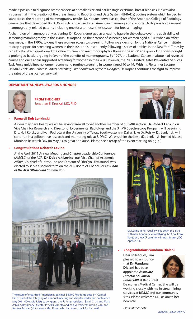
The future of organized Americian Medicine! BIDMC Residents pose on Capitol Hill as part of the lobbying ACR annual meeting and chapter leadership conference May 2011 400 radioligists to congress. L to R: 1st yr residents, Samir Shah and Mark Ashkan, Residency Director Priscilla Slanetz, and 2nd yr residents Yiming Gao, and Ammar Sarwar. (Not shown - Max Rosen who had to run back for his coat!)
• CongratulationsVandanaDialani Dear colleagues, I am
pleased to announce that Dr. Vandana Dialani has been appointed Associate Director of Clinical Breast MRI at Beth Israel Deaconess Medical Center. She will be working closely with me in streamlining services at BIDMC and our community sites. Please welcome Dr. Dialani to her new role.
- Priscilla Slanetz
June 2011 Radical Views /3
KUDOS - Please join us in congratulating the following staff for outstanding patient care and service
Support Services
Saliha Gardner is this month’s recipient of the Radiology Support Services Quality Spot on for Service Excellence Initiative Program. (Not shown)
Janice Kulas (left) recently received a wonderful complementary letter from a patient on her excellent customer service performance. "Janice is always gracious, courteous, efficient, and effective. I often take all of this for granted. This one time I wish to write to you to bring this level of service to your attention". Janice, who is the face of the IR unit on the East, has effectively made a difference in numerous patients’ lives that come into the department.
Nuclear MedicineLymphoscintigraphy patients come to the East nuclear medicine waiting room gowned. The East waiting room is filled with outpatients. Recognizing that it is uncomfortable for a gowned patient to mix with patients in street clothes, Mary Ritchie (right)checks them in and then wheels them down to the exam area so they can wait for their test in more privacy. Mary took on this duty on her own as a courtesy to the patients.
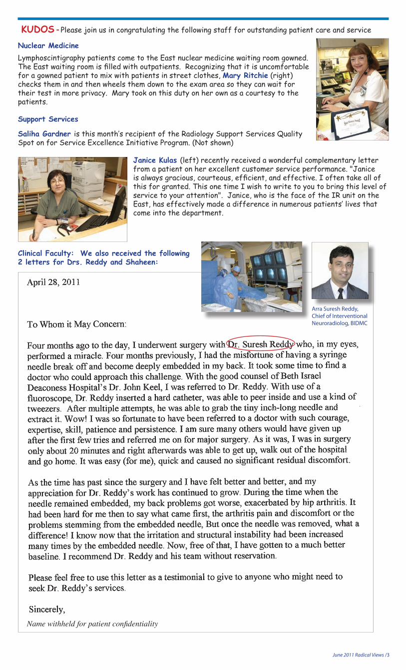
Clinical Faculty: We also received the following 2 letters for Drs. Reddy and Shaheen:
Name withheld for patient confidentiality
Arra Suresh Reddy, Chief of Interventional Neuroradiolog, BIDMC
June 2011 Radical Views /4
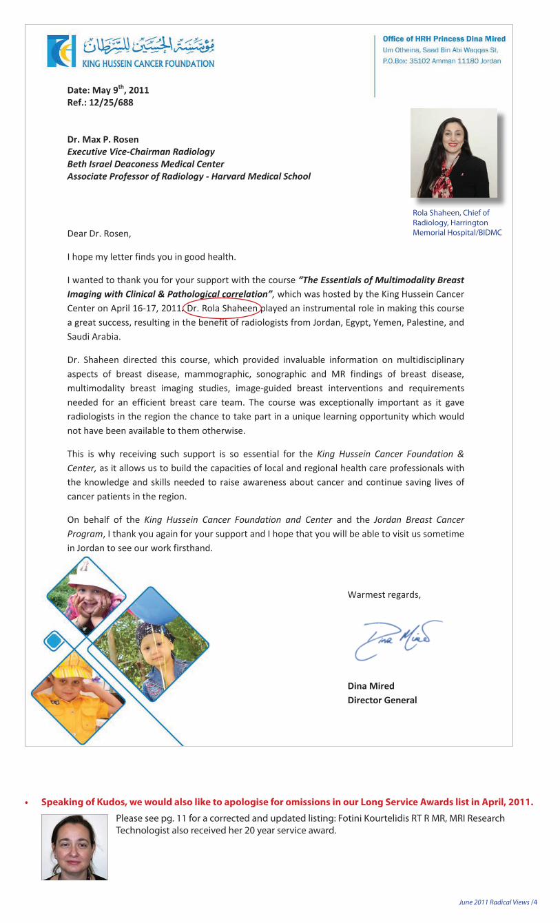
Date: May 9th, 2011 Ref.: 12/25/688 Dr. Max P. Rosen Executive Vice-Chairman Radiology Beth Israel Deaconess Medical Center Associate Professor of Radiology - Harvard Medical School
Dear Dr. Rosen,
I hope my letter finds you in good health.
I wanted to thank you for your support with the course “The Essentials of Multimodality Breast Imaging with Clinical & Pathological correlation”, which was hosted by the King Hussein Cancer Center on April 16-17, 2011. Dr. Rola Shaheen played an instrumental role in making this course a great success, resulting in the benefit of radiologists from Jordan, Egypt, Yemen, Palestine, and Saudi Arabia.
Dr. Shaheen directed this course, which provided invaluable information on multidisciplinary aspects of breast disease, mammographic, sonographic and MR findings of breast disease, multimodality breast imaging studies, image-guided breast interventions and requirements needed for an efficient breast care team. The course was exceptionally important as it gave radiologists in the region the chance to take part in a unique learning opportunity which would not have been available to them otherwise.
This is why receiving such support is so essential for the King Hussein Cancer Foundation & Center, as it allows us to build the capacities of local and regional health care professionals with the knowledge and skills needed to raise awareness about cancer and continue saving lives of cancer patients in the region.
On behalf of the King Hussein Cancer Foundation and Center and the Jordan Breast Cancer Program, I thank you again for your support and I hope that you will be able to visit us sometime in Jordan to see our work firsthand.
Warmest regards,
Dina Mired Director General
Rola Shaheen, Chief of Radiology, Harrington Memorial Hospital/BIDMC
Please see pg. 11 for a corrected and updated listing: Fotini Kourtelidis RT R MR, MRI Research Technologist also received her 20 year service award.
June 2011 Radical Views /5
MORRISONRESEARCHDAY2011
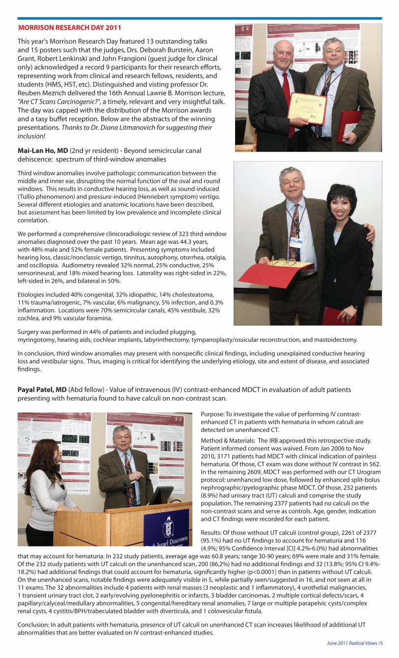
This year's Morrison Research Day featured 13 outstanding talks and 15 posters such that the judges, Drs. Deborah Burstein, Aaron Grant, Robert Lenkinski and John Frangioni (guest judge for clinical only) acknowledged a record 9 participants for their research efforts, representing work from clinical and research fellows, residents, and students (HMS, HST, etc). Distinguished and visting professor Dr. Reuben Mezrich delivered the 16th Annual Lawrie B. Morrison lecture, "Are CT Scans Carcinogenic?", a timely, relevant and very insightful talk. The day was capped with the distribution of the Morrison awards and a tasy buffet reception. Below are the abstracts of the winning presentations. Thanks to Dr. Diana Litmanovich for suggesting their inclusion!
Mai-Lan Ho, MD (2nd yr resident) - Beyond semicircular canal dehiscence: spectrum of third-window anomalies
Third window anomalies involve pathologic communication between the middle and inner ear, disrupting the normal function of the oval and round windows. This results in conductive hearing loss, as well as sound-induced (Tullio phenomenon) and pressure-induced (Hennebert symptom) vertigo. Several different etiologies and anatomic locations have been described, but assessment has been limited by low prevalence and incomplete clinical correlation.
We performed a comprehensive clinicoradiologic review of 323 third window anomalies diagnosed over the past 10 years. Mean age was 44.3 years, with 48% male and 52% female patients. Presenting symptoms included hearing loss, classic/nonclassic vertigo, tinnitus, autophony, otorrhea, otalgia, and oscillopsia. Audiometry revealed 32% normal, 25% conductive, 25% sensorineural, and 18% mixed hearing loss. Laterality was right-sided in 22%, left-sided in 26%, and bilateral in 50%.
Etiologies included 40% congenital, 32% idiopathic, 14% cholesteatoma, 11% trauma/iatrogenic, 7% vascular, 6% malignancy, 5% infection, and 0.3% inflammation. Locations were 70% semicircular canals, 45% vestibule, 32% cochlea, and 9% vascular foramina.
Surgery was performed in 44% of patients and included plugging, myringotomy, hearing aids, cochlear implants, labyrinthectomy, tympanoplasty/ossicular reconstruction, and mastoidectomy.
In conclusion, third window anomalies may present with nonspecific clinical findings, including unexplained conductive hearing loss and vestibular signs. Thus, imaging is critical for identifying the underlying etiology, site and extent of disease, and associated findings.
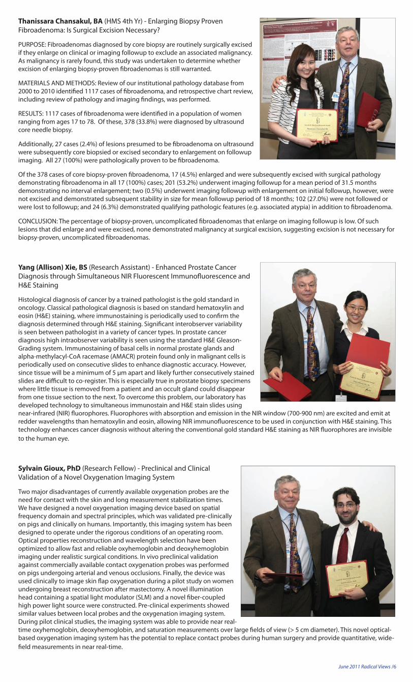
Payal Patel, MD (Abd fellow) - Value of intravenous (IV) contrast-enhanced MDCT in evaluation of adult patients presenting with hematuria found to have calculi on non-contrast scan.
Purpose: To investigate the value of performing IV contrast-enhanced CT in patients with hematuria in whom calculi are detected on unenhanced CT.
Method & Materials: The IRB approved this retrospective study. Patient informed consent was waived. From Jan 2006 to Nov 2010, 3171 patients had MDCT with clinical indication of painless hematuria. Of those, CT exam was done without IV contrast in 562. In the remaining 2609, MDCT was performed with our CT Urogram protocol: unenhanced low dose, followed by enhanced split-bolus nephrographic/pyelographic phase MDCT. Of those, 232 patients (8.9%) had urinary tract (UT) calculi and comprise the study population. The remaining 2377 patients had no calculi on the non-contrast scans and serve as controls. Age, gender, indication and CT findings were recorded for each patient.
Results: Of those without UT calculi (control group), 2261 of 2377 (95.1%) had no UT findings to account for hematuria and 116 (4.9%; 95% Confidence Interval [CI] 4.2%-6.0%) had abnormalities
that may account for hematuria. In 232 study patients, average age was 60.8 years; range 30-90 years; 69% were male and 31% female. Of the 232 study patients with UT calculi on the unenhanced scan, 200 (86,2%) had no additional findings and 32 (13.8%; 95% CI 9.4%-18.2%) had additional findings that could account for hematuria, significantly higher (p<0.0001) than in patients without UT calculi. On the unenhanced scans, notable findings were adequately visible in 5, while partially seen/suggested in 16, and not seen at all in 11 exams. The 32 abnormalities include 4 patients with renal masses (3 neoplastic and 1 inflammatory), 4 urothelial malignancies, 1 transient urinary tract clot, 2 early/evolving pyelonephritis or infarcts, 3 bladder carcinomas, 2 multiple cortical defects/scars, 4 papillary/calyceal/medullary abnormalities, 5 congenital/hereditary renal anomalies, 7 large or multiple parapelvic cysts/complex renal cysts, 4 cystitis/BPH/trabeculated bladder with diverticula, and 1 colovesicular fistula.
Conclusion: In adult patients with hematuria, presence of UT calculi on unenhanced CT scan increases likelihood of additional UT abnormalities that are better evaluated on IV contrast-enhanced studies.
PURPOSE: Fibroadenomas diagnosed by core biopsy are routinely surgically excised if they enlarge on clinical or imaging followup to exclude an associated malignancy. As malignancy is rarely found, this study was undertaken to determine whether excision of enlarging biopsy-proven fibroadenomas is still warranted.
MATERIALS AND METHODS: Review of our institutional pathology database from 2000 to 2010 identified 1117 cases of fibroadenoma, and retrospective chart review, including review of pathology and imaging findings, was performed.
RESULTS: 1117 cases of fibroadenoma were identified in a population of women ranging from ages 17 to 78. Of these, 378 (33.8%) were diagnosed by ultrasound core needle biopsy.
Additionally, 27 cases (2.4%) of lesions presumed to be fibroadenoma on ultrasound were subsequently core biopsied or excised secondary to enlargement on followup imaging. All 27 (100%) were pathologically proven to be fibroadenoma.
Of the 378 cases of core biopsy-proven fibroadenoma, 17 (4.5%) enlarged and were subsequently excised with surgical pathology demonstrating fibroadenoma in all 17 (100%) cases; 201 (53.2%) underwent imaging followup for a mean period of 31.5 months demonstrating no interval enlargement; two (0.5%) underwent imaging followup with enlargement on initial followup, however, were not excised and demonstrated subsequent stability in size for mean followup period of 18 months; 102 (27.0%) were not followed or were lost to followup; and 24 (6.3%) demonstrated qualifying pathologic features (e.g. associated atypia) in addition to fibroadenoma.
CONCLUSION: The percentage of biopsy-proven, uncomplicated fibroadenomas that enlarge on imaging followup is low. Of such lesions that did enlarge and were excised, none demonstrated malignancy at surgical excision, suggesting excision is not necessary for biopsy-proven, uncomplicated fibroadenomas.
Yang (Allison) Xie, BS (Research Assistant) - Enhanced Prostate Cancer Diagnosis through Simultaneous NIR Fluorescent Immunofluorescence and H&E Staining
Histological diagnosis of cancer by a trained pathologist is the gold standard in oncology. Classical pathological diagnosis is based on standard hematoxylin and eosin (H&E) staining, where immunostaining is periodically used to confirm the diagnosis determined through H&E staining. Significant interobserver variability is seen between pathologist in a variety of cancer types. In prostate cancer diagnosis high intraobserver variability is seen using the standard H&E Gleason-Grading system. Immunostaining of basal cells in normal prostate glands and alpha-methylacyl-CoA racemase (AMACR) protein found only in malignant cells is periodically used on consecutive slides to enhance diagnostic accuracy. However, since tissue will be a minimum of 5 μm apart and likely further consecutively stained slides are difficult to co-register. This is especially true in prostate biopsy specimens where little tissue is removed from a patient and an occult gland could disappear from one tissue section to the next. To overcome this problem, our laboratory has developed technology to simultaneous immunostain and H&E stain slides using near-infrared (NIR) fluorophores. Fluorophores with absorption and emission in the NIR window (700-900 nm) are excited and emit at redder wavelengths than hematoxylin and eosin, allowing NIR immunofluorescence to be used in conjunction with H&E staining. This technology enhances cancer diagnosis without altering the conventional gold standard H&E staining as NIR fluorophores are invisible to the human eye.
SylvainGioux,PhD (Research Fellow) - Preclinical and Clinical Validation of a Novel Oxygenation Imaging System
Two major disadvantages of currently available oxygenation probes are the need for contact with the skin and long measurement stabilization times. We have designed a novel oxygenation imaging device based on spatial frequency domain and spectral principles, which was validated pre-clinically on pigs and clinically on humans. Importantly, this imaging system has been designed to operate under the rigorous conditions of an operating room. Optical properties reconstruction and wavelength selection have been optimized to allow fast and reliable oxyhemoglobin and deoxyhemoglobin imaging under realistic surgical conditions. In vivo preclinical validation against commercially available contact oxygenation probes was performed on pigs undergoing arterial and venous occlusions. Finally, the device was used clinically to image skin flap oxygenation during a pilot study on women undergoing breast reconstruction after mastectomy. A novel illumination head containing a spatial light modulator (SLM) and a novel fiber-coupled high power light source were constructed. Pre-clinical experiments showed similar values between local probes and the oxygenation imaging system. During pilot clinical studies, the imaging system was able to provide near real-time oxyhemoglobin, deoxyhemoglobin, and saturation measurements over large fields of view (> 5 cm diameter). This novel optical-based oxygenation imaging system has the potential to replace contact probes during human surgery and provide quantitative, wide-field measurements in near real-time.
June 2011 Radical Views /7
JonathanMarmurek,BESc,MESc (HST PhD student) - A hydroxyapatite-targeted gadolinium contrast agent for MRI of microcalcification in malignant breast cancer
Detection of solid microcalcification by x-ray and computed tomography (CT) mammography is the primary clinical imaging finding for initial identification breast cancer tumors. Hydroxyapatite (HA) microcalcifications are a hallmark of malignant breast cancer, but the chemical composition of microcalcifications cannot be determined by x-ray/CT mammography. MRI has recently become a useful screening tool for women at high-risk of invasive breast cancer, but is unable to detect solid calcified structures using conventional techniques. We have developed a high-relaxivity gadolinium-bisphosphonate contrast agent with a short ligand-to-agent linker that exhibits specific adsorption to hydroxyapatite. Slow-rigid gadolinium (Gd) chelates with long rotational correlation times are expected to have high longitudinal relaxivities, and previous work has demonstrated that shorter linkers between a Gd-chelate and targeting-ligand reduce rotational freedom when bound. Ultra-short echo-time (UTE) MRI was performed to assess the specificity and sensitivity of the contrast agent in-vitro. The rigidly adsorbed contrast agent was detectable at 1 μM and had an apparent relaxivity approximately 102-fold higher than that of the free agent in solution and conventional small molecule T1 relaxation agents. Preliminary studies showed that the HA-bound agent can be detected and distinguished from calcium oxalate in-vivo.
Posters:
Nicholas J. Durr, PhD (Research Fellow) - Optical Systems for a Next Generation FLARE™ Device
Multiple Fluorescence-Assisted Resection and Exploration (FLARE™) systems are currently being used for research and clinical trials in operating rooms around the world. These novel systems provide real-time guidance to surgeons for targeting tissues of interest and avoiding sensitive structures with minimal impact on the typical operating room workflow. However, current FLARE™ systems are impractical for dissemination. We are investigating the use of several new lightweight and inexpensive technologies that will enable large-scale production of the FLARE™ systems. First, we will generate visible and near-infrared light in the instrument cart and deliver the light to the surgical field through a fiber optic bundle instead of using light emitting diodes mounted to the imaging head. This allows for the creation of a compact luminary and minimizes heat generation near the heat-sensitive detectors. Second, we are building a custom zoom lens that is corrected for high resolution at visible and near-infrared wavelengths. Finally, we are building a custom 3-channel imaging head, which will provide excellent performance at a low cost and eliminate the redundant parts associated with off-the-shelf solutions. Using these technologies, our next-generation FLARE™ system will be poised for large-scale distribution and have the potential to improve surgical outcomes.
Rachel Scheidegger, BS (HST PhD Student) - Amide Proton Transfer Imaging with Continuous Wave Dual Frequency Saturation Can Detect the Amide Proton Peak in the Z-Spectrum Acquired at 3T
We present a chemical exchange saturation transfer (CEST) imaging sequence with continuous wave saturation preparation relying on a 3-way subtraction between label frequency, control frequency, and simultaneous dual frequency RF irradiation to remove B0 inhomogeneity and intrinsic magnetization transfer (MT ) from in-vivo images. We demonstrate that this approach yields amide proton transfer (APT) images free of susceptibility artifacts and MT asymmetry, without any additional B0 correction. This allows clear and robust measurement of the amide proton peak in the z-spectrum acquired at 3T. This new method may improve the feasibility of quantifying exchange rates in-vivo and measuring pH. (Rachel was unable to accept her award due to her being on rotation)
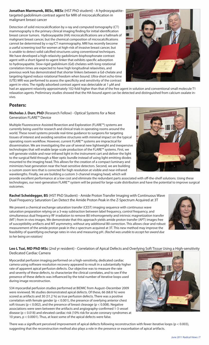
Leo L Tsai, MD PhD MSc (2nd yr resident) - Correlation of Apical Defects and Overlying Soft Tissue Using a High-sensitivity Dedicated Cardiac Camera
Myocardial perfusion imaging performed on a high-sensitivity, dedicated cardiac camera using software resolution recovery appeared to result in a substantially higher rate of apparent apical perfusion defects. Our objective was to measure the rate and severity of these defects, to characterize the clinical correlates, and to see if the presence of these defects was influenced by the total number of iterative loops used during image reconstruction.
534 myocardial perfusion studies performed at BIDMC from August–December 2009 were reviewed. 96 studies demonstrated apical defects. Of these, 66 (68.8 %) were scored as artifacts and 30 (31.2 %) as true perfusion defects. There was a positive correlation with female gender (p < 0.001), the presence of overlying anterior chest soft tissues (p < 0.002), and the presence of breast cleavage (p < 0.008). Negative associations were seen between the artifacts and angiography-confirmed 1-3 vessel disease (p < 0.018) and elevated cardiac risk (10% risk for acute coronary syndromes at 10 years, p < 0.0001). Thus, at least some of the apical defects were false.
There was a significant perceived improvement of apical defects following reconstruction with fewer iterative loops (p < 0.003), suggesting that the reconstruction method also plays a role in the presence or exacerbation of apical artifacts.
June 2011 Radical Views /8
OUTREACH2011-RadiologyattheBIDMC Health Fair
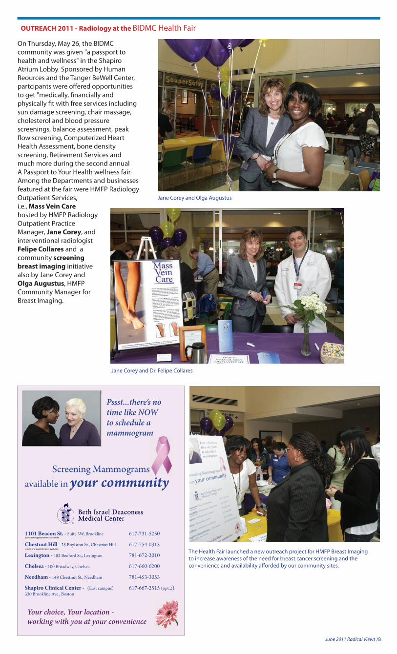
On Thursday, May 26, the BIDMC community was given "a passport to health and wellness" in the Shapiro Atrium Lobby. Sponsored by Human Reources and the Tanger BeWell Center, partcipants were offered opportunities to get "medically, financially and physically fit with free services including sun damage screening, chair massage, cholesterol and blood pressure screenings, balance assessment, peak flow screening, Computerized Heart Health Assessment, bone density screening, Retirement Services and much more during the second annual A Passport to Your Health wellness fair. Among the Departments and businesses featured at the fair were HMFP Radiology Outpatient Services, i.e., Mass Vein Care hosted by HMFP Radiology Outpatient Practice Manager, Jane Corey, and interventional radiologist Felipe Collares and a community screening breastimaging initiative also by Jane Corey and Olga Augustus, HMFP Community Manager for Breast Imaging.
Jane Corey and Olga Augustus
Jane Corey and Dr. Felipe Collares
Pssst...there’s no time like NOW to schedule a mammogram
1101 Beacon St. - Suite 3W, Brookline 617-731-5250
Chestnut Hill - 25 Boylston St., Chestnut Hill 617-754-0313
Shapiro Clinical Center - (East campus) 617-667-2515 (opt.2)330 Brookline Ave., Boston
Lunchtime appointments available
Lunchtime appointments available
Your choice, Your location - working with you at your convenience
available in your communityScreening Mammograms
The Health Fair launched a new outreach project for HMFP Breast Imaging to increase awareness of the need for breast cancer screening and the convenience and availability afforded by our community sites.
June 2011 Radical Views /9
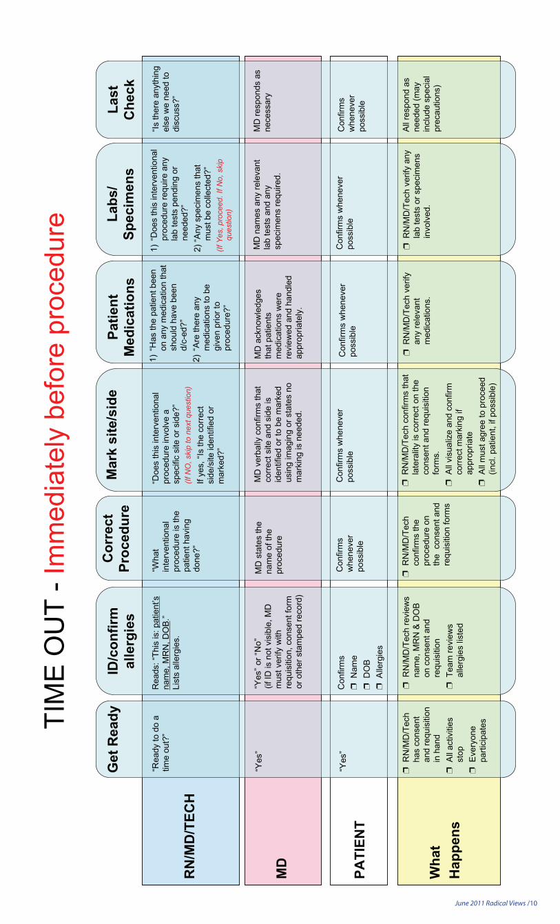
To streamline the submissions for RadReview, critical findings and QA cases, the InfoRadiology web site is now connected with GE Centricity.
A link called “InfoRadiology” will appeal at the right bottom corner of the Centricity window when an exam is opened.
One click of this link will transfer the clip number of the exam to InfoRadiology.
If your workstation does not have this link, please let me know and we can install this for you.
Or, you can do this yourself at your PACS workstation with the following steps:
[1] Go to http://Inforad <http://inforad/> at the web browser
[2] Click on “Install” and following the directions
– Sam Yam, PhD Director, Departmental Computing
Do you know... about the new InfoRad Link at PACS Workstations?
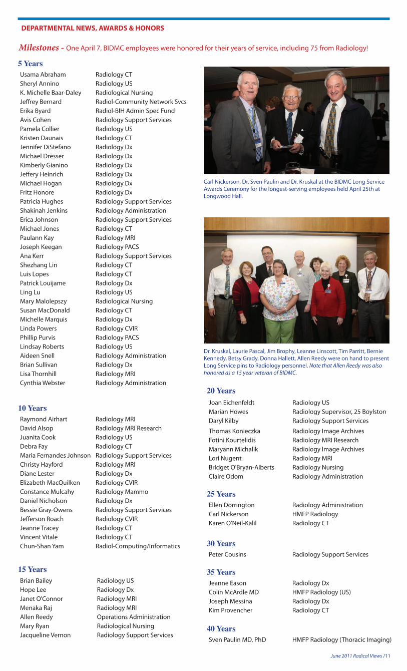
Medical Center Policy #CP-33 outlines our Universal Protocol, also know as the guidelines for the confirmation of correct patient, correct procedure and correct site for patients undergoing invasive or high-risk procedures being performed outside of the Operating Room. Despite present quality efforts, BIDMC continues to have a few wrong side/site procedures and many near misses. As we all know, one procedure error is too many and any of the near misses had the potential to cause harm. In response to this ongoing issue, the office of Healthcare Quality asked the Interventional Procedures Committee (IPC) to take part in a major immediate goal to standardize the approach to Time-Outs in the interventional areas. Within radiology, the section chiefs met with their representatives from the IPC to draft a script that would fit procedures carried out in our department. The draft had to meet the required elements of the time-out process which include the guidelines within Universal Protocol and additionally, medication, lab values and specimen issues. The time-out process is mandatory and must be carried out before the procedure begins and requires active participation on the part of all members of the professional team (MD/RN/Tech or MD/Tech). The new script (shown below) will be implemented on July 1, 2011.
– Misti Mullins, RN Radiology Quality
Do you know... about our Universal Protocol?
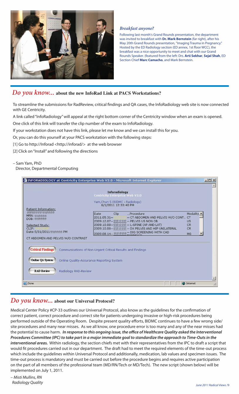
Breakfast anyone?Following last month's Grand Rounds presentation, the department was invited to breakfast with Dr.MarkBernstein (far right), after his May 20th Grand Rounds presentation, "Imaging Trauma in Pregnancy." Hosted by the ED Radiology section (ED annex, 1st floor WCC), the breakfast was a nice opportunity to meet and chat with our Grand Rounds Speaker. (featured from the left: Drs. ArtiSekhar, Sejal Shah, ED Section Chief MarcCamacho, and Mark Bernstein.
June 2011 Radical Views /10
PATI
ENT
TIM
E O
UT
- Im
med
iate
ly b
efor
e pr
oced
ure
Get
Rea
dyID
/con
firm
alle
rgie
sLa
stC
heck
Cor
rect
Proc
edur
eM
ark
site
/sid
e
“Rea
dy to
do
atim
e ou
t?”
“Yes
”
Rea
ds: “
This
is: p
atie
nt’s
nam
e, M
RN
, DO
B.”
List
s al
lerg
ies.
“Yes
” or “
No”
(if ID
is n
ot v
isib
le, M
Dm
ust v
erify
with
requ
isiti
on, c
onse
nt fo
rmor
oth
er s
tam
ped
reco
rd)
Con
firm
s❐
Nam
e❐
DO
B❐
Alle
rgie
s
“Yes
”
❐ R
N/M
D/T
ech
has
cons
ent
and
requ
isiti
onin
han
d❐
All
activ
ities
stop
❐ E
very
one
parti
cipa
tes
❐ R
N/M
D/T
ech
revi
ews
nam
e, M
RN
& D
OB
on c
onse
nt a
ndre
quis
ition
❐ T
eam
revi
ews
alle
rgie
s lis
ted
“Wha
tin
terv
entio
nal
proc
edur
e is
the
patie
nt h
avin
gdo
ne?”
MD
sta
tes
the
nam
e of
the
proc
edur
e
Con
firm
sw
hene
ver
poss
ible
❐ R
N/M
D/T
ech
conf
irms
the
proc
edur
e on
the
con
sent
and
requ
isiti
on fo
rms
“Doe
s th
is in
terv
entio
nal
proc
edur
e in
volv
e a
spec
ific
site
or s
ide?
”(If
NO
, ski
p to
nex
t que
stio
n)If
yes,
“Is
the
corre
ctsi
de/s
ite id
entif
ied
orm
arke
d?”
MD
ver
bally
con
firm
s th
atco
rrect
site
and
sid
e is
iden
tifie
d or
to b
e m
arke
dus
ing
imag
ing
or s
tate
s no
mar
king
is n
eede
d.
Con
firm
s w
hene
ver
poss
ible
❐R
N/M
D/T
ech
conf
irms
that
late
ralit
y is
cor
rect
on
the
cons
ent a
nd re
quis
ition
form
s.❐
All
visu
aliz
e an
d co
nfirm
corre
ct m
arki
ng if
appr
opria
te❐
All
mus
t agr
ee to
pro
ceed
(incl
. pat
ient
, if p
ossi
ble)
Labs
/Sp
ecim
ens
1)“D
oes
this
inte
rven
tiona
lpr
oced
ure
requ
ire a
nyla
b te
sts
pend
ing
orne
eded
?”2)
“Any
spe
cim
ens
that
mus
t be
colle
cted
?”(If
Yes
, pro
ceed
. If N
o, s
kip
ques
tion)
MD
nam
es a
ny re
leva
ntla
b te
sts
and
any
spec
imen
s re
quire
d.
❐R
N/M
D/T
ech
verif
y an
yla
b te
sts
or s
peci
men
sin
volv
ed.
Con
firm
s w
hene
ver
poss
ible
“Is th
ere
anyt
hing
else
we
need
todi
scus
s?”
All r
espo
nd a
sne
eded
(may
incl
ude
spec
ial
prec
autio
ns)
Con
firm
sw
hene
ver
poss
ible
1)“H
as th
e pa
tient
bee
non
any
med
icat
ion
that
shou
ld h
ave
been
d/c-
ed?”
2)“A
re th
ere
any
med
icat
ions
to b
egi
ven
prio
r to
proc
edur
e?”
Con
firm
s w
hene
ver
poss
ible
MD
ack
now
ledg
esth
at p
atie
nts
med
icat
ions
wer
ere
view
ed a
nd h
andl
edap
prop
riate
ly.
Patie
ntM
edic
atio
ns
RN
/MD
/TEC
H
MD
Wha
tH
appe
ns
❐R
N/M
D/T
ech
verif
yan
y re
leva
ntm
edic
atio
ns.
MD
resp
onds
as
nece
ssar
y
June 2011 Radical Views /11
DEPARTMENTAL NEWS, AWARDS & HONORS
Milestones - One April 7, BIDMC employees were honored for their years of service, including 75 from Radiology!
5 YearsUsama Abraham Radiology CTSheryl Annino Radiology USK. Michelle Baar-Daley Radiological NursingJeffrey Bernard Radiol-Community Network SvcsErika Byard Radiol-BIH Admin Spec FundAvis Cohen Radiology Support ServicesPamela Collier Radiology USKristen Daunais Radiology CTJennifer DiStefano Radiology DxMichael Dresser Radiology DxKimberly Gianino Radiology DxJeffery Heinrich Radiology DxMichael Hogan Radiology DxFritz Honore Radiology DxPatricia Hughes Radiology Support ServicesShakinah Jenkins Radiology AdministrationErica Johnson Radiology Support ServicesMichael Jones Radiology CTPaulann Kay Radiology MRIJoseph Keegan Radiology PACSAna Kerr Radiology Support ServicesShezhang Lin Radiology CTLuis Lopes Radiology CTPatrick Louijame Radiology DxLing Lu Radiology USMary Malolepszy Radiological NursingSusan MacDonald Radiology CTMichelle Marquis Radiology DxLinda Powers Radiology CVIRPhillip Purvis Radiology PACSLindsay Roberts Radiology USAideen Snell Radiology AdministrationBrian Sullivan Radiology DxLisa Thornhill Radiology MRICynthia Webster Radiology Administration
Brian Bailey Radiology USHope Lee Radiology DxJanet O'Connor Radiology MRIMenaka Raj Radiology MRIAllen Reedy Operations AdministrationMary Ryan Radiological NursingJacqueline Vernon Radiology Support Services
15 Years
Dr. Kruskal, Laurie Pascal, Jim Brophy, Leanne Linscott, Tim Parritt, Bernie Kennedy, Betsy Grady, Donna Hallett, Allen Reedy were on hand to present Long Service pins to Radiology personnel. Note that Allen Reedy was also honored as a 15 year veteran of BIDMC.
Sven Paulin MD, PhD HMFP Radiology (Thoracic Imaging)
40 Years
Carl Nickerson, Dr. Sven Paulin and Dr. Kruskal at the BIDMC Long Service Awards Ceremony for the longest-serving employees held April 25th at Longwood Hall.
Joan Eichenfeldt Radiology USMarian Howes Radiology Supervisor, 25 BoylstonDaryl Kilby Radiology Support Services
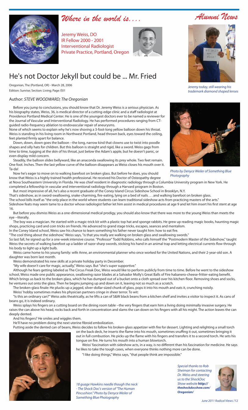
Jeremy Weiss, DOIR Fellow 2000 - 2001 Interventional RadiologistPrivate Practice, Portland, Oregon
Alumni News
He's not Doctor Jekyll but could be ... Mr. FriedOregonian, The (Portland, OR) - March 28, 2006
Edition: Sunrise; Section: Living; Page: E01
Author: STEVE WOODWARD; The Oregonian
Before you jump to conclusions, you should know that Dr. Jeremy Weiss is a serious physician. As his biography states, Weiss, 36, is medical director of a cutting-edge clinic and a staff radiologist at Providence Portland Medical Center. He is one of the youngest doctors ever to be named a reviewer for the Journal of Vascular and Interventional Radiology. He has performed procedures ranging from CT-guided radio-frequency ablation to endovascular repair of aneurysms.None of which seems to explain why he's now shoving a 3-foot-long yellow balloon down his throat.Weiss is standing in his living room in Northwest Portland, head thrown back, eyes toward the ceiling, feet planted firmly apart for balance. Down, down, down goes the balloon --the long, narrow kind that clowns use to twist into poodle shapes and silly hats for children. But this balloon is straight and rigid, like a sword. Weiss gags from time to time, tugging at the skin of his throat, just below the Adam's apple, but he doesn't panic, or even display mild concern. Steadily, the balloon slides bellyward, like an anaconda swallowing its prey whole. Two feet remain. One foot. Inches. Then the last yellow curve of the balloon disappears as Weiss closes his mouth over it.Ta-da! Now he's eager to move on to walking barefoot on broken glass. But before he does, you should know that Weiss is a highly trained health professional. He received his Doctor of Osteopathy degree at Nova Southeastern University in Florida. He was chief resident in diagnostic radiology through a Columbia University program in New York. He completed a fellowship in vascular and interventional radiology through a Harvard program in Boston. But most impressive of all, he's also a recent graduate of the Coney Island Circus Sideshow School in Brooklyn, N.Y.That's sideshow, as in sword-swallowing, snake-charming, fire-eating, lying on a bed of nails . . . and walking barefoot on broken glass.The school bills itself as "the only place in the world where students can learn traditional sideshow acts from practicing masters of the arts."Sideshow feats may seem tame to a doctor whose radiologist father let him assist in medical procedures at age 9 and let him insert his first stent at age 16. But before you dismiss Weiss as a one-dimensional medical prodigy, you should also know that there was more to the young Weiss than meets the eye --literally. The boy was a magician. He started with a magic-trick kit with a plastic top hat and sponge rabbits. He grew up reading magic books, haunting magic shops, practicing card and coin tricks on friends. He advanced to grand stage tricks, escapes, seances and mentalism.In the Coney Island school, Weiss saw his chance to learn something his father never taught him: how to eat fire."The cool thing about the sideshow," Weiss says, "is that you really are walking on broken glass and swallowing swords."So last fall, he signed up for a one-week intensive course. "Professor" Todd Robbins, who calls himself the "Postmodern Master of the Sideshow," taught Weiss the secrets of walking barefoot up a ladder of razor-sharp swords, sticking his hand in an animal trap and letting electrical currents flow through his body to light up a light bulb. Weiss came home to his young family: wife Anne, an environmental planner who once worked for the United Nations, and their 2-year-old son. A daughter was born last month. Weiss demonstrated his new skills at a private holiday party in December. "My wife doesn't care for magic, actually," Weiss says. But "she's super supportive." Although he fears getting labeled as The Circus Freak Doc, Weiss would like to perform publicly from time to time. Before he went to the sideshow school, Weiss made one public appearance, swallowing razor blades at a Salvador Molly's Great Balls of Fire habanero-cheese-fritter-eating benefit.Which brings us back to the broken glass, which he has dumped out of a bucket onto a cloth spread over his kitchen floor. Removing shoes and socks, he ventures out onto the glass. Then he begins jumping up and down on it, leaving not so much as a scratch. The broken-glass finale: He plucks up a jagged, silver-dollar-sized chunk of glass, pops it into his mouth and eats it, crunching noisily. Weiss' hobby sometimes makes his physician partners cringe in sheer terror. To wit: "Is this an ordinary can?" Weiss asks theatrically, as he lifts a can of S&W black beans from a kitchen shelf and invites a visitor to inspect it. As cans of beans go, it is indeed ordinary. Weiss splays his fingers on a cutting board on the dining room table --the very fingers that earn him a living doing minimally invasive surgery. He raises the can above his head, rocks back and forth in concentration and slams the can down on his fingers with all his might. The action leaves the can deeply dented. And his fingers? He smiles and wiggles them. He'll have no problem doing the next uterine fibroid embolization. Putting aside the dented can of beans, Weiss decides to follow his broken-glass appetizer with fire for dessert. Lighting and relighting a small torch
on the back deck, he inserts the flame into his mouth, sometimes snuffing it out, sometimes bringing it out in full combustion. He picks up the flame with his fingers and transfers it to a second torch. He sets his tongue on fire. He turns his mouth into a human blowtorch. Weiss' fascination with sideshow acts, in a way, is no different than his fascination for medicine. He says he likes to take the tough cases, when everyone thinks nothing more can be done. "I like doing things," Weiss says, "that people think are impossible."
Special thanks to Rob Sheiman for contacting Dr. Weiss and steering us to the ShockDoc Show website http://theshockdocshow.com/Oregonian/
Jeremy today, still wearing his trademark diamond shaped lenses
Photo by Denyce Weiler of Something Blue Photography
18 guage Hawkins needle though the neck - The Shock Doc's version of "The Human Pincushion." Photo by Denyce Weiler of Something Blue Photography
Where in the world is....
June 2011 Radical Views /13
2011 Publications from our Faculty Members [New citations in Blue]. We do a monthly PubMed search for new BIDMC publications and may miss those in which your affiliation is not noted. If we miss your paper, please send the reference to [email protected] to be included in next month’s issue. Please note that publications do not always appear in Pubmed in the same month they are acutally published.
Akçakaya M, Basha TA, Goddu B, Goepfert LA, Kissinger KV, Tarokh V, Manning WJ, Nezafat R. Low-dimensional-structure self-learning and thresholding: Regularization beyond compressed sensing for MRI Reconstruction. Magn Reson Med. 2011 Apr 4. doi: 10.1002/mrm.22841.
Akçakaya M, Hu P, Chuang ML, Hauser TH, Ngo LH, Manning WJ, Tarokh V, Nezafat R. Accelerated noncontrast-enhanced pulmonary vein MRA with distributed compressed sensing. J Magn Reson Imaging. 2011 May;33(5):1248-55. doi: 10.1002/jmri.22559. PMCID: PMC3081138.
Akcakaya M, Nam S, Hu P, Moghari MH, Ngo LH, Tarokh V, Manning WJ, Nezafat R. Compressed Sensing With Wavelet Domain Dependencies for Coronary MRI: A Retrospective Study. IEEE Trans Med Imaging. 2011 May;30(5):1090-9.
AhmedM, Brace CL, Lee FT Jr, GoldbergSN. Principles of and Advances in Percutaneous Ablation. Radiology. 2011 Feb;258(2):351-369.
Ahmed M, Goldberg SN. Basic science research in thermal ablation. Surg Oncol Clin N Am. 2011 Apr;20(2):237-58.
Appelbaum L, Sosna J, Nissenbaum Y, Benshtein A, GoldbergSN. Electromagnetic Navigation System for CT-Guided Biopsy of Small Lesions. AJR Am J Roentgenol. 2011 May;196(5):1194-200.
Ashitate Y, Tanaka E, Stockdale A, Choi HS, Frangioni JV. Near-infrared fluorescence imaging of thoracic duct anatomy and function in open surgery and video-assisted thoracic surgery. J Thorac Cardiovasc Surg. 2011 Apr 6. [Epub ahead of print]
Boiselle PM. The journal of thoracic imaging welcomes the European society of thoracic imaging. J Thorac Imaging. 2011 Feb;26(1):2.
Boiselle PM. In with the new! J Thorac Imaging. 2011 Feb;26(1):1.
Boiselle PM, Erasmus JJ, Ko JP, Ravenel JG, Vlahos I. Expert opinion: lung cancer staging. J Thorac Imaging. 2011 May;26(2):85.
Boiselle PM, Hurwitz LM, Mayo JR, Schoepf UJ, Tack D. Expert opinion: radiation dose management in cardiopulmonary imaging. J Thorac Imaging. 2011 Feb;26(1):3.
Boiselle PM, Reddy GP. Editors' recognition awards for distinction in reviewing in 2010. J Thorac Imaging. 2011 Feb;26(1):7.
Brook OR, Hakmon T, Brook A, Dudnik E, Kuten A, Engel A. The Effect of Radiology Conference Consultation on Cancer Patients Management. Ann Oncol. 2011 May;22(5):1204-8.
Brook OR, Kane RA, TyagiG, SiewertB, KruskalJB. Lessons Learned From Quality Assurance: Errors in the Diagnosis of Acute Cholecystitis on Ultrasound and CT. AJR Am J Roentgenol. 2011 Mar;196(3):597-604.
Brook OR, Mendiratta-Lala M, Brennan D, SiewertB, Faintuch S, GoldbergSN. Imaging findings after radiofrequency ablation of adrenal tumors. AJR Am J Roentgenol. 2011 Feb;196(2):382-8.
Cambers CE, Fetterly KA, Holzer R, Lin PJ, Blankenship JC, Balter S, Laskey WK. Radiation safety program for the cardiac catheterization laboratory. Catheter Cardiovasc Interv. 2011 Mar 1;77(4):546-56. doi: 10.1002/ccd.22867.
Cohen AB, Neema M, Arora A, Dell'oglio E, Benedict RH, Tauhid S, Goldberg-Zimring D, Chavarro-Nieto C, Ceccarelli A, Klein JP, Stankiewicz JM, Houtchens MK, Buckle GJ, Alsop DC, Guttmann CR, Bakshi R. The Relationships among MRI-Defined Spinal Cord Involvement, Brain Involvement, and Disability in Multiple Sclerosis. J Neuroimaging. 2011 Mar 29. doi: 10.1111/j.1552-6569.2011.00589.x.
Corwin MT, SiewertB, SheimanRG, Kane RA. Incidentally detected gallbladder polyps: is follow-up necessary?--Long-term clinical and US analysis of 346 patients. Radiology. 2011 Jan;258(1):277-82. Epub 2010 Aug 9.
Crema MD, Roemer FW, Marra MD, Burstein D, Gold GE, Eckstein F, Baum T, Mosher TJ, Carrino JA, Guermazi A. Articular Cartilage in the Knee: Current MR Imaging Techniques and Applications in Clinical Practice and Research1. Radiographics. 2011 Jan-Feb;31(1):37-61.
Dai W, Robson PM, ShankaranarayananA, Alsop DC. Sensitivity calibration with a uniform magnetization image to improve arterial spin labeling perfusion quantification. Magn Reson Med. 2011 Apr 26. doi: 10.1002/mrm.22954. [Epub ahead of print]
Dialani V, Hines N, Wang Y, Slanetz P. Breast Schwannoma. Case Report Med. 2011;2011:930841. Epub 2011 Feb 9.
Dialani V, LitmanovichD, BankierAA, Decamp M, Gangadharan SP, Boiselle PM. Subcarinal collection following mediastinoscopy: a normal post-procedural CT finding. Clin Radiol. 2011 Feb 8. [Epub ahead of print]
Douglas PS, Garcia MJ, Haines DE, Lai WW, Manning WJ, Patel AR, Picard MH, Polk DM, Ragosta M, Ward RP, Weiner RB. ACCF/ASE/AHA/ASNC/HFSA/HRS/SCAI/SCCM/SCCT/SCMR 2011 Appropriate Use Criteria for Echocardiography A Report of the American College of Cardiology Foundation Appropriate Use Criteria Task Force, American Society of Echocardiography, American Heart Association, American Society of Nuclear Cardiology, Heart Failure Society of America, Heart Rhythm Society, Society for Cardiovascular Angiography and Interventions, Society of Critical Care Medicine, Society of Cardiovascular Computed Tomography, and Society for Cardiovascular Magnetic Resonance Endorsed by the American College of Chest Physicians. J Am Coll Cardiol. 2011 Mar 1;57(9):1126-66.
Eisenberg RL, Ngo L, Boiselle PM, BankierAA. Honorary Authorship in Radiologic Research Articles: Assessment of Frequency and Associated Factors. Radiology. 2011 Mar 8.
Eisenberg RL, Yablon CM. Career development for residents and beyond: filling in the gaps. AJR Am J Roentgenol. 2011 Jan;196(1):W6-7.
Fong TG, Inouye SK, Dai W, Press DZ, Alsop DC. Association cortex hypoperfusion in mild dementia with Lewy bodies: a potential indicator of cholinergic dysfunction? Brain Imaging Behav. 2011 Mar;5(1):25-35.
Frangioni JV. The myth of multimodality diagnostic agents. Mol Imaging. 2011 Apr;10(2):79-80.
Fujii H, Idoine JD, Gioux S, Accorsi R, Slochower DR, Lanza RC, Frangioni JV. Optimization of Coded Aperture Radioscintigraphy for Sentinel Lymph Node Mapping. Mol Imaging Biol. 2011 May 13.
Gansler DA, Lee AK, Emerton BC, D'Amato C, Bhadelia R, Jerram M, Fulwiler C. Prefrontal regional correlates of self-control in male psychiatric patients: Impulsivity facets and aggression. Psychiatry Res. 2011 Jan 30;191(1):16-23. Epub 2010 Dec 9.
Gibbs-Strauss SL, Nasr KA, Fish KM, Khullar O, Ashitate Y, Siclovan TM, Johnson BF, Barnhardt NE, Tan Hehir CA, Frangioni JV. Nerve-highlighting fluorescent contrast agents for image-guided surgery. Mol Imaging. 2011 Apr;10(2):91-101.
GrantAK, VinogradovE, Wang X, LenkinskiRE, Alsop DC. Perfusion imaging with a freely diffusible hyperpolarized contrast agent. Magn Reson Med. 2011 Mar 22. doi: 10.1002/mrm.22860.
GreenmanRL, Smithline HA. The Feasibility of Measuring Phosphocreatine Recovery Kinetics in Muscle Using a Single-shot (31)P RARE MRI Sequence. Acad Radiol. 2011 Apr 30. [Epub ahead of print]
Hara AK, Blevins M, Chen MH, Dachman AH, Kuo MD, Menias CO, SiewertB, Cheema JI, Obregon RG, Fidler JL, Zimmerman P, Horton KM, Coakley KJ, Iyer RB, Halvorsen RA Jr, Casola G, Yee J, Herman BA, Johnson CD. ACRIN CT Colonography Trial: Does Reader's Preference for Primary Two-dimensional versus Primary Three-dimensional Interpretation Affect Performance? Radiology. 2011 Mar 1.
Hara AK, Kuo MD, Blevins M, Chen MH, Yee J, Dachman A, Menias CO, SiewertB, Cheema JI, Obregon RG, Fidler JL, Zimmerman P, Horton KM, Coakley K, Iyer RB, Halvorsen RA Jr, Casola G, Johnson CD. National CT Colonography Trial (ACRIN 6664): Comparison of Three Full-Laxative Bowel Preparations in More Than 2500 Average-Risk Patients. AJR Am J Roentgenol. 2011 May;196(5):1076-82.
Harrigan CJ, Peters DC, Gibson CM, Maron BJ, Manning WJ, Maron MS, Appelbaum E. Hypertrophic cardiomyopathy: quantification of late gadolinium enhancement with contrast-enhanced cardiovascular MR imaging. Radiology. 2011 Jan;258(1):128-33. Epub 2010 Nov 2.
Hu P, Chan J, Ngo LH, Smink J, Goddu B, Kissinger KV, Goepfert L, Hauser TH, RofskyNM, Manning WJ, Nezafat R. Contrast-enhanced whole-heart coronary MRI with bolus infusion of gadobenate dimeglumine at 1.5 T. Magn Reson Imaging. 2011 Feb;66(2):392-398.
June 2011 Radical Views /14
2011 Publications from our Faculty Members [New citations in Blue].
Hutteman M, Choi HS, Mieog JS, van der Vorst JR, Ashitate Y, Kuppen PJ, van Groningen MC, Löwik CW, Smit VT, van de Velde CJ, Frangioni JV, Vahrmeijer AL. Clinical Translation of Ex Vivo Sentinel Lymph Node Mapping for Colorectal Cancer Using Invisible Near-Infrared Fluorescence Light. Ann Surg Oncol. 2011 Apr;18(4):1006-14.
Hutteman M, Mieog JS, van der Vorst JR, Liefers GJ, Putter H, Löwik CW, Frangioni JV, van de Velde CJ, Vahrmeijer AL. Randomized, double-blind comparison of indocyanine green with or without albumin premixing for near-infrared fluorescence imaging of sentinel lymph nodes in breast cancer patients. Breast Cancer Res Treat. 2011 Mar 1.
Inoue K, Liu F, Hoppin J, Lunsford EP, Lackas C, Hesterman J, LenkinskiRE, Fujii H, Frangioni JV. High-resolution Computed Tomography Of Single Breast Cancer Microcalcifications In Vivo. Mol Imaging. 2011 Apr 1.
IuanowE, Kettler M, Slanetz PJ. Spectrum of Disease in the Male Breast. AJR Am J Roentgenol. 2011 Mar;196(3):W247-59. [CME. WEB. Pictorial Essay]
Khan A, Khosa F, Eisenberg RL. Cystic lesions of the pancreas. AJR Am J Roentgenol. 2011 Jun;196(6):W668-77.
Khan A, Nasir K, Khosa F, Saghir A, Sarwar S, Clouse ME. Prospective Gating With 320-MDCT Angiography: Effect of Volume Scan Length on Radiation Dose. AJR Am J Roentgenol. 2011 Feb;196(2):407-11.
Khosa F, Romney BP, Costa DN, RofskyNM, Manning WJ. Prevalence of noncardiac findings on clinical cardiovascular MRI. AJR Am J Roentgenol. 2011 Apr;196(4):W380-6.
Khurd P, Bahlmann C, Maday P, Kamen A, Gibbs-Strauss S, Genega EM, Frangioni JV. COMPUTER-AIDED GLEASON GRADING OF PROSTATE CANCER HISTOPATHOLOGICAL IMAGES USING TEXTON FORESTS. Proc IEEE Int Symp Biomed Imaging. 2010 Apr 17;14-17 April 2010:636-639. PMCID: PMC3017375.
Klein JP, Arora A, Neema M, Healy BC, Tauhid S, Goldberg-Zimring D, Chavarro-Nieto C, Stankiewicz JM, Cohen AB, Buckle GJ, Houtchens MK, Ceccarelli A, Dell'oglio E, Guttmann CR, Alsop DC, HackneyDB, Bakshi R. A 3T MR Imaging Investigation of the Topography of Whole Spinal Cord Atrophy in Multiple Sclerosis. AJNR Am J Neuroradiol. 2011 May 5. [Epub ahead of print]
Koshkareva YA, Weinstein GS, Feldman M, MoonisG. Steatocystoma simplex of the infratemporal fossa: An uncommon location for a rare entity. Ear Nose Throat J. 2011 Jan;90(1):E16-8.
Krajewski KM, Guo M, Van den Abbeele AD, Yap J, Ramaiya N, Jagannathan J, Heng DY, Atkins MB, McDermott DF, Schutz FA, Pedrosa I, Choueiri TK. Comparison of Four Early Posttherapy Imaging Changes (EPTIC; RECIST 1.0, Tumor Shrinkage, Computed Tomography Tumor Density, Choi Criteria) in Assessing Outcome to Vascular Endothelial Growth Factor-Targeted Therapy in Patients With Advanced Renal Cell Carcinoma. Eur Urol. 2011 Feb 1. [Epub ahead of print]
Kressel HY. Scientific dialogue: vestige of the past or hope for the future? Radiology. 2011 Jan;258(1):12-4.
Kressel HY, Dixon AK. Where Is the Honor in Honorary Authorship? Radiology. 2011 Mar 8.
Kung JW, Yablon CM, Eisenberg RL. Bone marrow signal alteration in the extremities. AJR Am J Roentgenol. 2011 May;196(5):W492-510.
Lee EY, Tracy DA, Eisenberg RL, Arellano CM, Mahmood SA, Cleveland RH, Zurakowski D, Boiselle PM. Screening of asymptomatic children for tuberculosis is a lateral chest radiograph routinely indicated? Acad Radiol. 2011 Feb;18(2):184-90. Epub 2010 Nov 20.
Lee EY, Tracy DA, Mahmood SA, Weldon CB, Zurakowski D, Boiselle PM. Preoperative MDCT Evaluation of Congenital Lung Anomalies in Children: Comparison of Axial, Multiplanar, and 3D Images. AJR Am J Roentgenol. 2011 May;196(5):1040-6.
LevensonRB, Pearson KM, Saokar A, Lee SI, Mueller PR, Hahn PF. Image-guided Drainage of Tuboovarian Abscesses of Gastrointestinal or Genitourinary Origin: A Retrospective Analysis. J Vasc Interv Radiol. 2011 May;22(5):678-86.
Li Y, Estroff JA, Mehta TS, Robertson RL, Robson CD, Poussaint TY, Feldman HA, Ware J, LevineD. Ultrasound and MRI of fetuses with ventriculomegaly: can cortical development be used to predict postnatal outcome? AJR Am J Roentgenol. 2011 Jun;196(6):1457-67.
Li Y, Sansgiri RK, Estroff JA, Mehta TS, Poussaint TY, Robertson RL, Robson CD, Feldman HA, Barnewolt C, LevineD. Outcome of fetuses with cerebral ventriculomegaly and septum pellucidum leaflet abnormalities. AJR Am J Roentgenol. 2011 Jan;196(1):W83-92.
MadhuranthakamAJ, SarkarSN, Busse RF, Bakshi R, Alsop DC. Optimized double inversion recovery for reduction of T(1) weighting in fluid-attenuated inversion recovery. Magn Reson Med. 2011 May 16. doi: 10.1002/mrm.22979.
Maleki N, Alsop DC, Dai W, Hudson C, Han JS, Fisher J, Mikulis D. The Effect of Hypercarbia and Hyperoxia on the Total Blood Flow to the Retina as Assessed by Magnetic Resonance Imaging. Invest Ophthalmol Vis Sci. 2011 Mar 29.
Maleki N, Dai W, Alsop DC. Blood flow quantification of the human retina with MRI. NMR Biomed. 2011 Jan;24(1):104-11. doi: 10.1002/nbm.1564. Epub 2010 Sep 22.
Mazhar A, Dell S, Cuccia DJ, Gioux S, Durkin AJ, Frangioni JV, Tromberg BJ. Wavelength optimization for rapid chromophore mapping using spatial frequency domain imaging. J Biomed Opt. 2010 Nov-Dec;15(6):061716.
McAlindon TE, Nuite M, Krishnan N, Ruthazer R, Price LL, Burstein D, Griffith J, Flechsenhar K. Change In Knee Osteoarthritis Cartilage Detected By Delayed Gadolinium Enhanced Magnetic Resonance Imaging Following Treatment With Collagen Hydrolysate: A Pilot Randomized Controlled Trial. Osteoarthritis Cartilage. 2011 Jan 17. [Epub ahead of print]
Melzer TR, Watts R, Macaskill MR, Pearson JF, Rüeger S, Pitcher TL, Livingston L, Graham C, Keenan R, Shankaranarayanan A, Alsop DC, Dalrymple-Alford JC, Anderson TJ. Arterial spin labelling reveals an abnormal cerebral perfusion pattern in Parkinson's disease. Brain. 2011 Mar;134(Pt 3):845-55. Epub 2011 Feb 9.
Mendiratta-Lala M, Brennan DD, Brook OR, Faintuch S, Mowschenson PM, SheimanRG, GoldbergSN. Efficacy of radiofrequency ablation in the treatment of small functional adrenal neoplasms. Radiology. 2011 Jan;258(1):308-16. Epub 2010 Oct 27.
Mieog JS, Troyan SL, Hutteman M, Donohoe KJ, van der Vorst JR, Stockdale A, Liefers GJ, Choi HS, Gibbs-Strauss SL, Putter H, Gioux S, Kuppen PJ, Ashitate Y, Löwik CW, Smit VT, Oketokoun R, Ngo LH, van de Velde CJ, Frangioni JV, Vahrmeijer AL. Toward Optimization of Imaging System and Lymphatic Tracer for Near-Infrared Fluorescent Sentinel Lymph Node Mapping in Breast Cancer. Ann Surg Oncol. 2011 Mar 1.
Moghari MH, Peters DC, Smink J, Goepfert L, Kissinger KV, Goddu B, Hauser TH, Josephson ME, Manning WJ, Nezafat R. Pulmonary vein inflow artifact reduction for free-breathing left atrium late gadolinium enhancement. Magn Reson Med. 2011 Feb 28. doi: 10.1002/mrm.22769.
Mohammed TL, Chowdhry A, Reddy GP, Amorosa JK, Brown K, Dyer DS, Ginsburg ME, Heitkamp DE, Jeudy J, Kirsch J, Macmahon H, ParkerJA, Ravenel JG, Saleh AG, Shah RD. ACR Appropriateness Criteria® Screening for Pulmonary Metastases. J Thorac Imaging. 2011 Feb;26(1):W1-W3.
Olmsted WW, Kressel HY. Challenges and opportunities in scientific publications in 2011: an analysis by the editors of the journals of the Radiological Society of North America (RSNA). Radiologia. 2011 Feb 9. [Epub ahead of print] English, Spanish.
Orcutt KD, Slusarczyk AL, Cieslewicz M, Ruiz-Yi B, Bhushan KR, Frangioni JV, Wittrup KD. Engineering an antibody with picomolar affinity to DOTA chelates of multiple radionuclides for pretargeted radioimmunotherapy and imaging. Nucl Med Biol. 2011 Feb;38(2):223-33. Epub 2010 Oct 27.
Pahade JK, Lebedis CA, Raptopoulos VD, Avigan DE, YamCS, KruskalJB, Pedrosa I. Incidence of Contrast-Induced Nephropathy in Patients With Multiple Myeloma Undergoing Contrast-Enhanced CT. AJR Am J Roentgenol. 2011 May;196(5):1094-101.
Pier DB, LevineD, Kataoka ML, Estroff JA, Werdich XQ, Ware J, Beeghly M, Poussaint TY, Duplessis A, Li Y, Feldman HA. Magnetic resonance volumetric assessments of brains in fetuses with ventriculomegaly correlated to outcomes. J Ultrasound Med. 2011 May;30(5):595-603.
Pillen S, van Alfen N, Sorenson EJ, Boon AJ, Wu JS, Darras BT, Rutkove SB. Assessing spinal muscular atrophy with quantitative ultrasound. Neurology. 2011Mar 8;76(10):933-4.
June 2011 Radical Views /15
2011 Publications from our Faculty Members [New citations in Blue].
Pritchett Y, Jemiai Y, Chang Y, Bhan I, Agarwal R, Zoccali C, Wanner C, Lloyd-Jones D, Cannata-Andía JB, Thompson T, Appelbaum E, Audhya P, Andress D, Zhang W, Solomon S, Manning WJ, Thadhani R. The use of group sequential, information-based sample size re-estimation in the design of the PRIMO study of chronic kidney disease. Clin Trials. 2011;8(2):165-74.
Rana RS, MoonisG. Head and neck infection and inflammation. Radiol Clin North Am. 2011 Jan;49(1):165-82.
Robich MP, Osipov RM, Chu LM, Han Y, Feng J, Nezafat R, Clements RT, Manning WJ, Sellke FW. Resveratrol modifies risk factors for coronary artery disease in swine with metabolic syndrome and myocardial ischemia. Eur J Pharmacol. 2011 May 7. [Epub ahead of print]
Santelli C, Nezafat R, Goddu B, Manning WJ, Smink J, Kozerke S, Peters DC. Respiratory bellows revisited for motion compensation: Preliminary experience for cardiovascular MR. Magn Reson Med. 2011 Apr;65(4):1097-102. doi: 10.1002/mrm.22687. Epub 2010 Nov 3.
Schaafsma BE, Mieog JS, Hutteman M, van der Vorst JR, Kuppen PJ, Löwik CW, Frangioni JV, van de Velde CJ, Vahrmeijer AL. The clinical use of indocyanine green as a near-infrared fluorescent contrast agent for image-guided oncologic surgery. J Surg Oncol. 2011 Apr 14. doi: 10.1002/jso.21943.
Scheidegger R, VinogradovE, Alsop DC. Amide proton transfer imaging with improved robustness to magnetic field inhomogeneity and magnetization transfer asymmetry using saturation with frequency alternating RF irradiation. Magn Reson Med. 2011 May 23. doi: 10.1002/mrm.22912. [Epub ahead of print]
Seth P, GrantAK, Tang J, VinogradovE, Wang X, LenkinskiRE, Sukhatme V. On-target inhibition of tumor fermentative glycolysis as visualized by hyperpolarized pyruvate. Neoplasia. 2011 Jan;13(1):60-71.
Shaheen R, Slanetz PJ, Raza S, Rosen MP. Barriers and opportunities for early detection of breast cancer in Gaza women. Breast. 2011 Feb 11.
Son JK, Lee EY, Eisenberg RL. Focal nonvascular thoracic masses in children. JR Am J Roentgenol. 2011 Mar;196(3):W224-39.
Sun X, Bhadelia R, Liebson E, Bergethon P, Folstein M, Zhu JJ, Mwamburi DM, Patz S, Qiu WQ. The relationship between plasma amyloid-β peptides and the medial temporal lobe in the homebound elderly. Int J Geriatr Psychiatry. 2011 Jun;26(6):593-601. doi: 10.1002/gps.2568.
Taylor-Phillips S, BankierAA, LevineD, Halpern EF, Kressel HY. Considerations of prevalence and reporting environment for achieving realistic test conditions. Radiology. 2011 Mar;258(3):959-60.
Thadhani R, Appelbaum E, Chang Y, Pritchett Y, Bhan I, Agarwal R, Zoccali C, Wanner C, Lloyd-Jones D, Cannata J, Thompson T, Audhya P, Andress D, Zhang W, Ye J, Packham D, Singh B, Zehnder D, Manning WJ, Pachika A, Solomon SD. Vitamin D Receptor Activation and Left Ventricular Hypertrophy in Advanced Kidney Disease. Am J Nephrol. 2011 Jan 18;33(2):139-149. [Epub ahead of print]
Thornton E, Brook OR, Mendiratta-Lala M, Hallett DT, KruskalJB. Application of failure mode and effect analysis in a radiology department. Radiographics. 2011 Jan-Feb;31(1):281-93. Epub 2010 Oct 27.
Toth R, Bloch BN, Genega EM, RofskyNM, LenkinskiRE, Rosen MA, Kalyanpur A, Pungavkar S, Madabhushi A. Accurate Prostate Volume Estimation Using Multifeature Active Shape Models on T2-weighted MRI. Acad Radiol. 2011 Jun;18(6):745-54.
Tokuda J, Mamata H, Gill RR, Hata N, Kikinis R, Padera RF Jr, LenkinskiRE, Sugarbaker DJ, Hatabu H. Impact of nonrigid motion correction technique on pixel-wise pharmacokinetic analysis of free-breathing pulmonary dynamic contrast-enhanced MR imaging. J Magn Reson Imaging. 2011 Apr;33(4):968-73. doi: 10.1002/jmri.22490. PMCID: PMC3069717.
Contact us:To submit news, comments, and publications, please email: [email protected] or call 617-754-2515
Tsao CW, Gona P, Salton C, Danias PG, Blease S, Hoffmann U, Fox CS, Albert M, Levy D, O'Donnell CJ, Manning WJ, Yeon SB. Subclinical and Clinical Correlates of Left Ventricular Wall Motion Abnormalities in the Community. Am J Cardiol. 2011 Jan 17. [Epub ahead of print]
Vachha B, Sun MR, SiewertB, Eisenberg RL. Cystic lesions of the liver. AJR Am J Roentgenol. 2011 Apr;196(4):W355-66.
van der Vorst JR, Hutteman M, Mieog JS, de Rooij KE, Kaijzel EL, Löwik CW, Putter H, Kuppen PJ, Frangioni JV, van de Velde CJ, Vahrmeijer AL. Near-Infrared Fluorescence Imaging of Liver Metastases in Rats using Indocyanine Green. J Surg Res. 2011 Feb 2.
Wu JS, Buettner C, Smithline H, Ngo LH, GreenmanRL. Evaluation of skeletal muscle during calf exercise by 31-phosphorus magnetic resonance spectroscopy in patients on statin medications. Muscle Nerve. 2011 Jan;43(1):76-81.
Xiao G, Bloch BN, Chappelow J, Genega EM, RofskyNM, LenkinskiRE, Tomaszewski J, Feldman MD, Rosen M, Madabhushi A. Determining histology-MRI slice correspondences for defining MRI-based disease signatures of prostate cancer. Comput Med Imaging Graph. 2011 Jan 19.
Zhang L, Bhasin M, Schor-Bardach R, Wang X, Collins MP, Panka D, Putheti P, Signoretti S, Alsop DC, Libermann T, Atkins MB, Mier JW, GoldbergSN, Bhatt RS. Resistance of renal cell carcinoma to sorafenib is mediated by potentially reversible gene expression. PLoS One. 2011 Apr 29;6(4):e19144. PMCID: PMC3084751.