A temperature-induced and shear-reversible assembly of latanoprost-loaded amphiphilic chitosan colloids: Characterization and in vivo glaucoma treatment Meng-Hsuan Hsiao a,1 , Shih-Hwa Chiou b,c,d,1 , Mikael Larsson e,1 , Kuo-Hsuan Hung f , Yi-Ling Wang a , Catherine Jui-Ling Liu c,d,⇑ , Dean-Mo Liu a,⇑ a Department of Materials Science and Engineering, BioICT Consortium, National Chiao Tung University, 1001 Ta-Hseuh Road, Hsinchu, Taiwan 300, ROC b Institute of Pharmacology, National Yang-Ming University and Department of Medical Research and Education, Taipei Veterans General Hospital, Taipei, Taiwan c National Yang-Ming University School of Medicine, No. 155, Sec. 2, Linong Street, Taipei, Taiwan d Department of Ophthalmology, Taipei Veterans General Hospital, No. 201, Shih-Pai Road, Sec. 2, Taipei, Taiwan e Ian Wark Research Institute, University of South Australia, Mawson Lakes Campus, Mawson Lakes SA 5095, Australia f Institute of Clinical Medicine, National Yang-Ming University, No. 155, Sec. 2, Linong Street, Taipei, Taiwan article info Article history: Received 10 December 2013 Received in revised form 20 February 2014 Accepted 19 March 2014 Available online 28 March 2014 Keywords: Amphiphilic chitosan Shear-reversible Thermogelling Glaucoma Colloidal gel abstract Hydrogels composed of assembled colloids is a material class that is currently receiving much interest and shows great promise for use in biomedical applications. This emerging material class presents unique properties derived from the combination of nanosized domains in the form of colloidal particles with a continuous gel network and an interspersed liquid phase. Here we developed an amphiphilic chitosan- based, thermogelling, shear-reversible colloidal gel system for improved glaucoma treatment and addressed how preparation procedures and loading with the anti-glaucoma drug latanoprost and com- monly used preservative benzalkonium chloride influenced the mechanical properties of and drug release from the colloidal gels. The results highlight that incorporated substances and preparation procedures have effects both on mechanical properties and drug release, but that the release of drug loaded in the colloidal carriers is mainly limited by transport out of the carriers, rather than by diffusion within the gel. The developed colloidal chitosan based gels hold outstanding biomedical potential, as confirmed by the ease of preparation and administration, low cytotoxicity in MTT assay, excellent biocompatibility and lowering of intraocular pressure for 40 days in a rabbit glaucoma model. The findings clearly justify further investigations towards clinical use in the treatment of glaucoma. Furthermore, the use of this shear-reversible colloidal gel could easily be extended to localized treatment of a number of critical con- ditions, from chronic disorders to cancer, potentially resulting in a number of new therapeutics with improved clinical performance. Ó 2014 Acta Materialia Inc. Published by Elsevier Ltd. All rights reserved. 1. Introduction Here a highly biocompatible, shear-reversible, injectable drug delivery system based on assembly of amphiphilic chitosan colloids was developed for improved glaucoma treatment. During the development important observations were recorded regarding how drug loading and preparation procedures influenced biomedi- cally relevant properties. Glaucoma is a major cause of irreversible vision loss and blind- ness worldwide [1]. It is characterized by permanent damage to the optic nerve, resulting in visual field loss. The damage to the optic nerve is commonly associated with high intraocular pressure (IOP), caused by abnormal drainage of fluid produced in the eye (aqueous humor). Current treatment alternatives are medications and surgeries [2–4], both aimed at lowering the IOP. The surgeries can substantially alleviate the symptoms of glaucoma but involve several latent risks, and many patients still require long-term medical treatment after the surgery [5–8]. Therefore, surgery is not the primary treatment in cases when IOP can be controlled by medications. However, the required medications are lifelong http://dx.doi.org/10.1016/j.actbio.2014.03.016 1742-7061/Ó 2014 Acta Materialia Inc. Published by Elsevier Ltd. All rights reserved. ⇑ Corresponding authors. Address: National Yang-Ming University School of Medicine, No. 155, Sec. 2, Linong Street, Taipei, Taiwan. Tel.: +886 2 2827 5657; fax: +886 2 287 61351 (C.J.-L. Liu), Department of Materials Science and Engineering, BioICT Consortium, National Chiao Tung University, 1001 Ta-Hseuh Road, Hsinchu, Taiwan 300, ROC. Tel.: +886 3 571 2121x55391; fax: +886 3 572 4727 (D.-M. Liu). E-mail addresses: [email protected](C.J.-L. Liu), [email protected](D.-M. Liu). 1 Contributed equally as first authors. Acta Biomaterialia 10 (2014) 3188–3196 Contents lists available at ScienceDirect Acta Biomaterialia journal homepage: www.elsevier.com/locate/actabiomat
Meng-Hsuan Hsiao a,1, Shih-Hwa Chiou b,c,d,1, Mikael Larsson e,1, Kuo-Hsuan Hung f, Yi-Ling Wang a,Catherine Jui-Ling Liu c,d,⇑, Dean-Mo Liu a,⇑a Department of Materials Science and Engineering, BioICT Consortium, National Chiao Tung University, 1001 Ta-Hseuh Road, Hsinchu, Taiwan 300, ROCb Institute of Pharmacology, National Yang-Ming University and Department of Medical Research and Education, Taipei Veterans General Hospital, Taipei, Taiwanc National Yang-Ming University School of Medicine, No. 155, Sec. 2, Linong Street, Taipei, Taiwand Department of Ophthalmology, Taipei Veterans General Hospital, No. 201, Shih-Pai Road, Sec. 2, Taipei, Taiwane Ian Wark Research Institute, University of South Australia, Mawson Lakes Campus, Mawson Lakes SA 5095, Australiaf Institute of Clinical Medicine, National Yang-Ming University, No. 155, Sec. 2, Linong Street, Taipei, Taiwan
a r t i c l e i n f o
Article history:Received 10 December 2013Received in revised form 20 February 2014Accepted 19 March 2014Available online 28 March 2014
Keywords:Amphiphilic chitosanShear-reversibleThermogellingGlaucomaColloidal gel
a b s t r a c t
Hydrogels composed of assembled colloids is a material class that is currently receiving much interestand shows great promise for use in biomedical applications. This emerging material class presents uniqueproperties derived from the combination of nanosized domains in the form of colloidal particles with acontinuous gel network and an interspersed liquid phase. Here we developed an amphiphilic chitosan-based, thermogelling, shear-reversible colloidal gel system for improved glaucoma treatment andaddressed how preparation procedures and loading with the anti-glaucoma drug latanoprost and com-monly used preservative benzalkonium chloride influenced the mechanical properties of and drug releasefrom the colloidal gels. The results highlight that incorporated substances and preparation procedureshave effects both on mechanical properties and drug release, but that the release of drug loaded in thecolloidal carriers is mainly limited by transport out of the carriers, rather than by diffusion within thegel. The developed colloidal chitosan based gels hold outstanding biomedical potential, as confirmedby the ease of preparation and administration, low cytotoxicity in MTT assay, excellent biocompatibilityand lowering of intraocular pressure for 40 days in a rabbit glaucoma model. The findings clearly justifyfurther investigations towards clinical use in the treatment of glaucoma. Furthermore, the use of thisshear-reversible colloidal gel could easily be extended to localized treatment of a number of critical con-ditions, from chronic disorders to cancer, potentially resulting in a number of new therapeutics withimproved clinical performance.
� 2014 Acta Materialia Inc. Published by Elsevier Ltd. All rights reserved.
1. Introduction
Here a highly biocompatible, shear-reversible, injectable drugdelivery system based on assembly of amphiphilic chitosancolloids was developed for improved glaucoma treatment. Duringthe development important observations were recorded regarding
how drug loading and preparation procedures influenced biomedi-cally relevant properties.
Glaucoma is a major cause of irreversible vision loss and blind-ness worldwide [1]. It is characterized by permanent damage tothe optic nerve, resulting in visual field loss. The damage to theoptic nerve is commonly associated with high intraocular pressure(IOP), caused by abnormal drainage of fluid produced in the eye(aqueous humor). Current treatment alternatives are medicationsand surgeries [2–4], both aimed at lowering the IOP. The surgeriescan substantially alleviate the symptoms of glaucoma but involveseveral latent risks, and many patients still require long-termmedical treatment after the surgery [5–8]. Therefore, surgery isnot the primary treatment in cases when IOP can be controlledby medications. However, the required medications are lifelong
and failure to comply will cause progression of the glaucoma, withworsened vision and possibly blindness as a consequence. Amongthe medications, the hydrophobic prostaglandin analogue latano-prost is the first-line treatment for glaucoma and ocular hyperten-sion, and was approved by FDA in 2003 [9]. Eye drop formulationsof latanoprost usually contain the quaternary ammonium com-pound benzalkonium chloride (BAK) as an antimicrobial preserva-tive [10–12]. Even though such eye drops are clinically approvedand effective, side effects such as ocular discomfort and temporaryburning sensation are common [12]. Those side effects and elderlypatients failing to follow punctual administration are the mostlikely reasons for poor patient compliance.
To overcome the side effects and reduce the need for frequentmedication, a reliable dosing technology with sustained release oflatanoprost over an extended time (weeks to months) would behighly beneficial. From a clinical perspective, an injectable drugdepot with a sustained release of latanoprost from the subcon-junctival region has been considered an attractive choice. In thevery limited literature available, the group of Professor Venkatr-aman has published two very promising studies where they usedan injectable liposome system for sustained latanoprost delivery[13,14]. Not taking anything away from the excellent results inthose studies, it is recognized that liposomal drug delivery sys-tems generally have some drawbacks, such as: multiple-steppreparation involving hazardous volatile organic solvents (in thelarge-scale pharmaceutical industry even ethanol can be a con-cern), changed properties upon storage and risk of fast-burstrelease from non-encapsulated drugs. In addition, liposomesmay enter the circulatory system with systemic and off-targeteffects as a consequence. To overcome and minimize such issues,a newly developed injectable carboxymethylhexanoyl chitosan(CHC)-based colloidal gel system was evaluated as a latanoprost-carrying depot formulation.
In water, the CHC self-assembles into nanocapsules of about200 nm in diameter, and the amphiphilic nature of the CHC allowsspontaneous and efficient encapsulation of both hydrophilic andhydrophobic drugs, as well as proteins [15–19]. This laboratoryhas previously demonstrated that, when mixed with b-glycero-phosphate (b-GP), the CHC nanocapsules form injectable thermo-gelling solutions which, upon increased temperature, aggregateinto a continuous colloidal network. The gels are composed of apolymer-rich CHC nanocapsule network phase and an aqueousinter-nanocapsule phase, both being continuous throughout thecolloidal gels. Furthermore, the gels are highly biocompatible andoffer excellent control of drug delivery through the encapsulationof drugs in the nanocapsules [20,21].
Unlike conventional hydrogels, where the continuous networkphase is constituted from crosslinked individual polymer chainsor phase-separated regions [22–24], colloidal hydrogels, such asthe one in this investigation, are formed as a result of colloidalassembly/aggregation of the constituting nanocapsules or nano-particles [20,25,26]. For such colloidal gels, the packing structuremay vary with gelling conditions and kinetics. The packing struc-ture may, in turn, determine or influence rheological, mechanicaland drug-release properties of the gels. To the authors’ knowl-edge, there is limited literature available on how gelation condi-tions and kinetics influence drug release and mechanicalproperties of drug-loaded colloidal gels. Therefore, in this article,while developing a colloidal CHC-based depot gel carrying latano-prost for glaucoma treatment, rheological properties and drug-release kinetics were investigated for different formulations andpreparation procedures. Selected formulations were brought for-ward for cytotoxicity tests using an MTT assay and in vivo evalu-ation of biocompatibility and therapeutic efficacy in a rabbitmodel.
2. Materials and methods
2.1. Materials
Acetonitrile was of HPLC grade and was bought from J.T. Baker.Latanoprost, HPLC-grade dimethyl sulfoxide (DMSO), triamcino-lone acetonide, hematoxylin, fetal bovine serum (FBS), trypsin–EDTA, trypan blue, eosin, MTT reagent, glycerol, b-GP and BAKwere bought from Sigma–Aldrich. SIRC cells derived from the Bio-resource Collection and Research Center (BCRC), Food IndustryResearch and Development Institute, Taiwan. Gibco minimumessential medium (MEM) and Gibco antibiotic antimycotic solutionwere bought from Life Technologies. Phosphate-buffered saline(PBS) solution was bought from UniRegion Bio-Tech (Taiwan).Deionized water was of Milli-Q grade. Carboxymethyl-hexanoylchitosan (CHC), Mw � 160,000 Da and viscosity � 120 cP (2% solu-tion), was purchased from Advanced Delivery Technology Inc.(http://www.adt-dds.com), Hsinchu, Taiwan, under the name AC-SAC (nanocarrier). Its chemical identity was confirmed to be simi-lar to previously reported CHC using nuclear magnetic resonanceimaging (Supplementary material). The molecular structures ofCHC, BAK and latanoprost are shown in Fig. 1S.
2.2. Preparation of colloidal CHC hydrogels containing latanoprost/BAK
Colloidal CHC gels containing latanoprost/BAK for glaucomatreatment were prepared as follows: CHC polymer (3 g) was dis-solved in deionized water (100 ml) and then cooled in an ice bath.Glycerol (0.5 ml) was added in a 3% CHC solution (8 ml) to prepare8.5 ml glycerol/CHC solution. Where applicable, latanoprost(500 lg or 5 mg) in DMSO (0.5 ml) was mixed with the glycerol/CHC solution. Subsequently, b-GP solution (33.3% b-GP in 1 mlwater), containing 0, 1 or 2 mg of BAK was added to the glycerol/CHC solution under stirring on ice to prepare CHC pre-gel solutioncontaining latanoprost/BAK (CHC gel-(b)). For the investigationinto how drug distribution affects the release properties, a differentencapsulation method was also used. Briefly, dry CHC (0.24 g) wasdissolved in latanoprost-containing solution (9 ml), prepared bymixing latanoprost (500 lg) in DMSO (0.5 ml) with glycerol(0.5 ml) and deionized water (8 ml), and was stirred for 1 day.BAK and b-GP were added to the latanoprost/CHC solution to pre-pare the CHC pre-gel solution containing 500 lg ml�1 latanoprost(CHC gel-(a)). The pre-gelling solutions were generally gelled at37 �C, to form solid-like CHC colloidal gels. However, to investigatethe effect of gelation time on the release properties, CHC gel-(b)was also gelled at 4 �C. To determine the drug encapsulation effi-ciency (EE), free latanoprost in supernatant and latanoprost encap-sulated in the CHC nanocapsules were separated using a centrifuge(Hermle Labortechnik GmbH, Germany) at 12,000 rpm and 20 �Cfor 15 min. The per cent EE was calculated as:
EE ¼ ðAtotal � AremainingÞAtotal
� 100 ð1Þ
where Atotal and Aremaining are the absorbance, determined using aHPLC system (Agilent Technologies, U.S.), at 210 nm of the totallatanoprost content and the latanoprost remaining in the superna-tant after centrifugation, respectively.
2.3. Rheological characterization
The dynamic viscoelastic properties of formed CHC gels withdifferent compositions were determined through rheological anal-ysis using an ARES strain-controlled rheometer (Rheological Scien-tific, NJ, U.S.) with a parallel-plate fixture (diameter = 41 mm,
gap = 2 mm). The test methods employed were oscillatory strainsweep, step strain analysis and frequency sweep, monitoring stor-age modulus (G0), loss modulus (G00) or viscosity. The strain sweepswere performed at fixed frequency (x = 10 rad s�1) and tempera-ture (37 �C), with the oscillatory strain being increased from 1 to200%. Step strain analysis was performed with the same settings,but with the strain directly alternating between 10 and 250%.The frequency sweeps were set up by holding the temperature at37 �C and applying strain with constant amplitude (c = 10%) whileincreasing the frequency from 0.1 to 100 rad s�1. The plots of G0, G00
or viscosity vs. strain or frequency from the two sweep tests wereobtained directly from the software controlling the rheometer.
2.4. In vitro release from CHC colloidal gels
From each formulation pre-gelling solution (0.5 ml) withlatanoprost was placed in three 2 ml Eppendorf tubes, which wereincubated at 37 �C for 1 day to form a solid gel. Subsequently, therelease experiment was carried out in 1 ml of PBS (pH 7.4, contain-ing 10% DMSO to accelerate the release) at room temperature. Atpredetermined times, 1 ml of the solution was sampled and thesame volume of fresh medium was added. The amount of releasedlatanoprost was determined using a HPLC system (1200 series,Agilent Technologies, U.S.) operating in the reversed-phase mode.Analysis was performed on an Eclipse XDB-C18 (Agilent Technolo-gies, U.S.) packed column (150 mm length � 4.6 mm inner diame-ter, 5 lm particle size). The mobile phase was a mixture ofacetonitrile and deionized water (55:45), the flow rate was0.8 ml min�1 and the UV detector was used at 210 nm. The cumu-lative amount of released latanoprost was calculated. The percent-age of released drug was calculated using the equation below:
drug released invitro ð%Þ ¼ Mt=Mtotal � 100 ð2Þ
where Mt is the amount of drug released at time t and Mtotal is thetotal amount of drug in the sample.
2.5. Cell culture cytotoxicity
SIRC cells (Statens Seruminstitut rabbit cornea, derived fromBCRC; BCRC number: 60093) were cultured in MEM containing10% FBS and antibiotic antimycotic solution (1%). The cells werecultured in the complete medium at 37 �C, in a 5% CO2 humidifiedatmosphere. For all experiments, cells were harvested from sub-confluent cultures using trypsin and were resuspended in freshcomplete medium before plating. The CHC colloidal gels for cellculture cytotoxicity were prepared from 1 cm � 1 cm thin films,which were put under UV light overnight for sterilization. Thein vitro cytotoxicity of CHC colloidal gel thin films with/withoutlatanoprost and BAK was evaluated with an in vitro proliferationmethod using the MTT assay, cultures without added CHC, latano-prost or BAK being used as the control. Briefly, 1 � 104 cells wereseeded into 24-well plates to allow the cells to attach, then CHCcolloidal gels of different composition were added. After incuba-tion at 37 �C, 5% CO2 in air, for 24 and 48 h, 200 ll of MTT solution(MTT reagent:medium = 1:9) was added and incubation was con-tinued for another 4 h. Subsequently, the MTT solution wasremoved and DMSO (200 ll) was added to each well to dissolvethe purple formazan salt crystals. The absorbance was measuredin a MicroELISA reader (Programmable MPT reader DV 990BV4,GDV, Italy) at 595 nm. Cell viability was determined by calculationaccording to the following equation:
cell viabilityð%Þ ¼ ðAsample=AcontrolÞ � 100% ð3Þ
where Asample is the absorbance of the sample and Acontrol is theabsorbance of control.
2.6. Animal studies
Male New Zealand white rabbits (12 weeks old with a weight ofabout 2 kg) were used in animal studies. The animals were treatedin accordance with the standards of Association for Research inVision and Ophthalmology. Approval for the study was given bythe Institutional Animal Care and Use Committee of NationalYang-Ming University, Taiwan. The animals were kept under thefollowing conditions: light (7am–7 pm) and dark (7 pm–7am);temperature (22–25 �C); humidity (55–60%); feeding (100 g stan-dard feed) twice per day (8AM, 6PM); no limit to water andactivity.
2.7. Glaucoma animal model and subconjunctival injection
Six rabbits were randomly (i.e. no screening of weight or age)assigned to each experimental group. The size of the groups(n = 6) was decided based on past experience in evaluation of bio-materials and IOP. The investigator was not blinded during thestudy. All the eyes were examined completely by portable slit-lamp to exclude abnormalities before any procedure and each timeIOP was measured. Glaucoma was induced in the rabbits’ right eyeby intravitreal injection of 0.1 ml of 4 mg ml�1 triamcinolone ace-tonide in PBS, the left eyes being used as normal controls. Twoinjections were performed 7 days apart (days 0 and 7) [27]. Atday 21, 500 ll of latanoprost-loaded CHC gel (2.4% CHC,500 lg ml�1 latanoprost, 0.02% BAK) and CHC gel without latano-prost was injected at subconjunctival sites in the right eye of thetest and control group, respectively. IOP was measured with aTonolab tonometer (Colonial Medical Supply, Franconia, NH) atpredetermined time points. Both eyes were measured five timesand the mean was calculated.
2.8. In vivo biosafety assessment
The in vivo biosafety and biocompatibility were evaluated byhistological survey of the effect of latanoprost–CHC gels, contain-ing 0.02% BAK, at subconjunctival and subcutaneous injection sitesin six rabbits. One month after injection, tissue including the injec-tion site was removed, and slices were stained with hematoxylinand eosin. The samples were investigated for inflammatory reac-tion, hemorrhagic angiogenesis, necrosis and scarring using anIX51 microscope (Olympus, U.S.).
2.9. Statistical analysis
The mean and standard deviation were used for presentation ofin vitro drug release. For cytotoxicity analysis and animal experi-ments, the median and interquartile range were used as the datacould not be assumed to be normally distributed and the samplesize was too small to test for normality with any statistical power.The significance of treatments was tested using the nonparametricWilcoxon sum rank test, with the null hypothesis that there was nodifference between compared populations.
3. Results
3.1. Formation of colloidal CHC gels containing latanoprost/BAK
Colloidal CHC-based thermogelling injectable hydrogels havebeen developed as a promising depot system for sustained drugdelivery [20]. Here, thermogelling formulations of CHC nanocap-sules were loaded simultaneously with hydrophobic latanoprostand/or the highly water-soluble stabilizer BAK. Scheme 1 showsthe procedure for preparation of the drug-loaded gels. In the
Scheme 1. Schematic illustration of the latanoprost-carrying CHC colloidal gels forglaucoma treatment.
presence of b-GP, the dispersions of CHC nanocapsules, with orwithout BAK or latanoprost, exhibited thermogelation, as reportedfor a number of chitosan-based hydrogels [28–30]. For such ther-mogelling systems the gelation rate is dependent on the tempera-ture, with higher temperatures generally corresponding toincreased gelation rate [20,31,32]. For the present formulationthe gelation took about 4 min at 37 �C and about 30 min at 4 �C,determined using the criterion of no flow upon tilting of the vial.The gel state was further confirmed by rheological analysis.
Fig. 1. Rheological characterization. (a) Viscosity and (b) storage (G0) and loss modulus (GNo BAK or latanoprost (CHC gel); 0.01% and 0.02% BAK (BAK gel); 0.02% BAK and 500 lgcontaining 0.02% BAK and 500 lg ml�1 latanoprost. (d) Continuous step-strain measuremThe inset shows the gel before injection, in a gauge 30 syringe and after being pushed t
3.2. Rheological behaviour of the colloidal CHC gels
The colloidal CHC gels were subjected to rheological analysis,monitoring the storage modulus (G0), loss modulus (G00) and viscos-ity. Frequency sweep analysis revealed that the viscosity of the gelsincreased when loaded with latanoprost and BAK (Fig. 1a). Under10% strain amplitude, a frequency of 10 rad s�1 and a temperatureof 37 �C, the viscosity of gels without latanoprost showed anincrease from 30 to 60 and 90 P with BAK concentrations of 0,0.01 and 0.02%, respectively. For the gel containing both latano-prost and BAK, the viscosity was even higher, at 130 P. In addition,the G0 and G00 values increased with BAK and latanoprost loading,and all investigated gels displayed solid-like gel behavior (G0 > G00)over the frequency range investigated (0.1–100 rad s�1), as seen inFig. 1b. The above observations indicate that BAK and latanoproststrengthen and/or alter the structure of the formed colloidalnetwork.
A highly relevant observation from the perspective of develop-ing an injectable drug delivery system was that the gels exhibited areversible shear-induced breakdown of the colloidal gel network.Above a critical strain, the gels transferred to quasi-liquid (tand = G00/G0 = 1). The behaviour of gel without latanoprost is shownin Supplementary material (Fig. S2). For gel containing both BAKand latanoprost, the gel–liquid transition occurred at a strain of70%, as seen in Fig. 1c. The recovery back to the gel state was veryrapid after removing the high strain, as revealed by step-strainanalysis (Fig. 1d). Under high shear strain (c = 250%; fre-quency = 10 rad s�1) and resulting high shear stress, G0 decreasedfrom 1000 to 20 dyn cm�2, resulting in a quasi-liquid state(tand � 4). However, when the strain was decreased to 10%, G0 rap-idly recovered to the initial value and the gel state (tand � 0.14)
00) depending on frequency (x = 0.1–100 rad s�1) for gels containing 2.4% CHC with:ml�1 latanoprost (Lata-BAK–CHC). (c) Large strain sweep (c = 1–200%) for CHC gel
ent (c = 10 and 250%) for CHC gel containing 0.02% BAK and 500 lg ml�1 latanoprost.hrough the syringe.
was restored. The extreme shear recovery of the colloidal gels is anexcellent property for an injectable biomedical material. Theextension of the shear reversibility to practical uses was confirmedby visually observing the gel before and after passing through a 30-gauge syringe (insert in Fig. 1d).
3.3. Effect of BAK concentration on drug release
In rheological tests, BAK was found to increase the hardness ofthe resulting colloidal gels. The influence of BAK on the release oflatanoprost from the gels was subsequently investigated. As shownin Fig. 2a, the CHC gel without BAK released 45% of the loaded drugover a period of 4 days, while the BAK–CHC gel (0.02% BAK)released only 35% over the same time period. However, for a test-ing period as long as 30 days, the total amounts of released drugfrom both the CHC gel and the BAK–CHC levelled off at similar lev-els, i.e. about 60%. The fact that the drug release levelled off at 60%indicates that a fraction of drug is released within the time-frameof the experiments, while another fraction is released very slowly.
Fig. 2. Release profiles of latanoprost (50 lg ml�1 gel) from colloidal CHC gels: (a)with different concentrations of BAK (0 and 0.02%); (b) with 0.02% BAK, preparedwith different gelling kinetics by controlling the temperatures (4 and 37 �C); and (c)with 0.02% BAK, prepared using different drug-encapsulation methods (CHC gel-(a),prepared using protocol (a), and CHC gel-(b), prepared using protocol (b); see thetext). Values are the mean from analysis of different samples; error bars indicate thestandard deviation (n = 3).
3.4. Effect of gelation temperature on drug release
For thermogelling systems, such as the present CHC gels, thegelation rate is dependent on the temperature. Higher tempera-tures generally correspond to increased gelation rates [20,31,32].For the colloidal CHC gels the gelation is virtually an aggregationof dispersed nanocapsules, and the gelation kinetics could thusinfluence the structure of the formed network. The nano/micro-structure could in turn influence the release of drugs from the gels.Therefore the influence of gelation kinetics on latanoprost releasewas investigated. Pre-gelation solutions (containing 0.02% BAK)were gelled at 4 �C or 37 �C and the drug release was investigated.The gelation took about 4 min at 37 �C and about 32 min at 4 �C.The drug release (evaluated at 37 �C) from the gel formed at 4 �Cwas slower than that from the gel formed at 37 �C, as shown inFig. 2b. However, the difference was relatively small. Interestingly,the difference became more pronounced at later times, and after30 days it seemed that the release had levelled off, with roughly10% less drug released from the gel formed at 4 �C than from thegel formed at 37 �C.
3.5. Effect of drug distribution on drug release
Only a fraction of the drug seemed to be released within theexperimental time-frame, with the other fraction being releasedvery slowly. Therefore, the correlation of drug release with fractionof drug loaded in the nanocapsules during preparation was inves-tigated. It is known that the EE depends on the loading procedure[15,16]. To achieve different EEs, latanoprost was loaded into theCHC nanocapsules by two different procedures prior to preparationof the thermogelling solutions (see Scheme 2). In procedure (a), dryCHC was dissolved in latanoprost-containing solution, resulting inan EE of 63 ± 7.8% (mean ± SD, n = 3). In procedure (b), latanoprostand CHC solutions were prepared separately and subsequentlymixed, resulting in an EE of 51 ± 5.3% (n = 3), i.e. less latanoprostwas loaded in the nanocapsules by procedure (b). As can be seenin Fig. 2c, the latanoprost release was initially somewhat fasterfrom the gels prepared using protocol (a) than from the gels pre-pared using protocol (b). However, after a release of about 35%
Scheme 2. Schematic illustration of the processes of preparing CHC colloidal gels.(a) The dry CHC sponge was dissolved in the solution containing latanoprost. (b)Separate CHC containing solution and latanoprost-containing solution were com-bined and thoroughly mixed.
Fig. 4. Cell viability of SIRC cells exposed to the colloidal gels and free latanoprost/BAK. (a) Gels of different CHC concentrations at 24 and 48 h; (b) 2.4% CHC colloidalgel containing 500 lg ml�1 latanoprost and 0.02% BAK, as well as pure CHC gel and
(4 days) the opposite trend was observed, with more latanoprostbeing released from the gel prepared using protocol (b). Therelease then levelled off for both gels. After 30 days, 61% of drughad been release from the gel prepared using protocol (b), whilefor the gel prepared using protocol (a) only 49% had been released.It seems that a higher EE of drug into the nanocapsules correlateswith a reduction of the drug fraction with fast release kinetics, asexpected.
3.6. Effect of injection on drug release
In applications the colloidal CHC gels will be pushed through asyringe upon injection into the target site. As the gel transformsinto a quasi-liquid under the high shear during injection, the finalstructure of the gel may be different from the pre-injection struc-ture. To investigate the effect of the injection on the drug releasefrom a clinically relevant formulation, the release was comparedwith and without passing through a gauge 30 syringe. The investi-gated formulation was the same as was used in the evaluation ofthe in vivo therapeutic effect (2.4% CHC, 500 lg ml�1 latanoprost,0.02% BAK). As seen in Fig. 3, the release from the gel passedthrough the syringe was somewhat accelerated compared to therelease from the as-prepared gel; however, for both gels the releaselevelled off around 70%. Even if the release was accelerated bypassing through a syringe, it is still very slow, especially when con-sidering that 10% DMSO was present in the dissolution medium toaccelerate the release.
3.7. Cytotoxicity of the CHC colloidal gels
To further prove the potential of the latanoprost-loaded colloi-dal CHC gels for biomedical applications, different formulationswere investigated for cytotoxicity by MTT assay, using SIRC cells.CHC gels without latanoprost and BAK exhibited very low cytotox-icity, as shown in Fig. 4a. The cell viability over the whole range ofinvestigated concentrations was above 80% after 48 h of treatment.The gel with 2.4% CHC concentration was further investigatedwhen loaded with BAK and/or latanoprost. As seen from Fig. 4b,the loading of BAK and/or latanoprost into the gels reduced cyto-toxicity compared to free latanoprost or BAK. After 48 h the gelcontaining only latanoprost displayed a cell viability of about70% and the gel containing both BAK and latanoprost displayed acell viability of about 30%, compared to cell viabilities of about15% for free latanoprost or BAK.
Given that the presented gels would allow for a very low fre-quency of administration, it may be economically and clinicallypossible to prepare sterile doses that can be stored and handledwithout the preservative effect of BAK. Therefore, the cytotoxicitywas further investigated for gels with 2.4% CHC containing
Fig. 3. Effect of injection on release profile. Release of latanoprost (500 lg ml�1 gel)from colloidal CHC gels containing 0.02% BAK, with and without passing through agauge 30 syringe. Values are the mean from analysis of different samples; error barsindicate the standard deviation (n = 3).
the corresponding amounts of free latanoprost and BAK; and (c) differentconcentrations of free latanoprost and latanoprost in 2.4% CHC gel at 24 h. Valuesare presented as the median from analysis of different samples; error bars indicatethe interquartile range (n = 4). Significance was tested for using the Wilcoxon sumrank test; ⁄ indicates significantly reduced cytotoxicity (p 6 0.05).
different latanoprost concentrations but no BAK. The cell viabilityat 24 h was compared to the corresponding amount of free latano-prost. As seen in Fig. 4c, the cell viability was as low as 22% with500 lg ml�1free latanoprost. However, when the same amount oflatanoprost was loaded in a CHC gel, the corresponding cell viabil-ity was 78%.
3.8. In vivo therapeutic effect in glaucoma model
To evaluate the therapeutic efficacy (pharmacodynamics effect)of latanoprost-loaded CHC gels for reducing IOP, triamcinolone
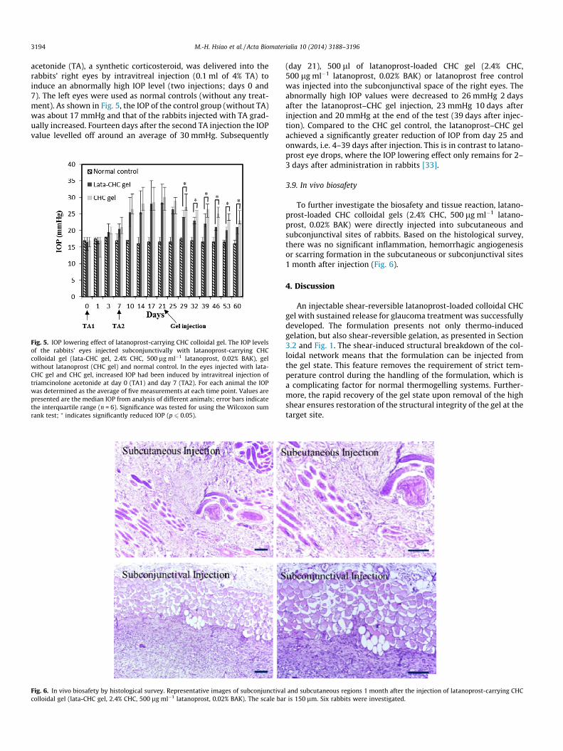
acetonide (TA), a synthetic corticosteroid, was delivered into therabbits’ right eyes by intravitreal injection (0.1 ml of 4% TA) toinduce an abnormally high IOP level (two injections; days 0 and7). The left eyes were used as normal controls (without any treat-ment). As shown in Fig. 5, the IOP of the control group (without TA)was about 17 mmHg and that of the rabbits injected with TA grad-ually increased. Fourteen days after the second TA injection the IOPvalue levelled off around an average of 30 mmHg. Subsequently
Fig. 5. IOP lowering effect of latanoprost-carrying CHC colloidal gel. The IOP levelsof the rabbits’ eyes injected subconjunctivally with latanoprost-carrying CHCcolloidal gel (lata-CHC gel, 2.4% CHC, 500 lg ml�1 latanoprost, 0.02% BAK), gelwithout latanoprost (CHC gel) and normal control. In the eyes injected with lata-CHC gel and CHC gel, increased IOP had been induced by intravitreal injection oftriamcinolone acetonide at day 0 (TA1) and day 7 (TA2). For each animal the IOPwas determined as the average of five measurements at each time point. Values arepresented are the median IOP from analysis of different animals; error bars indicatethe interquartile range (n = 6). Significance was tested for using the Wilcoxon sumrank test; ⁄ indicates significantly reduced IOP (p 6 0.05).
Fig. 6. In vivo biosafety by histological survey. Representative images of subconjunctivacolloidal gel (lata-CHC gel, 2.4% CHC, 500 lg ml�1 latanoprost, 0.02% BAK). The scale ba
(day 21), 500 ll of latanoprost-loaded CHC gel (2.4% CHC,500 lg ml�1 latanoprost, 0.02% BAK) or latanoprost free controlwas injected into the subconjunctival space of the right eyes. Theabnormally high IOP values were decreased to 26 mmHg 2 daysafter the latanoprost–CHC gel injection, 23 mmHg 10 days afterinjection and 20 mmHg at the end of the test (39 days after injec-tion). Compared to the CHC gel control, the latanoprost–CHC gelachieved a significantly greater reduction of IOP from day 25 andonwards, i.e. 4–39 days after injection. This is in contrast to latano-prost eye drops, where the IOP lowering effect only remains for 2–3 days after administration in rabbits [33].
3.9. In vivo biosafety
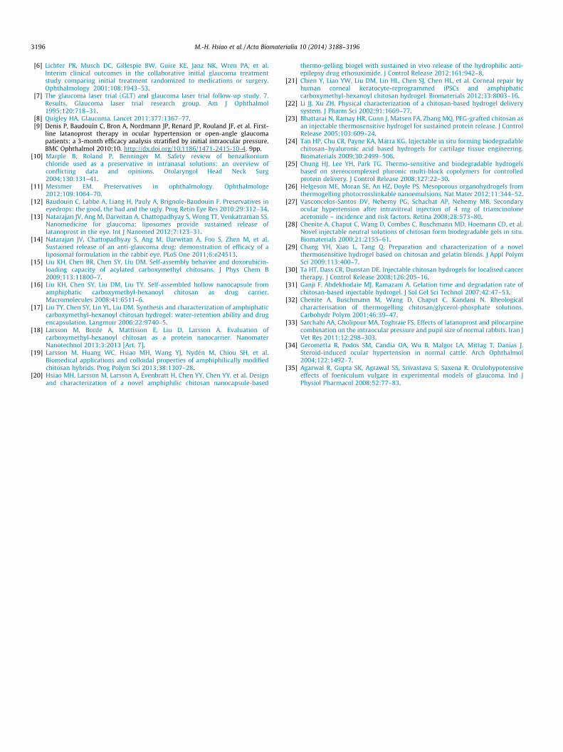
To further investigate the biosafety and tissue reaction, latano-prost-loaded CHC colloidal gels (2.4% CHC, 500 lg ml�1 latano-prost, 0.02% BAK) were directly injected into subcutaneous andsubconjunctival sites of rabbits. Based on the histological survey,there was no significant inflammation, hemorrhagic angiogenesisor scarring formation in the subcutaneous or subconjunctival sites1 month after injection (Fig. 6).
4. Discussion
An injectable shear-reversible latanoprost-loaded colloidal CHCgel with sustained release for glaucoma treatment was successfullydeveloped. The formulation presents not only thermo-inducedgelation, but also shear-reversible gelation, as presented in Section3.2 and Fig. 1. The shear-induced structural breakdown of the col-loidal network means that the formulation can be injected fromthe gel state. This feature removes the requirement of strict tem-perature control during the handling of the formulation, which isa complicating factor for normal thermogelling systems. Further-more, the rapid recovery of the gel state upon removal of the highshear ensures restoration of the structural integrity of the gel at thetarget site.
l and subcutaneous regions 1 month after the injection of latanoprost-carrying CHCr is 150 lm. Six rabbits were investigated.
The in vitro drug release studies revealed that, for all formula-tions and preparation conditions, one fraction of the loaded drugwas released over the 30 days of the experiment, while anotherfraction remained in the gels (Figs. 2 and 3). We hypothesize thatthe fraction of drug available for fast drug release was inverselydependent on the drug actually loaded into the nanocapsules. Thisis supported by the drug release profiles from gels prepared usingdifferent drug loading protocols (Fig. 2c). For the gels with a nano-capsule EE of 51%, the drug release levelled off at 61%, while forgels with an EE of 63% the drug release levelled off at 49%. It wasalso found that the total drug loading of the gels influenced thefraction of drug with a fast release rate. As seen in supportingFig. S3, a tenfold increase in drug content increased the plateauvalue at day 30 from 60 to 70%. This behaviour is logical, as ahigher drug content should lead to a reduced drug EE of the nano-capsules. The gelation temperature and presence of BAK were bothfound to slightly influence the release profiles, and a lower gelationtemperature also led to a somewhat lower plateau value for thefraction of drug released at after 30 days (Fig. 2a and b). The exactmechanisms behind this behaviour are yet to be elucidated andmerit further investigation. The drug release profile after injectionis highly relevant for applications. As seen in Fig. 3, the injectionprocess caused an accelerated initial release. However, the releasewas still extended, and after 30 days the release had levelled off atabout 70% for both the gel pushed through the syringe and the as-prepared control. The results from the in vitro drug release clearlysuggest that the formulations have the potential for extendedrelease in vivo, especially when considering that the in vitrorelease was performed in the presence of 10% DMSO to acceleratethe kinetics.
The formulations did not only present extended release ofloaded latanoprost, the loading of BAK and latanoprost into theCHC gels reduced their cytotoxicity, as presented in Section 3.4and shown in Fig. 4. A small decrease in cell viability was observedfor the pure CHC gels, but the viability remained above 80% evenfor the highest investigated CHC concentration of 2.7%. The loadingof BAK and latanoprost into the CHC gels did improve cell viabilitycompared to the free substances, but the cell viability was still aslow as about 30% after 48 h for gels containing both BAK andlatanoprost (Fig. 4b). The low cell viability may be problematicfor clinically relevant applications. However, it was observed that,for gels loaded only with latanoprost, the cell viability was greatlyimproved compared to the free substance (Fig. 4c). For a dose of500 lg ml�1, the viability was improved from 22% for free latano-prost to 78% when encapsulated in CHC. This may be a key featurefor avoiding cytotoxicity in the development of formulations carry-ing high drug loads to be released over extended times. The obser-vation that gels without BAK were much less cytotoxic indicatesthat a more clinically feasible formulation may be sterile-packedone-dose preparations that can be stored and handled withoutthe preservative effect of BAK. For a depot formulation like in thepresent study, this would be a viable alternative, as the frequencyof administration is very low.
In vivo experiments revealed excellent performance for theselected formulation (2.4% CHC, 500 lg ml�1 latanoprost, 0.02%BAK), despite the low cell viability observed in vitro. The extendedtherapeutic effect of the latanoprost-loaded colloidal CHC gel wasproved in a rabbit glaucoma model, where a single administrationlowered the IOP for up to 40 days (Fig. 5). It is worth mentioningthat the IOP of the control animals, to which colloidal CHC gelwithout latanoprost was administered, also recovered from thepeak value over time. However, this recovery was slower and lev-elled off at a higher value. This recovery of the control group isexplained by the steroid-induced increase in IOP results in a peakvalue with slow recovery over time, as demonstrated by others[34]. The sustained lowering of IOP for the latanoprost-loaded
CHC formulation is highly relevant as the effect of free latanoprostonly remains for 2–3 days after administration in rabbits [33] andother IOP-lowering substances reduced IOP in steroid-induced rab-bit models for less than 8 h [35]. In addition to the extendedrelease, the drug-loaded gels also presented excellent biocompati-bility, as determined by histological survey 1 month after adminis-tration (Fig. 6).
5. Conclusion
The drug used in this investigation, latanoprost, is currently oneof the most powerful ocular hypotensive drugs available and thepatent expired in March 2011. Improved glaucoma treatmentsare highly relevant, but no new classes of glaucoma drugs haveemerged since the introduction of latanoprost. There are, however,a number of ongoing developments, the depot latanoprost deliverysystem being one approach [1,13,14]. Therefore, the results of thisstudy, a new biocompatible injectable formulation with local sus-tained release of latanoprost and proven efficiency in vivo, areextremely interesting. The presented formulation allows for easyadministration, easy preparation without volatile organic solvents,no-burst release and localization to site of injection. The results ofthis study suggest that the formulations should be further investi-gated with a view to clinical use. It is also easily foreseen that theuse of these novel gels, or the same concept with different compo-nents, could be extended to the treatment of a number of criticalconditions, from chronic disorders to cancer. The highly desirableproperties suggest that the presented shear-reversible thermogel-ling colloidal CHC gels could offer improved clinical performance ina number of localized therapies.
Acknowledgements
The authors give their heartfelt thanks to National ScienceCouncil, Taiwan, for financial support under contract number ofNSC-101-3011-P-010-002.
Appendix A. Figures with essential colour discrimination
Certain figures in this article, particularly Figs. 1, 3 and 6 are dif-ficult to interpret in black and white. The full colour images can befound in the on-line version, at http://dx.doi.org/10.1016/j.actbio.2014.03.016.
Appendix B. Supplementary data
Supplementary data associated with this article can be found, inthe online version, at http://dx.doi.org/10.1016/j.actbio.2014.03.016.
References
[1] Zhang K, Zhang L, Weinreb RN. Ophthalmic drug discovery: novel targets andmechanisms for retinal diseases and glaucoma. Nat Rev Drug Discov2012;11:541.
[2] Weinreb RN, Khaw PT. Primary open-angle glaucoma. Lancet 2004;363:1711–20.
[3] Shingleton B, Tetz M, Korber N. Circumferential viscodilation and tensioning ofschlemm canal (canaloplasty) with temporal clear corneal phacoemulsificationcataract surgery for open-angle glaucoma and visually significant cataract:one-year results. J Cataract Refract Surg 2008;34:433–40.
[4] Camras CB, Alm A, Watson P, Stjernschantz J. Latanoprost, a prostaglandinanalog, for glaucoma therapy. Efficacy and safety after 1 year of treatment in198 patients. Latanoprost study groups. Ophthalmology 1996;103:1916–24.
[5] Ederer F, Gaasterland DE, Dally LG, Kim J, VanVeldhuisen PC, Blackwell B, et al.The advanced glaucoma intervention study (agis). 13. Comparison oftreatment outcomes within race: 10-year results. Ophthalmology 2004;111:651–64.
[6] Lichter PR, Musch DC, Gillespie BW, Guire KE, Janz NK, Wren PA, et al.Interim clinical outcomes in the collaborative initial glaucoma treatmentstudy comparing initial treatment randomized to medications or surgery.Ophthalmology 2001;108:1943–53.
[7] The glaucoma laser trial (GLT) and glaucoma laser trial follow-up study. 7.Results. Glaucoma laser trial research group. Am J Ophthalmol1995;120:718–31.
[8] Quigley HA. Glaucoma. Lancet 2011;377:1367–77.[9] Denis P, Baudouin C, Bron A, Nordmann JP, Renard JP, Rouland JF, et al. First-
line latanoprost therapy in ocular hypertension or open-angle glaucomapatients: a 3-month efficacy analysis stratified by initial intraocular pressure.BMC Ophthalmol 2010;10. http://dx.doi.org/10.1186/1471-2415-10-4. 9pp.
[10] Marple B, Roland P, Benninger M. Safety review of benzalkoniumchloride used as a preservative in intranasal solutions: an overview ofconflicting data and opinions. Otolaryngol Head Neck Surg2004;130:131–41.
[11] Messmer EM. Preservatives in ophthalmology. Ophthalmologe2012;109:1064–70.
[12] Baudouin C, Labbe A, Liang H, Pauly A, Brignole-Baudouin F. Preservatives ineyedrops: the good, the bad and the ugly. Prog Retin Eye Res 2010;29:312–34.
[13] Natarajan JV, Ang M, Darwitan A, Chattopadhyay S, Wong TT, Venkatraman SS.Nanomedicine for glaucoma: liposomes provide sustained release oflatanoprost in the eye. Int J Nanomed 2012;7:123–31.
[14] Natarajan JV, Chattopadhyay S, Ang M, Darwitan A, Foo S, Zhen M, et al.Sustained release of an anti-glaucoma drug: demonstration of efficacy of aliposomal formulation in the rabbit eye. PLoS One 2011;6:e24513.
[15] Liu KH, Chen BR, Chen SY, Liu DM. Self-assembly behavior and doxorubicin-loading capacity of acylated carboxymethyl chitosans. J Phys Chem B2009;113:11800–7.
[16] Liu KH, Chen SY, Liu DM, Liu TY. Self-assembled hollow nanocapsule fromamphiphatic carboxymethyl-hexanoyl chitosan as drug carrier.Macromolecules 2008;41:6511–6.
[17] Liu TY, Chen SY, Lin YL, Liu DM. Synthesis and characterization of amphiphaticcarboxymethyl-hexanoyl chitosan hydrogel: water-retention ability and drugencapsulation. Langmuir 2006;22:9740–5.
[18] Larsson M, Borde A, Mattisson E, Liu D, Larsson A. Evaluation ofcarboxymethyl-hexanoyl chitosan as a protein nanocarrier. NanomaterNanotechnol 2013;3:2013 [Art. 7].
[19] Larsson M, Huang WC, Hsiao MH, Wang YJ, Nydén M, Chiou SH, et al.Biomedical applications and colloidal properties of amphiphilically modifiedchitosan hybrids. Prog Polym Sci 2013;38:1307–28.
[20] Hsiao MH, Larsson M, Larsson A, Evenbratt H, Chen YY, Chen YY, et al. Designand characterization of a novel amphiphilic chitosan nanocapsule-based
thermo-gelling biogel with sustained in vivo release of the hydrophilic anti-epilepsy drug ethosuximide. J Control Release 2012;161:942–8.
[21] Chien Y, Liao YW, Liu DM, Lin HL, Chen SJ, Chen HL, et al. Corneal repair byhuman corneal keratocyte-reprogrammed iPSCs and amphiphaticcarboxymethyl-hexanoyl chitosan hydrogel. Biomaterials 2012;33:8003–16.
[22] Li JJ, Xu ZH. Physical characterization of a chitosan-based hydrogel deliverysystem. J Pharm Sci 2002;91:1669–77.
[23] Bhattarai N, Ramay HR, Gunn J, Matsen FA, Zhang MQ. PEG-grafted chitosan asan injectable thermosensitive hydrogel for sustained protein release. J ControlRelease 2005;103:609–24.
[24] Tan HP, Chu CR, Payne KA, Marra KG. Injectable in situ forming biodegradablechitosan–hyaluronic acid based hydrogels for cartilage tissue engineering.Biomaterials 2009;30:2499–506.
[25] Chung HJ, Lee YH, Park TG. Thermo-sensitive and biodegradable hydrogelsbased on stereocomplexed pluronic multi-block copolymers for controlledprotein delivery. J Control Release 2008;127:22–30.
[26] Helgeson ME, Moran SE, An HZ, Doyle PS. Mesoporous organohydrogels fromthermogelling photocrosslinkable nanoemulsions. Nat Mater 2012;11:344–52.
[27] Vasconcelos-Santos DV, Nehemy PG, Schachat AP, Nehemy MB. Secondaryocular hypertension after intravitreal injection of 4 mg of triamcinoloneacetonide – incidence and risk factors. Retina 2008;28:573–80.
[28] Chenite A, Chaput C, Wang D, Combes C, Buschmann MD, Hoemann CD, et al.Novel injectable neutral solutions of chitosan form biodegradable gels in situ.Biomaterials 2000;21:2155–61.
[29] Chang YH, Xiao L, Tang Q. Preparation and characterization of a novelthermosensitive hydrogel based on chitosan and gelatin blends. J Appl PolymSci 2009;113:400–7.
[30] Ta HT, Dass CR, Dunstan DE. Injectable chitosan hydrogels for localised cancertherapy. J Control Release 2008;126:205–16.
[31] Ganji F, Abdekhodaie MJ, Ramazani A. Gelation time and degradation rate ofchitosan-based injectable hydrogel. J Sol Gel Sci Technol 2007;42:47–53.
[32] Chenite A, Buschmann M, Wang D, Chaput C, Kandani N. Rheologicalcharacterisation of thermogelling chitosan/glycerol-phosphate solutions.Carbohydr Polym 2001;46:39–47.
[33] Sarchahi AA, Gholipour MA, Toghraie FS. Effects of latanoprost and pilocarpinecombination on the intraocular pressure and pupil size of normal rabbits. Iran JVet Res 2011;12:298–303.
[35] Agarwal R, Gupta SK, Agrawal SS, Srivastava S, Saxena R. Oculohypotensiveeffects of foeniculum vulgare in experimental models of glaucoma. Ind JPhysiol Pharmacol 2008;52:77–83.