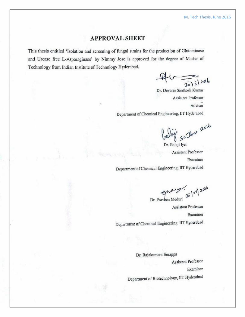
M. Tech Thesis, June 2016 Isolation and Screening of Glutaminase & Urease Free Novel Fungal Strains for the Production of L-Asparaginase A thesis submitted to Indian Institute of Technology Hyderabad in partial fulfilment of the requirements for the degree of Master of Technology By NIMMY JOSE Under the supervision of Dr. Devarai Santhosh Kumar Department of Chemical Engineering Indian Institute of Technology Hyderabad June 2016
Transcript
M. Tech Thesis, June 2016
Isolation and Screening of Glutaminase & Urease Free Novel Fungal
Strains for the Production of L-Asparaginase
A thesis submitted to Indian Institute of Technology Hyderabad in partial
fulfilment of the requirements for the degree of Master of Technology
By
NIMMY JOSE
Under the supervision of
Dr. Devarai Santhosh Kumar
Department of Chemical Engineering
Indian Institute of Technology Hyderabad
June 2016
M. Tech Thesis, June 2016
M. Tech Thesis, June 2016
M. Tech Thesis, June 2016
ACKNOWLEDGEMENT
I would like to express my sincere gratitude to my advisor Dr. Devarai Santhosh Kumar, Assistant Professor,
IIT Hyderabad for giving me an opportunity to pursue this research work and for his valuable guidance
throughout the research.
I thank Department of Science and Technology (SERB No. SB/EMEQ-048/2014) India for their financial
support.
I extend my sincere thanks to Department of Biotechnology for allowing me to do spectrophotometric analysis.
My gratitude goes to other Industrial Bioprocess and Bio prospecting Laboratory members Kruthi Doriya,
Jyothi Rao, Anup Ashok, Haritha P and Vaibhav Lendekar for their immeasurable support and constant help in
my works.
I also thank my parents, brothers and friends for their love and constant support without which this project
would have been incomplete.
Nimmy Jose
M. Tech Thesis, June 2016
i
ABSTRACT
L-Asparaginase is an amidohydrolase that catalyzes the hydrolysis of amino acid L-
asparagine into aspartic acid and ammonia. It is used in the treatment of Acute
Lymphoblastic Leukemia (ALL) and some other malignant lymphoid abnormalities. It is
also used in food industry to prevent the formation of acrylamide, a carcinogenic substance
in carbohydrate rich fried and baked foods. Naturally L-Asparaginase is present in plants,
animals and microbes but microorganisms such as bacteria, yeast and fungi are generally
used for the production of L-Asparaginase as it is difficult to obtain the same from plants
and animals. It is found that the L-Asparaginase from bacteria causes side effects in humans
including anaphylaxis and serious allergic reactions which can be fatal in some cases. To
overcome this, eukaryotic organisms such as fungi can be used for the production of L-
Asparaginase. But sometimes the fungi produces L-glutaminase and urease enzymes along
with L-Asparaginase which is difficult to remove in the purification stage. In order to
prevent this fungal strains which can produce L-Asparaginase free of L-glutaminase and
urease are isolated from different sources using standard protocols.
In the current study four novel fungal strains (C3-Aspergillus sps, C7-Aspergillus sps, W3-
Rhizopus sps, W5-Rhizopus sps) producing L-Asparaginase free of L-glutaminase and
urease are screened from a total of 40 fungal sps isolated from various soil samples and
agricultural substrates collected from different locations. Activity studies are conducted for
all these species according to standard protocols. Fungus with high enzyme index (C7)
1.57 was then subjected to Solid State Fermentation (SSF) studies in flasks and the results
were compared with that of flask level Submerged Fermentation (SmF). The strain C7 is
M. Tech Thesis, June 2016
ii
found to have the highest activity of 44.09 U/ml in SmF and 22.41 U/ml in SSF at 72 hour
of incubation at 35 ° C and 180 rpm.
M. Tech Thesis, June 2016
iii
CONTENTS
Abstract i
Contents iii
List of figures v
List of tables vi
Abbreviations and notations vii
1. Introduction
1.1 Generalities 1
1.2 L-Asparaginase in tumor treatment and its mechanism 2
1.3 Applications in food industries 3
1.4 Methods of production : Comparison of SSF and SmF 4
1.5 Objectives and scopes 6
1.6 Organization of thesis 6
2. Literature Review
2.1 L-Asparaginase 8
2.2 Historical development 8
2.3 Chemistry and structural aspects of L-Asparaginase 10
2.4 Sources of L-Asparaginase 12
2.4.1 Bacterial L-Asparaginase 12
2.4.2 Fungal L-Asparaginase 12
2.4.3 Actinomycetes sources 12
2.5 Clinical availability of L-Asparaginase 14
2.6 Treatment and Side effects 16
2.7 Large scale production of L-Asparagine 17
2.7.1 Production of L-Asparaginase by Submerged fermentation 17
2.7.2 Production of L-Asparaginase by Solid state fermentation 18
M. Tech Thesis, June 2016
iv
3. Materials and methods
3.1 Chemicals and reagents 21
3.2 Fungal species 21
3.3 Collection of soil samples 21
3.4 Plate assay for the screening of L-Asparaginase production 21
3.5 Plate assay for L-Glutaminase production 22
3.6 Plate assay for Urease production 22
3.7 Isolation and screening of fungi from soil 23
3.8 Analytical methods
3.8.1 Assay of L-Asparaginase 23
3.8.2 Protein determination 24
3.9 SSF studies 25
4. Results and discussion
4.1 Isolation of fungal strains from different sources 26
4.2 Screening studies of the isolated fungal sps 26
4.3 Semi quantitative studies of the isolated fungal strains 30
4.4 Quantitative studies of L-Asparaginase activity 33
4.5 Protein estimation studies 34
4.6 L-Asparaginase activity studies in SSF 36
5. Conclusion and future studies 38
6. References 40
7. Appendix
M. Tech Thesis, June 2016
v
LIST OF FIGURES
Figure no
Title of figure
Figure 1 Hydrolysis reaction of L-Asparaginase on asparagine
Figure 2 Maillard reaction leading to the formation of acrylamide
Figure 3 Schematic illustration of the reaction mechanism of L-Asparaginase
Figure 4 Screening of isolated strains for multiple enzyme activity using
MCD plates amended with 0.009% phenol red
Figure 5 Screening of isolated strains for multiple enzyme activity using
MCD plates amended with 0.007% BTB
Figure 6 Picture showing zone diameter and colony diameter
Figure 7 Microscopic pictures of isolated strains
Figure 8 Activity plots of isolated fungal strains
Figure 9 Specific activity plots of isolated fungal strains
Figure 10 Comparison of SSF and SmF activity values for W5
Figure 11 Comparison of SSF and SmF activity values for C7
M. Tech Thesis, June 2016
vi
LIST OF TABLES
Table no Title of table
Table 1 Comparison of SSF and SmF for enzyme production
Table 2 Various microbial sources of L-Asparaginase
Table 3 Available commercial forms of L-Asparaginase
Table 4 Summary of fermentation conditions and microbial cultures
for production of L-Asparaginase using SmF
Table 5 Summary of microbial cultures and fermentation conditions
for production of L-Asparaginase using SSF
Table 6 List of isolation sources and strains
Table 7 Fungal species screened for multi enzyme production
Table 8
L-Asparaginase enzyme index measurement using phenol red
and Bromothymol blue amended in MCD medium and species
observed under Light microscope
Table 9 Activity values of isolated fungal strains
Table 10 Protein content of isolated fugal strains
Table 11 Specific activity values of isolated fungal strains
Table 12 Comparison of SSF and SmF activity values for W5
Table 13 Comparison of SSF and SmF activity values for C7
M. Tech Thesis, June 2016
vii
ABBREVIATIONS AND NOTATIONS
ALL Acute lymphoblastic leukemia
BSA Bovine serum albumin
BTB Bromothymol blue
IU International unit
MCDM Modified Czapek Dox Medium
MTCC Microbial Type Culture Collection and Gene Bank
OD Optical density
PDA Potato dextrose agar
PR Phenol red
SmF Submerged fermentation
SSF Solid state fermentation
TCA Trichloro acetic acid
M. Tech Thesis, June 2016
viii
M. Tech Thesis, June 2016
1
Chapter 1
INTRODUCTION
1.1 Generalities
L-Asparaginase (E.C. 3.5.1.1) is an enzyme which is found in a wide range of organisms
including plants, microbes, animals and in the serum of certain rodents but not in human
beings. It is an amidohydrolase, which catalyzes the hydrolysis of the amide group on the
side chain of asparagine, an amino acid into aspartic acid and ammonia. It was first found
to be present in the serum of guinea pigs by J G Kidd in 1953. He observed that the enzyme
has tumor inhibitory properties and showed that transplanted lymphomas of mice and rat
are repressed in vivo by repeated injections of guinea pig serum [1]. Because of its anti-
tumor activities L-Asparaginase is used mainly in the treatment of Acute Lymphoblastic
Leukemia (ALL). It is also used in the food industry to prevent the formation of acrylamide
in fried food items [2]. L-Asparaginase is present in plants and mammals, since the
extraction is difficult microbial sources especially bacteria and fungi are evaluated as
potential source of enzyme production [3].
Figure 1. Hydrolysis reaction of L-Asparaginase on asparagine
M. Tech Thesis, June 2016
2
1.2 L-Asparaginase in tumor treatment and its mechanism
Acute lymphoblastic leukemia (ALL) which mostly affects children is a form of cancer in
which the bone marrow produces too many immature lymphocytes leading to reduced
immunity. L-Asparaginase enzyme is used as a chemotherapy drug for the treatment of
ALL. It is also used in the treatment of a number of lymphocytic cancers including
Hodgkin’s disease, non-Hodgkin’s lymphoma, melanosarcoma etc. Normal cells can
synthesize L-asparagine by itself because of the presence of the enzyme asparagine
synthetase, whereas certain sensitive malignant cells cannot synthesize it by itself and
require an external source of L-asparagine for optimal growth. During the treatment of
ALL with L-Asparaginase, all the circulating asparagine in the body of the patient get
hydrolyzed to aspartic acid and ammonia preventing the absorption of asparagine by
tumor cells and hence depriving the dependent tumor cells of their extracellular source of
L-asparagine. The asparagine deficiency rapidly impairs the protein synthesis and leads
to delay in DNA and RNA synthesis and hence impairs the cell functioning finally
resulting in cell death [4, 5]. L-Asparaginase is commonly used as a combination
chemotherapy drug for the treatment of acute lymphoblastic leukemia (ALL) in children.
Unfortunately, despite the wide use of L-Asparaginase, most of the treatments have been
interrupted due to severe side effects and immunological reactions in the patients. The side
effects include anaphylaxis, coagulation abnormality, thrombosis, liver dysfunction,
pancreatitis, hyperglycemia, cerebral dysfunction etc. These side effects are developed
either due to the production of anti-asparaginase antibody in the body or due to multiple
enzymatic activity of the produced enzyme [6]. Toxicity of L-Asparaginase is mainly due
to the fact that the enzyme preparations are amidohydrolase, not L-Asparaginase. L-
M. Tech Thesis, June 2016
3
Glutaminase and urease are usually associated with the L-Asparaginase isolated from most
of the bacteria and fungi and it is very difficult to separate them in the purification stage
[7]. These enzymes hydrolyze L-glutamine and urea in the body, thereby preventing
kidney, central nervous system and other vital organs from normal functioning thus leading
to serious side effects [8, 9].
1.3 Applications in food industry
This Enzyme is also used in the food industry to prevent the formation of acrylamide, a
carcinogenic substance during frying or baking of food items containing starch at high
temperatures [10]. The reaction is a result of heat induced Maillard reaction (or non-
enzymatic browning reaction) between amino acid group of asparagine and carbonyl
group of reducing sugar which provides desirable flavor to the food. On addition of the
enzyme the asparagine in the food gets converted to aspartic acid and ammonia hence
preventing the formation of acrylamide.
Figure 2. Maillard reaction of asparagine and glucose leading to the formation of
acrylamide
M. Tech Thesis, June 2016
4
L-Asparaginase production throughout the world is carried out either by submerged
fermentation (SmF) or solid state fermentation (SSF). SSF is defined as the growth of
microorganism on solid substrate which acts as an energy source in the absence of free
flowing water. SSF is a substitute to submerged fermentation for the large scale production
of industrial enzymes. The solid substrates used in SSF are mainly agricultural or
industrial wastes which are cheap and has resistance to contamination especially for the
large scale production of fungal enzymes. Therefore SSF can be used as a better method
for the large scale production of L-Asparaginase.
1.4 Methods of production: Comparison of SSF and SmF
L-Asparaginase production throughout the world is carried out either by submerged
fermentation (SmF) or solid state fermentation (SSF). Submerged fermentation is a process
in which the growth of microorganisms takes place in liquid broth medium which is
optimized with required nutrients to have a better cultivation of micro-organisms. This
involves growing carefully the selected microorganisms in closed reactor containing the
fermentation medium and a high concentration of oxygen. Submerged fermentation has
well established equipment that make use of the existing micro-organisms. Bacteria is
commonly used as source in this process as it requires high moisture content.
SSF is defined as the growth of microorganism on solid substrate which acts as an energy
source in the absence of free flowing water [11]. SSF is a substitute to submerged
fermentation for the large scale production of industrial enzymes. The solid substrates used
in SSF are mainly agricultural or industrial wastes which are cheap and has resistance to
contamination especially for the large scale production of fungal enzymes.
M. Tech Thesis, June 2016
5
Compared to submerged fermentation SSF has many advantages, among those the most
important thing is that it provides high yield and activity of the enzyme and the process is
eco-friendly because it makes use of agricultural waste as the substrate and since the
moisture content is low it avoids the need to treat a huge amount of effluent water. These
factors avoid environmental pollution to a considerable extent.
SSF has disadvantages as well. The heat produced in SSF reactor is difficult to dissipate
effectively hence it often leads to heat buildup which affects the growth of the fungi. The
solid mass prevents effective diffusion of oxygen and the controlling of process parameters
are really difficult.
Table 1. Comparison of SSF and SmF for enzyme production
Advantages Limitations
Submerged
Fermentation
Solid state fermentation Submerged
Fermentation
Solid state
fermentation
Better heat and
mass transfer
can be achieved
Low water requirement,
resistance to
contamination
Complex in
operation, Low
yield.
Heat build up
Difficulties to ensure
proper oxygen diffusion
Better diffusion
of
microorganism
Better diffusion
of oxygen
No effluent water
Substrate are agricultural
wastes
High energy
consumption and
cost intensive
Large scale inoculums
and difficult to control
process parameters
Commercially
available in
large scale
High yield and product
activity
High release of
effluents
Difficulties in scale-up
M. Tech Thesis, June 2016
6
1. 5 Objectives and scopes
Based on an extensive literature survey on the production of L-Asparaginase and
characterization, the present study focused on isolation of a novel fungal strain for the
production of glutaminase and urease free L-Asparaginase. The following objectives have
been envisaged in the present investigation:
Isolation and screening of potential glutaminase and urease free L-Asparaginase
producing fungal strains from soil and agricultural samples.
Identification of the strain with the maximum enzyme index.
Comparison of activity studies in SmF and SSF.
These four strains C3, C7, W3 and W5 are free of glutaminase and urease and are found to
have good L-Asparaginase activity and hence have high potential in the treatment of ALL.
This is the first report on L-Asparaginase producing strain free of glutaminase and urease
elsewhere reported in the literature.
1.6 Organization of thesis
The presentation of the work has been divided into five chapters. The current Chapter 1
presents a general introduction, objective and scope of the present work. While the
literature that supports the work is presented in Chapter 2. Chapter 3 includes the details
of the materials and methods adopted in the present study. It explains the procedures and
protocols used in the study. Chapter 4 contains the results and discussions. This chapter
discusses in detail about the four isolates which are free of glutaminase and urease activity
M. Tech Thesis, June 2016
7
and its SmF and SSF activity studies. Chapter 5 draws summary and appropriate
conclusions based on the previous results and discussions. It also provides some useful
recommendations to carry out further work in this field.
M. Tech Thesis, June 2016
8
Chapter 2
LITERATURE REVIEW
2.1 L-Asparaginase
L-Asparaginase (L-Asparaginase amidohydrolase EC 3.5.1.1) is the enzyme having
antitumor activity and obtained from various biological sources viz., plants, animals and
many other microorganisms (fungus, yeast, bacteria etc.). The enzyme acquired clinical
importance in 1961 when the antitumor effect of Guinea pig serum originally discovered
by Kidd. It has been used in leukemia treatment last four decades. The most common
therapeutic indications are treatment of Hodgkin disease, acute lymphocytic leukemia
(mainly in children), acute myelocytic leukemia, acute myelomonocytic leukemia, and
chronic lymphocytic leukemia, lymphosarcoma treatment, reticle sarcoma and
melanosarcoma. Recently, some more applications of L-Asparaginase have been reported
in acrylamide free food production.
2.2 Historical development
The pioneer observation that turned out to be important for the development of L-
Asparaginase as a potential antineoplastic agent was made by Clementi in 1922 revealing
the presence of high activity of L-Asparaginase in the serum of guinea pig. High L-
Asparaginase activity was observed only in guinea pig serum, whereas other mammals
were found devoid of this enzyme [12]. Later in 1953 J G Kidd showed that transplanted
lymphomas of mice and rat are repressed in vivo by repeated injections of guinea pig serum
and found that some active constituent in serum is responsible for the selective necrosis of
lymphoma cells [13]. The studies took another turn when Neumann and McCoy has
M. Tech Thesis, June 2016
9
observed in 1956 that the basically non-essential amino acid asparagine is needed to grow
the Walker carcinosarcoma 256 in vitro [14]. Haley and co-workers have found that murine
L5178Y leukemia cells also require asparagine for in vitro growth in 1961 [15]. Broome
also observed the same results in 1961 with his experiments with 6C3HED cell lines [16].
It was Broome who later in 1963 came up with the theory that the antitumor activities of
guinea pig serum is due to the presence of the enzyme L-Asparaginase in it [17]. Looking
at the biochemical reactions involved in these experiments it became evident that certain
leukemic blast cells are sometimes unable to synthesize enough asparagine for their own
metabolism, so that the asparaginase-induced deficiency in asparagine will impair cellular
function and eventually cause cellular death. So the specificity of L-Asparaginase towards
L-asparagine is the reason behind this therapeutic effect.
Furthermore, a major advancement resulted when Mashburn and Wriston in 1963 reported
that asparaginase can be extracted from E.coli bacteria and it can inhibit the growth of
tumor cell just like guinea pig serum [18].This opened the possibilities to produce and
utilize the enzyme in larger quantities. It also leads to number of clinical studies [19]. The
first clinical trials in patients with acute lymphoblastic leukemia were carried out with
asparaginase preparations both from guinea pig serum and E. coli. Both enzymes showed
clinical efficacy [20].
In the later years further studies identified more bacterial species with L-Asparaginase
producing capability. Among those isolates Erwinia Chrisanthemi showed maximum
activity and it was used for large scale production of the enzyme [21]. Even though a large
number of strains were reported to have L-Asparaginase activity in the following years
M. Tech Thesis, June 2016
10
including only E Coli and Erwinia Chrisanthemi species were widely used for large scale
production.
Treatment with E Coli protein was always found to be associated with hypersensitivity
reactions. Whereas the Erwinia Chrisanthemi protein was found to have negligible or lesser
side effects in clinical trials [22]. But both the protein have certain level of
immunogenicity. Later it was found that coupling the derived protein with Poly Ethylene
Glycol (PEG) group could preserve the activity of the enzyme for a longer time and could
reduce the immunogenicity to certain extend [23]. It helped to reduce the hypersensitivity
of the enzyme and allowed much less frequent administration of PEG-asparaginase
compared to normal asparaginase.
2.3 Chemistry and structural aspects of L-Asparaginase
Enzymes with L-Asparaginase activity can be generally classified into two groups, the
bacterial-type and the plant-type L-Asparaginases, characterized by different structural and
biochemical features. The bacterial-type enzymes are further grouped into type I and type
II depending on their cellular localization and substrate specificity. Type I includes
cytosolic enzymes that exhibit low affinity for L-Asparaginase, whereas type II enzymes
are localized in the periplasm and show considerably higher affinity for L-Asparaginase
[24]. These enzymes from various sources have been purified and its biochemical
properties are studied extensively over the last 4 decades. Type II asparaginase has a stable
tetrameric structure composed of 4 identical sub units and each subunit contains 326 amino
acid residues [25]. E.Coli asparaginase has molecular weight of approximately 130 kDa
and the affinity constants for L-asparagine and L-glutamine are 1.15 x 10⁻⁵ and 6.25 X 10-
3 M, respectively. The isoelectric point of crystalline type varies from 4.8 to 5.6 [26, 27].
M. Tech Thesis, June 2016
11
Whereas the molecular weight of Erwinia L-Asparaginase is between 135 - 138 kDa and
specific activity of the purified enzyme lies between 300 and 400 mole of the substrate per
minute per milligram of protein. The isoelectric point ranges between pH 4.6 and 5.5 for
E. coli enzyme, and is around 8.7 for the Erwinia enzyme [28].
The amidohydrolase L-Asparaginase helps in the hydrolysis of non-essential amino acid
asparagine into aspartic acid and ammonia. L-asparagine hydrolysis is known to proceed
in two steps. In the first step a covalent intermediate, beta-acyl-enzyme intermediate is
formed through nucleophilic attack by the threonine group on L-Asparaginase as shown in
figure 3. In the second step, a water molecule attacks the acyl-enzyme intermediate to
produce L-aspartate and ammonia [29]. The structure of E. coli L-Asparaginase was
studied by Swain et al., (1993) and two domains were observed [25]. Location of the active
site was found to be between the N and C terminals. Structure of the enzyme with bound
L-aspartate indicated a threonine residue as a catalytic nucleophile by Miller et al. in 1993
[30]. Hydrolysis reaction is assayed by measuring the release of ammonia using Nessler’s
reagent or by measuring the release of L-aspartate.
Figure 3. Schematic illustration of the reaction mechanism of L-Asparaginases. The
proposed covalent intermediate is formed through nucleophilic attack by the enzyme. Bold
arrows indicate nucleophilic attack
M. Tech Thesis, June 2016
12
2.4 Sources of L-Asparaginase
Microorganisms are considered as effective sources for the production of therapeutic
enzymes since microbes are easy to manipulate. Broad range of microorganisms such as
filamentous fungi, yeast, actinomycetes and marine organisms are isolated from different
sources.
2.4.1 Bacterial L-Asparaginase
L-Asparaginase production from various bacterial sources have been studied extensively
over decades due to the flexibility with which bacteria’s can be manipulated. L-
Asparaginase from E.coli and Erwinia chrysanthemi are clinically used for the treatment
of ALL. Bacterial asparaginase derived from various bacteria differ in pH, molecular
weight, stability and affinity and they are serologically and biochemically different even
though the toxicity, anti-neoplasticity and immunogenicity are similar. Bacterial
formulations are found to have high immunogenicity in ALL treatment. Different bacterial
isolates with L-Asparaginase activity reported in the literature are given in table 2.
2.4.2 Fungal L-Asparaginase
Bacterial L-Asparaginase is often associated with hypersensitive reactions in patients
which can be fatal in some cases. This leads to the studies to identify fungal strains which
are free of allergic and immunogenic reactions. Since the fungi are eukaryotic organisms
and evolutionarily more close to human cell line the immunogenic side reactions are
comparatively lesser for fungal asparaginase. The mitosporic fungi genera such as
Aspergillus, Penicillium and Fusarium are commonly reported in the literature to produce
asparaginase [31, 32, 33, 34]. Imada et al. observed that amidase activity is present in
M. Tech Thesis, June 2016
13
fungal strains, Penicillium clavgorme and P. expansum. Sarquis et al., (2004) and Mishra,
(2006) reported that the L-Asparaginase production by A. terreus and A. niger, respectively
[8, 35]. Other isolates are given in the table.
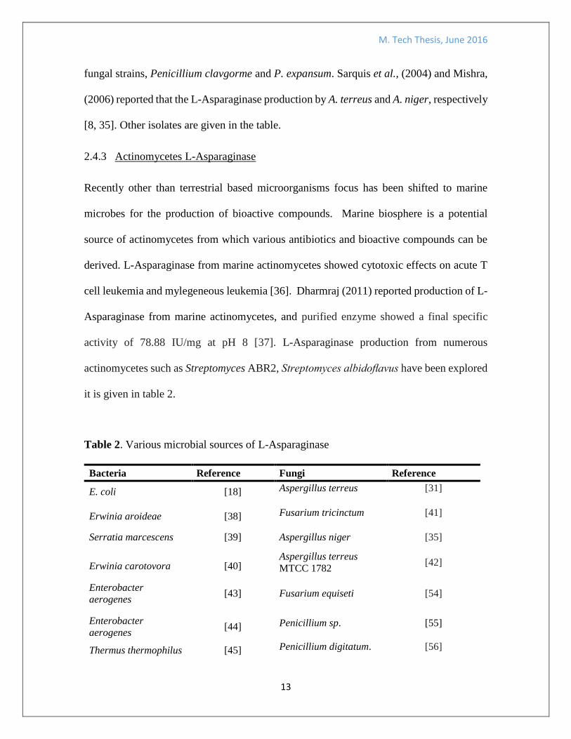
2.4.3 Actinomycetes L-Asparaginase
Recently other than terrestrial based microorganisms focus has been shifted to marine
microbes for the production of bioactive compounds. Marine biosphere is a potential
source of actinomycetes from which various antibiotics and bioactive compounds can be
derived. L-Asparaginase from marine actinomycetes showed cytotoxic effects on acute T
cell leukemia and mylegeneous leukemia [36]. Dharmraj (2011) reported production of L-
Asparaginase from marine actinomycetes, and purified enzyme showed a final specific
activity of 78.88 IU/mg at pH 8 [37]. L-Asparaginase production from numerous
actinomycetes such as Streptomyces ABR2, Streptomyces albidoflavus have been explored
it is given in table 2.
Table 2. Various microbial sources of L-Asparaginase


![Judy Lewis Masters Thesis Submitted[1]](https://static.documents.pub/doc/80x56/577cc7761a28aba711a10230/judy-lewis-masters-thesis-submitted1.jpg)
![THESIS TITLE A THESIS SUBMITTED TO THE MIDDLE EAST ...ii.metu.edu.tr/system/files/documents/thesis... · [SAMPLE 1] Approval of the thesis: THESIS TITLE Submitted by STUDENT NAME](https://static.documents.pub/doc/80x56/6019035f39977162fc4f0b03/thesis-title-a-thesis-submitted-to-the-middle-east-iimetuedutrsystemfilesdocumentsthesis.jpg)
![THESIS TITLE A THESIS SUBMITTED TO THE MIDDLE EAST ... · [SAMPLE 1] Approval of the thesis: THESIS TITLE Submitted by STUDENT NAME SURNAME in partial fulfillment of the requirements](https://static.documents.pub/doc/80x56/5ead9906bfb8c70db70cb309/thesis-title-a-thesis-submitted-to-the-middle-east-sample-1-approval-of-the.jpg)


