1 A Virtual Heart Model for Formal and Functional Medical Device Verification NSF Summer Undergraduate Fellowship in Sensor Technologies Allison Connolly, Biomedical Engineering, Johns Hopkins University Advisor: Rahul Mangharam, Ph.D. ABSTRACT Currently, there is no formal method for the development and testing of medical device software, such as that used in pacemakers and implantable cardioverter-defibrillators (ICD). A large majority of device recalls are due to failures in the software that went undiscovered during product testing. For example, safety recalls of pacemakers and implantable cardioverter defibrillators due to firmware (i.e. software) problems between 1990 and 2000 affected over 200,000 devices, comprising 41% of the devices recalled. In order to preempt these failures, the device companies and the regulatory agencies, such as the FDA, need a better way to formally and functionally verify these devices before bringing them to the market. The heart model outlined in this paper is a tool used to simulate, test and validate these devices across multiple modalities in a plug-and-play manner. By synthesizing a large number and variety of intra- cardiac electrogram and derived external electrocardiogram signals, the model will create a database well beyond the scope of the MIT-BIH ECG database, the current standard for most cardiac medical device algorithm testing. This heart model allows for more extensive formal and functional testing of pre-market cardiac medical devices to detect flaws before the devices are implanted in patients.
Transcript
1
A Virtual Heart Model for Formal and Functional Medical Device Verification
NSF Summer Undergraduate Fellowship in Sensor Technologies Allison Connolly, Biomedical Engineering, Johns Hopkins University
Advisor: Rahul Mangharam, Ph.D.
ABSTRACT Currently, there is no formal method for the development and testing of medical device software, such as that used in pacemakers and implantable cardioverter-defibrillators (ICD). A large majority of device recalls are due to failures in the software that went undiscovered during product testing. For example, safety recalls of pacemakers and implantable cardioverter defibrillators due to firmware (i.e. software) problems between 1990 and 2000 affected over 200,000 devices, comprising 41% of the devices recalled. In order to preempt these failures, the device companies and the regulatory agencies, such as the FDA, need a better way to formally and functionally verify these devices before bringing them to the market. The heart model outlined in this paper is a tool used to simulate, test and validate these devices across multiple modalities in a plug-and-play manner. By synthesizing a large number and variety of intra-cardiac electrogram and derived external electrocardiogram signals, the model will create a database well beyond the scope of the MIT-BIH ECG database, the current standard for most cardiac medical device algorithm testing. This heart model allows for more extensive formal and functional testing of pre-market cardiac medical devices to detect flaws before the devices are implanted in patients.
2
Table of Contents
1. Introduction p. 3 2. Background p. 3
2.1.Population of Medical Device Users p. 3 2.2.Current Patient Models p. 4 2.3.Current Heart Models p. 4
3. Virtual Patient p. 4 3.1.Medical History p. 4 3.2.Data Storage p. 5
4. Design and Development p. 5 4.1.Electrophysiology p. 6 4.2.Inverse Problem p. 6 4.3.Hardware p. 7
5. Implementation p. 7 6. Discussion p. 9 7. Future Work p. 10
7.1.Integration of the Virtual Patient Model p. 10 7.1.1.Context of Signal p. 10 7.1.2.Patient Specific Signal p. 10
7.2.Signal Database p. 11 8. Applications p. 11
8.1.Research and Development p. 11 8.2.Medical p. 11
9. Acknowledgements p. 12 10. References p. 12 11. Appendices p. 14
11.1. Appendix A: VHM version 1.0 – Virtual ICD and Heart State Machine p. 14 11.2. Appendix B: VHM version 1.1 – Pacemaker/Sinus Rhythm State Diagram p. 15 11.3. Appendix C: Pacemaker Flow Diagram p. 16 11.4. Appendix D: Labview Code for Heart Model and Virtual Medical Devices p. 17
3
1. INTRODUCTION As the medical device industry grows, more devices contain software without a person in the loop. However, the Food and Drug Administration (FDA) and other regulatory agencies have no formal method for testing and verifying that the software performs properly and does not malfunction. For this reason, as the medical device industry grows so does the number of medical device recalls. The percentage of device recalls due to software problems is exceptionally high in cardiology and in vitro diagnostics due to the heavy reliance on software in these fields [1][2]. Many recalls in cardiac devices are due to inappropriate delivery of treatment or inability to intervene when necessary [3]. According to Compton, 25-33% of implantable cardioverter-defibrillator (ICD) shocks delivered are inappropriate, causing unnecessary pain and damage to the patient [4]. Firmware problems go undiscovered during FDA testing and approval because the algorithms are completely proprietary to the device companies. Each company has its own method for testing and validating its own algorithms, but there is no standardized, formal method that the FDA can utilize to ensure all ICD’s deliver shocks appropriately and all pacemakers maintain proper cardiac rhythms. The development of a virtual heart model (VHM) that can be applied in research and medical environments allows for the betterment of medical device development techniques. The VHM is part of a larger virtual patient model (VPM). Data from every patient entering a hospital and undergoing tests will be stored and used to create an individualized patient model. Incorporating this data into a generic body model, such as the VHM, calls for the creation of interactive signals for use by doctors and researchers alike. Currently, a patient who enters the hospital to have a cardiac ablation procedure for treatment of an arrhythmia undergoes hours of monitoring and several pre-operative tests [5]. These data are stored only for the duration of the patient’s visit and are discarded afterwards. If this data could be saved and incorporated into a database, it would enhance future patient diagnostics and provide a more extensive database for device validation and verification. This paper focuses on the development plan for the VHM with the primary aim of testing medical devices for premarket approval. Section 2 provides further background on the growing issue of software failures as well as a summary of current patient models. Section 3 overviews the virtual patient project. Section 4 details the design components of the VHM. Section 5 addresses how the current version of the VHM is implemented. Sections 6 and 7 present conclusions reached during the development of the project as well as future directions. Section 8 lists possible applications of the complete VHM and VPM. 2. BACKGROUND 2.1. Population of Medical Device Users Every year, over 100,000 ICD’s are implanted in patients throughout the United States [6]. This number will continue to increase as the baby boomers age and as the rate of obesity-induced cardiac disease climbs, as predicted by [7] and [8]. Since 1990, the number of pacemakers implanted has doubled and the number of ICD’s implanted has increased 11-fold. These devices
4
are implanted in patients with arrhythmias and those who are at risk of sudden cardiac death. These conditions are usually associated with obesity, diabetes, and hypertension [9]. It is predicted that 1% or less of the devices that are implanted will malfunction [10]. However, this statistic is surprisingly high when the raw number of deaths is considered. This number will only increase as the number of implanted devices increases in the near future. 2.2. Current Patient Models Patient models have many uses, including device testing, medical staff training, and scenario modeling. The MIT-BIH database contains collections of signals from real patients. Some of the waveform data contains simultaneous ECG, blood pressure, arterial pressure, CO2 level, and respiration recordings [11]. This allow for the analysis of multiple aspects of a patient’s health at once, without the presence of an actual patient. These recordings contain all the variability characteristic of real-time signals, but are often too short and do not contain the interaction to be useful in comprehensive device testing. An alternative model is the MedSim300 patient simulator by Fluke [12]. It is a signal generator that can be used for medical personnel training as well as medical device testing. This device can synthesize signals of cardiac arrhythmias, oxygen levels, respiration, and more. However, these signals are highly simplified and do not contain the variability inherent in true patient data. The virtual patient model outlined in this paper contains the variability of true data with the freedom of a synthesized signal. 2.3. Current Heart Models Models of cardiac tissue excitation and contraction have been developed at institutions across the globe. However, many of these are complex, based on intracellular electrical conduction and action potentials. Boulakia et al. have developed a model to simulate external ECG recordings based on the electrical activity across the membranes of a network of interconnected cells [13]. Trayanova et al. have modeled the cardiac structure and its response to electrical stimulation, ranging from arrhythmias to depolarization [14]. These models contain both mechanical and electrical components on the micro level, but are highly complicated and too computationally heavy for real-time processing. The model developed in this paper is on a macro-level, modeling the global electrical activity of the atria and ventricles of the heart. 3. VIRTUAL PATIENT Creating the virtual patient has a dual purpose, to create a complete medical history for each patient, and to provide a larger number of medical recordings to the public domain for research purposes. This project is developed separately from but has potential to be included in the electronic health record [15]. 3.1. Medical History The virtual patient model will compile all medically-relevant information from a patient into a single location to enhance treatment capabilities. Data and results from all tests will be stored here, even if they are not deemed relevant to a specific issue at the time of recording. As
5
medical testing and screening becomes less invasive, even healthy patients will have detailed recordings taken, such as electrophysiology studies of their hearts. The maps created during these tests can be stored in the VPM and provide a healthy baseline for future comparison if the patient develops a heart abnormality. The medical history will also incorporate details about a patient’s lifestyle. This includes physical activity levels, living environment, and daily habits. Previous medical treatments can also be relevant to the VPM. Usage of certain cardio-active drugs affects the performance of the heart and circulatory system. Data such as prescribed drugs, medical treatments or therapies used, the number of hours per week a patient exercises, and the quality of air in the patient’s living environment are all important factors that can alter his or her health status, both temporarily and in the long run. 3.2. Data Storage The MIT-BIH database is now the largest and most widely used database for real signals. This data is used to test and verify research-based devices and algorithms as well as devices developed for the market. The database is not the ideal resource for pre-market testing because of the limited number and variety of signals available. One of the main uses of the database is for electrocardiogram (ECG) algorithm testing. However, most of the signals within the database contain only two leads of pre-recorded data, while standard ECG’s contain 12 leads. Doctors are only able to make a satisfactory diagnosis by examining recordings from multiple locations on the patient’s chest. Two leads do not contain enough information to make a diagnosis. When the database was created in the 1980’s, it contained a wealth of signals compared to other sources [16]. Because the database is not updated regularly, many of these recordings have become outdated and are of low quality compared to those taken with new devices. The data stored in the VPM will be updated regularly as the patient’s health is monitored over the years. By making the data stored in the VPM anonymous and available to the public, it will provide a variety of high quality signals for all who desire to use them. The VPM has the potential to be a more powerful algorithm testing tool for all types of medical devices than the MIT-BIH database. 4. DESIGN AND DEVELOPMENT The first iteration of the virtual heart model was recently developed by the authors for the purpose of virtual and real medical device testing. For this reason, the VHM had to produce electrical signals similar to those recorded by medical devices, namely electrogram and ECG signals. In order to minimize the complexity of the model while making it as rich as possible, it was necessary to translate between the electrogram signals (measured from the interior of the heart) and the ECG signals (measured from the skin on the torso). Transforming electrogram data into multiple-lead ECG signals is known as the forward problem, while transforming the surface signals to the intracardiac signals is known as the inverse problem [17]. This iteration of
6
the model utilizes the inverse problem, as ECG signals are more readily available for testing and manipulation. 4.1. Electrophysiology
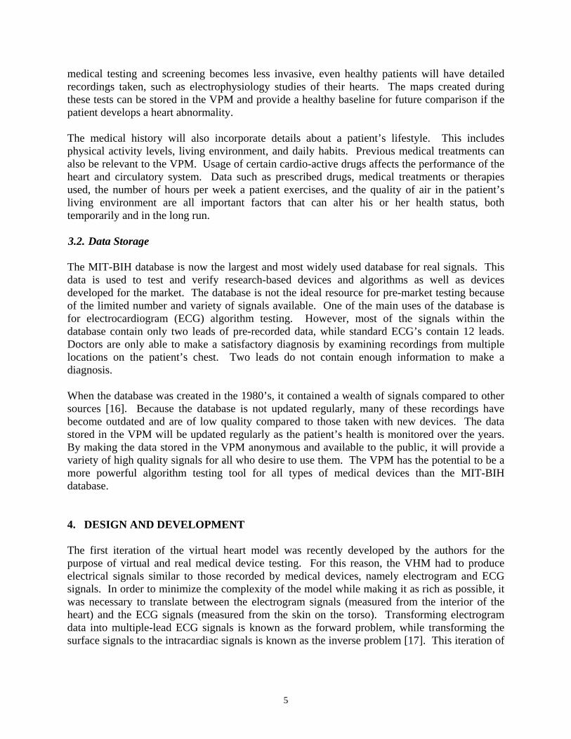
The electrocardiogram is the most readily available recording of heart activity because it is a non-invasive and relatively inexpensive monitoring procedure. Using ten electrodes placed across the surface of the upper torso, the ECG records the global electrical activation of the heart muscle that correlates with the contraction of the heart and expulsion of blood. The components of lead II of the standard ECG are shown in Figure 4.1. When the heart is beating in normal sinus rhythm, this pattern is repeated continuously at a constant rate [18]. However, when the heart functions abnormally, the morphology of the ECG is altered in relation to the type of arrhythmia.
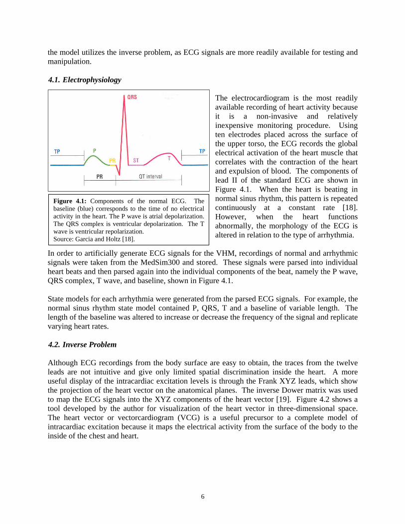
In order to artificially generate ECG signals for the VHM, recordings of normal and arrhythmic signals were taken from the MedSim300 and stored. These signals were parsed into individual heart beats and then parsed again into the individual components of the beat, namely the P wave, QRS complex, T wave, and baseline, shown in Figure 4.1. State models for each arrhythmia were generated from the parsed ECG signals. For example, the normal sinus rhythm state model contained P, QRS, T and a baseline of variable length. The length of the baseline was altered to increase or decrease the frequency of the signal and replicate varying heart rates. 4.2. Inverse Problem Although ECG recordings from the body surface are easy to obtain, the traces from the twelve leads are not intuitive and give only limited spatial discrimination inside the heart. A more useful display of the intracardiac excitation levels is through the Frank XYZ leads, which show the projection of the heart vector on the anatomical planes. The inverse Dower matrix was used to map the ECG signals into the XYZ components of the heart vector [19]. Figure 4.2 shows a tool developed by the author for visualization of the heart vector in three-dimensional space. The heart vector or vectorcardiogram (VCG) is a useful precursor to a complete model of intracardiac excitation because it maps the electrical activity from the surface of the body to the inside of the chest and heart.
Figure 4.1: Components of the normal ECG. The baseline (blue) corresponds to the time of no electrical activity in the heart. The P wave is atrial depolarization. The QRS complex is ventricular depolarization. The T wave is ventricular repolarization. Source: Garcia and Holtz [18].
7
-0.50
0.51
1.52
0
0.
1
1.
2
2.
0
0.5
1
1.5
2
X component
Z co
mpo
nent
-1 0 1 2 3-1
0
1
2
3Frontal Plane
X
Y
-1 0 1 2 3-0.5
0
0.5
1
1.5Left Sagital Plane
Z
Y
-1 0 1 2 3-0.5
0
0.5
1
1.5Bottom Horizontal Plane
X
Z
4.5 4.55 4.6 4.65 4.7 4.75 4.8 4.85
-0.5
0
0.5
1
Lead I
4.5 4.55 4.6 4.65 4.7 4.75 4.8 4.85
0
1
2
3
Lead II
4.5 4.55 4.6 4.65 4.7 4.75 4.8 4.85
-1
0
1Lead III
4.5 4.55 4.6 4.65 4.7 4.75 4.8 4.85
0
1
2
Lead aVR
4.5 4.55 4.6 4.65 4.7 4.75 4.8 4.85
-1
-0.5
0
0.5
Lead aVL
4.5 4.55 4.6 4.65 4.7 4.75 4.8 4.85-1
0
1
Lead aVF
4.5 4.55 4.6 4.65 4.7 4.75 4.8 4.85-2
-1
0
Lead V1
4.5 4.55 4.6 4.65 4.7 4.75 4.8 4.85
-2
-1
0
1
Lead V2
4.5 4.55 4.6 4.65 4.7 4.75 4.8 4.85
-2
0
2
Lead V3
4.5 4.55 4.6 4.65 4.7 4.75 4.8 4.85
-1
0
1
2
Lead V4
4.5 4.55 4.6 4.65 4.7 4.75 4.8 4.85
-10123
Lead V5
4.5 4.55 4.6 4.65 4.7 4.75 4.8 4.85
0
2
4
Lead V6
.5
.5
.5
Y component
4.3. Hardware The model was implemented on a programmable hardware platform, allowing the user to directly interface with analog and digital devices. For this project, the VHM was implemented on the National Instrument sbRIO-9641 FPGA board, shown in Figure 4.3 [20]. The virtual heart model is embedded in the real-time microcontroller. The signals are fed through the FPGA chip to the ADC and DAC onboard, allowing for the transmission of signals between the patient simulator on the computer, the MedSim300, an oscilloscope, and medical devices. 5. IMPLEMENTATION All aspects of the heart model software were created in Labview and embedded in the NI FPGA board. The Labview GUI is shown in Figure 5.1 and the code is shown in appendix D. The heart model responded to three inputs: atrial pacing, ventricular pacing, and an ICD shock. A
Figure 4.2: a) 3D Heart Vector Loop Derived from 12-Lead ECG for one heart beat, b) Projection of the heart vector in the frontal XY plane, c) Projection of the heart vector in the left sagital YZ plane, d) Projection of the heart vector in the horizontal XZ plane, e) Tracings of the 12 ECG leads for one heart beat.
Figure 4.3: National Instrument sbRIO-9641 FPGA
a
b c d
e
8
state diagram of the arrhythmia heart model is depicted in appendix A. The more detailed state diagram of the pieces of sinus rhythm and parts of the ECG are depicted in appendix B. In this version of the GUI for the heart model, pacing and shocking are manually implemented. This allows for comprehensive testing of the heart model to aid development of the algorithms. For normal sinus rhythm (NSR), the heart rate can be adjusted at the start of signal generation. The construction of the ECG signal in real time is done on the real-time microcontroller. The signal is output through the FPGA board and the DAC to the virtual pacemaker or ICD medical device. The medical device software reacts similarly to how a real medical device would react. The software contains a QRS detector, allowing for heart rate detection and thus identification of the state of the heart. The medical device can then output a pace or shock signal, which is fed back through an ADC to the FPGA and into the heart model.
Because the system contains the heart and device in a closed loop, the heart model can react to inputs from the medical devices. This gives the entire system a dynamic nature that one does not find in recorded signals from real patients. For example, if the initial state of the heart is ventricular tachycardia (VTach), the ICD would normally pace in order to restore NSR. However, if the ICD does not detect the VTach for some reason, the state of the heart will degrade to ventricular fibrillation (VFib). For this state, the ICD detects the chaotic rhythm and delivers a shock. This shock causes the heart model to restore NSR.
Figure 5.1: Interactive user interface for the heart model version 1.0. a) the signal being input from the heart model to the virtual medical device, b) the signal output from the virtual medical device, c) control box for the ICD shocking or pacing functions of the medical device, d) display of the current state of the heart model, either normal sinus rhythm (NSR), bradychardia (BRAD), ventricular tachycardia (VTACH), or ventricular fibrillation (VFIB) and the hear rate measured by QRS detector.
a
b
c
d
9
Not every situation with medical device intervention is ideal, and some can lead to unhealthy results. For example, if the ICD shock or pace is delivered when the ventricles are undergoing repolarization, the heart will not reset to NSR, but will instead go into VFib. This is implemented in the heart model to ensure that the medical device software can detect when the heart is in the T wave (ventricular repolarization), and will thus withhold shock until an appropriate time. 6. DISCUSSION The virtual heart model was successfully created to interact with actuations from medical devices and medical device software. This actuation will allow for direct testing of medical devices by the FDA and by the medical device companies themselves. The heart model can respond to pacing located in the right atrium and the right ventricle, as well as shocks delivered in the right ventricle. The location of the virtual electrodes that deliver the pace or shock is not variable within the model as it would be in a real patient. Adjusting the location of electrode attachment in the heart wall will need to be incorporated in later iterations of the model. The current version of the model incorporates a comprehensive set of arrhythmias that are important for medical device testing, namely VTach and VFib. The model does not, however, contain more complex arrhythmias such as heart block or atrial fibrillation, which would be necessary for more in depth analysis and testing of software. Each arrhythmia is represented as a separate state in this version of the model. In order to make the model more physiologically accurate, the model must become based on probabilities and other factors. This can be done using a Hidden Markov process to train the heart model to create more realistic signals. The use of electrocardiogram signals in the heart model is not realistic for integration with medical devices, as most devices are implanted inside the patient’s chest and respond to electrogram signals. For this reason, the use of the vectorcardiogram transform or another inverse model solution is necessary to transform between the external ECG signals and the internal electrogram signals. Once this transformation is made, real medical devices can be plugged in to the model hardware and interact in a closed-loop fashion. In conclusion, version 1 of the virtual heart model outlined in this paper provides a solid foundation on which to build a more complex and realistic model. The closed loop nature allows for interaction between the heart and devices. A complete model will provide medical device companies with a tool for proper software development and testing. It will also provide the FDA and other regulatory agencies a method to formally validate and verify this software before approving it for the market. With both the FDA and the device companies working to improve software development, device malfunctions due to software issues are hoped to decrease significantly and reduce the rate of device-related mortality.
10
7. FUTURE WORK 7.1. Integration of the Virtual Patient Model By combining the recorded signals from patients with the synthesized signals from the VHM, the model will be able to create an infinite array of cardiac electrical signals. A purely synthesized signal, such as that created by the MedSim300, is not sufficient for testing and validation of medical devices. Those signals do not contain the variability and unpredictability of signals recorded from real patients. 7.1.1. Context of Signal While standard ECG signals contain useful features due to redundancy and quality of signal, they are limited by the context of the recordings. Because the patient being monitored must be hooked up to the ECG through ten electrodes with connection wires, the patient must remain relatively still. For this reason, nearly all ECG signals are recorded while the patient is stationary. They contain no artifacts due to movement or exercise exertion. Recordings taken from Holter monitors [21] or Loop monitors [17] are taken while the patient is ambulatory. This means the signals recorded by these monitors contain artifacts associated with patient motion, environment, and activity level. The VHM will be able to extract these artifacts from recorded signals and superimpose them on the signals produced by the modeling, creating a more realistic and more widely applicable model. Artifacts can arise from activity level, such as running, walking, sleeping, and emotionally and physically stressful situations. The patient’s environment and surroundings, such as being underwater, at high altitudes, or at extreme temperatures, can affect the operation of his or her cardiac system. The patient’s internal state, such as hormone levels or any drugs that are in his or her system, will also alter the signals, making them more complex for interpretation by software algorithms. A basic example of artifact integration is breath rate. Breathing causes a baseline variation in the ECG because the distance from the heart to the torso wall changes as lung diameter increases and decreases. The breath rate also introduces variability in heart rate. As the lungs expand, they put pressure on the major veins and arteries in the chest, causing blood pressure to increase, which then triggers heart rate to decrease [18]. The VHM must be able to introduce the wandering baseline and the variable heart rate into synthesized signals. 7.1.2. Patient-Specific Signal Just as the context artifacts can be integrated into synthesized signals, so can patient specific artifacts. This includes elevations in the ST-level, morphology changes in the P wave, and lengthened PR interval due to diseased conduction pathways. A combination signal containing context and patient artifacts superimposed on the general VHM has uses in medical care. This virtual patient can be used to test medical devices specifically with the patient in mind to see how he or she will respond and if the device will function properly
11
7.2. Signal Database The combination signals give the heart model user the freedom to create a complex signal that will respond to actuation or feedback from an ICD or pacemaker. This provides a better testing environment for software validation and verification than other databases, which are static and provide no response. In addition, multiple forms of cardiac excitation signals need to be generated. This includes ECG, endocardial and epicardial electrograms, and cellular-level action potentials. Using these signals, the heart model can be used to test both implantable and on-body medical devices. Future iterations of the VHM will contain spatial information on the muscular contraction and deformation of the heart, allowing mechanical-based medical devices to be tested as well. 8. APPLICATIONS The VHM can be used in both research and medical environments. The formal model can be used by the FDA for rapid premarket testing, validation, and certification. The model can be used in clinical trials for cardio-active drugs and other pharmaceuticals. The patient-specific model is useful for pre-operative diagnosis, potentially reducing the number of cardiac procedures. It also has uses in post-operative recovery monitoring, to ensure that treatment was complete and complications do not arise. 8.1. Research and Development The heart model can be used by the FDA for rapid premarket testing, validation and verification of medical devices. Currently, the FDA entrusts the validation of medical device software to the companies that make the devices. These companies run the software through a set of signals that was determined by the company itself to be sufficient for testing. The FDA is not involved in choosing the set of signals, so they cannot be sure the device is actually validated or compare performance across different devices. Examples of devices that require software validation and verification are implantable loop monitors, pacemakers, and ICD’s. In conjunction with the design of the heart model, the authors will design an electrocardiogram monitoring adhesive strip. The strip, which will be placed on the patient’s chest above the heart, will contain two electrodes for signal acquisition and embedded programmable hardware for arrhythmia detection. The software on the monitoring strip will be verified and validated using the heart model. It will provide useful feedback during the model development to make the model interact properly with device software. 8.2. Medical In many situations, insufficient patient history and data are recorded to make a diagnosis. In these cases, the doctor can conduct new tests, which are time consuming and costly, or he or she might perform a procedure or surgery the patient may not need. Integrating a medical history and patient lifestyle with a more general heart model will provide the physician with a predictive tool to diagnose what ailments a patient has without doing invasive testing.
12
A second medical application is for cardiac ablation surgeries. These surgeries are complicated procedures that require the patient to be under general anesthesia for eight or more hours [22]. Much of this time is spent creating an intracardiac map of the excitation timing within the myocardium. If a general map could be produced using the VHM and patient specific data before the procedure, the electrophysiologist would need less time for mapping and thus reduce the overall surgical time. After the surgery is completed, the patient is at risk for post-operative complications or recurrence of the arrhythmia. The VPM would also have the capability to evaluate the effectiveness of the ablation procedure and predict the likelihood of a recurrence or transference of the arrhythmia from one site to another [23]. The collection of post-operative recordings from a large number of patients will give the heart model the capability to predict both normal and abnormal outcomes, allowing the doctor to preemptively choose the next step in the patient’s treatment. 9. ACKNOWLEDGEMENTS The author would like to thank her advisor, Professor Rahul Mangharam, for his invaluable guidance and advice throughout the summer. She would also like to thank Zhihao Jiang for his long hours of work and dedication to the project and wishes him luck as the project continues. Thanks to all the members of mLab and Dr. Ed Gerstenfeld for their support and collaboration on the project. Finally, thanks to Jan Van der Spiegel and the rest of the SUNFEST administration, along with the National Science Foundation, for providing this opportunity. 10. REFERENCES [1] Z. Bliznakov, G. Mitalas and N. Pallikarakis, “Analysis and Classification of Medical Device
Recalls,” in IFMBE Proc., 2006, pp. 3782-3785. [2] W. H. Maisel, M. O. Sweeney and W. G. Stevenson, “Recalls and safety alerts involving
pacemakers and implantable cardioverter-defibrillator generators,” JAMA vol. 286(7), pp. 793-799, 2001.
[3] FDA. (2009, July 23). Medical Devices: List of Device Recalls. [Online]. http://www.fda.gov /MedicalDevices/Safety/RecallsCorrectionsRemovals/ListofRecalls/default.htm
[4] A. J. Compton, H. Bolouri and A. W. Nathan, “Arrhythmia recognition strategies and hardware decisions for the implantable cardioverter-defibrillator- A review,” Med. Eng. Phys., vol. 17(2), pp. 96-103, Mar. 1995.
[5] E. P. Gerstendfeld, private communication, June, 2009. [6] J. Coromilas, “Physician credentials and ICD implantation: Certified “electricians” best deal
with electrical problems,” JAMA vol. 301(16), pp. 1713-1714, 2009. [7] D. E. Arterburn, P. K. Crane and S. D. Sullivan, “The coming epidemic of obesity in elderly
Americans,” J. Am. Geriatrics Society, vol. 52(11), pp. 1907-1912, Nov. 2004. [8] K. Bibbins-Domingo, P. Coxson, M. J. Pletcher, J. Lightwood and L. Goldman, “Adolescent
overweight and future adult coronary heart disease,” New England J. of Med., vol. 357, pp. 2371-2379, Dec. 6, 2007.
13
[9] K. P. Anderson, “The changing epidemiology of ventricular arrhythmias,” Clin. Cardiol., vol.
26(3), pp. 321-333. [10] Food and Drug Administration. FDA Enforcement Report. Rockville, Md: National Press
[Online]. http://assets.fluke.com/manuals/medsim__omeng0000.pdf [13] M. Boulakia, M. A. Fernandez, J. F. Gerbeau and N. Zemzemi, “Towards the numerical
simulation of electrocardiograms,” LNCS, vol. 4466, pp. 240-249, 2007. [14] N. Trayanova, K. Skouibine and F. Aguel, “The role of cardiac tissue structure in
defibrillation,” Chaos vol. 8, pp. 221, 1998. [15] “Electronic Health Records Overview,” National Institutes of Health , National Center for
Research Resources, McClean, VA, 2006. [16] G. B. Moody and R. G. Mark, “The impact of the MIT-BIH database,” IEEE Eng. In Med
and Biol., May/June 2001. [17] B. Taccardi and B. B. Punske, “Body Surface Potential Mapping,” in Cardiac
Electrophysiology: From Cell to Bedside, D. P. Zipes and J. Jalife, Ed. Philadelphia: Saunders, 2004, pp. 803-811.
[18] T. B. Garcia and N. E. Holtz, 12-Lead ECG: The Art of Interpretation, Sudbury, MA: Jones & Bartlett Pub., 2001.
[19] B. Bojovic, L. Hadzievski and P. Belicev, “Device and procedure for visual three-dimensional presentation of ECG data,” U.S. Patent 72266408, Sept. 4, 2007.
[20] (2008). NI Single-Board RIO Embedded Control and Acquisition Devices. National Instruments. Austin, TX. [Online]. http://www.ni.com/pdf/products/us/cat_sbRIO_96xx.pdf
[21] J. P. DiMarco and J. T. Philbrick, “Use of Ambulatory Electrocardiographic (Holter) Monitoring,” Annals of Internal Medicine, vol. 113, pp. 53-68, 1990.
[22] P. J. Zimetbaum and M. E. Josephson, Practical Clinical Electrophysiology, Philadelphia: Lippincott Williams and Wilkins, 2009.
[23] A. S. Go, E. M. Hylek, K. A. Phillips, Y. Chang, L. E. Henault, J. V. Selby, D. E. Singer, “Prevalence of Diagnosed Atrial Fibrillation in Adults,” JAMA., vol. 285(18), pp. 2370-2375, May 9, 2001.
14
11. APPENDICES
11.1. Appendix A: VHM version 1.0 – Virtual ICD and Heart State Machine
Normal Sinus Rhythm
Bradychardia Ventricular Tachycardia
Ventricular Fibrillation
Death
pace pace
shock
shock on T wave
do nothing for s1 seconds
do nothing for s2 seconds
15
11.2. Appendix B: VHM version 1.1 – Pacemaker/Sinus Rhythm State Diagram
Normal P wave
Paced P wave
Paced QRS complex
Normal QRS complex
Tp interval
T wave
Ventricular Fibrillation
pace atria
pace atria
pace ventricle
pace ventricle
pace ventricle
pace ventricle
16
11.3. Appendix C: Pacemaker Flow Diagram
AVI
100 ms
Pace Ventricle
Pace Atria
PVARP
Wait 500 ms
VRP
Wait 400 ms
LRI‐AVI‐VRP
count 600 ms
LRI‐AVI‐PVARP
count 500 ms
sense QRS
do not sense QRS
sense QRS sense P
do not sense P
17
11.4. Appendix D: Labview Code for Heart Model and Virtual Medical Devices